HAL Id: tel-00989767
https://tel.archives-ouvertes.fr/tel-00989767
Submitted on 12 May 2014
HAL is a multi-disciplinary open access archive for the deposit and dissemination of sci-entific research documents, whether they are pub-lished or not. The documents may come from teaching and research institutions in France or abroad, or from public or private research centers.
L’archive ouverte pluridisciplinaire HAL, est destinée au dépôt et à la diffusion de documents scientifiques de niveau recherche, publiés ou non, émanant des établissements d’enseignement et de recherche français ou étrangers, des laboratoires publics ou privés.
Frontal and parietal contributions to visual perception
in humans
Lorena Chanes Puiggros
To cite this version:
Lorena Chanes Puiggros. Frontal and parietal contributions to visual perception in humans. Agri-cultural sciences. Université Pierre et Marie Curie - Paris VI, 2014. English. �NNT : 2014PA066027�. �tel-00989767�
Thèse de Doctorat de l’Université Pierre et Marie Curie (UPMC) École Doctorale Cerveau Cognition Comportement
Spécialité : Neurosciences
Frontal and parietal contributions to
visual perception in humans
Lorena CHANES
Directeur:
Dr. Antoni VALERO-CABRE
Membres du jury :
Dr. Marisa CARRASCO Examinateur
Dr. Karim JERBI Rapporteur
Dr. Michel LE VAN QUYEN Examinateur
Dr. Pascale PRADAT-DIEHL Présidente du jury
Dr. Juha SILVANTO Rapporteur
ACKNOWLEDGEMENTS
This thesis would not have been possible without the generosity and love of those who were always there, my mother, my father, my sister Camila, Yago, Jose and Marc.
I deeply and sincerely thank my advisor Antoni Valero-Cabré for his commitment all along this process and for everything he taught me along these years.
I would like to thank Ana Chica for teaching me and helping me, together with Toni, when I took my first steps.
I also thank Bruno Dubois and Paolo Bartolomeo for opening me the doors to the group and introduce me to the team.
I am also grateful to Felipe Fregni for having me in his lab as part of my PhD, and to Lotfi Merabet.
I also thank Catherine Tallon-Baudry for her contribution to some of the work presented here.
I would like to thank the members of my defense committee, Marisa Carrasco, Michel Le Van Quyen and Pascale Pradat-Diehl, as well as Karim Jerbi and Juha Silvanto, who were already part of my half-thesis committee, for kindly contributing to this work with their expertise and their feedback.
I would also like to thank Lionel Naccache, member of my half-thesis committee, for his feedback.
I sincerely thank Romain Quentin and Marine Vernet for their friendship and companionship, for being there when it had to do with the boulot and also when it didn’t.
I thank Deniz Doruk for being always ready to help and to share.
I also want to thank the rest of my labmates in Paris and Boston, especially Seth Elkin-Frankston, Dimitri Bayle and León Morales, for their collaboration, their fellowship and for everything we learned together and the great time we had.
I thank Isabel Seror for her help and her patience.
I would like to sincerely thank the Ecole de Neuroscience de Paris (ENP) and all the people involved, especially Jean-Antoine Girault, Patricia Gaspar, Yvette Hénin, Laura Peeters and André Sobel, for the great opportunity they gave me and the great scientific quality of the program but also for being there every time I needed it, for
standing by the students’ side when we had to start a new life in a new city far away from our families and friends, for making things always easy.
I also thank La Caixa and all the people of the program, especially Emília Jordi and Paul Fogleman, for giving me the opportunity to do a part of my PhD in the United States, for their funding and support and their readiness to contribute to my scientific and personal development.
I could not forget to thank the rest of my family in Uruguay and my friends outside the lab, thanks so much Uri, Anna, Dom, Frances, Loli, Rodrigo, Fran, Lupa, Marcelo and my other dear companions because
Des bateaux j'en ai pris beaucoup, Mais le seul qui ait tenu le coup, Qui n'ai jamais viré de bord, Mais viré de bord,
Naviguait en père peinard Sur la grand-mare des canards, Et s'app'lait Les Copains d'Abord Les Copains d'Abord.
SUMMARY
Frontal and parietal areas have been shown to subtend different cognitive processes such as attentional orienting, decision making and access to consciousness, with bearing on visual performance. In spite of prior evidence supporting an implication of those regions in visual cognition, their contributions to the processing of low-contrast unmasked stimuli and the characteristic spatiotemporal activity patterns underlying them remain to be fully explored and causation is lacking. We here addressed a thorough exploration of such contributions in humans, with an emphasis on the dynamics of neural activity and visual performance enhancements as probed by patterns of noninvasive manipulation of local brain oscillatory activity. To this end, we tested in healthy participants the effects of either single pulses or short bursts of
active vs. sham transcranial magnetic stimulation (TMS), delivered to the frontal eye field (FEF) and the intraparietal sulcus (IPS) prior to the presentation of a lateralized
low-contrast near-threshold Gabor stimulus, on the visual discrimination and conscious detection of such stimulus. In Chapter II we show that single TMS pulses delivered to the right FEF alone or in combination with visuo-spatial cues have the ability to increase perceptual sensitivity, effects that proved strongly modulated by cue validity. In Chapter III we report that induced pre-target high-beta (30 Hz) activity in this same region selectively enhances perceptual sensitivity (d’) whereas gamma band (50 Hz) bursts selectively shift response criterion (beta). This result shows that neural oscillations could be a general mechanism to multiplex functions, with specific behavioral effects, in the same region or network. In Chapter IV we provide evidence of stochastic facilitation of perceptual performance, showing perceptual sensitivity
enhancements by the induction of neural noise to the left FEF, and demonstrate that the left and right hemispheres modulate visual performance through different coding strategies possibly reflecting different cognitive processes. In Chapter V we show that the effects of right-FEF high-beta activity on perceptual sensitivity are phase independent and that additional perceptual phase-dependent effects can be observed. Finally, in Chapter VI, we report for the right IPS similar frequency-specific effects to the ones observed in the right FEF as well as additional perceptual modulations when the pulses are delivered in a short time window. Our findings contribute to better substantiate the oscillatory basis of visual cognition and its associated behaviors and to set the stage for the development of novel therapies based on noninvasive manipulation of dysfunctional brain oscillatory activity.
RESUME
Les aires cérébrales frontales et pariétales sont impliquées dans différents processus cognitifs importants pour la performance visuelle, tels que l’attention ou la conscience. Malgré les preuves existantes en faveur d’une implication de ces régions dans la cognition visuelle, leurs contributions dans le traitement de stimuli non masqués de faible contraste ainsi que l’activité spatio-temporelle sous-tendant ces contributions restent largement inexplorées, tout particulièrement en termes de causalité. Nous avons mené une exploration approfondie de ces contributions chez l’humain, en mettant l’accent sur la dynamique de l’activité neurale et les améliorations perceptives potentielles qui peuvent résulter de la manipulation non invasive de l’activité cérébrale. À cette fin, nous avons testé chez des sujets sains les
effets d’impulsions simples ou de rafales courtes de stimulation magnétique
transcrânienne (SMT) réelle versus fausse, délivrée sur le champ oculomoteur frontal ou le sillon intrapariétal avant la présentation d’un filtre de Gabor de faible contraste, sur la discrimination et la détection consciente de ce filtre de Gabor. Nos résultats montrent que chez l’humain, la distribution spatio-temporelle de l’activité frontale et pariétale joue un rôle causal dans la performance visuelle. Nos recherches contribuent à mieux comprendre les bases oscillatoires de la cognition visuelle et les comportements associés et à préparer le terrain pour le développement de nouvelles thérapies basées sur la manipulation non-invasive de l’activité cérébrale oscillatoire avec, pour objectif ultime, l’amélioration des pathologies neuropsychiatriques.
TABLE OF CONTENTS
CHAPTER I: Introduction and Aims 1
I.I. Visual perception 2
I.II. Neural bases of cognitive processes involved in visual
performance 4
I.II.1. Neural bases of visuo-spatial attention 5
I.II.2. Neural bases of conscious access 7
I.III. Neural dynamics of visuo-spatial attention and conscious
access 12
I.III.1. Neural dynamics of visuospatial attention 12
I.III.2. Neural dynamics of conscious access 14
I.IV. Noninvasive brain stimulation methods and approaches 15
I.IV.1. Transcranial magnetic stimulation (TMS) 16
I.IV.2. TMS and the visual system 18
I.IV.3. TMS and brain oscillations 20
I.V. The frontal eye fields, the intraparietal sulcus and their manipulation
through noninvasive brain stimulation 23
I.VI. Aims of the present dissertation 27
REFERENCES 32
CHAPTER II : Manipulation of pre-target activity on the right frontal eye field
enhances conscious visual perception in humans 60
CHAPTER III: Causal frequency-specific contributions of frontal spatiotemporal
performance 70
CHAPTER IV: Stochastic facilitation by left frontal arrhythmic activity patterns reveals
hemisphere-specific coding strategies for conscious visual perception 77
CHAPTER V : Causal contributions of induced frontal beta oscillation phase to the
modulation of visual performance in humans 98
CHAPTER VI: Distinct causal contributions of frequency and non-frequency-specific
posterior parietal activity patterns to visual performance 120
CHAPTER VII: Discussion 139
VII.I. Overview and summary of the main results 140
VII.II. Frontal and parietal contributions to perceptual performance through characteristic spatiotemporal activity patterns: Implications of our results for the current ideas on neural dynamics as a key factor for brain function and its
noninvasive manipulation in humans 143
VII.III. Pending questions and future directions 146
VII.IV. Perspectives 151
1
Chapter I
Introduction and Aims
2
At every moment of our lives, our senses are hit by much more information than we can treat. To succeed in using sensory inputs to act, cognitive processes such as attentional orienting help us prioritize those data that are behaviorally meaningful, selecting stimuli involuntarily because of their physical saliency or voluntarily according to their granted relevance to achieve our behavioral goals in a given context. Moreover, information that accesses consciousness will be able to be largely stored and strategically used and, in order to produce goal-directed behavior, evidence will need to be flexibly translated into motor actions through decision-making processes. All these operations shape the way we act and perform.
In this introduction we will present a brief overview of the current knowledge on the neural basis of visual perception, and some of the processes relevant for perceptual performance such as attention and conscious access. We will then address how noninvasive neurostimulation techniques can be used to manipulate activity in different brain regions and their associated networks and provide new insights on their role and processing features.
I.I. Visual perception
Vision is one of the main senses in humans and it has been extensively studied in virtually all organisms. In order to produce a visual percept, light reaches the retina, which contains a large number of photoreceptors. These photoreceptors convert this incoming radiation into electrical signals that are conveyed to the brain through the optic nerve. Most part of the retinal ganglionar cell axons (about 90%) relay on the lateral geniculate nucleus (LGN) of the thalamus to then reach the primary visual cortex (V1 or striate cortex). A few projections (about 10%) relay on the superior colliculus in the midbrain (Perry and Cowey 1984), which is known as
3
the indirect retino-tectal pathway. The superior colliculus influences the visual and posterior parietal regions indirectly through the pulvinar nucleus of the thalamus and receives modulatory projections from the primary visual cortex (Shipp 2004).
The primary visual cortex is retinotopically organized (Hubel and Wiesel 1959) and devoted to the processing of basic visual features, as for example spatial frequency (De Valois et al. 1982a; Maffei and Fiorentini 1973; Tootell et al. 1981), line orientation (De Valois et al. 1982b) and stimulus contrast (Hawken and Parker 1984). From V1 information is transmitted to the extrastriate cortex (V3, V4 and MT/V5), where higher order processing takes place (Orban 2008). Lesions to V1 involve a decrease of visually evoked activity in extrastriate visual areas and temporal cortices (Gross 1991; Rocha-Miranda et al. 1975). Moreover, visual perception is thought to involve feedback and feed-forward activations within and amongst V1 and higher order visual areas (Hupe et al. 1998).
Two pathways have been identified for the flow of information from the primary visual cortex to higher order areas: the dorsal and the ventral streams. The ventral stream (also known as the “what” stream) travels from the occipital to the inferior temporal cortex, including V2 and V4 and it is associated with the visual recognition and categorization of objects and faces (Kanwisher et al. 1997; Kourtzi and Kanwisher 2000). Lesions to the ventral visual stream result in visual agnosias (Ffytche et al. 2010). For example, in the early 90s an influential study described the case of a patient with a lesion of the ventral stream who presented a severe visual form agnosia (Goodale et al. 1991). This patient, however, had preserved guidance of hand movements with regards to objects the qualities of which (e.g. orientation) he failed to perceive. This preserved visuomotor ability would be coded in the dorsal visual stream. Indeed, the dorsal stream (also known as the “where”/“how” stream) is
4
associated with spatial localization and visuomotor control and projects from the occipital to the posterior parietal cortex, including areas V2, V3a, middle temporal (MT) and middle superior temporal (MST) cortices. Lesions to the dorsal visual stream result in a variety of cognitive dysfunctions including spatial neglect (Heilman et al. 1993), which involves inability to perceive and/or attend to stimuli contralateral to the lesion, and optic ataxia (Battaglia-Mayer and Caminiti 2002; Pisella et al. 2000; Pisella et al. 2009), which involves failure to integrate visual and motor information and thus to perform adapted visuomotor plans. The two streams are thought to interact to contribute to action and perception (Goodale and Westwood 2004; Milner et al. 2003; Westwood and Goodale 2003). For example, a patient with a bilateral lesion of the ventral stream presenting visual form agnosia, has impaired ability to adjust hand aperture to different object sizes (Goodale et al. 1994) and increased pointing errors to remembered target positions (Milner et al. 1999) when the action is delayed for a few seconds. Conversely, dorsal-stream lesion patients with optic ataxia improve when there is a delay between target and movement onset (Himmelbach and Karnath 2005; Milner et al. 2003; Milner et al. 1999; Revol et al. 2003). It has been suggested that such impairment and enhancement are mediated by the spared stream.
I.II. Neural bases of cognitive processes involved in visual performance
Cognitive processes such as attention and conscious access shape our performance with regards to sensory stimuli. In the last decades, research at different levels, from invasive single neuron recordings in nonhuman primates to noninvasive neuroimaging studies, have focused on mapping the brain systems of these complex processes, which tend to overlap at least partially and thus are difficult to dissect out.
5
I.II.1. Neural bases of visuo-spatial attention
Attentional orienting is crucial for an adaptive interaction of organisms with their environment, allowing them to pursue their behavioral goals while being capable of responding to unexpected but behaviorally relevant events. For example, the allocation of attentional resources in a particular region of the space improves the processing of stimuli presented in such region (Carrasco et al. 2000; Yeshurun and Carrasco 1998). Attention can be oriented in space either endogenously (voluntarily or top-down) or exogenously (involuntarily or bottom-up), and both types interact dynamically to allow appropriate perceptual behavior. In the mid-eighties, Posner and collaborators made popular the cue-target paradigm, which since then has been widely used to study visuospatial attention (Posner 1980; Posner et al. 1980; Posner et al. 1984). In this paradigm, participants have to detect or discriminate, as fast and as accurately as possible, a visual target presented on a screen that is preceded by a spatial cue displayed either centrally (e.g. an arrow) or peripherally (e.g. a salient dot near one of the potential locations of the target) and informative or not about target location. Peripheral non-informative cues engage exogenous attention, since the cue captures attention by its physical saliency, whereas central informative cues can be used strategically by participants to endogenously orient their attention towards the expected location of the target. All these cues typically decrease reaction times when they correctly predict the location of the target (valid trials) as compared to when target location is incorrectly predicted (invalid trials).
Early evidence from neurological patients and physiological studies monkeys has indicated the importance of frontal and parietal regions for visuospatial attention (e.g. (Goldberg and Bruce 1985; Mesulam 1981; Posner et al. 1984)). More recently,
6
investigators have searched for the neural basis of spatial attention in the healthy human brain using correlational techniques such as functional MRI (fMRI). One of the first studies to dissociate the brain activity related to attention from the activity related to the processing of the target revealed the superior frontal, inferior parietal and superior temporal cortices as the regions activated by the cues (Hopfinger et al. 2000). Taken together, neuroimaging studies have led to the proposal of two cortical networks that are involved in visuospatial attention (Corbetta et al. 2002). According to this model, a bilateral dorsal fronto-parietal network, including the intraparietal sulcus (IPS) and the frontal eye fields (FEF), would be in charge of orienting our attention in space. Indeed, the FEF and the IPS have been shown in humans and non-human primates to reflect the locus of attention (Armstrong et al. 2009; Bisley and Goldberg 2003; Kelley et al. 2008; Thompson et al. 1997; Thompson et al. 2005). A right-lateralized ventral network, including the temporoparietal junction (TPJ) and the inferior frontal gyrus (IFG), would be involved in the reorientation of attention towards unexpected and task-relevant events (Corbetta et al. 2008).
Recent studies have pointed out the importance of white matter tracts linking these cortical regions, in particular the superior longitudinal fascicle (SLF) and the inferior fronto-occipital fascicle (IFOF), for spatial attention. The SLF appears to present a dorsal to ventral increase in lateralization, with its dorsal-most branch (SLF I), overlapping with the dorsal attentional network, being symmetrically distributed in both hemispheres and its most ventral branch (SLF III), overlapping the ventral attentional network, being right-lateralized (Thiebaut de Schotten et al. 2011). It has also been hypothesized that the middle branch of the SLF (SLF II), overlapping with the anterior portion of the dorsal network and the posterior portion of the ventral network, would link both networks (Thiebaut de Schotten et al. 2011).
7
Spatial attention has also been shown to involve subcortical regions, particularly the superior colliculus (SC) and the pulvinar nucleus of the thalamus (Shipp 2004). The SC contains a retinotopic map of the external world. It receives direct input from the frontal, parietal and visual cortices and it is thought to be involved in both endogenous and exogenous attention (Fecteau et al. 2004; Kustov and Robinson 1996; Rafal et al. 1988). Moreover, it has recently been shown to present sustained activity in those neurons coding for the attended location (Ignashchenkova et al. 2004). The pulvinar is widely interconnected with the cortex and has been proposed to coordinate activity within multiple cortical regions (Petersen et al. 1987; Saalmann et al. 2012; Snow et al. 2009).
I.II.2. Neural bases of conscious access
In the last decades, a growing body of experimental work has revealed that sensory stimuli that do not access consciousness are however treated by the brain and can influence our behavior. As an early example, in the seventies, it was discovered that some patients with lesions to the primary visual cortex and associated visual deficits could perform over chance levels in simple tasks on stimuli presented in the blind part of his visual field where they reported not to see anything (Poppel et al. 1973; Weiskrantz et al. 1974). Weiskrantz and colleagues named this condition blindsight (Weiskrantz et al. 1974).
More recently, several investigators have studied the cerebral processing of stimuli that do not access consciousness in healthy participants using different paradigms that allow rendering stimuli subliminal and study conscious vs. unconscious processing, including near-threshold stimuli, masking, bistable images, binocular rivalry, inattentional blindness and change blindness. Using masked stimuli,
8
it has been shown for example that the emotional valence of subliminal stimuli can be treated by the amydgala (Whalen et al. 1998). This evidence has been supported by further data in a cortically blind patient (named G.Y.) who had massive damage to the left occipital lobe, sparing the occipital pole only, as well as a destruction of the optic radiation (Barbur et al. 1993). This patient was capable of discriminating over chance levels emotional facial expressions presented to his blind visual field (de Gelder et al. 1999) and showed activation of the amygdala (without activation of the fusiform face area) for fear vs. happy faces presented in such region (Morris et al. 2001). Moreover, studies have reported the existence of subliminal priming at different levels, visual, (Dehaene et al. 2001), semantic (Naccache and Dehaene 2001; Van den Bussche et al. 2009), and have shown that subliminal monetary incentives can influence subjects’ motivation (Pessiglione et al. 2007) indicating that subliminal information is not only treated by the brain but can also affect our behavior. Such influence is, however, potentially very limited (Dehaene and Changeux 2011; Dehaene and Naccache 2001). For example, it appears to decrease with time, as suggested by subliminal priming studies showing that the effect is observed only when the prime-to-target time is under 500 ms (Dupoux et al. 2008; Greenwald et al. 1996; Mattler 2005), and it is often unable to flexibly modulate cognitive control (e.g. (Kinoshita et al. 2008; Kunde et al. 2003). So, the brain treats sensory information that does not access consciousness and such information is able to affect our behavior.
Conscious access of sensory information is characterized by a subjective reportable experience, which constitutes an operational definition. We can consider that a sensory stimulus has gained access to consciousness when the subject is able to report it (Dehaene and Changeux 2011; Tallon-Baudry 2011). Several fMRI
9
studies have shown that the activation of cortical sensory regions is not sufficient for information to be conscious, but that conscious experience is often accompanied of an amplification of the activity in those regions (Polonsky et al. 2000; Williams et al. 2008), as well as fronto-parietal activations (Carmel et al. 2006; Dehaene et al. 2001). EEG, MEG and intracortical recordings have helped characterize temporally conscious experience, revealing robust conscious correlates at late stages (>300 ms) of information processing (Babiloni et al. 2006; Del Cul et al. 2007; Fernandez-Duque et al. 2003; Lamy et al. 2009; Niedeggen et al. 2001; Pins and Ffytche 2003; Quiroga et al. 2008; Sergent et al. 2005), although early correlations have also been reported (Pins and Ffytche 2003). Other studies have also suggested that consciousness is an all-or-none process (Dehaene et al. 2003; Quiroga et al. 2008; Sergent et al. 2005; Sergent and Dehaene 2004), meaning that there would be a sharp rather than continuous transition between unconscious and conscious perception.
Interestingly, the above-mentioned studies in humans are in agreement with prior monkey data. In particular, recordings from several lower and higher visual areas during binocular rivalry showed that cells in those regions increase their firing rate when their preferred stimulus is perceived, supporting the idea of a distributed neural substrate for visual awareness (Logothetis et al. 1996; Sheinberg and Logothetis 1997). A first neuronal response period is thought to reflect sensory evidence while a later response period has been shown to correlate with stimulus detection in the primary visual, inferotemporal and frontal cortices (Kovacs et al. 1995; Lamme et al. 2002; Super et al. 2001; Thompson and Schall 1999; Thompson and Schall 2000), supporting the idea of late correlates of conscious access.
Several models of conscious access have used reportability as an operational definition for consciousness. Of all of them, the global workspace remains one of the
10
most influential ones (Baars 1989). According to this model, a dominant coalition of specialized neural processers is selected amongst several that perform their operations in parallel due to its pertinence for the organism’s behavioral goals. This coalition sends its result to the global workspace, from which it can be broadcasted to the rest of the system, what would constitute conscious access. Based on this idea, the global neuronal workspace model proposed a neural substrate for the workspace (Dehaene and Changeux 2005; Dehaene et al. 2006; Dehaene et al. 1998; Dehaene and Naccache 2001; Dehaene et al. 2003), which included long-range axonal projection neurons, densely distributed in the prefrontal and parietal cortices, capable of conveying information to distant regions. It is the sudden ‘ignition’ of such systems what would correspond to conscious access and they would then stay in a reverberant state that would allow retaining the information. Another model for which reverberant, reentering loops are a key element is Victor Lamme’s proposal (Lamme 2006). He proposes that it is precisely the reentering loops what creates consciousness. When information arrives at V1, it rapidly progresses anteriorly and then there is reverberation and that reverberation, particularly in ventral regions, creates an integrated state in which according to Lamme the information is already conscious. For Lamme this state would correspond to phenomenal consciousness, which precedes and exceeds reportability. Then that reverberation would extend to fronto-parietal regions and we would have reportability. Another influential model, the
information integration theory (Tononi 2008; Tononi and Edelman 1998), has focused
on two general properties of conscious experience: integration (i.e. a conscious scene is unified) and differentiation or complexity (i.e., a conscious scene involves the selection of a given conscious ‘state’ among a huge repertoire). According to this model, differentiation (i.e. the availability of a rich and diverse repertoire of neural
11
activity patterns) would be reflected by low-voltage, fast activity characteristic of waking and REM sleep, whereas integration would result from effective and rapid reentrant interactions in the thalamocortical system. This way, there would be a large functional complex cluster of neuronal groups that would constitute, on a temporal scale under the second, a unified neural process. They named this cluster the ‘dynamic core’, which would typically include posterior and anterior corticothalamic regions. Another proposal has suggested that conscious access involves the formation of a stable coalition of neurons (similar to the above mentioned ‘dynamic core’) and the key contribution of reverberating gamma oscillations, although in later versions they emphasized a role for connections with the prefrontal cortex (Crick and Koch 1990; Crick and Koch 1995; Crick and Koch 2003; Crick and Koch 2005).
Other processes, such as decision-making, are also relevant for perceptual performance. Decision-making is a complex flexible process, e.g., the same sensory information can lead to different actions or different sensations can lead to the same action, depending on the individual’s goals (Siegel et al. 2011). Research in monkeys has provided rich evidence on the involvement of sensory, parietal and frontal activity in the encoding of sensory evidence, its accumulation over time and the planning of motor action (Glimcher 2003; Gold and Shadlen 2007; Kable and Glimcher 2009; Romo and Salinas 2003; Schall 2001). Consistently with monkey studies, fMRI studies in humans in different sensory modalities have provided proof that sensory neurons encode the representation of sensory evidence used in decision-making (Binder et al. 2004; Heekeren et al. 2004). As in monkeys, the accumulation of sensory evidence in regions like the dlPFC and the IPS also appear to occur in humans. Studies have shown that activity in the dlPFC may integrate the output from
12
lower-level sensory regions and use the comparison between activity of selectively tuned neuronal populations to compute decisions (Heekeren et al. 2004; Krawczyk 2002). Finally, studies in humans have also shown, similarly to the work in monkeys, that motor areas involved in motor response actions (e.g. the FEF in the case of an occulomotor response) are also implicated in decision-making processes (e.g. (Heinen et al. 2006) in the case of occulomotor reponses).
I.III. Neural dynamics of visuospatial attention and conscious access
The processes and subprocesses reviewed in the previous section involve highly co-localized cortical networks. For example, resources within regions such as the FEF or the IPS are involved in several operations. How do the same sets of brain regions subtend different functions and how are these coordinated? In the last years, the idea of dynamic functional circuits that arise to flexibly map brain functions, investigated by means of high temporal resolution techniques at different spatial scales (intractortical recordings, EEG and MEG), has gained weight, together with the notion that these networks and their interactions are regulated through cerebral oscillations (Engel et al. 2001; Salinas and Sejnowski 2001; Varela et al. 2001). Research using these correlational techniques has helped characterize the temporal dynamics of the perceptual relevant processes described in previous sections.
I.III.1. Neural dynamics of visuospatial attention
Local and long-range cerebral oscillations at different frequency bands, including alpha, beta and gamma, have been related to attentional processes. In particular, visuospatial attention has been shown in occipital and parieto-occipital cortices to decrease alpha activity contralaterally to attended locations and increase it
13
contralaterally to unattended ones (Gould et al. 2011; Rihs et al. 2007; Sauseng et al. 2005; Thut et al. 2006; Worden et al. 2000). It has also been shown in visual areas that attention improves gamma oscillatory activity in humans (Doesburg et al. 2008; Tallon-Baudry et al. 2005) and monkeys (Bichot et al. 2005; Fries et al. 2001; Womelsdorf et al. 2006).
Long-range coherence between frontal, parietal and visual cortices and more specifically, increases of gamma band activity (35-51 Hz) on parieto-occipital areas by endogenous attention have been reported (Gruber et al. 1999). Also, endogenous attention in a motion direction discrimination task selectively enhanced gamma-band (35-60 Hz) synchronization between the posterior parietal cortex (PPC) and mediotemporal cortex (MT) and between the FEF and MT during the delay period for the hemisphere that processed the attended stimulus (Siegel et al. 2008). This is in accordance with non-human primates data, reflecting enhanced oscillatory coupling in the gamma band (40-60 Hz) between the FEF and V4 with endogenous attention (Gregoriou et al. 2009). In this study, the coupling appeared shifted by about 10 ms, being initiated by the FEF. Other studies in monkeys have also linked gamma- and beta-range oscillations to attention. Increases in the high-beta/low-gamma band (25-45 Hz) coherence have been observed between posterior parietal cortex (LIP) and MT neuronal populations coding for the attended location (Saalmann et al. 2007). Between frontal (FEF) and parietal (LIP) regions, activity has been shown to be enhanced at different frequency bands associated to different types of attentional orienting. Synchrony was enhanced in the beta-band (22-34 Hz) in a visual search task requiring endogenous attention, whereas in a pop-out task in which a salient stimulus captured attention exogenously, activity was enhanced at higher oscillation frequencies (35-55 Hz) (Buschman and Miller 2007). Moreover, recently, using
14
diffusion tensor imaging to track pulvino-cortical networks and invasive electrophysiological recordings in monkeys performing a visuo-spatial attentional task, it has been suggested that synchrony between cortical areas according to attentional demands is regulated through the pulvinar (Saalmann et al. 2012).
I.III.2. Neural dynamics of conscious access
Non-invasive neuroimaging methods have shown that conscious access in humans involves local and long-range synchronization in different frequency bands. Indeed, in humans late (>300 ms) local and long-distance synchronization increases in the gamma band (>30 Hz) across occipital, parietal and prefrontal cortices have been observed (Doesburg et al. 2009; Gaillard et al. 2009; Melloni et al. 2007; Rodriguez et al. 1999; Schurger et al. 2006; Wyart and Tallon-Baudry 2009). Intracortical recording evidence in monkeys is consistent with these findings (e.g. (Panagiotaropoulos et al. 2012) for gamma oscillations increases in the prefrontal cortex). In the beta band, decrease of local power but increase of long-range phase synchronization has been reported (12-30 Hz) (Gaillard et al. 2009; Hipp et al. 2011). Although some early increases of gamma power appearing 150-200 ms with regards to target onset have also been reported to correlate with conscious access (Fisch et al. 2009), the time window after 200 ms (300-500 ms) seems to be a more specific marker of conscious access.
Several studies have shown that gamma-band activity in the visual cortex depends on stimulus strength and features (Berens et al. 2008; Frien et al. 2000; Henrie and Shapley 2005; Hoogenboom et al. 2006; Kayser and Konig 2004; Liu and Newsome 2006; Siegel et al. 2007; Siegel and Konig 2003) and that pre-stimulus
15
gamma activity in lateral-occipital regions biases perceptual decisions (Wyart and Tallon-Baudry 2009). Taken together, these studies indicate that gamma frequencies in specialized regions of the sensory cortex reflect the encoding of sensory evidence during perceptual decisions (Siegel et al. 2011).
I.IV. Noninvasive brain stimulation methods and approaches
Non-invasive electrophysiological and neuroimaging methods such as EEG, MEG and fMRI have contributed important data to the neural bases of human cognition. However, these techniques lack the ability to demonstrate a causal involvement of specific brain regions and networks in different cognitive processes and behavioral outputs. Causal relationships between the contribution of specific brain areas and cognition have been traditionally established in patient lesion studies. However, factors such as the lack of focality of the damage, the high degree of interindividual variability and the interfering role of neural reorganization and behavioral compensation (which are difficult to properly control for) limit the conclusions driven by these data.
In this context, noninvasive brain stimulation technologies have emerged in the last decades as novel methods to explore causal contributions of specific brain regions and their associated networks to behavior in healthy human participants and neuropsychiatric patients. Importantly, they also allow investigators to establish causal relationships between behavioral outputs and specific brain activity patterns and states. Last but not least, these same tools can be employed to characterize the functional connectivity of brain regions, probe its sensitivity and responsiveness to perturbations and the possibility to induce lasting changes in local and network activity, which has proven therapeutic in some conditions. Several techniques (which
16
can be eventually combined with brain anatomical and functional methods like EEG, MEG, fMRI or DTI) have been developed in the last decades. The most established and widely used ones are transcranial magnetic stimulation (TMS), transcranial direct current stimulation (tDCS) and transcranial alternate current stimulation (tACS). Moreover, TMS and tDCS are currently used already not only for research but also for clinical purposes to treat or potentially treat different neuropsychiatric conditions (e.g. (Brunoni et al.; Loo and Mitchell 2005)). More recently, the use of other techniques such as transcranial ultrasound stimulation (TUS), transcranial pulsed current stimulation (tPCS) and transcranial random noise stimulation (tRNS) has considerably increased (Dayan et al. 2013). Although all of these techniques have the potential to provide causal evidence on the involvement of cortical regions in behavior, they differ considerably in focality, temporal resolution, portability, safety and easiness of use. For example, techniques such as tDCS and tACS can be easily used and transported and have proved safe to date, but they have a relatively poor spatial resolution and cannot be used to precisely characterize neural processes in time. On the other hand, TMS, although more difficult to transport and rarely reported to induce epileptic crises, offers a relatively good focality and an excellent temporal resolution and can be used safely when doing it according to established guidelines.
I.IV.1. Transcranial magnetic stimulation (TMS)
Developed in the mid-eighties by Anthony Barker and collaborators, transcranial magnetic stimulation (TMS) is still today the most focal noninvasive brain stimulation technique, largely accepted and used both in fundamental research and clinical applications. Its operating principle is based on Faraday’s law of electromagnetic induction. A stimulating coil is placed on the participant’s scalp. When a short
17
intense current is discharged through the coil, it generates a brief (0.1-1 ms) and intense (1-4 T) magnetic field perpendicular to the coil surface (Wagner et al. 2007). This pulsed field reaches the brain tissue placed under the coil and induces in it electric currents parallel to the coil surface. The spatial resolution of the technique for a standard 70 mm figure-of-eight coil is about 12 to 14 mm (Valero-Cabre et al. 2005) and the magnitude of the induced current depends on several factors including tissue conductivity, distance between the coil and the targeted brain region and coil design (Valero-Cabre et al. 2005; Wagner et al. 2007).
Different TMS modalities can generate immediate or long-lasting modulations of cortical activity and assess different aspects of brain function. Single TMS pulses, doublets or short bursts yield time-specific immediate “online” effects and are typically employed to probe the causal involvement of a given cortical area in a specific behavioral task and to assess the time window in which the contribution of that area is crucial. The delivery of a single pulse on a trial-by-trial basis at specific time windows over a particular brain area (single-pulse TMS) has been used to causally explore with high temporal resolution the contribution of specific brain regions to human behavior. The highly precise timing at which the pulse can be delivered with regards to specific task events during a trial makes this tool particularly suited to perform the so called chronometric studies. With the aim to disrupt cortical activity during longer but still specific periods of time, doublets or triplets of TMS pulses have been used in online or trial-by-trial TMS designs in many studies (e.g. (Kalla et al. 2008; Koch et al. 2005; O'Shea et al. 2004; Silvanto et al. 2005)).
In contrast, long trains of TMS pulses, known as repetitive TMS (rTMS), yield long-lasting “offline” effects that outlast the duration of stimulation and reflect changes in cortical excitability. Repetitive TMS is typically used to probe the
18
contribution of different areas to a certain brain process measured through a specific behavioral task without an emphasis on temporal resolution or the potential of the technique to yield long-lasting effects that could be relevant for therapy. The most commonly used patterns are 1 Hz as low frequency and 10 and 20 Hz as higher frequency. Newer patterns of rTMS (e.g. theta burst stimulation, TBS) have combined different frequencies (e.g. 3 pulses at 50 Hz embedded in 5 Hz for TBS) (Huang et al. 2005). Finally, multiple sessions of long trains of stimulation have been shown to yield even longer lasting effects that can be useful for therapeutic purposes.
TMS is known to induce local but also connectivity mediated network effects when used both in the online (Ruff et al. 2006; Ruff et al. 2009) and offline (Valero-Cabre et al. 2007; Valero-(Valero-Cabre et al. 2005; Wagner et al. 2007) modalities and such network effects appear to depend on the strength of the interactions between areas (Valero-Cabre et al. 2005). Also, its impact has been proven dependent on the ongoing activity levels of the neurons within the stimulated region (Cattaneo et al. 2008; Perini et al. 2012; Silvanto and Muggleton 2008b; Silvanto et al. 2007). Accordingly, paradigms such as neural adaptation (Silvanto et al. 2007) or visuo-spatial cues (Armstrong et al. 2009), which shape the amount of activation of different subpopulations within a given brain area, could be used to shape the effects of TMS (online and offline) and increase its selectivity both in basic research and clinical applications, as well as to further investigate the mechanisms of action of this technique (Silvanto et al. 2008; Silvanto and Muggleton 2008a; Silvanto and Pascual-Leone 2008).
I.IV.2. TMS and the visual system
19
system. Single-pulse TMS over the occipital pole has been shown to yield perceptual modulations. When delivered after the presentation of a visual target, a TMS pulse typically disrupts visual perception (Amassian et al. 1989; Kastner et al. 1998; Maccabee et al. 1991; Miller et al. 1996) although perceptual enhancements have also been reported (Abrahamyan et al. 2011). In contrast, when a single TMS pulse is delivered prior to target onset over occipital regions perceptual enhancements have been described (Mulckhuyse et al. 2011). Perception has also been reported to be enhanced by 1-Hz rTMS over V1 (Waterston and Pack 2010). Moreover, the delivery of a single TMS to higher order visual areas can also modulate visual perception (e.g. for V5, (Amassian et al. 1998; Beckers and Homberg 1992; Beckers and Zeki 1995; Hotson et al. 1994)). TMS has also been used to provide causal evidence for cortico-cortical loops relevant for visual awareness, showing for example that if activity in V1 is modulated using TMS right after V5 activation, visual awareness of motion is disrupted (Pascual-Leone and Walsh 2001). Several studies have used TMS to study the dorsal visual streams in healthy participants. For example, TMS over V5 alters motion perception (Theoret et al. 2002). The use of TMS for the study of the ventral stream has been importantly limited by the inaccessibility of the regions involved (e.g. the fusiform gyrus), and the presence of skeletal muscles, which make the stimulation uncomfortable obliging investigators to work at lower intensities. Nonetheless, stimulation of lateral occipital regions has been shown to affect shape discrimination and distance judgments (Ellison and Cowey 2006) as well as facial recognition (Gilaie-Dotan et al. 2010).
Finally, perceptual modulations can be also yielded by TMS over areas that are not necessarily part of the visual system but involved in processes that are known to be relevant for perceptual performance, such as attention and conscious access
20
described in prior sections. Two particularly relevant regions for these processes, the FEF and the IPS, and the studies modulating perception from these cortical sites are described in section I.V.
I.IV.3. TMS and brain oscillations
In the last years, brain oscillations have been proven to be extremely important for cognition. As discussed above, neuronal synchronization at different frequency bands could be a general mechanism underlying brain function (Fries 2005). Brain oscillations have been typically studied using methods such as EEG and MEG, able to record brain activity with high temporal resolution. These studies have provided very valuable information on the dynamics of brain activity involved in different cognitive processes. However, causal relationships can be suggested but not established with these techniques.
Single-pulse TMS has been shown to modulate ongoing oscillations and it is likely that it does it through phase resetting (Moliadze et al. 2003). Repetitive TMS has also shown an ability to locally modulate oscillatory activity (Brignani et al. 2008; Fuggetta et al. 2008). Furthermore, the simultaneous stimulation of two cortical sources at a specific TMS frequency can result in lasting enhancements of long-range coupling (Plewnia et al. 2008) as indicated by EEG recordings performed after the stimulation. Although technically challenging, the concurrent use of TMS and EEG (TMS-EEG) has recently provided extremely valuable information on the impact of TMS on brain activity and its potential to manipulate oscillation patterns. The most important limitation of this technique is that each TMS pulse produces an artifact, which can last from hundreds of milliseconds (Thut et al 2003) to 5 ms (Veniero et al. 2009), during which no usable EEG is recorded. Indeed, TMS-EEG requires the
21
implementation of a system preventing the signal to go into the amplifier when the pulse is delivered to avoid its saturation and the consequent long artifact until the signal recovers. Moreover, a special EEG cap with flat electrodes is also required so that the coil does not stand too far from the scalp and the brain weakening the cortical impact of the stimulation. The combination of TMS and online EEG has provided insights on the relationship between brain oscillations and cortical excitability (Romei et al. 2008; Sauseng et al. 2009; Zarkowski et al. 2006) and shown that single TMS pulses modulate EEG oscillatory activity (Komssi et al. 2002). Moreover, recently, short bursts of TMS have been shown to entrain cerebral oscillations. In particular, it has been reported that TMS bursts at the parietal alpha oscillator preferred frequency entrains local alpha oscillations in a narrow band around it (10-12 Hz), reaching significance after the third of the five pulses of the burst (Thut et al. 2011). Whether the fact that the stimulation is delivered at the preferred frequency of the region plays a key role in the TMS-induced entrainment of oscillations or not remains unclear.
Bursts at different frequencies have been used to noninvasively explore oscillatory phenomena in the human brain and provide causal evidence of their involvement in different cognitive tasks with an acceptable chronometry. For example, occipito-parietal TMS at alpha but not theta or beta control frequencies have been shown to modulate target visibility in a visual detection task (Romei et al. 2010). Similarly, theta and beta TMS over the parietal cortex have been shown to favor processing of global versus local features, respectively (Romei et al. 2011). In spite of their undeniable interest, the comparison of effects across oscillation frequencies in these pioneering studies has been limited by the different duration of the trains. Indeed, trains at higher frequencies are significantly shorter than those
22
with the same number of pulses at lower frequencies (e.g. 5 pulses at 20 Hz, interstimulus interval of 50 ms, total duration of 200 ms vs. 5 pulses at 10 Hz, interstimulus interval of 100 ms, total duration of 400 ms) and thus one cannot rule out the possibility that different effects result from the different amount of activity induced within a critical time rather than being specific to the frequency used. To the best of our knowledge there is to date a single study that has causally isolated the effect of oscillation frequency on behavior. The investigators of this study, performed in honeybees, pharmacologically induced desynchronization in sensory evidence encoding neurons while keeping unaffected their average firing rate, isolating this way the specific effect of frequency from the level of activation of those neurons. They observed impaired odor discrimination (Stopfer et al. 1997), providing evidence on the role of frequency on behavior.
The use of short TMS bursts to probe the contributions of specific frequencies to behavior in relatively short time windows provides an interesting starting point to causally explore in humans the oscillatory basis of cognition. Furthermore, it opens the door to critically review under new light the results of past online TMS studies in which more than one TMS pulses (typically 2 or 3) were used with the purpose of disrupting activity during longer yet relatively short time windows. Since the pulses in such studies were equally distributed in time, activity was being induced rhythmically, thus if the frequency of the induced activity is proved relevant for behavior the effects could be reinterpreted in terms of oscillations.
Moreover, if local and interregional oscillatory activity proves to be a key element for brain function, pathologies could result from the impairment of these processes. Indeed, altered oscillations have been already associated with several neuropsychiatric diseases including Parkinson’s (e.g. (Brown 2003)), depression (e.g.
23
(Linkenkaer-Hansen et al. 2005)) and schizophrenia (e.g. (Uhlhaas and Singer 2010)). New insights of the specific alterations involved have been revealed by means of the TMS-EEG approaches indicated above (e.g. for schizophrenia see (Farzan et al. 2010; Farzan et al. 2009; Ferrarelli et al. 2008)). This breadth of knowledge will pave the way for a future use of noninvasive neurostimulation to transiently restore normal oscillatory patterns in patients and, if this proved efficient, use such stimulation techniques in longer lasting modality regimes to improve their clinical condition (e.g. for schizophrenia see (Barr et al. 2011)).
I.V. The frontal eye fields, the intraparietal sulcus and their manipulation through noninvasive brain stimulation
Two key regions stand out as involved in the processes relevant for perceptual performance described above, the frontal eye field (FEF) and the intraparietal sulcus (IPS), which have been at the center of our investigations.
The FEF is a cortical area located in the prefrontal cortex of both hemispheres, between the pre-central sulcus and the superior frontal gyrus, and it has been defined according to its role in saccadic activity. Nonetheless, there is controversy with regards to its exact cortical location, which shows some level of interindividual variability and dependence on the mapping technique used (low-current intracortical stimulation studies, fMRI) and the task (sensory guided saccades, antisaccades, memory guided saccades, paradigms with or without a gap between fixation and target onset, lateralized spatial cues as signal for saccades) employed to define it (Amiez et al. 2008; Blanke et al. 2000; Petit et al. 2009; Rasmussen and Penfield 1948).
24
Some investigators have attempted a causal functional localization of this region as the cortical site in which TMS yields delays in saccade onset, and have reported the FEF to be located 6 cm lateral to the vertex and 0.2 cm posterior to the inter-aural line (Thickbroom et al. 1996) and 1.5 cm anterior to the motor hand area (Olk et al. 2006; Ro et al. 2002). A review of studies using cerebral blood-flow measures obtained with positron emission tomography (PET), localized this region in the vicinity of the precentral sulcus and/or the caudalmost region of the superior frontal sulcus (Paus 1996). More precisely, this study provided averaged Talairach coordinates for the right and the left FEF, which were x=31, y=-2, z=47 and x=-32, y=-2, z=46, respectively. These locations have been successfully employed in TMS studies demonstrating saccadic, attentional and awareness effects (Grosbras and Paus 2002; Grosbras and Paus 2003; Smith et al. 2004).
The frontal eye field is part of a rich brain network of cortical and subcortical sites, sustaining reciprocal connections with other eye field regions (supplementary eye field, parietal eye field), with the intraparietal sulcus (IPS) and the superior temporal sulcus (STS) (Huerta et al. 1987; Stanton et al. 1993; Stanton et al. 1995; Tian and Lynch 1996a; Tian and Lynch 1996b). It also receives afferent connections from the middle superior temporal area (MST) (Tian and Lynch 1996a; Tian and Lynch 1996b) and efferent connections to V2, V3, V4, the occipitotemporal cortex (Stanton et al. 1995), the midbrain (in particular the superior colliculus) and the pons (Leichnetz 1981). The FEF plays a crucial role in the planning and control of eye movements (Rivaud et al. 1994; Schiller et al. 1980). It is also known to contribute significantly in monkeys and humans to the orienting of visuospatial attention, even when eye movements are not involved (i.e. covert attention) (Corbetta et al. 2002; Corbetta et al. 2008) and some recent studies have emphasized a role in the
25
interaction between attentional orienting and conscious access (Chica et al. 2012). Indeed, electrophysiological recordings in monkeys have shown modulations of neuronal activity in the FEF by attention independently on eye movements (Kodaka et al. 1997) and inactivation of the FEF affects covert attention (Wardak et al. 2006). In humans, TMS studies have provided causal evidence of visuo-spatial attention disruption by modulations of FEF activity during cueing (Grosbras and Paus 2002; Smith et al. 2004) and visual search (Muggleton et al. 2003). Moreover, the FEF has also been shown to modulate activity in visual areas and visual perception. In monkeys, the threshold for detection of a luminance change in an attended target is decreased by microstimulation of FEF neurons coding for the attended location (Moore and Fallah 2001) and this type of interventions enhances responses in striate (V1-V4) and extrastriate (MST, MT) visual and posterior parietal (LIP) areas (Armstrong and Moore 2007; Ekstrom et al. 2008; Ekstrom et al. 2009; Moore and Armstrong 2003). Similarly, using TMS in humans, studies have reported decreases of reaction time (Grosbras and Paus 2002) and awareness enhancements (Grosbras and Paus 2003) in visual detection tasks by pre-target FEF TMS and disruption of discrimination in a visual conjunction search (O'Shea et al. 2004). Used concurrently with fMRI or EEG, FEF TMS has been shown to modulate activity in parietal and visual areas (Capotosto et al. 2009; Ruff et al. 2006) and using occipital TMS evoked phosphenes as a measure of cortical excitability, changes have been reported when FEF was ipsilaterally stimulated 20-30 ms before the visual region (Silvanto et al. 2006). Finally, single TMS pulse in this region has been shown to improve high-beta oscillations around 30 Hz, a likely natural oscillatory activity taking place in this region (Rosanova et al. 2009).
26
The intraparietal sulcus (IPS) is located in the lateral surface of the posterior parietal cortex (PPC), demarcating the superior and the inferior parietal lobule. Recent TMS studies have used the averaged Talairach coordinates (x=16, y=-63, z=47) provided by a fMRI paper (Kincade et al. 2005) to stimulate the IPS (Bourgeois et al. 2013; Chica et al. 2011).
Its likely homologue in monkeys, the lateral intraparietal area (LIP), has been shown to withhold reciprocal connections with several extrastriate visual regions including V3, V4, MT and MST, as well as with the FEF, the SC and the pulvinar (Andersen et al. 1990; Grieve et al. 2000; Schall et al. 1995). It is involved in spatial representation and visuo-motor processes (Culham and Kanwisher 2001; Grefkes and Fink 2005). Like the FEF, the IPS (or the LIP) plays an important role in visuospatial attention (Corbetta et al. 2002; Corbetta et al. 2008). Moreover, microstimulation of this region has been recently shown to bias saccade direction (Mirpour et al. 2010). Similarly to the FEF, the IPS has been shown to modulate activity and cortical excitability of visual areas (Ruff et al. 2009; Saalmann et al. 2007) and its disruption using TMS alters the occipital phosphene threshold (Silvanto et al. 2009). IPS TMS has also been shown to yield modulations of visual search (Chambers et al. 2004), perception (Bjoertomt et al. 2002; Kanai et al. 2008) and mental rotation (Feredoes and Sachdev 2006).
Finally, some degree of lateralization has been described for cortical networks and processes involving both the FEF and the IPS. For example, although the left and right FEF and the left and right IPS are part of a bilaterally distributed attentional network (Shulman et al. 2010; Thiebaut de Schotten et al. 2011), there is strong evidence of righ-hemisphere dominance for visuo-spatial processes (Bartolomeo et al. 2008; Grosbras and Paus 2002). Similarly, interhemispheric differences have
27
been reported with regards to prefrontal contributions to conscious access (Del Cul et al. 2009; Rastelli et al.) and perceptual decision-making (Heekeren et al. 2006), with a relevant role of the left hemisphere in such processes.
I.VI. Aims of the present dissertation
Together, the research briefly reviewed in this introduction provides evidence that frontal and parietal areas influence neural activity in the visual cortex and perceptual performance through the different processes in which they are involved. Although the FEF has been shown to be relevant for conscious access of masked stimuli, its causal implication in conscious access of low-contrast unmasked stimuli remains unexplored and conscious access is often associated with the parietal cortex. Moreover, the literature reviewed shows that the temporal dynamics of brain activity is a key element for brain function and that the development of novel procedures and methods to manipulate it can be very useful for exploratory or therapeutic purposes. In particular, available correlational data obtained with fMRI, MEG and EEG needs to be supplemented with causal evidence about the impact of cerebral oscillations in human behavior. In order to contribute to fill this gap, the experiments presented in this dissertation manipulated frontal and parietal activity and provided causal evidence of their relevance for perceptual performance. In particular, we studied how spatiotemporal activity patterns emerging from the FEF and the IPS (known to be involved in several processes such as visuospatial attention and conscious access) may ultimately modulate performance. We focused on visual perception because the visual system constitutes an excellent testing bench thanks to the broad existing wealth of knowledge on its anatomy and function and the possibility to put it to test by means of well-established behavioral paradigms and
28
measures. We used TMS to induce transient selective non-invasive modulations of neural activity in the human FEF and IPS to provide causal evidence of their role in visual performance, particularly in visual discrimination and conscious visual detection. Importantly, we put special emphasis on the temporal dynamics of neural activity in such regions, by testing the impact of a series of trial-by-trial single pulses or short TMS patterns at specific frequencies and comparing their effects to those yielded by sham TMS, non-frequency-specific active patterns and ‘random’ noise stimulation.
Our overall hypothesis is that fronto-parietal areas contribute to perception through specific spatiotemporal activity patterns, likely to reflect different cognitive processes, and yield selective behavioral contributions to visual performance.
AIM 1: Chapter II: We aimed to explore the causal contribution of frontal pre-target activity to visual discrimination and conscious detection
The modulation of neural FEF activity using single-pulse TMS has been shown to decrease reaction times in response to suprathreshold visual stimuli and to facilitate the detection of masked stimuli. However, causal evidence for the contribution of such area to visual discrimination and conscious detection of low-contrast near-threshold targets remained unclear. Moreover, neuroimaging studies have provided evidence that the orientation of spatial attention using visuo-spatial cues is likely to engage activity within a fronto-parietal network involving the FEF and the IPS. In monkeys, invasive electrophysiological recordings showed that neuronal subpopulations of the FEF coding for attended locations increased their activity and kept them increased during the cue to target period with regards to those units coding for other locations.
29
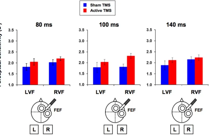
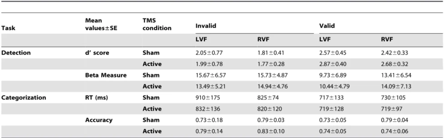
In a first experiment, single TMS pulses were delivered to the right FEF at different timings prior to the onset of a low-contrast near-threshold target which participants had to subsequently discriminate (line orientation discrimination) and consciously detect. We aimed to causally explore the chronometric contributions of this activity to visual performance. We hypothesized that pre-target FEF activity would be relevant for conscious perception and thus its modulation by means of single TMS pulses would yield perceptual performance modulations. In a second experiment, single TMS pulses were delivered after a peripheral predictive visuo-spatial cue engaging attention, likely to modulate FEF activity. An interaction of TMS effects with visuo-spatial attentional orienting, i.e. distinct effect in valid (cue signals target location) and invalid trials (cue does not signal target location) would constitute causal evidence of cue-induced differences in activation of FEF neuronal subpopulations coding for attended vs unattended locations.
AIM 2: Chapters III, IV and VI: We aimed to provide causal evidence of the role played by specific spatiotemporal activity patterns emerging from frontal and parietal areas to visual conscious detection and discrimination
In a scientific context characterized by growing interest in the oscillatory and synchrony basis of cognition, the specific contribution of oscillatory activity to different aspects of brain function is a topic of major significance and, often, the role for oscillatory phenomena in visual performance and consciousness remains to be explored causally.
In the experiments presented in these three chapters, we delivered 4-pulse TMS bursts at well-controlled interpulse intervals over two cortical regions, the FEF or the IPS, and measured their impact on the discrimination and conscious detection
30
of low-contrast near-threshold targets. The use of frequency-specific patterns vs. non-frequency-specific ones with equivalent amount of activity induced, i.e. same number of pulses in the same time window at the same stimulation intensity, but slightly differently distributed in time, allowed us to isolate the effects of frequency from those resulting from the activation itself. We hypothesized that the dynamics of frontoparietal activity is a key factor in its ability to contribute to visual performance.
In a first study (Chapter III), we explored the frequency-specific oscillatory basis of visual performance. We delivered real or sham frequency-specific TMS bursts at high-beta (30 Hz) and gamma (50 Hz) frequencies, as well as control non-frequency-specific patterns matched in duration and number of pulses, to the right FEF. We hypothesized that these two frequencies, known to reflect different types of attentional processes, would yield selective effects on visual performance that would not be observed by active non-frequency-specific matched patterns.
In a second study (Chapter IV), we explored potential hemisphere-specific basis of frontal perceptual performance modulations. Real or sham frequency-specific or non-frequency-frequency-specific TMS bursts were delivered to the left FEF. We hypothesized that the different involvement of left and right homotopic frontal regions in perceptually relevant processes such as attention and conscious access could yield differences either in the behavioral effects that they yield or in the patterns proving able to do so.
Finally, in a third study (Chapter VI), we assessed the role of high-beta and gamma parietal activity in perceptual performance modulations. Following the same design of the first study we assessed the effects of 30 and 50 Hz in the right IPS on visual performance. We hypothesized that, since the parietal cortex is more directly