HAL Id: hal-00023079
https://hal.archives-ouvertes.fr/hal-00023079
Submitted on 20 Apr 2006HAL is a multi-disciplinary open access archive for the deposit and dissemination of sci-entific research documents, whether they are pub-lished or not. The documents may come from teaching and research institutions in France or abroad, or from public or private research centers.
L’archive ouverte pluridisciplinaire HAL, est destinée au dépôt et à la diffusion de documents scientifiques de niveau recherche, publiés ou non, émanant des établissements d’enseignement et de recherche français ou étrangers, des laboratoires publics ou privés.
Autonomous regulation of free Ca2+ concentrations in
isolated plant cell nuclei: A mathematical analysis
Christian Brière, Tou Cheu Xiong, Christian Mazars, Raoul Ranjeva
To cite this version:
Christian Brière, Tou Cheu Xiong, Christian Mazars, Raoul Ranjeva. Autonomous regulation of free Ca2+ concentrations in isolated plant cell nuclei: A mathematical analysis. Cell Calcium, Elsevier, 2006, 39, pp.293-303. �10.1016/j.ceca.2005.11.005�. �hal-00023079�
AUTONOMOUS REGULATION OF FREE CA
2+CONCENTRATIONS
IN ISOLATED PLANT CELL NUCLEI: A MATHEMATICAL
ANALYSIS
Christian Brière, Tou Cheu Xiong, Christian Mazars, Raoul Ranjeva
UMR CNRS-UPS 5546, Pôle de Biotechnologie végétale, 31326 Castanet-Tolosan, France Running Title: Modelling nuclear calcium homeostasis
Corresponding author: Christian Brière, UMR CNRS-UPS 5546, Pôle de Biotechnologie végétale, BP42617 Auzeville, 27 chemin de Borde Rouge, 31326 Castanet-Tolosan, France; Tel : +33(0)5 62193590 ; fax : +33(0)5 62193502 ; E-Mail: briere@scsv.ups-tlse.fr
Keywords: calcium homeostasis, nucleus, nucleoplasm, plant cell, modelling
Abbreviations: ER, endoplasmic reticulum; NE, nuclear envelope; INM, inner nuclear
membrane; NPC, nuclear pore complex; [Ca2+]nuc , nucleoplasmic free calcium concentration;
[Ca2+]nuc_total , nucleoplasmic total calcium concentration; [Ca2+]store , nuclear store free
calcium concentration; [Ca2+]store_total , nuclear store total calcium concentration. Abstract
Experiments performed on nuclei isolated from animal or plant cells have provided evidence that the nucleus generates directly specific nucleoplasmic calcium transients in response to external stimuli. Recent data suggest that isolated plant nuclei might be considered as a closed system where the nuclear concentration of free calcium wouldbe regulated by reversible movements between the nucleoplasm and nuclear stores. We have addressed the relevance of this hypothesis by developing a mathematical approach to simulate
nucleoplasmic calcium dynamics generated under various pH and temperature conditions. Here, we show that the experimental results could be explained provided that calcium-channels as well as systems transporting calcium are present on the inner nuclear membrane. The putative channels would allow the entry of calcium into the nucleoplasm whereas the elusive transporting system(s) would contribute to replenish the nuclear stores. The simple proposed model is versatile enough to explain and predict autonomous changes in free calcium in the nucleoplasm of isolated plant nuclei.
1. Introduction
In both plants and animals, the calcium ion is fully recognized as a second messenger that regulates a wide variety of biological processes triggered by biotic and abiotic external stimuli [1]. Changes in free calcium concentration do not proceed in a stereotypical manner. Rather, fluctuations of free calcium have time-, space-, intensity-, and frequency-
characteristics which depend upon the nature and strength of the stimulus [2]. Concerning the spatial changes in free calcium, it is now established that calcium concentrations vary in cell organelles like chloroplasts [3], mitochondria [4] or nuclei [5-7]. Nuclei are separated from the other cell compartments by a double membrane system that is punctuated by nuclear pore complexes (NPC), which allow trafficking of molecules and ions between the nucleoplasm and the cytosol [8-10]. Indeed, the capacity of NPC to allow free diffusion of calcium from the cytosol to the nucleoplasm is largely accepted [11-13]. Thus, simple diffusion has been reported to explain fully the pattern of nuclear calcium upon stimulation in rat cardiac
myocytes [14]. However, a series of experimental evidence suggests that the situation is more complex [13] and other mechanisms may be involved in the generation of nuclear calcium signals, depending on the cell type and/or the stimulus. Firstly, kinetic measurements of intranuclear calcium gradients have shown that calcium permeates through NPC in a strictly controlled manner [15]. Secondly, nuclei contain specific calcium stores in the nuclear envelope and in the recently identified nucleoplasmic reticulum which forms a continuum with the endoplasmic reticulum [16].
Recent experimental evidence, in both animals and plants, show that nuclear and cytosolic calcium may be regulated independently. In nuclei of HepG2 cells, nuclear calcium signals may be generated before any variation in the cytosol [17]. In tobacco cells, a hyper-osmotic shock induces an increase in the calcium concentration in the cytosol but not in the nucleus [6]. Cryptogein, an oomycete elicitor, induces calcium transients in both the cytosol and the nucleus, but the nuclear calcium peaks 15 min after the cytosolic peak [7]. According to Meyer et al. [18] if calcium signal kinetics in the cytosol and the nucleoplasm differ from each other by at least 1 sec, then the nuclear envelope is a substantial barrier to Ca2+ fluxes. Furthermore, isolated nuclei from either myocyte [19] or tobacco cells [5, 20] are able to respond to external stimuli by generating nucleoplasmic calcium signals directly and thus independently of the cytosolic concentration. Xiong et al. [20] showed further that high concentrations of calcium in the external medium or treatment with EGTA do not induce any particular calcium variation in isolated tobacco nuclei, thus showing that the nuclei are impermeable to external calcium.
Different calcium channels or transporters have been shown to localize to the nuclear envelope. The presence of Ca2+-ATPases pumps on the outer membrane of the NE is well documented in animals [21-24] as well as in plants [25, 26]. InsP4- receptors that allow the InsP4-dependent entry of calcium into the NE calcium store have been located to the outer membrane of rat liver nuclei [24].
Systems mobilizing calcium from the NE and subsequently inducing calcium increases in the nucleoplasm have also been characterized. In animals, the inner membrane of the NE has been shown to contain InsP3-dependent calcium channels [12, 22, 24, 27] as well as
ryanodin-receptors [28]. Mechano-sensitive Ca2+-channels responsible for calcium elevation in the nucleus have been characterized by patch-clamping of nuclei isolated from MC3T3-E1 cells [19].
In plants, a Ca2+- and voltage-dependent cation channel has been characterized by
electrophysiology in nuclei isolated from beet [29], and a pharmacological approach suggests the existence of TRP-like channels in nuclei of tobacco cells [20].
Taken together, these data show that the nuclear envelope is able to store calcium and that the stored ion may be mobilized to increase the nucleoplasmic free calcium concentration.
In order to understand and to explain calcium dynamics in the nucleoplasm, it is essential to consider calcium exchanges between the different sub-nuclear compartments (nucleoplasm, nuclear envelope stores, calcium buffers) taken as a whole. In this context, development of mathematical models may be instrumental for testing different hypotheses in silico, by verifying their consistency with experimental data. Here, we present a simple model of nuclear calcium dynamics that may explain most characteristics of the calcium patterns in isolated plant cell nuclei.
Because most of the molecular components which are likely to be implicated in the
entry/mobilization of calcium remain to be identified in plants, the model is built on general assumptions based on known or well-accepted data (mostly in animal cells), and on more speculative hypotheses which emerged from the analysis of specific experimental data [20].
2. Material and methods
2.1. Calcium measurements
Nuclei from BY2 tobacco cells expressing apoaequorin in the nucleus were isolated as
described previously [20]. Coelentarazin (2.5 µM final concentration) was added to the nuclei suspension and aequorin was allowed to reconstitute for at least 2 hours. Aliquots of 100µl of nuclei suspension containing about 30 000 nuclei per tube were prepared and stored on ice in the dark before measurements. Luminescence was measured using a Berthold Sirius
luminometer and calcium concentrations calibrated as previously reported [30]. In a typical experiment, a 100µl nuclei sample was transferred into the luminometer at room temperature and luminescence recording was started immediately (t=0). At t=20s a mechanical stimulus was applied by injecting (injection duration = approx. 1s) two volumes of buffer (0.4M sucrose, 5mM Bis-Tris, 10mM NaCl, 5mM MgCl2, pH adjusted at the required value) in the
luminometer cuvette. At t=5 min 300µl of lysis buffer containing 2% Nonidet were injected to discharge total aequorin in the sample.
2.2. Model simulation
Differential equations were solved by a 4th order Runge-Kutta method using the modeling software package Berkeley-Madonna (University of California, Berkeley, USA).
3. Model description
3.1. Experimental background
The present mathematical model relies on experimental data from the work of Xiong et al. [20] who analyzed nucleoplasmic free calcium transients in isolated nuclei of tobacco BY2 cells, challenged with mechanical stimuli. These data have been completed by additional experiments reported in figure 1. The main conclusions are as follows:
a) Under acidic conditions, the resting value of the nucleoplasmic free calcium concentration [Ca2+]nuc is low (about 0.1µM) and a mechanical stimulus provokes a rapid (<1sec) increase
in the free calcium level which is followed by a slow decrease (>1min) back to the
background level. The intensity of the response is positively correlated to the acidity of the bathing medium and the nuclei responded to a series of stimuli without a refractory period or a loss in response intensity. At low pH the resting concentration is not dependent on
b) As shown in figure 1A, addition of 10 mM calcium in the incubation medium had no effect on the resting level of nucleoplasmic free calcium, confirming that isolated nuclei are not freely permeable to calcium. Upon mechanical stimulation, there was no difference in either the kinetic characteristics or the intensity of the calcium response between the control (no addition of calcium) and the assay (addition of calcium). Consequently, the presence of calcium in the external milieu did not increase the nuclear response, suggesting that no additional entry of calcium was elicited by the stimulus.
c) Under neutral or alkaline pH conditions, a mechanical stimulus has no effect on [Ca2+]nuc
whose resting level becomes sensitive to temperature (Fig. 1B). After thermal equilibration at 20°C, the resting level is higher (about 0.4µM) than under acidic conditions. A cold shock lowers rapidly [Ca2+]nuc to about 0.1µM. [Ca2+]nuc increases gradually to 0.4 µM upon thermal
equilibration.
From these data, we conclude that isolated nuclei from BY2 cells constitute a closed system (no calcium exchanges with the external medium). They respond to mechanical stimulation in a pH dependent manner and the resting levels of [Ca2+]nuc are regulated by temperature.
A rapid increase of the free calcium concentration in the nucleoplasm might be explained by the opening of Ca2+-channels located on the INM, inducing a calcium influx from nuclear stores (nuclear envelope, nucleoplasmic reticulum). The slow decreasing phase of the process, which took up to 3 min to return to the initial calcium level, cannot be explained in such a simple way. A possible explanation would be the binding of free calcium to calcium chelators (proteins or negatively charged compounds). However, interactions between calcium and buffers proceed with time constants in the millisecond range [31], that are not thermodynamically consistent with the experimental data unless the existence of very slow calcium buffers is postulated. Another possibility would be to suppose a release of calcium from the nucleoplasm to the external medium by free diffusion through the nuclear pore. This would suit reports suggesting that ions can pass freely through NPC channels. However, this hypothesis is contradicted by the data reported in figure 1A. Indeed, increasing the calcium concentration in the external medium to reverse the calcium gradient between the
nucleoplasm and the bathing medium has no effect on the nuclear responses. Thus,
experiments with isolated nuclei show that (1) control of calcium diffusion through nuclear pores must exist to maintain the resting nucleoplasmic concentration at its observed level (about 0.1µM), and (2) there must be a mechanism by which calcium ions are transported actively from the nucleoplasm to the nuclear store, in order to restore the resting level after stimulation.
In order to check the consistency of this hypothesis with experimental data, we developed a simple mathematical model to simulate the dynamics of calcium in isolated nuclei.
3.2. Model hypotheses
We will consider here the isolated nucleus as a closed system, where no calcium exchange with the environment takes place, as suggested by the experiments reported above. This system is thus composed of only two physical compartments, the nucleoplasm and the nuclear envelope. For the sake of simplicity, we will consider the NE and other putative nuclear stores as a single compartment. In each compartment, calcium is either in a free form or bound to calcium buffers. Following Neher [32] “calcium buffer” refers here to chemical species acting as calcium ligands with a rapid equilibrium between the free and bound forms of calcium. Free calcium can be mobilized from the NE to the nucleoplasm through calcium channels, some of them being activated by a mechanical stimulus. We will not consider here the mechanisms of channel activation, we just assume that a stimulus provokes a calcium influx. Therefore the model will be valid regardless of the (direct or indirect) activation
mechanism. Conversely, we suppose that calcium ions are actively transported from the nucleoplasm to the nuclear envelope by still hypothetical transporters.
3.3. Model formulation
Under these assumptions, the rates of change of the free Ca2+ concentration in the nucleoplasm [Ca2+]nuc and in the stores [Ca2+]store are represented by
(
Jin Jout dt d + = − β ] [Ca2 nuc)
(1a)(
Jin Jout dt d[Ca2+]store =−αρ −)
(1b) withJin = inward flux from the stores to the nucleoplasm,
Jout = outward flux from the nucleoplasm to the stores,
β is in first approximation equal to the ratio of free versus total calcium in the nucleoplasm [Ca2+]
nuc / [Ca]nuc_total,
α is in first approximation equal to the ratio of free versus total calcium in the store
[Ca2+]store / [Ca]store_total.
Because fluxes are expressed in term of nucleoplasmic concentration the rate of change of
[Ca2+]store is weighted by the volume ratio ρ= Volnucleoplasm / Volstore
For Ca2+ buffering, we considered fast reactions between calcium and buffers and used the
rapid equilibrium approximation [31] in order to simplify the model (see appendix). Calcium ratios depend on free calcium concentration, but as only small variations of [Ca2+] are
involved, in most simulations we assumed constant values for the free calcium ratios α and β. An extended version of the model, accounting for variable buffering capacities, is described in the appendix.
From (1) we can derive the conservation equation expressing that the total quantity of calcium is constant: Q = + + + γ β store 2 nuc 2 ] [Ca ] [Ca (2) where γ = αρ.
Formulations of the inward flux Jin and of the outward flux Jout used in the numerical
simulations were derived from various models of calcium exchanges between the
endoplasmic reticulum (ER) and the cytosol [33-36]. Jin is the sum of the effects of (1) a
calcium leak from nuclear stores to the nucleoplasm, and (2) of a mechanical stimulus which is supposed to open specific calcium channels. A calcium leak current has been observed between the ER and the cytosol in animal cells [37], and we assumed a similar leak between the nuclear store and the nucleoplasm. This leak is assumed to be proportional to the
difference of concentrations between the two compartments, such that:
(
2 2)
store nuc
[Ca ] [Ca ]
leak s
We assumed also that a mechanical stimulus induces a transient opening of Ca2+-channels,
resulting in a rapid and transient influx of calcium in the nucleoplasm from the nuclear stores:
(
nuc)
2 store 2 ] [Ca ] [Ca ) ( + − + =F t Jstimulus (4)The time-dependent function F(t) is related to the fractional activity of the channels [34, 36], and is modeled as : 2 1 1 1 ) ( t t t t t t s s e e F t F − − − − − = (5)
where ts is the time at which a stimulus is applied.
For calcium uptake from the nucleoplasm to the nuclear store we assumed a simple Hill function, such that:
p p p out K V J nuc 2 nuc 2 ] [Ca ] [Ca + + + = (6)
Such a function has been used often to model Ca2+ pumps [33, 34, 38] either in the plasma membrane or in the ER membrane.
Finally, the simplified model reduces to the conservation equation (2) and one differential equation
(
)
+ − − + = + + + + + p p p K V ks t F dt d nuc 2 nuc 2 nuc 2 store 2 nuc 2 ] [Ca ] [Ca ] [Ca ] [Ca ) ) ( ( ] [Ca β (7) 3.4. Steady-state analysisWhen F = 0, the system equilibrates to a steady-state, whose value is the solution of the following equation: p nuc p p nuc s nuc C K C k V C Q + = + −(1 ) β γ γ (8)
The equilibrium level is strongly dependent upon the ratio V/ks, which determines the
balance between inward flux and outward flux, and upon the free calcium ratio β. Its value reaches an upper limit Q
γ β
βγ
+ when V/ks →0, and for which Cnuc = Cstore .
An increase of the equilibrium level may thus be obtained either by increasing the influx or by decreasing the uptake. But the kinetics of convergence towards the equilibrium are quite different according to whether ks is high or V is low. In the case of a weak uptake, the
transient to the steady-state is slow. In contrast, in the case of a large basal influx, the system jumps immediately to the steady-state (Fig. 2A). If one simulates a stimulus, according to equation (5), in both cases the system evolves rapidly to the steady-state, and becomes essentially insensitive to a second stimulus (Fig. 2B).
3.5. Parameter values
The average nucleus size of BY2 cells was derived from microscopic measurements of
isolated nuclei labeled with DAPI (average width = 6.5±0.83 µm, average length = 7.5±1 µm, n=66). Considering an ellipsoid shape for the nucleus, the mean volume of a nucleus was estimated to be 160 µm3 and the surface area to 150 µm2. For a 50 nm thick nuclear envelope this gives a volume ratio ρ ≈ 20.
The total amount of calcium in a nucleus of plant cells has been estimated to be about 1nmol/mg protein [26]. In BY2 cell nuclei we assessed the quantity of proteins at 150 pg / nucleus. Thus, for a mean volume of 160 µm3 the total calcium concentration in the nucleus
is approximately 900 µM. This figure falls into the same order of magnitude as values reported for animal cell nuclei, namely, 225µM in nuclei of NIH 3T3 cells [39] and 600 µM in breast epithelial cells [40]. Free calcium concentration in the nucleoplasm of plant nuclei at rest has been assessed at 150nM [26]; here, we found approximately 100nM. These values lead to an estimation of the nucleoplasmic free calcium ratio to be in the range 1000 – 5000. By comparison, the calcium binding ratio in the cytosol was found to vary from 1500 to 2000 in pancreatic cells [41], and was assessed at 50 in bovine adrenal chromaffin cells [42]. Because of the NE-ER continuity, expected calcium concentrations in the nuclear envelope should be in the same range as in the reticulum lumen, namely 100-1000µM [free calcium] and 5-50mM [total calcium] [28, 43, 44]. This leads to a calcium binding ratio between 10 and 50 in the ER lumen, which is lower than the ratio in the cytosol [42, 45].
From these published data, we assumed resting concentrations of total and free calcium in the nucleoplasm of about 200µM and 100nM respectively (β = 1/2000). In the nuclear envelope
the total calcium concentration was set to 10mM and the free calcium concentration to 200 µM (α = 1/50), giving a total calcium concentration in the whole nucleus of approximately 400µM, a value close to the above mentioned estimate.
Kinetic parameter values used in mathematical models vary greatly according to the cell type and to the authors [33-36, 38, 41, 45, 46]. Table 1 summarizes the parameter values used in these various models. In our simulations, kinetic parameter values (table 2) were chosen such as to fit the model to experimental data.
4. Results
4.1. Simulation of changes in nucleoplasmic free calcium in isolated nuclei challenged with mechanical stimuli
In the following simulations, a low free calcium level ([Ca2+]nuc = 0.1 µM) was set as an
initial condition to mimic experiments in which nuclei were maintained at low temperature before being stimulated by addition of buffer at room temperature [20]. The effect of this mechanical stimulus was simulated by a rapid and transient influx of calcium from the store into the nucleoplasm (Fig3A, dotted line), according to equation (5). Figure 3A shows an example of the calcium response in isolated nuclei, after mechanical stimulation at acidic pH conditions (thin line), and the corresponding simulation (thick solid line). Nucleoplasmic concentration [Ca2+]
nuc increased rapidly up to a maximum and then returned slowly to the
initial equilibrium as expected. This variation of [Ca2+]nuc corresponds to a concomitant
variation of [Ca2+]store, but in the opposite direction. After a rapid decrease, due to the
outward flux from the store to the nucleoplasm, the calcium level in the store returns slowly to its initial level under the action of Ca2+-transporter(s) (Fig. 3B). In this simulation, the free
calcium level in the store decreased dramatically, from 160µM to about 10µM. Because of the large volume ratio between the two compartments and of the high buffering capacity, a weak increase in the nucleoplasmic free calcium required a large influx of calcium from the store. The response of the model to a stimulus is qualitatively and quantitatively in good agreement with experiments.
The model predicts that repeated stimuli will induce a stable variation of [Ca2+]nuc as
observed experimentally. After an increase due to the initial stimulation, subsequent stimulations do not modify the average value of [Ca2+]nuc . There is no accumulating or
exhausting effect of repeated stimuli (Fig. 3C). This results from the large decrease of the Ca2+-level in the store initiated by the first stimulus and by the effect of the re-uptake
mechanism which restores partially the calcium level of the store (Fig. 3D). Such a behavior was observed for parameter values which fit experimental data shown in Figure 3A. When parameters values are set such that either the store level does not decrease enough or the outward flux is too low, the average level of [Ca2+]nuc increases progressively after each
subsequent stimulus before reaching a plateau (result no shown).
At room temperature, the response of isolated nuclei to a mechanical stimulus depends upon the pH of the bathing buffer [20]. At acid pH, nuclei respond as described above, whereas at alkaline pH they are insensitive to stimulation. This is illustrated in Figure 4, showing the experimental responses of nuclei stored on ice before measurement, and subsequent effect of adding two volumes of buffer adjusted to acid or alkaline pH at room temperature. The stimulus induced a rapid increase of [Ca2+]nuc reaching a pH-dependent peak value, followed
by a slow transient towards equilibrium values positively correlated to the pH of the medium. Experimental plots were simulated using the same procedure as above (Fig. 4) and coefficient values given in table 2. A good adjustment of the various curves was obtained by changing only three coefficients between different pH conditions. These include the rate constant ks of
the resting inward flux, and the two free calcium ratios β and γ. Changing ks alone had an
effect on the equilibrium value only, while changing the values of β and γ modified also the peak value. Thus, the pH value of the bathing buffer would influence both the kinetics of the calcium leak and the calcium binding capacity of the nuclei.
When repeated stimuli were simulated (Fig. 4B), different responses were obtained according to the parameter values. For values simulating a neutral or alkaline pH, the model was
insensitive to stimulation, while for values corresponding to an acid pH, the amplitude of the response was pH dependent.
4.2. Simulation of temperature effects on the resting calcium level at alkaline pH
Xiong et al (23) have shown that, the calcium resting level is independent upon temperature variations at acid pH, and positively correlated with temperature in alkaline conditions. When nuclei, stored at 0°C in an alkaline buffer (pH 7.5) before measurement, are transferred to room temperature, there is a progressive increase of the nucleoplasmic calcium concentration with temperature, reaching a plateau after about 5 min.
Temperature variations may affect the kinetics of calcium release and/or calcium uptake, or the kinetics of binding to calcium binding proteins. In the model (eq. 7) these kinetics are controlled essentially by the coefficients ks, V, and K and by the buffering capacity,
represented by β. For example, according to eq. (8), an increase of ks or a decrease of V will
induce an increase of [Ca2+]nuc. As shown in Fig. 2A, a high value of ks induces a rapid
increase of [Ca2+]nuc to the steady-state, while a low value of V induces only a slow increase
of [Ca2+]nuc towards the steady-state. [Ca2+]nuc may also increase if the affinity of calcium to
In order to simulate the effect of temperature variations on calcium dynamics we used the extended version of the model that is described in the appendix. The extended model accounts for time-dependent buffering coefficients.
Variations in temperature were simulated by time-variations of ks, V, K and/or the
calcium-buffer dissociation coefficient Kd (Fig5a-e) and the simulated response compared to
experimental curves (Fig. 5f). To simulate a cold shock, following a slow increase in temperature, coefficients were reversed rapidly to their initial value. The responses of the model to variations of V or K, which control calcium re-uptake, are not in good qualitative agreement to the experimental data (Fig. 5a and 5c). The measured increase of [Ca2+]
nuc with
temperature is simulated adequately by a progressive increase of the influx rate constant ks
(Fig. 5a), but the effect of a cold shock is better predicted by a rapid decrease of Kd (Fig. 5d).
The best fit to experimental measurements was obtained by simultaneously increasing ks and
the dissociation constant Kd (Fig. 5e).
Thus, according to these simulations, temperature might have a double effect: i) on the activity of Ca2+-channels, by changing membrane dynamics, and ii) on the calcium-buffering capacity, by modifying the calcium-buffer dissociation constant.
5. Discussion
Active trafficking of information between the nucleus and the cytosol in intact cells has been largely established [15, 47, 48]. Moreover, it has become apparent that nuclei isolated either from plant cells [20] or from human cells [19] are able to convert mechanical stimuli into changes in the free calcium concentration, which then controls downstream
calcium-dependent events. In nuclei isolated from tobacco cells, the calcium response depends highly on (i) the pH and (ii) the temperature of the incubation medium. Thus, at acidic pH, a train of mechanical stimuli induces a periodic stable variation of [Ca2+]nuc even at low temperature. In
contrast, at neutral-alkaline pH, the system is apparently desensitized to mechanical
stimulation. Rather, under these conditions, the concentration of nucleoplasmic free calcium depends on the temperature of the incubation medium which determines the steady state concentration level of calcium at equilibrium [20].
We have simulated these data by a simple mathematical model to describe calcium homeostasis and disturbance in isolated plant cell nuclei. This model is based upon the
assumption that nucleoplasmic calcium is regulated by the balance between Ca2+-channel and
Ca2+-transporter activities, both located on the inner nuclear membrane of the nuclear envelope.
Isolated nuclei from tobacco BY2 cells behave like a closed system, the nucleoplasmic calcium variations being independent from the external calcium concentration. Therefore, calcium diffusion through nuclear pores as a possible mechanism of regulation can be ruled out. A similar behavior has already been observed in isolated nuclei from pancreatic beta cells in which blockade of a nuclear KATP channel triggers nuclear Ca2+ transients [49].
Calcium binding to proteins or to negatively charged compounds could be a way of reducing free calcium level after stimulation. However, the kinetics of calcium buffering is very fast, with a time constant in the millisecond range [31] which is not compatible with observed kinetics, with a characteristic time higher than 1 min. Thus, to be consistent with kinetic data, the existence of a very slow calcium buffer in the nucleus should be assumed. Another argument against the “buffering” hypothesis comes from experiments with a train of mechanical stimuli. Essentially, we have observed that the maximal calcium concentration reached in the nucleoplasm after a stimulus remains constant whatever the frequency of the stimuli. A few minutes after stimulation, the system seems to be restored to its resting state and able to respond to a new stimulus. Such behavior cannot be explained only by calcium
buffering in the nucleoplasm, because in such a case the calcium stores would be rapidly depleted. For example, it has been shown that Ca2+-concentration in the ER decreased by a
factor of 10 after Ca2+ mobilization [50]. In the nucleus, because of the high ratio (> 1000) between total and free calcium in the nucleoplasm and the low volume ratio (< 0.05) between nuclear envelope and nucleoplasm, a small increase (e.g. 300nM) of [Ca2+] in the
nucleoplasm must correspond to a large decrease (> 6mM) of [Ca2+] in the nuclear envelope. Considering that the total calcium concentration in the nuclear envelope is in the 5-50mM range [28, 43, 44], such a calcium release after each stimulus should empty rapidly the calcium stores, leading to a decrease of the nucleoplasmic response to subsequent stimuli. This is not consistent with experimental data, which showed a sustained response after successive stimuli [20].
In the “calcium re-uptake” hypothesis, the nuclear calcium stores are continuously refilled, making it possible to respond to another stimulus. Thus, the present model, based on such a hypothesis, formally explains the response of isolated nuclei to repeated stimuli. Furthermore, calcium transporters like sarco/endoplasmic reticulum calcium ATPases have time constants in the minute range [51] that are in the time constant range of the calcium decrease measured in the nucleoplasm of isolated nuclei.
Variations of resting [Ca2+]nuc with temperature or pH result from modifications of the
balance between inward and outward calcium fluxes, i.e. by a modulation of Ca2+-channel or/and Ca2+-transporter activities. Numerical simulations have shown that temperature effects on calcium kinetics are better described by an increase of the Ca2+-channel kinetic constant with temperature rather than by a decrease in transport activity. An active transport activity that would be lower at room temperature than at low temperature seems very unlikely. Thus, ATPase activity increases with temperature [52] whereas Ca2+-permeable channels are primary sensors of temperature in plants [53]. Plant cells respond to a cold shock by a rapid increase of [Ca2+]cyt but do not respond to a slow variation of temperature [54]. In
mammalian cells, primary sensors of temperature are Ca2+-channels of the TRP family [55] which can be activated either by cold (TRPM8) or by heat (TRPV1). Xiong et al [20] suggested the presence of TRP-like Ca2+-channels on the inner membrane of the nuclear envelope. These channels would be activated by a slow increase rather than by a rapid variation of temperature. This hypothesis fits with the proposed model which simulates the effect of temperature increase by a progressive activation of Ca2+-channels.
The numerical simulations suggest that the pH-dependence of the nuclear calcium dynamics may be due mainly to simultaneous changes of the Ca2+-binding capacity and the modulation of Ca2+-channel activity, rather than to Ca2+ re-uptake. Proton concentration is known to have a profound influence on a variety of biological processes and especially on ion channel, transporter activities, and the calcium-binding capacity of calcium-binding proteins.
Acidification of the extracellular medium reduces the conductivity of many voltage-gated and ligand-gated channels [56-59]. This is the case particularly for L-type calcium channels [56, 60] and sodium channels [61]. Conversely, it has been shown that acidification activates a particular class of channels referred to as Acid Sensitive Ion Channels (ASIC) [62] and stimulation of ASIC1a provides a pathway for calcium entry in neuronal cells [63].
The experimental data used to propose the model presented here show clearly that acidic pH values inhibit changes in nucleoplasmic calcium, this suggesting that ASICs are not key players in the process. Moreover, the pharmacological profile of the putative channels is more compatible with the channels being TRP-like rather than L-type channels. The putative channels become highly sensitive to activation by mechanical stimulations at acidic pH and not at alkaline pH. Changes in ionic charges of the channels may be the mechanism that controls their sensitivity to either mechanical or thermal stimulation. Clearly,
the molecular nature of the channels and the mechanism of their activation remain to be clarified.
In isolated nuclei, the resting calcium concentration [Ca2+]nuc is temperature independent at
low pH (5.3). At low temperature (approx. 0°C), [Ca2+]nuc becomes pH independent.
Consequently, if temperature controls channel opening and pH controls uptake activity, the two processes must be coupled. The simple way to achieve such coupling is that both factors regulate the same process.
Acknowledgements: The authors thank Dr J.V. Cullimore, (INRA Castanet-Tolosan, France)
for helpful discussion and for editing the English version of the manuscript.
References
1. Hetherington AM, Brownlee C. (2004) The generation of Ca2+ signals in plants. Annu Rev Plant Biol, 55, 401-427.
2. Berridge MJ. (1997) The AM and FM of calcium signalling. Nature, 386, 759-60. 3. Johnson CH, Knight MR, Kondo T, Masson P, Sedbrook J, Haley A, Trewavas A.
(1995) Circadian oscillations of cytosolic and chloroplastic free calcium in plants. Science, 269, 1863-5.
4. Logan DC, Knight MR. (2003) Mitochondrial and cytosolic calcium dynamics are differentially regulated in plants. Plant Physiol, 133, 21-4.
5. Pauly N, Knight MR, Thuleau P, Van der Luit AH, Moreau M, Trewavas AJ, Ranjeva R, Mazars C. (2000) Control of free calcium in plant cell nuclei. Nature, 405, 754-755.
6. Pauly N, Knight MR, Thuleau P, Graziana A, Muto S, Ranjeva R, Mazars C. (2001) The nucleus together with the cytosol generates patterns of specific cellular calcium signatures in tobacco suspension culture cells. Cell Calcium, 30, 413-21.
7. Lecourieux D, Lamotte O, Bourque S, Vendehenne D, Mazars C, Ranjeva R, Pugin A. (2005) Proteinaceous and oligosaccharidic elicitors induce different calcium signatures in the nucleus of tobacco cells. Cell Calcium, (in press).
8. Goldberg MW, Allen TD. (1995) Structural and functional organization of the nuclear envelope. Curr Opin Cell Biol, 7, 301-9.
9. Mazzanti M, Bustamante JO, Oberleithner H. (2001) Electrical dimension of the nuclear envelope. Physiol Rev, 81, 1-19.
10. Brandizzi F, Irons SL, Evans DE. (2004) The plant nuclear envelope: new prospects for a poorly understood structure. New Phytologist, 163, 227-246.
11. Brini M, Murgia M, Pasti L, Picard D, Pozzan T, Rizzuto R. (1993) Nuclear Ca2+ concentration measured with specifically targeted recombinant aequorin. Embo J, 12, 4813-9.
12. Gerasimenko OV, Gerasimenko JV, Tepikin AV, Petersen OH. (1995)
ATP-dependent accumulation and inositol trisphosphate- or cyclic ADP-ribose-mediated release of Ca2+ from the nuclear envelope. Cell, 80, 439-44.
13. Bootman MD, Thomas D, Tovey SC, Berridge MJ, Lipp P. (2000) Nuclear calcium signalling. Cell Mol Life Sci, 57, 371-8.
14. Genka C, Ishida H, Ichimori K, Hirota Y, Tanaami T, Nakazawa H. (1999)
rat cardiac myocytes with an ultra-fast confocal imaging system. Cell Calcium, 25, 199-208.
15. Santella L, Carafoli E. (1997) Calcium signaling in the cell nucleus. Faseb J, 11, 1091-1109.
16. Echevarria W, Leite MF, Guerra MT, Zipfel WR, Nathanson MH. (2003) Regulation of calcium signals in the nucleus by a nucleoplasmic reticulum. Nat Cell Biol, 5, 440-6.
17. Leite MF, Thrower EC, Echevarria W, Koulen P, Hirata K, Bennett AM, Ehrlich BE, Nathanson MH. (2003) Nuclear and cytosolic calcium are regulated independently. Proc Natl Acad Sci U S A, 100, 2975-80.
18. Meyer T, Allbritton NL, Oancea E. (1995) Regulation of nuclear calcium concentration. Ciba Found Symp, 188, 252-62; discussion 262-6.
19. Itano N, Okamoto S, Zhang D, Lipton SA, Ruoslahti E. (2003) Cell spreading controls endoplasmic and nuclear calcium: a physical gene regulation pathway from the cell surface to the nucleus. Proc Natl Acad Sci U S A, 100, 5181-6.
20. Xiong TC, Jauneau A, Ranjeva R, Mazars C. (2004) Isolated plant nuclei as mechanical and thermal sensors involved in calcium signalling. Plant J, 40, 12-21. 21. Nicotera P, McConkey DJ, Jones DP, Orrenius S. (1989) ATP stimulates Ca2+ uptake
and increases the free Ca2+ concentration in isolated rat liver nuclei. Proc Natl Acad Sci U S A, 86, 453-7.
22. Gerasimenko OV, Gerasimenko JV, Tepikin AV, Petersen OH. (1996) Calcium transport pathways in the nucleus. Pflugers Arch, 432, 1-6.
23. Abrenica B, Gilchrist JS. (2000) Nucleoplasmic Ca(2+)loading is regulated by mobilization of perinuclear Ca(2+). Cell Calcium, 28, 127-36.
24. Humbert JP, Matter N, Artault JC, Koppler P, Malviya AN. (1996) Inositol 1,4,5-trisphosphate receptor is located to the inner nuclear membrane vindicating regulation of nuclear calcium signaling by inositol 1,4,5-trisphosphate. Discrete distribution of inositol phosphate receptors to inner and outer nuclear membranes. J Biol Chem, 271, 478-85.
25. Downie L, Priddle J, Hawes C, Evans DE. (1998) A calcium pump at the higher plant nuclear envelope? FEBS Lett, 429, 44-8.
26. Bunney TD, Shaw PJ, Watkins PA, Taylor JP, Beven AF, Wells B, Calder GM, Drobak BK. (2000) ATP-dependent regulation of nuclear Ca(2+) levels in plant cells. FEBS Lett, 476, 145-9.
27. Stehno-Bittel L, Luckhoff A, Clapham DE. (1995) Calcium release from the nucleus by InsP3 receptor channels. Neuron, 14, 163-7.
28. Gerasimenko JV, Maruyama Y, Yano K, Dolman NJ, Tepikin AV, Petersen OH, Gerasimenko OV. (2003) NAADP mobilizes Ca2+ from a thapsigargin-sensitive store in the nuclear envelope by activating ryanodine receptors. J Cell Biol, 163, 271-82. 29. Grygorczyk C, Grygorczyk R. (1998) A Ca2+- and voltage-dependent cation channel
in the nuclear envelope of red beet. Biochim Biophys Acta, 1375, 117-30.
30. Van Der Luit AH, Olivari C, Haley A, Knight MR, Trewavas AJ. (1999) Distinct calcium signaling pathways regulate calmodulin gene expression in tobacco. Plant Physiol, 121, 705-14.
31. Wagner J, Keizer J. (1994) Effects of rapid buffers on Ca2+ diffusion and Ca2+ oscillations. Biophys J, 67, 447-56.
32. Neher E. (1998) Usefulness and limitations of linear approximations to the understanding of Ca++ signals. Cell Calcium, 24, 345-357.
33. Kargacin GJ. (2003) Responses of Ca2+-binding proteins to localized, transient changes in intracellular [Ca2+]. J Theor Biol, 221, 245-58.
34. Korngreen A, Goldshtein V, Priel Z. (1997) A realistic model of biphasic calcium transients in electrically nonexcitable cells. Biophysical Journal, 73, 659-673. 35. Sneyd J, Tsaneva-Atanasova K, Bruce JI, Straub SV, Giovannucci DR, Yule DI.
(2003) A model of calcium waves in pancreatic and parotid acinar cells. Biophys J, 85, 1392-405.
36. Sneyd J, Tsaneva-Atanasova K, Yule DI, Thompson JL, Shuttleworth TJ. (2004) Control of calcium oscillations by membrane fluxes. Proc Natl Acad Sci U S A. 37. Camello C, Lomax R, Petersen OH, Tepikin AV. (2002) Calcium leak from
intracellular stores--the enigma of calcium signalling. Cell Calcium, 32, 355-61. 38. Jafri MS, Keizer J. (1997) Agonist-induced calcium waves in oscillatory cells: A
biological example of Burgers' equation. Bulletin of Mathematical Biology, 59, 1125-1144.
39. Chandra S, Gross D, Ling Y-C, Morrison GH. (1989) Quantitative imaging of free and total intracellular calcium in cultured cells. Proc. Natl. Acad. Sci. USA, 86, 1870-1874.
40. Chandra S, Lorey DR, 2nd. (2001) SIMS ion microscopy in cancer research: single cell isotopic imaging for chemical composition, cytotoxicity and cell cycle
recognition. Cell Mol Biol (Noisy-le-grand), 47, 503-18.
41. Mogami H, Tepikin AV, Petersen OH. (1998) Termination of cytosolic Ca2+ signals: Ca2+ reuptake into intracellular stores is regulated by the free Ca2+ concentration in the store lumen. Embo J, 17, 435-42.
42. Zhou Z, Neher E. (1993) Mobile and immobile calcium buffers in bovine adrenal chromaffin cells. J Physiol, 469, 245-73.
43. Meldolesi J, Pozzan T. (1998) The endoplasmic reticulum Ca2+ store: a view from the lumen. Trends Biochem Sci, 23, 10-4.
44. Meldolesi J, Pozzan T. (1998) The heterogeneity of ER Ca2+ stores has a key role in nonmuscle cell signaling and function. J Cell Biol, 142, 1395-8.
45. Mogami H, Gardner J, Gerasimenko OV, Camello P, Petersen OH, Tepikin AV. (1999) Calcium binding capacity of the cytosol and endoplasmic reticulum of mouse pancreatic acinar cells. J Physiol, 518 ( Pt 2), 463-7.
46. Jafri MS. (1995) A theoretical study of cytosolic calcium waves in Xenopus oocytes. J. theor. Biol., 172, 209-216.
47. Gerasimenko J, Maruyama Y, Tepikin A, Petersen OH, Gerasimenko O. (2003) Calcium signalling in and around the nuclear envelope. Biochem Soc Trans, 31, 76-8. 48. Greber UF, Carafoli E. (2002) Signalling takes control of nucleo-cytoplasmic
trafficking. Workshop on signal-regulated nuclear transport. EMBO Rep, 3, 410-4. 49. Quesada I, Rovira JM, Martin F, Roche E, Nadal A, Soria B. (2002) Nuclear KATP
channels trigger nuclear Ca(2+) transients that modulate nuclear function. Proc Natl Acad Sci U S A, 99, 9544-9.
50. Miyawaki A, Llopis J, Heim R, McCaffery JM, Adams JA, Ikura M, Tsien RY. (1997) Fluorescent indicators for Ca2+ based on green fluorescent proteins and calmodulin. Nature, 388, 882-7.
51. Zima AV, Copello JA, Blatter LA. (2004) Effects of cytosolic NADH/NAD(+) levels on sarcoplasmic reticulum Ca(2+) release in permeabilized rat ventricular myocytes. J Physiol, 555, 727-41.
52. Caldwell CR, Haug A. (1981) Temperature dependence of the barley root plasma membrane-bound Ca2+- and Mg2+-dependent ATPase. Physiol. Plant., 53, 117-124. 53. Plieth C. (1999) Temperature sensing by plants: calcium-permeable channels as
54. Plieth C, Hansen UP, Knight H, Knight MR. (1999) Temperature sensing by plants: the primary characteristics of signal perception and calcium response. Plant J, 18, 491-7.
55. Voets T, Droogmans G, Wissenbach U, Janssens A, Flockerzi V, Nilius B. (2004) The principle of temperature-dependent gating in cold- and heat-sensitive TRP channels. Nature, 430, 748-54.
56. Klöckner U, Isenberg G. (1994) Calcium channel current of vascular smooth muscle cells: extracellular protons modulate gating and single channel conductance. J Gen Physiol, 103, 665-78.
57. Horvath F, Wodala B, Erdei L, Moroni A, Van Etten J, Thiel G. (2002) pH-dependent regulation of a potassium channel protein encoded by a Chlorella virus PBCV-1. In: Proc. 7th Hungarian Congress Plant Physiol. Vol. 46, pp 21-22.
58. Hordejuk R, Lobanov NA, Kicinska A, Szewczyk A, Dolowy K. (2004) pH modulation of large conductance potassium channel from adrenal chromaffin granules. Mol Membr Biol, 21, 307-13.
59. Liu D, Zhang Z, Liman ER. (2005) Extracellular Acid Block and Acid-enhanced Inactivation of the Ca2+-activated Cation Channel TRPM5 Involve Residues in the S3-S4 and S5-S6 Extracellular Domains. J. Biol. Chem., 280, 20691-20699.
60. Smirnov SV, Knock GA, Belevych AE, Aaronson PI. (2000) Mechanism of effect of extracellular pH on L-type Ca(2+) channel currents in human mesenteric arterial cells. Am J Physiol Heart Circ Physiol, 279, H76-85.
61. Veresov VG. (1999) A theoretical analysis of pH dependence of sodium channel conductance. Membr Cell Biol, 13, 95-110.
62. Krishtal O. (2003) The ASICs: Signaling molecules? Modulators? Trends in Neurosciences, 26, 477-483.
63. Yermolaieva O, Leonard AS, Schnizler MK, Abboud FM, Welsh MJ. (2004)
Extracellular acidosis increases neuronal cell calcium by activating acid-sensing ion channel 1a. Proc Natl Acad Sci U S A, 101, 6752-7.
64. Wagner J, Li Y-X, Pearson J, Keizer J. (1998) Simulation of the fertilization Ca2+ wave in Xenopus laevis eggs. Biophysical J., 75, 2088-2097.
Tables and figure legends
Table 1: Kinetic parameter values taken from the literature Ref. Ca buffer (µM) k1 (µM-1s-1) k2 (s-1) Ca2+ ratio Vp (µM.s-1) Kp (µM) [46] 120 5 1 [38] 300 0.02 45 0.1 [34] 300 601 97 500 0.1 [64] 0.053 11.2 0.4 [33] 195 10 10 350 0.219
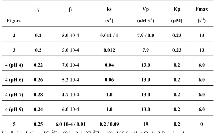
Table 2. Kinetic parameter values used in the simulations. Figure γ β ks (s-1) Vp (µM s-1) Kp (µM) Fmax (s-1) 2 0.2 5.0 10-4 0.012 / 1 7.9 / 0.0 0.23 13 3 0.2 5.0 10-4 0.012 7.9 0.23 13 4 (pH 4) 0.22 7.0 10-4 0.04 13.0 0.2 6.0 4 (pH 6) 0.26 5.2 10-4 0.06 13.0 0.2 6.0 4 (pH 7) 0.28 4.7 10-4 1.0 13.0 0.2 6.0 4 (pH 9) 0.24 6.0 10-4 1.0 13.0 0.2 6.0 5 0.25 6.0 10-4 / 0.01 0.2 / 0.09 19 0.2 0 In all simulations: [Ca2+]nuc(0 )= 0.1, [Ca2+]store(0)=160 (so that Q=1mM) and p=1.
Fig. 1. (A) Nucleoplasmic free calcium ([Ca2+]nuc) variations in isolated nuclei at rest (U and %) or after mechanical stimulation () and #) applied at t=20 sec (arrow). Nuclei were
incubated at room temperature in a buffered medium (pH 5.3) containing no (% and #) or 10
mM (U and )) calcium. (B) pH-dependence of nucleoplasmic free calcium concentration.
Nuclei were stored on ice before measurement in a buffer adjusted to pH 5.3, 6.5 or 7.5, and thereafter transferred at room temperature in a luminometer for recording of light emission. Fig. 2. (A) Simulations of calcium transients from the initial state to the steady-state value of model eq. (7), (B) Effect of repeated stimuli simulated by eq. (4) on the simulated calcium transient. Parameter values: ( ) ks=1 and V=8 (large basal influx), and ( - - - ) ks=0.012
and V=0 (absence of calcium re-uptake). γ = 0.2, β = 0.0005, Kp = 0.228294, p = 2, Fmax = 12.9521, initial conditions at t=0: [Ca2+]nuc = 0.1 µM, [Ca2+]store = 160 µM.
Fig. 3. Simulations of nucleoplasmic free calcium ([Ca2+]nuc) variation by eq. (7) after single
(A and B) or repeated (C and D) mechanical stimulation (eq. 5). Thin line (A, C) = observed data for [Ca2+]nuc, thick line (A-D) = [Ca2+]nuc solution of eq. 7, dashed line (B, D) =
[Ca2+]store , dotted line (A, C) = influx of Ca2+ after a stimulus simulated by eq. 4. Parameter
values used in simulations (A,B) : γ = 0.2, β = 0.0005, ks = 0.012, V = 8, Kp = 0.23, p = 2, Fmax = 13, (C,D) γ = 0.2, β = 0.00054, ks = 0.025, V = 13.3, Kp = 0.2, p = 2, initial conditions at t=0: [Ca2+]nuc = 0.1 µM, [Ca2+]store = 160 µM.
Fig. 4. Calcium variations induced by mechanical stimulation at different pH values. (A) Adjustment of the model (thick line) to experimental data (thin line). Stimulation was simulated according to eq. (7). Parameter values as in table 2. (B) Simulation of the response to repeated stimuli (same parameter values as in (A)).
Fig. 5. Effect of temperature variations on the nucleoplasmic calcium level. Temperature effect was simulated by time-dependent variations of single parameters: ks (A), V (B), K (C),
Kd (D), or both parameters ks and Kd (E). Solid line = calcium level, dotted line = parameter
value. (F) Experimental data showing the effect of temperature on the nucleoplasmic calcium level of nuclei in a basic buffer (pH 7.5). T was allowed to increase from 0 to 20°C, before applying a cold shock at t=200 s (arrow).
Appendix
A full model for nuclear calcium homeostasis reads:
nuc out in nuc J J Jb dt Ca d[ 2+] = − − (A1) nuc nuc Jb dt CaB d = ] [ (A2) store out in store J J Jb dt Ca d + =− − − ) ( ] [ 2 ρ (A3) store store Jb dt CaB d = ] [ (A4) with (A5) nuc nuc nuc nuc nuc nuc
nuc k Ca B CaB k CaB
Jb = 1 [ 2+] ([ ] −[ ] )− 2 [ ] (A6) store store store store store store
store k Ca B CaB k CaB
Jb = 1 [ 2+] ([ ] −[ ] )− 2 [ ]
where [Ca2+]nuc and [B]nuc are free calcium and total buffer concentrations in the nucleosol,
[Ca2+]store and [B]store are free calcium and total buffer concentrations in the nuclear stores,
and ρ is the volume ratio between the nucleosol and the nuclear stores. Summing (A1)+(A2), and (A3)+(A4) implies :
out in total nuc J J dt Ca d − = _ ] [ and [ ] _ ( ) out in total store J J dt Ca d − − = ρ (A7)
Assuming fast reaction kinetics for calcium buffering, the following equilibrium equation holds: nuc nuc nuc nuc nuc K Ca B Ca CaB + = ++ ] [ ] [ ] [ 22 (A8)
where Knuc is the dissociation constant. It follows:
nuc nuc nuc nuc nuc nuc total nuc Ca K Ca B CaB Ca Ca )[ ] ] [ 1 ( ] [ ] [ ] [ 2 2 2 _ + + + + + = + = (A9)
(
2)
2 1 2 2 2 2 2 2 ) ] [ 1 ( where ) ] ([ ] [ ] [ ] [ ] [ − + + + + + + + × + = + + + − − = nuc nuc nuc nuc nuc nuc nuc nuc nuc nuc nuc nuc nuc out in nuc K Ca B K dt dK K Ca Ca B dt dB K Ca Ca J J dt Ca d β β (A10)In the case where the buffering coefficients Bnuc and Knuc are constant, (A10) reduces to:
(
2)
2 1 2 ) ] [ 1 ( where ) ( ] [ − + + + × + = − = nuc nuc nuc nuc out in nuc K Ca B K J J dt Ca d β β (A11)A similar equation can be derived for the nuclear stores:
(
2)
2 1 2 ) ] [ 1 ( where ) ( ] [ − + + + × + = − − = store store store store out in store K Ca B K J J dt Ca d α αρ (A12)For low affinity buffers and small variations of [Ca2+], the two parameters α and β can be considered as approximately constant, with:
nuc nuc nuc store store store B K K B K K + ≅ + ≅ β α and (A13)
![Table 1: Kinetic parameter values taken from the literature Ref. Ca buffer (µM) k1 (µM-1 s -1 ) k2 (s-1 ) Ca 2+ ratio Vp (µM.s -1 ) Kp (µM) [46] 120 5 1 [38] 300 0.02 45 0.1 [34] 300 601 97 500 0.1 [64] 0.053 11.2 0.4](https://thumb-eu.123doks.com/thumbv2/123doknet/14412238.512017/16.892.103.716.163.485/table-kinetic-parameter-values-taken-literature-buffer-ratio.webp)
![Figure 4 00.10.20.30.40.50.60.7 0 30 60 90 120 150 180 t (s)[Ca2+]nuc (µM) pH 9pH 7pH 6 pH 4A 00.10.20.30.40.50.60.7 0 30 60 90 120 150 180 t (s)[Ca2+]nuc (µM) pH 9pH 7pH 4pH 6B](https://thumb-eu.123doks.com/thumbv2/123doknet/14412238.512017/24.892.295.606.237.648/figure-ca-nuc-µm-ph-ca-nuc-µm.webp)
