HAL Id: hal-03171012
https://hal.sorbonne-universite.fr/hal-03171012
Submitted on 16 Mar 2021
HAL is a multi-disciplinary open access
archive for the deposit and dissemination of
sci-entific research documents, whether they are
pub-lished or not. The documents may come from
teaching and research institutions in France or
abroad, or from public or private research centers.
L’archive ouverte pluridisciplinaire HAL, est
destinée au dépôt et à la diffusion de documents
scientifiques de niveau recherche, publiés ou non,
émanant des établissements d’enseignement et de
recherche français ou étrangers, des laboratoires
publics ou privés.
Association of Clinical, Biological, and Brain Magnetic
Resonance Imaging Findings With
Electroencephalographic Findings for Patients With
COVID-19
Virginie Lambrecq, Aurélie Hanin, Esteban Munoz-Musat, Lydia Chougar,
Salimata Gassama, Cecile Delorme, Louis Cousyn, Alaina Borden, Maria
Damiano, Valerio Frazzini, et al.
To cite this version:
Virginie Lambrecq, Aurélie Hanin, Esteban Munoz-Musat, Lydia Chougar, Salimata Gassama, et al..
Association of Clinical, Biological, and Brain Magnetic Resonance Imaging Findings With
Electroen-cephalographic Findings for Patients With COVID-19. JAMA Network Open, American Medical
Association, 2021, 4 (3), pp.e211489. �10.1001/jamanetworkopen.2021.1489�. �hal-03171012�
Association of Clinical, Biological, and Brain Magnetic Resonance Imaging Findings
With Electroencephalographic Findings for Patients With COVID-19
Virginie Lambrecq, MD, PhD; Aurélie Hanin, PharmD; Esteban Munoz-Musat, MD; Lydia Chougar, MD; Salimata Gassama, MD; Cécile Delorme, MD; Louis Cousyn, MD; Alaina Borden, MD; Maria Damiano, MD, PhD; Valerio Frazzini, MD, PhD; Gilles Huberfeld, MD, PhD; Frank Landgraf, MD; Vi-Huong Nguyen-Michel, MD; Phintip Pichit, MD; Aude Sangare, MD; Mario Chavez, PhD; Capucine Morélot-Panzini, MD, PhD; Elise Morawiec, MD; Mathieu Raux, MD, PhD; Charles-Edouard Luyt, MD, PhD;
Pierre Rufat, MD; Damien Galanaud, MD, PhD; Jean-Christophe Corvol, MD, PhD; Catherine Lubetzki, MD, PhD; Benjamin Rohaut, MD, PhD; Sophie Demeret, MD; Nadya Pyatigorskaya, MD, PhD; Lionel Naccache, MD, PhD; Vincent Navarro, MD, PhD; for the Cohort COVID-19 Neurosciences (CoCo Neurosciences) Study Group
Abstract
IMPORTANCE There is evidence of central nervous system impairments associated with
coronavirus disease 2019 (COVID-19) infection, including encephalopathy. Multimodal monitoring of patients with COVID-19 may delineate the specific features of COVID-19–related encephalopathy and guide clinical management.
OBJECTIVES To investigate clinical, biological, and brain magnetic resonance imaging (MRI) findings in association with electroencephalographic (EEG) features for patients with COVID-19, and to better refine the features of COVID-19–related encephalopathy.
DESIGN, SETTING, AND PARTICIPANTS This retrospective cohort study conducted in Pitié-Salpêtrière Hospital, Paris, France, enrolled 78 hospitalized adults who received a diagnosis of severe acute respiratory syndrome coronavirus 2 (SARS-Cov2) and underwent EEG between March 30 and June 11, 2020.
EXPOSURES Detection of SARS-CoV-2 from a nasopharyngeal specimen using a reverse transcription–polymerase chain reaction assay or, in the case of associated pneumonia, on a computed tomography scan of the chest.
MAIN OUTCOMES AND MEASURES Data on the clinical and paraclinical features of the 78 patients with COVID-19 were retrieved from electronic patient records.
RESULTS Of 644 patients who were hospitalized for COVID-19, 78 (57 men [73%]; mean [SD] age, 61 [12] years) underwent EEG. The main indications for EEG were delirium, seizure-like events, and delayed awakening in the intensive care unit after stopping treatment with sedatives. Sixty-nine patients showed pathologic EEG findings, including metabolic-toxic encephalopathy features, frontal abnormalities, periodic discharges, and epileptic activities. Of 57 patients who underwent brain MRI, 41 showed abnormalities, including perfusion abnormalities, acute ischemic lesions, multiple microhemorrhages, and white matter–enhancing lesions. Fifty-five patients showed biological abnormalities, including dysnatremia, kidney failure, and liver dysfunction, the same day as the EEG. The results of cerebrospinal fluid analysis were negative for SARS-Cov-2 for all tested patients. Nine patients who had no identifiable cause of brain injury outside COVID-19 were further isolated; their brain injury was defined as COVID-19–related encephalopathy. They represented 1% (9 of 644) of patients with COVID-19 requiring hospitalization. Six of these 9 patients had movement disorders, 7 had frontal syndrome, 4 had brainstem impairment, 4 had periodic EEG discharges, and 3 had MRI white matter–enhancing lesions.
(continued)
Key Points
Question Can electroencephalography
(EEG), combined with clinical, biological, and magnetic resonance imaging (MRI) analyses, help to better characterize patients with neurologic coronavirus disease 2019 (COVID-19) and diagnose specific COVID-19–related
encephalopathy?
Findings Neurologic manifestations,
biological findings, EEG findings, and brain MRI images were analyzed in a cohort study of 78 adult patients with COVID-19. Nine patients had no identified cause of brain injury, as revealed by biological and MRI findings; their injury was defined as COVID-19– related encephalopathy.
Meaning This study suggests that,
although neurologic manifestations, EEG findings, and MRI findings may appear heterogeneous and nonspecific, multimodal monitoring may better identify patients with COVID-19–related encephalopathy and guide treatment strategy.
+
Supplemental contentAuthor affiliations and article information are listed at the end of this article.
Abstract (continued)
CONCLUSIONS AND RELEVANCE The results from this cohort of patients hospitalized with COVID-19 suggest there are clinical, EEG, and MRI patterns that could delineate specific COVID-19– related encephalopathy and guide treatment strategy.
JAMA Network Open. 2021;4(3):e211489. doi:10.1001/jamanetworkopen.2021.1489
Introduction
Severe acute respiratory syndrome coronavirus 2 (SARS-CoV-2) may damage the central nervous system (CNS).1,2
Brain magnetic resonance imaging (MRI) results or cerebrospinal fluid findings may be suggestive of encephalitis or may be normal for patients with CNS symptoms.3,4
Electroencephalography (EEG) is a tool to identify neurologic injury and understand underlying mechanisms. At the beginning of the coronavirus disease 2019 (COVID-19) pandemic, periodic EEG discharges with triphasic morphologic characteristics were reported in 1 patient with alteration of consciousness,5
with unremarkable results of cerebrospinal fluid analysis and brain MRI. Frontal periodic EEG discharges were further reported in 5 critically ill patients with COVID-19.6
To our knowledge, few studies have evaluated EEG findings together with clinical, biological, and MRI findings in patients with COVID-19, and these studies did not show evidence of specific patterns.7-9
Here, we aimed to better characterize patients with neurologic COVID-19 and, possibly, to identify a subgroup of patients with COVID-19–related encephalopathy (CORE). We combined EEG with clinical, biological, and MRI findings in a cohort study of 78 patients. We had 3 main goals: (1) to provide a description of the clinical symptoms and the biological, EEG, and MRI patterns observed in these patients, including their frequency and their prognostic value; (2) to analyze EEG patterns in light of MRI, clinical, and biological findings; and (3) to further define CORE.
Methods
Study Design and Participants
We included all consecutive adult inpatients with confirmed COVID-191
(based on the results of a nasopharyngeal reverse transcription–polymerase chain reaction test or a chest computed tomography scan) who underwent EEG for neurologic symptoms in the Pitié-Salpêtrière
Neurophysiology Department between March 30 and June 11, 2020. This study received approval from the Sorbonne Université Ethic Committee. All patients or relatives provided written consent. The study design and report are in accordance with the Strengthening the Reporting of
Observational Studies in Epidemiology (STROBE) reporting guideline.
Data Collection
Electroencephalography was performed over 20 minutes with SystemPlus Evolution (Micromed) using 8 to 21 channels, and the results were analyzed prospectively in a longitudinal bipolar montage.10
Demographic, clinical, and biological data were extracted from the electronic medical records. Clinical evaluation was performed before EEG, and then we reviewed neurologic symptoms and summarized into syndromes. For patients who had another neurologic evaluation before hospital discharge, we reported the proportion of patients with a total recovery of neurologic symptoms and the association of persistent neurologic symptoms with patient autonomy. Magnetic resonance imaging scans were performed using the 3.0-T MRI system (Premier; GE Healthcare) with a 48-channel receive head coil.3
Electroencephalographic features were analyzed in light of clinical and biological characteristics and the therapeutics received on the day of EEG. We performed a detailed analysis of biological findings and focused on all disturbances (hyponatremia, hypocalcemia, renal insufficiency, hepatic
dysfunction, hypercapnia, and hyperosmolarity; Table 111
) that were able to induce an
encephalopathy pattern during EEG. If one of these disturbances was reported, the patient was classified as a patient with biological abnormality. Similarly, we reported all drugs taken by the patient at time of EEG. If the patient was under sedation or taking drugs with possible CNS adverse effects (antibiotic, pain medication, or psychotropic medications), we considered the possibility that the EEG findings may be affected by these drugs.12
Statistical Analysis
Pairwise comparisons were performed using t tests or Mann-Whitney tests when appropriate to evaluate the association of an intensive care unit (ICU) stay with EEG findings or the prognostic significance of EEG alterations. We performed univariable logistic regression analyses to identify paraclinical variables that differ between patients with CORE and other patients. The sequential rejective Benjamini-Hochberg test procedure was used to correct for multiple comparisons. Next, in the cohort of the 57 patients who underwent both EEG and brain MRI, we performed a backward stepwise logistic regression procedure to select the variables most associated with CORE (n = 5).
We then used a multivariable logistic regression model with the 5 variables previously selected. The performance of the model was evaluated according to the area under the receiver operating characteristic curve. We also reported sensitivity and specificity. To evaluate the classification performance, we performed a 100-fold cross-validation. Our data set was partitioned into 2 folds: 70% of the patients were used for training, and 30% for testing. All P values were from 2-sided tests and results were deemed statistically significant at P < .05. Analyses were performed with R software, version 3.5.0 (R Foundation for Statistical Computing).
Results
Population Characteristics
During the inclusion period, 644 patients were hospitalized for COVID-19 in Pitié-Salpêtrière Hospital and 78 underwent EEG (57 men and 21 women; mean [SD] age, 61 [12] years; and mean [SD] delay after COVID-19 onset, 29 [21] days). Seven of the 78 patients (9%) died at hospital discharge. Forty-three of the 71 surviving patients (61%) underwent a new neurologic evaluation before hospital discharge; 15 patients (35%) had a total recovery of neurologic symptoms, while 28 patients (65%) had persistent neurologic symptoms. Among these patients, 22 were studied for autonomy at discharge, and 8 (36%) were considered fully dependent.
Clinical Findings
Before the patients underwent EEG, the most frequent neurologic manifestations were delirium (n = 44), movement disorders (n = 15, including tremor [n = 3], dyskinesia [n = 2], akathisia [n = 2], myorrhythmia [n = 2], and myoclonus [n = 8]), anosmia (n = 12), seizures (n = 10, including status epilepticus [n = 3], focal seizures [n = 2], and generalized seizures [n = 5]), and oculomotor disorders (n = 6) (Table 111
). At hospital discharge, all neurologic manifestations were summarized into syndromes, such as disorder of consciousness (n = 28), frontal syndrome (n = 15), brainstem impairment (n = 7), and cerebellar syndrome (n = 5).
EEG Findings
The main indications for EEG were delirium (24 of 78 [31%]), seizure-like events (22 of 78 [28%]), and delayed awakening after stopping sedatives (17 of 78 [22%]). Electroencephalographic abnormalities were identified in 69 patients: periodic discharges (n = 6); epileptic activities (n = 4); an abnormal EEG background without periodic EEG discharges, epileptic activities, or frontal slow waves (n = 12); and frontal slow waves (n = 47). The latter included an encephalopathy pattern (ie, reactive triphasic or rhythmic diffuse waves with bifrontal predominance; n = 23) and frontal slow waves (unilateral or bilateral and symmetric or nonsymmetric; n = 24) (Figure 1).
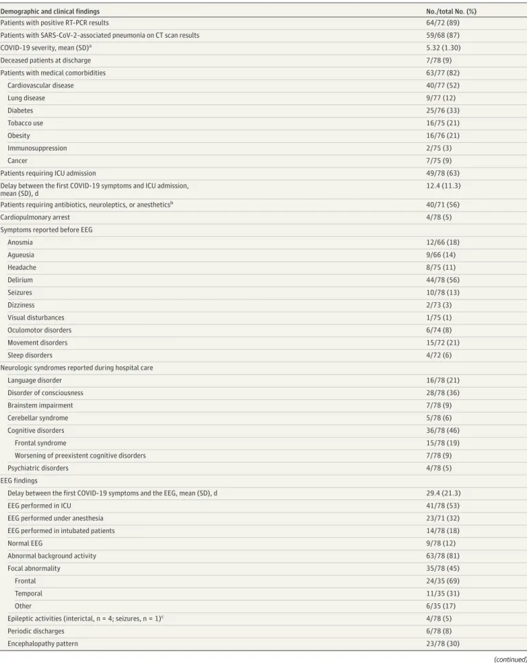
Table 1. Characteristics of Patients
Demographic and clinical findings No./total No. (%) Patients with positive RT-PCR results 64/72 (89) Patients with SARS-CoV-2–associated pneumonia on CT scan results 59/68 (87)
COVID-19 severity, mean (SD)a 5.32 (1.30)
Deceased patients at discharge 7/78 (9)
Patients with medical comorbidities 63/77 (82)
Cardiovascular disease 40/77 (52) Lung disease 9/77 (12) Diabetes 25/76 (33) Tobacco use 16/75 (21) Obesity 16/76 (21) Immunosuppression 2/75 (3) Cancer 7/75 (9)
Patients requiring ICU admission 49/78 (63)
Delay between the first COVID-19 symptoms and ICU admission, mean (SD), d
12.4 (11.3) Patients requiring antibiotics, neuroleptics, or anestheticsb 40/71 (56)
Cardiopulmonary arrest 4/78 (5)
Symptoms reported before EEG
Anosmia 12/66 (18) Agueusia 9/66 (14) Headache 8/75 (11) Delirium 44/78 (56) Seizures 10/78 (13) Dizziness 2/73 (3) Visual disturbances 1/75 (1) Oculomotor disorders 6/74 (8) Movement disorders 15/72 (21) Sleep disorders 4/72 (6)
Neurologic syndromes reported during hospital care
Language disorder 16/78 (21) Disorder of consciousness 28/78 (36) Brainstem impairment 7/78 (9) Cerebellar syndrome 5/78 (6) Cognitive disorders 36/78 (46) Frontal syndrome 15/78 (19)
Worsening of preexistent cognitive disorders 7/78 (9)
Psychiatric disorders 4/78 (5)
EEG findings
Delay between the first COVID-19 symptoms and the EEG, mean (SD), d 29.4 (21.3)
EEG performed in ICU 41/78 (53)
EEG performed under anesthesia 23/71 (32)
EEG performed in intubated patients 14/78 (18)
Normal EEG 9/78 (12)
Abnormal background activity 63/78 (81)
Focal abnormality 35/78 (45)
Frontal 24/35 (69)
Temporal 11/35 (31)
Other 6/35 (17)
Epileptic activities (interictal, n = 4; seizures, n = 1)c 4/78 (5)
Periodic discharges 6/78 (8)
Encephalopathy pattern 23/78 (30)
Table 1. Characteristics of Patients (continued)
Demographic and clinical findings No./total No. (%) MRI findings
Patients who underwent MRI 57/78 (73)
Unremarkable MRI findings 16/57 (28)
Hemorrhages 21/57 (37)
Multiple microhemorrhages 10/21 (48)
Corpus callosum injury 4/21 (19)
Acute ischemic lesions 13/57 (23)
Gray matter injury 13/57 (23)
White matter enhancing lesions 5/57 (9)
Basal ganglia abnormalities 4/57 (7)
Hypoxic-ischemic lesions 3/57 (5)
Metabolic abnormalities 3/57 (5)
PRES lesions 2/57 (4)
Leptomeningeal contrast enhancement 2/57 (4)
CLOCC 1/57 (2)
Perfusion abnormalities 20/40 (50)
Hypoperfusion 19/20 (95)
Frontal hypoperfusion 14/19 (74)
Temporal hypoperfusion 10/19 (53)
Other location hypoperfusion 15/19 (79)
Hyperperfusion 4/20 (20)
Frontal hyperperfusion 3/4 (75)
Temporal hyperperfusion 4/4 (100)
Other location hyperperfusion 2/4 (50)
Biological findings the day of the EEG
Patients with biological abnormalitiesd 55/77 (71)
Hyponatremia (sodium, <135 mEq/L) 11/76 (15) Hypernatremia (sodium, >145 mEq/L) 13/76 (17) Hypocalcemia (calcium, <8 mg/dL) 25/61 (41) Renal insufficiency (according to creatinine clearance) 32/67 (48) Hepatic dysfunction (AST and/or ALT 3 times higher than
the standard)
10/65 (15)
Hypercapnia (>45 mm Hg) 13/45 (29)
Hyperosmolarity (>310 mOsm/kg) 13/40 (33)
Patients with lumbar puncture 30/78 (39)
Delay between the first COVID-19 symptoms and the lumbar puncture, mean (SD), d
29.2 (25.3) Patients with positive RT-PCR results in CSF 0/26 Patients with increased CSF elements (>5/mm3) 3/30 (10)
Patients with increased CSF proteins (>0.65 g/L) 4/30 (13)
Abbreviations: ALT, alanine aminotransferase; AST, aspartate aminotransferase; CLOCC, cytotoxic lesion of the corpus callosum; COVID-19, coronavirus disease 2019; CSF, cerebrospinal fluid; CT, computed tomography; EEG, electroencephalogram; ICU, intensive care unit; MRI, magnetic resonance imaging; PRES, posterior reversible encephalopathy syndrome; RT-PCR, reverse transcription–polymerase chain reaction; SARS-CoV-2, severe acute respiratory syndrome coronavirus 2.
SI conversion factors: To convert sodium to millimoles per liter, multiply by 1.0; and calcium to millimoles per liter, multiply by 0.25.
aCOVID-19 severity was evaluated according to the World Health Organization nadir scale11: 1, nonhospitalized patients without activity limitation; 2, nonhospitalized patients with
activity limitation; 3, hospitalized patients without oxygen requirement; 4, hospitalized patients with oxygen requirement; 5, hospitalized patients with noninvasive ventilation; 6, hospitalized patients with invasive ventilation; and 7, deceased patient at discharge.
b
We evaluated all drugs taken by patient the day of the EEG.
c
One patient had both interictal epileptic activities and seizures.
dPatients were assessed as having biological abnormalities if they presented with one of the following abnormalities: hyponatremia, hypernatremia, hypocalcemia, renal
Patients in the ICU experienced more abnormal background activity than patients not in the ICU (39 of 41 [95%] vs 24 of 37 [65%]; P < .001). Nevertheless, the periodic discharges, epileptic activities, focal abnormalities, or encephalopathy patterns were seen in both ICU patients and non-ICU patients.
Among the 35 patients with focal abnormalities, 17 were studied for autonomy at discharge. Those with focal frontal abnormalities had a less-frequent total recovery of neurologic symptoms at hospital discharge than those with other abnormalities (1 of 10 [10%] vs 4 of 7 [57%]; P = .05). No EEG pattern was associated with death at hospital discharge.
MRI Findings
Of 57 patients who underwent MRI, 41 had abnormalities: acute ischemic lesions (n = 13), white matter–enhancing lesions (n = 5), basal ganglia abnormalities (n = 4), and metabolic lesions (ie, central pontine myelinolysis) (n = 3). Twenty patients had perfusion abnormalities—almost entirely
Figure 1. Examples of Electroencephalogram Recordings and Magnetic Resonance Imaging Findings
Intermittent slow biphasic delta waves in bifrontal areas
A Bilateral lesions in the
supratentorial white matter D
Diphasic and triphasic anterior slow waves
B Multiple microhemorrhages
involving the corpus callosum E
Periodic discharges in bifrontal areas
C Left frontotemporal hypoperfusion F 100 μV 1 s 100 μV 1 s 100 μV 1 s Fp2 C4 C4 02 Fp2 T4 T4 02 Fp1 C3 C3 01 Fp1 T3 T3 01 ECG Fp2 C4 C4 02 Fp2 T4 T4 02 Fp1 C3 C3 01 Fp1 T3 T3 01 ECG Fp2 C4 C4 02 Fp2 T4 T4 02 Fp1 C3 C3 01 Fp1 T3 T3 01 ECG
Eight electrodes, longitudinal bipolar montage, 20-second epoch, low frequency filter 0.53 Hz, high frequency filter 70 Hz. A, Intermittent slow biphasic delta waves in bifrontal areas, with low-voltage continuous background activity. B, Diphasic and triphasic anterior slow waves, with slow continuous background activity, C, Periodic discharges in the bifrontal areas, with low voltage background activity, D, Bilateral lesions in the supratentorial white matter (arrowhead), hyperintense on axial fluid-attenuated inversion recovery images, E, Multiple
microhemorrhages (arrowheads) involving the corpus callosum on T2 star images, F, Left frontotemporal hypoperfusion (arrowhead). ECG indicates electrocardiogram.
hypoperfusion (n = 19) (Figure 1). The results of MRI scans were more frequently unremarkable than EEG findings (16 of 57 [28%] vs 9 of 78 [12%]; P = .02).
Drugs and Biological Findings
Electroencephalographic features were explained according to major confounders at the time of EEG. Fifty-five patients showed biological abnormalities, including dysnatremia, kidney failure, and liver dysfunction, the same day as the EEG procedure. Of 23 patients with encephalopathy, 7 received antibiotics, 1 received a neuroleptic drug, and 4 received light sedation on the day of EEG.13
Eighteen patients had biological abnormalities (moderate to severe renal insufficiency [n = 5], hypernatremia [n = 3], and hyponatremia [n = 1]).
No patients had positive reverse transcription–polymerase chain reaction results from cerebrospinal fluid samples (n = 26). No patients received immunomodulatory treatments before EEG.
EEG Results and Related Clinical and Paraclinical Findings
The neurologic manifestations and MRI abnormalities were described according to EEG patterns (eTable in theSupplement). Owing to the large heterogeneity of clinical, MRI, and EEG findings, we were not able to show specific correlations between those findings. Three of 4 patients with epileptic activities detected by EEG had previous seizures, and 5 of 24 patients with EEG frontal abnormalities had frontal syndrome.
An overview of the most specific EEG and brain MRI findings is represented according to clinically defined syndromes in Figure 2. In our cohort, patients with disorder of consciousness, brainstem impairment, or frontal syndrome seemed to more frequently have EEG or MRI abnormalities than those with cerebellar syndrome or psychiatric disorders.
Patients With COVID-19–Related Encephalopathy
To isolate a subgroup of patients with specific COVID-19–related brain injury, we distinguished patients with an identified cause of central neurologic disorders from those without. Based on clinical and paraclinical findings, the causes were as follows: ICU complications (n = 37), isolated metabolic or toxic encephalopathy (n = 8), cerebrovascular disorders (n = 6), previous mild cognitive impairment (n = 3), intracranial tumors (n = 2), isolated seizures and epilepsy (n = 6), history of psychiatric disorders (n = 3), cardiorespiratory arrest (n = 3), multiorgan failure (n = 2), associated varicella zoster virus encephalitis (n = 1), and headache (n = 1).
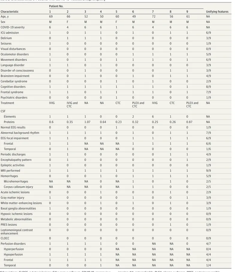
The 9 remaining patients who had acute neurologic injuries with duration more than 48 hours, without any identified cause of encephalopathy (clinical, MRI, or biological), were assessed as patients with CORE (Figure 2; Table 2). Compared with patients without CORE, those with CORE presented more frequently with movement disorders (6 of 9 [67%] vs 9 of 63 [14%]; P = .002), frontal syndrome (7 of 9 [78%] vs 8 of 69 [12%]; P < .001), brainstem impairment (4 of 9 [44%] vs 3 of 69 [4%]; P < .001), periodic EEG discharges (4 of 9 [44%] vs 2 of 69 [3%]; P < .001), and white matter–enhancing MRI lesions (3 of 9 [33%] vs 2 of 48 [4%]; P = .03).
Using clinical, EEG, and MRI data, we developed a model to identify patients with CORE, taking into account variable risk. The regression model included periodic EEG discharges, movement disorders, brainstem impairment, frontal syndrome, and white matter–enhancing MRI lesions. The model resulted in an area under the receiver operating characteristics curve of 0.94 (95% CI, 0.88-1.00; P < .001) (Figure 3). After a 100-fold cross-validation, the model was able to estimate the risk for a new patient to present with CORE with a sensitivity of 76% (95% CI, 33%-100%), specificity of 93% (95% CI, 86%-100%), positive predictive value of 65% (95% CI, 33%-100%), negative predictive value of 95% (95% CI, 86%-100%), and accuracy of 91% (95% CI, 76%-100%).
Discussion
We report on a cohort of 78 patients with COVID-19 who underwent EEG for a wide range of CNS manifestations. Recent case reports, small series, and meta-analyses assessing the value of EEG for patients with COVID-195-9,14-17
showed (1) nonspecific patterns, reflecting the diversity of SARS-CoV-2 infection complications,1,7,9,14-17
or (2) a striking periodic EEG pattern,5,6,8,17
suggestive of COVID-19– specific brain complications. Nevertheless, a systematic correlation of EEG findings with biological findings and brain MRI findings was lacking, precluding a better understanding of pattern origins.
We performed a multimodal evaluation of patients with COVID-19. Their neurologic
complications were sometimes associated with ICU complications, preexisting pathologic conditions, toxic or metabolic encephalopathies, or strokes.1,14
The existence of specific COVID-19 brain complications is still being debated. Eight patients with SARS-CoV-2 infection and irritability, delirium, drowsiness, and new-onset epilepsy were reported.14
Additional reports further reinforced the hypothesis of brain-specific COVID-19 involvement, including marked brain metabolism changes detected on fluorodeoxyglucose positron emission tomography scans.18
We defined this brain involvement as CORE. In our study, we showed that patients with CORE mostly had movement
Figure 2. Representation of Electroencephalogram (EEG) and Brain Magnetic Resonance Imaging (MRI) Findings According to Neurologic Syndromes
36% 0% 14% 43% 23% 5% 14% 14% 41% Frontal abnormality Epileptic activities Periodic discharges Encephalopathy pattern Multiple microhemorrhages Corpus callosum injury White matter– enhancing lesions Basal ganglia abnormalities Hypoperfusion 57% 0% 29% 43% 33% 17% 33% 0% 50% Frontal abnormality Epileptic activities Periodic discharges Encephalopathy pattern Multiple microhemorrhages Corpus callosum injury White matter– enhancing lesions Basal ganglia abnormalities Hypoperfusion 20% 0% 0% 60% 0% 25% 0% 0% 33% Frontal abnormality Epileptic activities Periodic discharges Encephalopathy pattern Multiple microhemorrhages Corpus callosum injury White matter– enhancing lesions Basal ganglia abnormalities Hypoperfusion 33% 7% 20% 20% 15% 15% 15% 8% 60% Frontal abnormality Epileptic activities Periodic discharges Encephalopathy pattern Multiple microhemorrhages Corpus callosum injury White matter– enhancing lesions Basal ganglia abnormalities Hypoperfusion 50% 0% 25% 75% 0% 0% 0% 0% 25% Frontal abnormality Epileptic activities Periodic discharges Encephalopathy pattern Multiple microhemorrhages Corpus callosum injury White matter– enhancing lesions Basal ganglia abnormalities Hypoperfusion 67% 11% 44% 22% 22% 22% 33% 11% 67% Frontal abnormality Epileptic activities Periodic discharges Encephalopathy pattern Multiple microhemorrhages Corpus callosum injury White matter– enhancing lesions Basal ganglia abnormalities Hypoperfusion
Disorder of consciousness Brainstem impairment
Frontal syndrome Cerebellar syndrome
Psychiatric disorders COVID-19–related encephalopathy
Each radar chart is a graphical representation of the proportion of patients with the selected neurologic syndrome who had EEG findings (frontal abnormality, epileptic activities, periodic discharges, and encephalopathy pattern; percentage in orange) or MRI findings (multiple microhemorrhages, corpus callosum injury, white matter–enhancing lesions, basal ganglia abnormalities, and hypoperfusion; percentage in black). Each concentric circle represents a proportion of 20%. A blue line is drawn connecting the values (percentages) for each finding (EEG or MRI), giving the blue polygon appearance. The polygon represents the prevalence of EEG and MRI findings: the larger the area of the polygon is, the more abnormal EEG or MRI findings the patients had. Data are represented as proportion to take into account missing data. COVID-19 indicates coronavirus disease 2019.
Table 2. Characteristics of Patients With COVID-19–Related Encephalopathy Characteristic Patient No. Unifying features 1 2 3 4 5 6 7 8 9 Age, y 69 66 52 50 60 49 72 56 61 NA Sex M F M M F M M M M NA COVID-19 severity 6 4 6 6 1 6 4 6 6 NA ICU admission 1 0 1 1 0 1 0 1 1 6/9 Delirium 0 1 1 1 0 0 0 0 0 3/9 Seizures 1 0 0 0 0 0 0 0 0 1/9 Visual disturbances 0 0 0 0 0 0 0 0 0 0/9 Oculomotor disorders 1 0 0 0 0 1 1 1 1 5/9 Movement disorders 1 0 1 0 1 1 1 0 1 6/9 Language disorder 1 1 0 1 0 0 0 0 0 3/9 Disorder of consciousness 0 0 1 0 0 0 0 1 1 3/9 Brainstem impairment 0 0 1 0 0 1 0 1 1 4/9 Cerebellar syndrome 0 0 0 0 1 0 1 0 0 2/9 Cognitive disorders 1 1 1 1 1 1 1 0 1 8/9 Frontal syndrome 1 1 0 1 1 1 1 0 1 7/9 Psychiatric disorders 0 0 0 0 1 0 0 0 0 1/9
Treatment IVIG IVIG and CTC
NA NA CTC PLEX and CTC
IVIG CTC PLEX and CTC
NA CSF
Elements 1 1 1 0 0 2 6 1 0 NA
Proteins 0.6 0.35 1.07 0.64 0.23 0.32 0.25 0.26 0.87 NA
Normal EEG results 0 0 0 0 1 0 0 0 0 1/9
Abnormal background rhythm 1 1 1 1 0 1 0 1 1 7/9
EEG focal impairment 1 1 0 0 0 1 1 1 1 6/9
Frontal 1 1 NA NA NA 1 1 1 1 6/6 Temporal 0 1 NA NA NA 0 0 0 0 1/6 Periodic discharges 1 1 0 0 0 0 0 1 1 4/9 Encephalopathy pattern 0 1 0 0 0 0 0 0 1 2/9 Epileptic activities 1 0 0 0 0 0 0 0 0 1/9 MRI performed 1 1 1 1 1 1 1 1 1 9/9 Hemorrhages 0 0 0 1 0 1 1 1 1 5/9 Microhemorrhages NA NA NA 0 NA 1 0 1 0 2/5
Corpus callosum injury NA NA NA 0 NA 1 1 0 0 2/5
Acute ischemic lesions 0 0 0 1 0 0 0 1 0 2/9
Gray matter injury 1 0 0 0 0 1 0 0 1 3/9
White matter–enhancing lesions 0 0 0 1 0 1 0 1 0 3/9 Basal ganglia abnormalities 1 0 0 0 0 0 0 0 0 1/9
Hypoxic ischemic lesions 0 0 0 0 0 0 0 0 0 0/9
Metabolic abnormalities 0 0 0 0 0 0 0 0 0 0/9 PRES lesions 0 0 0 0 0 0 0 1 0 1/9 Leptomeningeal contrast enhancement 0 0 0 0 0 0 0 0 0 0/9 CLOCC 0 0 0 0 0 0 0 0 0 0/9 Perfusion disorders 1 1 1 1 0 0 NA NA 0 4/7 Hyperperfusion 0 0 0 0 NA NA NA NA NA 0/4 Hypoperfusion 1 1 1 1 NA NA NA NA NA 4/4 Frontal 1 1 1 1 NA NA NA NA NA 4/4 Temporal 0 0 0 1 NA NA NA NA NA 1/4
Abbreviations: CLOCC, cytotoxic lesions of the corpus callosum; COVID-19, coronavirus disease 2019; CSF, cerebrospinal fluid; CTC, corticosteroids; EEG, electroencephalogram; ICU, intensive care unit; IVIG, intravenous immunoglobulins; MRI, magnetic resonance
imaging; NA, not applicable; PLEX, plasma exchange; PRES, posterior reversible encephalopathy syndrome.
disorders (mainly seizures and/or myorrhythmia), and brainstem impairment (oculomotor disorders such as bobbing) and frontal syndrome (disinhibition and grasping).
Similarly, MRI findings showed both (1) unspecific lesions, such as perfusion abnormalities, and (2) more specific lesions, such as basal ganglia abnormalities, microhemorrhages, corpus callosum injury, and white matter–enhancing lesions.3
The latter abnormality was the most significant lesion detected on MRI scans in patients with CORE (Table 2).
The most frequent EEG findings were abnormal background activity (81%) and frontal slow waves (60%). The latter were associated with metabolic and toxic encephalopathies—for which we identified 1 or several factors in most cases—or frontal lesions. Six patients (8%) showed a periodic EEG pattern, predominating in frontal lobes and not explained by MRI findings.
Our results are in accordance with previous reports; a recent meta-analysis reported abnormal background activity in almost all patients (96.1%),16
while half of all patients had focal slowing that involved the frontal region.17
A more specific periodic EEG pattern was also reported, with an incidence ranging from 0% to 38% according to the etiologic characteristics.6-9,15,16,19-23
Nevertheless, we found that this periodic EEG pattern had no prognostic value.
Epileptiform discharges and seizures have been reported in patients with COVID-19, with an incidence ranging from 0% to 63% for epileptiform discharges and from 0% to 25% for seizures.7-9,15,16,19-25
In our cohort, 4 patients (5%) had epileptiform discharges, and seizures occurred in 1 patient (1%) during EEG.
Patients with CORE had a periodic EEG pattern more frequently than other patients. All EEG abnormalities from the frontal lobe, coupled with the frontal syndrome noted in patients with CORE, suggest frontal lobe dysfunction, which is reminiscent of the hypothesis of a neuroinvasive entry of SARS-CoV-2 into the brain via the olfactory nerves or via the nasopharyngeal mucosa.18,26
A change in neuronal excitability, perhaps mediated by specific cytokines, may occur in brain areas close to the nasopharynx, such as the orbitofrontal lobe and the brainstem. Because inflammatory mechanisms, such as cytokine-mediated response or postviral autoimmune process, are suspected, immunomodulator treatments, such as plasma exchanges or intravenous immunoglobulins, may be proposed as early treatment for patients with CORE (Table 2).26-28
Limitations
This study has some limitations. A relatively small number of patients underwent both EEG and MRI in a single center. There was a lack of systematic follow-up after hospital discharge. There was also a
Figure 3. Clinical, Electroencephalogram (EEG), and Magnetic Resonance Imaging (MRI) Disturbances in Patients With Coronavirus Disease 2019 (COVID-19)–Related Encephalopathy
1.0 0.8 0.6 0.4 0.2 0 Sensitivit y 1–Specificity 1.0 0 0.2 0.4 0.6 0.8 AUC, 0.94
Receiver operating characteristic curve for the model, evaluating the performance of movement disorders, brainstem impairment, frontal syndrome, EEG periodic discharges, and white matter–enhancing MRI lesions to identify patients with COVID-19–related encephalopathy. AUC indicates area under the curve.
risk of underestimating the number of patients with CORE owing to other COVID-19–related comorbidities or pathologic conditions.
Conclusions
Despite different clinical presentations, our study suggests that EEG is a valuable procedure for patients with COVID-19 and neurologic symptoms, to better identify different brain dysfunctions and CORE. We further emphasize the benefit associated with combining EEG and brain MRI for patients with neurologic symptoms concomitant with COVID-19. It remains to be clarified whether treatment strategies could be optimized with earlier identification of patients with CORE.
ARTICLE INFORMATION
Accepted for Publication: January 22, 2021.
Published: March 15, 2021. doi:10.1001/jamanetworkopen.2021.1489
Open Access: This is an open access article distributed under the terms of theCC-BY License. © 2021 Lambrecq V et al. JAMA Network Open.
Corresponding Author: Virginie Lambrecq, MD, PhD, Sorbonne Université, Assistance Publique des Hôpitaux de
Paris, Neurophysiology Department, GH Pitié-Salpêtrière-Charles Foix, 47-83 Boulevard de l’Hôpital, Paris 75013, France (virginie.lambrecq@aphp.fr).
Author Affiliations: Sorbonne Université, Paris Brain Institute, Institut du Cerveau, Institut National de la Santé et
de la Recherche Médicale U 1127, Centre National de la Recherche Scientifique, Unité Mixte de Recherche 7225, Paris, France (Lambrecq, Hanin, Munoz-Musat, Chougar, Cousyn, Frazzini, Sangare, Chavez, Raux, Galanaud, Corvol, Lubetzki, Rohaut, Pyatigorskaya, Naccache, Navarro); Assistance Publique des Hôpitaux de Paris, Clinical Neurophysiology Department, Pitié-Salpêtrière Hospital, Paris, France (Lambrecq, Munoz-Musat, Borden, Damiano, Frazzini, Huberfeld, Landgraf, Nguyen-Michel, Pichit, Sangare, Naccache, Navarro); Neurophysiology Department, Sorbonne Université, Paris, France (Lambrecq, Munoz-Musat, Huberfeld, Naccache); Neuroradiology Department, Sorbonne Université, Paris, France (Chougar, Galanaud, Pyatigorskaya); Assistance Publique des Hôpitaux de Paris, Neuroradiology Department, Pitié-Salpêtrière Hospital, Paris, France (Chougar, Galanaud, Pyatigorskaya); Assistance Publique des Hôpitaux de Paris, Neurology Department, Pitié-Salpêtrière Hospital, Paris, France (Gassama, Delorme, Cousyn, Damiano, Frazzini, Pichit, Corvol, Lubetzki, Rohaut, Demeret, Navarro); Neurology Department, Sorbonne Université, Paris, France (Cousyn, Lubetzki, Rohaut, Navarro); Service de Pneumologie, Sorbonne Université, Paris, France (Morélot-Panzini, Morawiec); Assistance Publique des Hôpitaux de Paris, Service de Pneumologie, Médecine Intensive et Réanimation, Pitié-Salpêtrière Hospital, Paris, France (Morélot-Panzini, Morawiec); Department of Anesthesia, Critical Care and Peri-Operative Medicine, Sorbonne Université, Paris, France (Raux); Assistance Publique des Hôpitaux de Paris, Department of Anesthesia, Critical Care and Peri-Operative Medicine, Pitié-Salpêtrière Hospital, Paris, France (Raux); Institut de Cardiologie, Sorbonne Université, Paris, France (Luyt); Sorbonne Université, Institut National de la Santé et de la Recherche Médicale, Unité Mixte de Recherche, 1166–Institute of Cardiometabolism and Nutrition, Paris, France (Luyt); Assistance Publique des Hôpitaux de Paris, Service de Médecine Intensive Réanimation, Institut de Cardiologie, Pitié-Salpêtrière Hospital, Paris, France (Luyt); Assistance Publique des Hôpitaux de Paris, Biostatistic Department, Pitié-Salpêtrière Hospital, Paris, France (Rufat); Center of Reference for Rare Epilepsies, Pitié-Salpêtrière Hospital, Paris, France (Navarro).
Author Contributions: Drs Lambrecq and Hanin had full access to all of the data in the study and take
responsibility for the integrity of the data and the accuracy of the data analysis. Drs Lambrecq and Hanin contributed equally to this work as co–first authors.
Concept and design: Lambrecq, Hanin, Munoz-Musat, Damiano, Lubetzki, Naccache, Navarro.
Acquisition, analysis, or interpretation of data: Lambrecq, Hanin, Munoz-Musat, Chougar, Gassama, Delorme,
Cousyn, Borden, Frazzini, Huberfeld, Landgraf, Nguyen-Michel, Pichit, Sangare, Chavez, Morélot-Panzini, Morawiec, Raux, Luyt, Rufat, Galanaud, Corvol, Rohaut, Demeret, Pyatigorskaya, Navarro.
Critical revision of the manuscript for important intellectual content: Lambrecq, Chougar, Gassama, Delorme,
Cousyn, Borden, Damiano, Frazzini, Huberfeld, Landgraf, Nguyen-Michel, Pichit, Sangare, Chavez, Morélot-Panzini, Morawiec, Raux, Luyt, Rufat, Galanaud, Corvol, Lubetzki, Rohaut, Demeret, Pyatigorskaya, Naccache, Navarro.
Statistical analysis: Hanin, Cousyn, Pichit, Rufat, Pyatigorskaya. Obtained funding: Corvol.
Administrative, technical, or material support: Lambrecq, Munoz-Musat, Morélot-Panzini, Galanaud, Corvol. Supervision: Lambrecq, Hanin, Frazzini, Landgraf, Corvol, Naccache, Navarro.
Conflict of Interest Disclosures: Dr Huberfeld reported receiving personal fees from Advicenne, GW Pharma, and
EISAI outside the submitted work. Dr Morélot-Panzini reported receiving personal fees from Astra-Zeneca, GSK, SOS Oxygène, ADEP, ISIS, Resmed, Chiesi, Menarini, Vivisol, Air Liquide, and Lowenstein outside the submitted work. Dr Raux reported receiving personal fees from Chiesi outside the submitted work. Dr Luyt reported receiving personal fees from Bayer Healthcare, Merck, ThermoFischer Brahms, Carmat, and Biomérieux outside the submitted work. Dr Corvol reported receiving grants from Fédération Internationale pour l’Automobile and Investissements d’avenir program during the conduct of the study; personal fees from Biogen, UCB, Prevail Therapeutic, Idorsia, Ever Pharma, Denali, Air Liquide, and Theranexus; and grants from Sanofi outside the submitted work. Dr Lubetzki reported receiving personal fees from Biogen, Merck-Serono, Roche, Rewind, and Ipsen outside the submitted work. Dr Pyatigorskaya reported receiving personal fees from GE Healthcare and Biogen; and grants from ANR outside the submitted work. Dr Navarro reported receiving personal fees from UCB Pharma, Liva Nova, and EISAI outside the submitted work. No other disclosures were reported.
Funding/Support: The Cohort COVID-19 Neurosciences (CoCo Neurosciences) study was sponsored by Assistance
Publique des Hôpitaux de Paris and funded by the Fédération Internationale pour l’Automobile Foundation and donors of the Paris Brain Institute–ICM. This work received support from the Investissements d’avenir program (grant ANR-10-IAIHU-06), from the Fondation pour la Recherche Médicale (grant FDM20170839111), and from the Fondation Assistance Publique-Hôpitaux de Paris (EPIRES-Marie Laure PLV Merchandising) for the conduct of the study.
Role of the Funder/Sponsor: The funding sources had no role in the design and conduct of the study; collection,
management, analysis, and interpretation of the data; preparation, review, or approval of the manuscript; and decision to submit the manuscript for publication.
The Cohort COVID-19 Neurosciences (CoCo Neurosciences) Study Group Members: Steering Committee
(Pitié-Salpêtrière Hospital, Paris): Cecile Delorme, MD; Jean-Christophe Corvol, MD, PhD; Jean-Yves Delattre, MD, PhD; Stephanie Carvalho; and Sandrine Sagnes. Scientific Committee (Pitié-Salpêtrière Hospital, Paris): Bruno Dubois, MD, PhD; Vincent Navarro, MD, PhD; Celine Louapre, MD, PhD; Tanya Stojkovic, MD; Ahmed Idbaih, MD, PhD; Charlotte Rosso, MD, PhD; David Grabli, MD, PhD; Ana Zenovia Gales, MD; Bruno Millet, MD, PhD; Benjamin Rohaut, MD, PhD; Eleonore Bayen, MD, PhD; Sophie Dupont, MD, PhD; Gaelle Bruneteau, MD, PhD; Stephane Lehericy, MD, PhD; Danielle Seilhean, MD, PhD; Alexandra Durr, MD, PhD; Aurelie Kas, MD, PhD; Foudil Lamari, PharmD, PhD; Marion Houot; and Vanessa Batista Brochard. Principal investigators: Pitié-Salpêtrière Hospital,
Paris: Sophie Dupont, MD, PhD; Catherine Lubetzki, MD, PhD; Danielle Seilhean, MD, PhD; Pascale Pradat-Diehl,
MD, PhD; Charlotte Rosso, MD, PhD; Khe Hoang-Xuan, MD, PhD; Bertrand Fontaine, MD, PhD; Lionel Naccache, MD, PhD; Philippe Fossati, MD, PhD; Isabelle Arnulf, MD, PhD; Alexandra Durr, MD, PhD; Alexandre Carpentier, MD, PhD; Stephane Lehericy, MD, PhD; and Yves Edel, MD; Foch Hospital, Suresnes: Anna Luisa Di Stefano, MD, PhD;
Rothschild Hospital, Paris: Gilberte Robain, MD, PhD; and Philippe Thoumie, MD, PhD; Avicenne Hospital, Bobigny:
Bertrand Degos, MD, PhD; Sainte-Anne Hospital, Paris: Tarek Sharshar, MD, PhD; Saint-Antoine Hospital, Paris: Sonia Alamowitch, MD, PhD; Emmanuelle Apartis-Bourdieu, MD, PhD; and Charles-Siegried Peretti, MD, PhD;
Saint-Louis Hospital, Paris: Renata Ursu, MD; Tenon Hospital, Paris: Nathalie Dzierzynski, MD; Charles Foix Hospital, Ivry: Kiyoka Kinugawa Bourron, MD, PhD; Joel Belmin, MD, PhD; Bruno Oquendo, MD; Eric Pautas, MD, PhD; and
Marc Verny, MD, PhD. Co-investigators: Pitié-Salpêtrière Hospital, Paris: Cecile Delorme, MD; Jean-Christophe Corvol, MD, PhD; Jean-Yves Delattre, MD, PhD; Yves Samson, MD, PhD; Sara Leder, MD; Anne Leger, MD; Sandrine Deltour, MD; Flore Baronnet, MD; Ana Zenovia Gales, MD; Stephanie Bombois, MD, PhD; Mehdi Touat, MD; Ahmed Idbaih, MD, PhD; Marc Sanson, MD, PhD; Caroline Dehais, MD, PhD; Caroline Houillier, MD; Florence Laigle-Donadey, MD; Dimitri Psimaras, MD; Agusti Alenton, MD, PhD; Nadia Younan, MD, PhD; Nicolas Villain, MD, PhD; David Grabli, MD, PhD; Maria del Mar Amador, MD; Gaelle Bruneteau, MD, PhD; Celine Louapre, MD, PhD; Louise-Laure Mariani, MD, PhD; Nicolas Mezouar, MD; Graziella Mangone, MD, PhD; Aurelie Meneret, MD, PhD; Andreas Hartmann, MD, PhD; Clement Tarrano, MD; David Bendetowicz, MD; Pierre-François Pradat, MD, PhD; Michel Baulac, MD, PhD; Sara Sambin, MD, PhD; Phintip Pichit, MD; Florence Chochon, MD; Adele Hesters, MD; Bastien Herlin, MD; An Hung Nguyen, MD, PhD; Valerie Pourcher, MD, PhD; Alexandre Demoule, MD, PhD; Elise Morawiec, MD; Julien Mayaux, MD; Morgan Faure, MD; Claire Ewenczyk, MD, PhD; Giulia Coarelli, MD; Anna Heinzmann, MD; Perrine Charles, MD, PhD; Tanya Stojkovic, MD; Marion Masingue, MD; Guillaume Bassez, MD, PhD; Vincent
Navarro, MD, PhD; Isabelle An, MD; Yulia Worbe, MD, PhD; Rabab Debs, MD; Esteban Munoz Musat, MD; Timothee Lenglet, MD; Virginie Lambrecq, MD, PhD; Aurelie Hanin, PharmD; Lydia Chougar, MD; Nathalia Shor, MD; Nadya Pyatigorskaya, MD, PhD; Damien Galanaud, MD, PhD; Delphine Leclercq, MD; Sophie Demeret, MD; Benjamin Rohaut, MD, PhD; Albert Cao, MD; Clemence Marois, MD; Nicolas Weiss, MD, PhD; Salimata Gassama, MD; Loic Le Guennec, MD, PhD; Vincent Degos, MD, PhD; Alice Jacquens, MD; Thomas Similowski, MD, PhD; Capucine Morelot-Panzini, MD, PhD; Jean-Yves Rotge, MD, PhD; Bertrand Saudreau, MD; Bruno Millet, MD, PhD; Victor Pitron, MD; Nassim Sarni, MD; Nathalie Girault, MD; Redwan Maatoug, MD; Ana Zenovia Gales, MD; Smaranda Leu, MD; Eleonore Bayen, MD, PhD; Lionel Thivard, MD, PhD; Karima Mokhtari, MD; and Isabelle Plu, MD, PhD;
Sainte-Anne Hospital, Paris: Bruno Gonçalves; Saint-Antoine Hospital, Paris: Laure Bottin, MD; and Marion Yger, MD; Rothschild Hospital, Paris: Gaelle Ouvrard, MD; and Rebecca Haddad, MD; Charles Foix Hospital, Ivry: Flora Ketz,
MD; Carmelo Lafuente, MD, PhD; and Christel Oasi, MD. Other contributors—associated centers: Lariboisière
Hospital, Paris: Bruno Megarbane, MD, PhD; and Dominique Herve, MD. Clinical research associates: ICM, Pitié-Salpêtrière Hospital, Paris: Haysam Salman; Armelle Rametti-Lacroux; Alize Chalançon; Anais Herve; Hugo Royer;
Florence Beauzor; Valentine Maheo; Christelle Laganot; Camille Minelli; Aurelie Fekete; Abel Grine; Marie Biet; Rania Hilab; Aurore Besnard, PhD; Meriem Bouguerra; Gwen Goudard; Saida Houairi; Saba Al-Youssef; Christine Pires; Anissa Oukhedouma; Katarzyna Siuda-Krzywicka; and Tal Seidel Malkinson; Saint-Louis Hospital, Paris: Hanane Agguini; Foch Hospital, Suresnes: Hassen Douzane; Data manager: ICM, Paris: Safia Said; and Statistician:
ICM, Paris: Marion Houot.
Additional Contributions: Aurore Besnard, PhD, Neurophysiology Department, Pitié-Salpêtrière Hospital, and
Meriem Bouguerra, Paris Brain Institute, assisted with data recording; they were not compensated for their contributions. We thank the EEG technicians, the Cohort COVID-19 Neurosciences (CoCo Neurosciences) and the COVID SMIT PSL study groups for their participation to conduct the study and to data collection.
Additional Information: All anonymized data are available on request.
REFERENCES
1. Ellul MA, Benjamin L, Singh B, et al. Neurological associations of COVID-19. Lancet Neurol. 2020;19(9):767-783.
doi:10.1016/S1474-4422(20)30221-0
2. Nepal G, Rehrig JH, Shrestha GS, et al. Neurological manifestations of COVID-19: a systematic review. Crit Care.
2020;24(1):421. doi:10.1186/s13054-020-03121-z
3. Chougar L, Shor N, Weiss N, et al; CoCo Neurosciences Study Group. Retrospective observational study of brain
MRI findings in patients with acute SARS-CoV-2 infection and neurologic manifestations. Radiology. 2020;297(3): E313-E323. doi:10.1148/radiol.2020202422
4. Paterson RW, Brown RL, Benjamin L, et al. The emerging spectrum of COVID-19 neurology: clinical, radiological
and laboratory findings. Brain. 2020;143(10):3104-3120. doi:10.1093/brain/awaa240
5. Flamand M, Perron A, Buron Y, Szurhaj W. Pay more attention to EEG in COVID-19 pandemic. Clin Neurophysiol.
2020;131(8):2062-2064. doi:10.1016/j.clinph.2020.05.011
6. Vespignani H, Colas D, Lavin BS, et al. Report on electroencephalographic findings in critically ill patients with
COVID-19. Ann Neurol. 2020;88(3):626-630. doi:10.1002/ana.25814
7. Petrescu A-M, Taussig D, Bouilleret V. Electroencephalogram (EEG) in COVID-19: a systematic retrospective
study. Neurophysiol Clin. 2020;50(3):155-165. doi:10.1016/j.neucli.2020.06.001
8. Pellinen J, Carroll E, Friedman D, et al. Continuous EEG findings in patients with COVID-19 infection admitted to
a New York academic hospital system. Epilepsia. 2020;61(10):2097-2105. doi:10.1111/epi.16667
9. Roberto KT, Espiritu AI, Fernandez MLL, Gutierrez JC. Electroencephalographic findings in COVID-19 patients:
a systematic review. Seizure. 2020;82:17-22. doi:10.1016/j.seizure.2020.09.007
10. Hirsch LJ, LaRoche SM, Gaspard N, et al. American Clinical Neurophysiology Society’s standardized critical care
EEG terminology: 2012 version. J Clin Neurophysiol. 2013;30(1):1-27. doi:10.1097/WNP.0b013e3182784729 11. World Health Organization. WHO R&D blueprint: novel coronavirus: COVID-19 therapeutic trial synopsis.
Published February 18, 2020. Accessed January 31, 2021. https://www.who.int/blueprint/priority-diseases/key-action/COVID-19_Treatment_Trial_Design_Master_Protocol_synopsis_Final_18022020.pdf
12. Bhattacharyya S, Darby RR, Raibagkar P, Gonzalez Castro LN, Berkowitz AL. Antibiotic-associated
encephalopathy. Neurology. 2016;86(10):963-971. doi:10.1212/WNL.0000000000002455
13. Devlin JW, Skrobik Y, Gélinas C, et al. Clinical practice guidelines for the prevention and management of pain,
agitation/sedation, delirium, immobility, and sleep disruption in adult patients in the ICU. Crit Care Med. 2018; 46(9):e825-e873. doi:10.1097/CCM.0000000000003299
14. Helms J, Kremer S, Merdji H, et al. Neurologic features in severe SARS-CoV-2 infection. N Engl J Med. 2020;
15. Galanopoulou AS, Ferastraoaru V, Correa DJ, et al. EEG findings in acutely ill patients investigated for
SARS-CoV-2/COVID-19: a small case series preliminary report. Epilepsia Open. 2020;5(2):314-324. doi:10.1002/ epi4.12399
16. Kubota T, Gajera PK, Kuroda N. Meta-analysis of EEG findings in patients with COVID-19. Epilepsy Behav.
2020;107682. Published online December 4, 2020. doi:10.1016/j.yebeh.2020.107682
17. Antony AR, Haneef Z. Systematic review of EEG findings in 617 patients diagnosed with COVID-19. Seizure.
2020;83:234-241. doi:10.1016/j.seizure.2020.10.014
18. Delorme C, Paccoud O, Kas A, et al; CoCo-Neurosciences study group and COVID SMIT PSL study group.
COVID-19–related encephalopathy: a case series with brain FDG-positron-emission tomography/computed tomography findings. Eur J Neurol. 2020;27(12):2651-2657. doi:10.1111/ene.14478
19. Canham LJW, Staniaszek LE, Mortimer AM, Nouri LF, Kane NM. Electroencephalographic (EEG) features of
encephalopathy in the setting of Covid-19: a case series. Clin Neurophysiol Pract. 2020;5:199-205. doi:10.1016/j. cnp.2020.06.001
20. Pastor J, Vega-Zelaya L, Martín Abad E. Specific EEG encephalopathy pattern in SARS-CoV-2 patients. J Clin
Med. 2020;9(5):E1545. doi:10.3390/jcm9051545
21. Ayub N, Cohen J, Jing J, et al. Clinical electroencephalography findings and considerations in hospitalized
patients with coronavirus SARS-CoV-2. Preprint. Posted July 15, 2020. medRxiv 20152207. doi:10.1101/2020.07. 13.20152207
22. Louis S, Dhawan A, Newey C, et al. Continuous electroencephalography characteristics and acute
symptomatic seizures in COVID-19 patients. Clin Neurophysiol. 2020;131(11):2651-2656. doi:10.1016/j.clinph.2020. 08.003
23. Pilato MS, Urban A, Alkawadri R, et al. EEG Findings in Coronavirus Disease. J Clin Neurophysiol. 2020.
Published online July 1, 2020. doi:10.1097/WNP.0000000000000752
24. Cecchetti G, Vabanesi M, Chieffo R, et al. Cerebral involvement in COVID-19 is associated with metabolic and
coagulation derangements: an EEG study. J Neurol. 2020;267(11):3130-3134. doi:10.1007/s00415-020-09958-2 25. Pasini E, Bisulli F, Volpi L, et al. EEG findings in COVID-19 related encephalopathy. Clin Neurophysiol. 2020;131
(9):2265-2267. doi:10.1016/j.clinph.2020.07.003
26. Muccioli L, Pensato U, Cani I, et al. COVID-19–related encephalopathy presenting with aphasia resolving
following tocilizumab treatment. J Neuroimmunol. 2020;349:577400. doi:10.1016/j.jneuroim.2020.577400 27. Muccioli L, Pensato U, Bernabè G, et al. Intravenous immunoglobulin therapy in COVID-19–related
encephalopathy. J Neurol. 2020. doi:10.1007/s00415-020-10248-0
28. Cao A, Rohaut B, Le Guennec L, et al; CoCo-Neurosciences study group. Severe COVID-19-related encephalitis
can respond to immunotherapy. Brain. 2020;143(12):e102. doi:10.1093/brain/awaa337
SUPPLEMENT.