SCIENTIFIC ARTICLE
Therapeutic impact of [
18
F]fluoride positron-emission
tomography/computed tomography on patients with unclear
foot pain
Dorothee Rita Fischer&Gerardo J. Maquieira&Norman Espinosa&Marco Zanetti&
Rolf Hesselmann&Anass Johayem&Thomas F. Hany&Gustav K. von Schulthess&
Klaus Strobel
Received: 24 September 2009 / Revised: 28 December 2009 / Accepted: 5 January 2010 / Published online: 23 February 2010 # ISS 2010
Abstract
Purpose To evaluate the therapeutic impact of [18F]fluoride positron-emission tomography/computed tomography ([18F]fluoride PET/CT) imaging on patients with unclear foot pain.
Methods Twenty-eight patients were prospectively included in this study. Therapeutic management was defined by two experienced dedicated foot surgeons before and after [18F] fluoride PET/CT imaging. Twenty-six patients underwent cross-sectional imaging [CT, magnetic resonance (MR)] prior to PET/CT. A retrospective analysis of the magnetic resonance imaging (MRI) diagnoses was performed when a therapy change occurred after PET/CT imaging.
Results In 13/28 (46%) patients therapeutic management was changed due to PET/CT results. Management changes occurred in patients with the following diagnoses: os trigonum syndrome; sinus tarsi syndrome; os tibiale externum syndrome; osteoarthritis of several joints;
non-consolidated fragments; calcaneo-navicular coalition; plantar fasciitis; insertional tendinopathy; suggestion of periostitis; neoarticulations between metatarsal bones. Os trigonum, os tibiale externum, subtalar osteoarthritis and plantar fasciitis were only seen to be active on PET/CT images but not on MR images.
Conclusion [18F]fluoride PET/CT has a substantial thera-peutic impact on management in patients with unclear foot pain.
Keywords [18F]fluoride PET/CT . Foot pain . Fluoride uptake . Therapeutic impact
Introduction
Despite advanced imaging technology, diagnostic results after magnetic resonance (MR) and/or computed tomogra-phy (CT) imaging in patients suffering from foot pain may remain inconclusive. The combination of morphological and functional imaging using fluoride positron-emission tomography/computed tomography (PET/CT) bears the potential to allow us to obtain a better three-dimensional appreciation of morphology, together with metabolism, thereby arriving at a more specific diagnosis. As a result, it may alter and/or improve therapeutic management.
[18F]-labeled sodium fluoride (NaF) had already been used for bone scintigraphy by Blau et al. in the 1960s. Owing to the technical limitations of positron imaging and the widespread availability of molybdenum-99/technetium-99m (99Mo/99mTc) generators,99mTc-labeled bone seeking agents such as methylene diphosphonate (MDP) were subsequently preferred for bone scanning. In the early 1990s [18F]fluoride was readopted for PET scanning [1].
D. R. Fischer (*)
:
R. Hesselmann:
A. Johayem:
T. F. Hany:
G. K. von Schulthess:
K. StrobelDepartment of Nuclear Medicine, University Hospital of Zurich, Rämistrasse 100,
8091 Zurich, Switzerland e-mail: [email protected] G. J. Maquieira
:
N. EspinosaFoot and Ankle Surgery, Department of Orthopaedics, University of Zurich,
Balgrist,
Zurich, Switzerland M. Zanetti
Department of Radiology, University of Zurich, Balgrist,
Zurich, Switzerland
[18F]fluoride is a tracer that mainly depicts blood flow and osteoblastic activity. After intravenous (i.v.) adminis-tration, [18F]fluoride runs through the bone capillaries and diffuses into the bone extracellular fluid (ECF). Its plasma clearance is more rapid, and its single-passage extraction efficiency is higher, than that of 99mTc-MDP due to its smaller molecular weight and its negligible protein binding. Uptake is into hydroxyapatite crystals, which form the mineral fluoroapatite within the bone, especially at sites of bone remodeling. Its bone uptake is approximately two-times greater than that of the conventional tracer 99m Tc-MDP, and its blood-to-bone clearance reaches almost 100%, with the fast bone clearance resulting in a better target-to-background ratio. As a further advantage, [18F] fluoride allows imaging shortly (approximately 30 min) after i.v. administration, in contrast to the significantly longer uptake times of99mTc-MDP [2,3].
Several studies have analyzed [18F]fluoride uptake in patients with malignant and benign diseases in various regions of the human skeleton using PET only or PET/CT. Encouraging results have been published regarding the detection of bone metastasis from breast cancer, lung cancer, and thyroid and prostate cancers [4–8].
It has also been shown that planar bone scintigraphy has a significantly lower accuracy in detecting bone metastases than has combined bone scintigraphy and single-photon emission computed tomography (SPECT) or [18F] PET [5]. However, while there are many studies that have investigated the role of [18F]fluoride PET for the detection of benign bone abnormalities, addressing the use of quantitative PET and the evaluation of [18F]fluoride kinetics [3, 9, 10], few studies are available that have defined the role of static [18F]fluoride PET/(CT) for the evaluation of non-malignant bone disorders [3, 11–14]. Recently promising results of SPECT/CT imaging in patients with foot pain have been published [15, 16]. Owing to a worldwide shortage of molybdenum, [18F] fluoride has become more and more attractive as an alternative bone tracer [17,18].
The purpose of our study was to evaluate the therapeutic impact of [18F]fluoride PET/CT imaging on patients with unclear foot pain. It was hypothesized that [18F]fluoride PET/CT may be able to specify the diagnosis better and, as a consequence, influence further treatment strategies.
Patients and methods
Patient selection
Twenty-eight patients (14 female and 14 male; mean age 47 years, age range 33–70 years) with foot pain were prospectively enrolled in this study between November
2007 and February 2009. For these patients the specific diagnosis for this condition had remained inconclusive after clinical examination and/or imaging with magnetic reso-nance imaging (MRI) and/or CT. The study was approved by our institutional review board (IRB), and the written informed consent of each patient was obtained.
Nine (32%) patients had a history of both trauma and surgery; three patients (10%) had a history of surgery only, one of them due to hemophilic arthropathy/implantation of a talotibial joint arthrodesis, one due to debridement because of a subtalar arthropathy, one due to osteochond-rosis dissecans of the talus and implantation of a talotibial joint prosthesis; nine (32%) patients had a history of trauma without surgery, and seven (25%) had had neither a history of surgery nor trauma. Patients had foot pain either in the left foot (14/28; 50%), right foot (11/28; 40%), or both feet (3/28; 11%). Twenty-one patients had undergone prior MRI; two patients had undergone CT, and three both MRI and CT (one of the latter had had a CT scan with intra-articular injection of contrast medium), which all remained inconclusive regarding the origin of the foot pain.
Radiochemistry
[18F]fluoride was produced by proton irradiation of oxygen-18 (18O)-enriched water (RWE Nukem) in a cyclotron (PETtrace 200, GE Health Care). After irradiation for other [18F] tracers, the cyclotron target was rinsed with water for injection to recover the remaining [18F]fluoride. The recovered amount was sufficient for up to eight patients. The [18F]fluoride in the target flush water was then purified and formulated in 0.9% sodium chloride for injection by an automated synthesis unit (FASTlab, GE Health Care). The quality control for the sodium [18F]fluoride solution for injection was in accordance with the European Pharmaco-poeia. The radiochemical and radionuclide purity was >98.5% and >99.9%, respectively.
Fluoride-PET/CT scanning
For this study, we used a combined PET/CT system (Discovery STE or Discovery RX, GE Health Systems, Milwaukee, WI, USA). This device integrates a PET scanner with multi-slice helical CT (16 or 64 slices) and permits the acquisition of co-registered CT and PET images in the same session. Scanning was started approximately 30–45 min after the i.v. injection of a dose of 97–303 MBq, mean 165 MBq of [18F]fluoride. The patients were examined in a supine position. Immediately following the low-dose CT image acquisition of ankles and feet [field of view (FOV) 50 cm, tube current 80 mA, tube voltage 120 kV, slice thickness 3.75 mm, rotation time 0.5 s,
standard reconstruction type], three-dimensional (3D) PET emission data were acquired for 2 min per bed position. The CT data were used for attenuation correction, and the images were reconstructed with a standard iterative algo-rithm (OSEM).
Additionally, a dedicated thin slice CT image [0.625 mm, medium FOV 36 cm, 0.5 s rotation time, smartmA (maximum 120 mA), 120 kV] was acquired, covering the same region (standard and bone reconstruction type).
The acquired images were post-processed with dedicated software (AW workstation, GE Health Systems) providing multiplanar reformatted images for PET alone, CT alone and fused PET/CT with linked cursors.
PET/CT evaluation
PET/CT images were analyzed by a dual board certified radiologist and nuclear physician with special training in musculoskeletal radiology and 5 years’ experience in PET/ CT reading. The reader was unaware of the results of prior imaging examinations. Attenuation-corrected PET images were used for analysis. The PET images were evaluated for the presence and nature of focal lesions with increased [18F]fluoride uptake in both feet and lower limbs. Increased uptake of [18F]fluoride higher than by normal appearing bone was interpreted as pathologic, regardless whether or not pathological changes were visible on CT scans. The thin-slice CT data were used for exact anatomic correlation with fluoride uptake and were analyzed for additional fluoride-PET negative important morphological findings.
Patient treatment
Two experienced foot and ankle surgeons were asked to define, first, the therapeutic management without the data from the PET/CT examination, and second, the therapeutic management with the data from the PET/ CT-examination.
All 28 patients had had foot pain for between 4 months and 84 months (mean 27.4 months) prior to undergoing PET/CT. As their foot pain remained unclear after the clinical examination and the suggestion of soft-tissue pathology was raised, the patients were primarily sent for MRI ( 24 patients). In four patients the indication for [18F] fluoride PET/CT was given, due to clinical suggestion of degenerative joint disease (two of hindfoot osteoarthritis, two of loosening of a talotibial joint prosthesis with possible osteoarthritis of the subtalar joint).
After all 28 patients had undergone [18F]fluoride PET/ CT, the diagnoses were compared with the diagnoses before the PET/CT, and the two surgeons decided whether the therapeutic management had to be changed.
Retrospective analysis of MR diagnoses when a therapy change occurred after PET/CT
A radiologist with 16 years experience in musculoskeletal imaging retrospectively evaluated the MR images of those 13 patients for whom therapeutic management had been changed after PET/CT. In 11/13 of the patients an MRI scan was available with a time interval of 2 days to 6 months prior to the PET/CT imaging. First, the MR images were evaluated with the reader unaware of the clinical data and PET/CT findings. Second, the PET/CT findings were retrospectively compared with those of the MRI. Nine of the 11 MR examinations were performed according to the in-house protocol, consisting of five MR pulse sequences [short-tau inversion recovery (STIR) sagittal; T1-weighted (T1-w) and T2-weighted (T2-w) coronal; T2-w axial, and one angled T1-w axial]. Two MR examinations had been performed in an outside institution with a similar MR protocol, including a STIR sequence and one with the i.v. administration of contrast agent (Table 2).
Results
PET/CT findings
In 13/28 (46%) patients therapeutic management was changed, based on the results of [18F]fluoride PET/CT imaging. Of these 13 patients, nine (69%) had pain in their left foot, 1/13 (8%) in the right foot, and 3/13 (23%) in both feet. Four of the 13 (31%) had a history of trauma, 4/13 (31%) a history of trauma and surgery, and 5/13 (38%) had had neither trauma nor surgery.
The main diagnoses in those patients with pain in their left foot (9/13) were as follows: os trigonum syndrome; sinus tarsi syndrome; osteoarthritis of the talotibial joint, including the talofibular space and a non-consolidated fragment of the anterior calcaneal process; non-union after distal tibial fracture; increased activity at the lateral calcaneal bone (possibly a periostitis); a non-consolidated fragment of the left anterior calcaneal process; a calcaneo-navicular coalition without significant activity; osteoarthri-tis of the talonavicular joint and a subtalar osteoarthriosteoarthri-tis and plantar fasciitis. One patient out of 13 who had pain in his right foot revealed osteoarthritis of the talotibial and subtalar joints and os tibiale externum syndrome. Three patients out of 13 had bilateral foot pain. Of these, one patient suffered from os trigonum syndrome on the left side and from bilateral plantar fasciitis. The second patient with bilateral pain showed proximal intermetatarsal neoarticula-tions (between metatarsal bones III and IV) associated with increased fluoride uptake. The third patient revealed
predominant pain in the right foot and showed right-sided osteoarthritis of the third metatarso-phalangeal joint and osteoarthritis of the first metatarsal sesamoid bone, as well as insertional tendinopathy of the anterior fibulotalar ligament and increased fluoride uptake in a degenerative lesion of the medial cuneiform on the left side (Table 1, Figs.1,2,3 and4).
MRI findings
Of the 13 patients with changed therapeutic treatment, 11 had MR images available for retrospective analysis.
There were 15 findings from PET/CT to be compared with those from MRI. Unaware of the PET/CT results, the reviewer found that 6/15 (40%) of the findings from the PET/CT were prospectively visible in the MR images, 9/15 (60%) findings were new in the PET/CT images. After the reviewer was informed of the PET/CT findings, two of the nine (22%) new findings in the PET/CT images could, though less prominently, be retrospectively confirmed in the MR images. The other seven (78%) PET/CT findings could also not be confirmed retrospec-tively on MRI. One of these was rather discrete on PET/ CT (Table 2).
The nine new findings by PET/CT were (time between MRI and PET/CT in parentheses): an active os trigonum (6 months); an active sinus tarsi (8 days); an active non-union after a distal tibial fracture (6 months); twice a plantar fasciitis (3 months, 22 days); subtalar osteoarthritis (2 days); an active os tibiale externum (2 days); an active neoarticulation between a third and forth metatarsal bone (3 months), and osteoarthritis of a third metatarsophalan-geal joint (5 months). The active sinus tarsi and one active plantar fasciitis were retrospectively visible by MRI after the reviewer became aware of the findings. The six findings which were diagnosed by both modalities (‘blind’ evalua-tion) were as follows: an active calcaneal periostitis in PET/ CT medially to peroneal tendons, with concomitant tenosynovitis of the peroneal tendons on MRI (only visible after i.v. injection of contrast agent) (5 months); an active fragment of the left anterior calcaneal process (2 months); a calcaneo-navicular coalition (active on MRI, not active on PET/CT) (2 months); subtalar osteoarthritis, clearly active only on PET/CT (3 months); one osteoarthritis of the talotibial joint (2 days), and a osteoarthritis of a first metatarsal sesamoid bone (5 months).
Patient treatment
Prior to PET/CT in 6/13 patients, no specific therapy had been provided; in the other 7/13 patients infiltrations of different joints were performed.
Therapeutic management changes induced by PET/CT in those 13 patients were as follows:
In four patients PET/CT found increased fluoride uptake in joints, and, subsequently, therapeutic infiltrations were performed in a more focused way or in a different location. Three patients without any special therapy prior to PET/CT received infiltrations afterwards. One patient who had received an infiltration prior to PET/CT was prescribed orthotics. One patient without specific therapy prior to PET-CT was treated with an infiltration on the right side in addition to insoles, due to the PET/CT findings. In three patients the PET/CT findings led to surgery: arthrodesis of the subtalar joint, and resection of a non-consolidated calcaneal fragment and of a calcaneo-navicular coalition. In one patient the PET/CT results prevented the foot surgeons from performing further foot-specific therapy and caused them to refer the patient to pain specialists (Table1).
Discussion
In our study fluoride PET/CT, using a specific foot protocol, had a substantial impact on therapeutic decision making by foot and ankle surgeons. PET/CT delivered important additional information compared to prior mor-phologic imaging by MRI and/or CT. Therapeutic manage-ment was changed in almost one-half of all patients in our study (46%).
Recently, Rüdisüli et al. published a study investigating SPECT/CT in 54 patients with foot pain as their main symptom. In their study 24 patients showed osteoarthritis, nine patients showed osteochondrosis dissecans and 18 patients revealed miscellaneous findings such as fractures, osteonecroses, a bone tumor, plantar fasciitis, talocalcanear coalitions, osteomyelitis and stress reactions. Three patients had completely normal findings. The therapeutic impact of SPECT/CT on patient treatment was not assessed [15]. With the worldwide shortage of molybdenum [17,18], the increasing availability of PET scanners and the superior image quality [2] of fluoride–PET imaging generates
increasing interest.
The dedicated foot–fluoride PET/CT protocol is highly sensitive for the detection of metabolically active bone lesions, especially osteoarthritis. Combination with a thin-slice CT scan provides precise anatomic location of lesions with increased bone turnover and can be used, for instance, to guide therapeutic local infiltrations.
In the majority of our patients PET/CT led to therapeutic joint infiltrations with anesthetics and corticosteroids (in eight patients) but also to surgery (in three patients) and the prescription of insoles/orthotics (in two patients). It could be hypothesized that patients with fluoride-active joints in the foot would respond better to local therapeutic injections
T able 1 Characteristics of the 13 patients whose therapy was changed because of the [ 18 F]fluoride PET/CT findings. If not explicitly mentioned, the side of diagnosis and intervention corresponds to the side with the pain (m male, f female, MT metatarsal) Patient no. Gender Age (years) Activity [MBq] Side of pain MRI prior to examination CT prior to examination Diagnosis after PET/CT Intervention before PET/CT Intervention after PET/CT 1 m 39 290 left Y es N o O s trigonum syndrome None Infiltration of os trigonum 2 m 55 213 left Y es N o Sinus tarsi syndrome Several local infiltrations Infiltration of sinus tarsi 3 f 35 1 1 0 left No Y es Osteoarthritis of the talotibial joint including talofibular space; non-consolidated fragment of anterior calcaneal process Infiltration of talotibial joint Infiltration of subtalar and of anterior calcaneal process 4 m 41 153 left Y es N o Non-union after distal tibial fracture Infiltration of subtalar joint Orthotics 5 f 46 129 left Y es N o Possibly calcaneal periostitis Infiltration of subtalar joint and sinus tarsi; operation for sinus tarsi 8 years previously No more foot-specific therapy; referral to pain specialists 6 f 36 1 1 7 left Y es N o Non-consolidated fragment of the left anterior calcaneal process None Resection of fragment of anterior calcaneal process 7 m 40 133 left Y es N o Calcaneo-navicular coalition Infiltration of coalition Resection of coalition 8 m 53 156 left No No Osteoarthritis of the talonavicular joint Infiltration of talotibial joint Infiltration of talonavicular joint 9 f 70 220 left Y es N o Subtalar osteoarthritis; plantar fasciitis None Subtalar joint arthrodesis 10 m 3 9 218 right Y es Y es Osteoarthritis of the talotibial joint and subtalar joint; os tibiale externum syndrome None Infiltration of talotibial joint 1 1 m 4 6 185 both sides Y es N o O s trigonum syndrome of the left side and plantar fasciitis of both sides None Infiltration of os trigonum, left side 12 f 5 6 161 left>right Y es N o Bilateral proximal intermetatarsal neoarticulation MT III/IV Infiltration of sinus tarsi and Morton ’s neuroma Infiltration of proximal intermetatarsal space III/IV , both sides 13 f 6 8 139 right>left Y es N o Right side: osteoarthritis of the third metatarso-phalangeal joint and first metatarsal sesamoid bone osteoarthritis None Insoles for both sides; infiltration of metatarsophalangeal joint III, right side Left side: insertional tendinopathy anterior fibulotalar ligament; increased uptake in a degenerative lesion of medial cuneiform
than those with inactive joints, as has been shown in facet joint osteoarthritis with conventional bone scanning [19].
Our study has several limitations. First, the time interval between PET/CT and MRI as standard of reference showed a wide range in this patient population
(i.e., from 2 days to 6 months in those patients whose therapeutic management had been changed, Table 2). Our study would have been more rigorous if the MRI had been performed closer in time to the PET/CT and with a standardized imaging protocol.
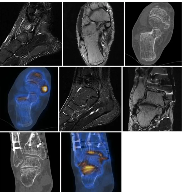
Fig. 2 Non-consolidated fragment of the left anterior calcaneal process in a 36-year-old woman with pain in her left foot (patient no. 6). a Sagittal STIR image shows the fracture of the anterior calcaneal process with stress-like changes/edema in the adjacent bones
and joints. b Sagittal CT image shows the fracture of the anterior calcaneal process. c Fused PET/CT-image demonstrates increased [18F]fluoride uptake in the region of the fracture
Fig. 1 Os trigonum syndrome in a 39-year-old man with pain in his left foot (patient no. 1). a Sagittal STIR image does not show increased signal in the os trigonum. b Three-dimensional PET/CT fusion demonstrates the increased activity as a red spot in the os trigonum. c
Sagittal CT image shows the os trigonum. d Fused PET/CT image demonstrates the increased activity in the os trigonum region. e Infiltration after PET-/CT
Surprisingly, PET/CT in this study delivered 9/15 new findings compared to previous MR examinations, two of them being retrospectively confirmed but less prominent on MRI.
Six findings could be seen both on PET/CT and MRI (‘blinded’ evaluation). In one case no active lesion at all was seen on PET/CT, although morphologic information was confirmed (patient no. 7)
Interestingly, one finding on PET/CT of possible periostitis corresponded to a soft-tissue finding on MRI after i.v. injection of contrast agent (tenosynovitis of peroneal tendons, patient no. 5) as an indirect sign of concomitant increased bone activity.
The results of this study suggest the complementary usefulness of PET/CT and MRI for patients with unclear foot pain.
Second, this study evaluated impact on management rather than outcome—an accepted approach in fluoro-2-deoxy-D
-glucose (FDG) PET/CT oncology literature, as outcome studies are more difficult to perform [20,21]. Further studies have to confirm that [18F]fluoride PET/CT examinations have not only an impact on therapeutic decision making but also on patient outcome.
Third, the study has a selection bias, because patients were only referred to PET/CT when imaging and/or clinical diagnosis had failed to lead to an obvious diagnosis.
However, it is a standard approach to use a new imaging technology first on patients for whom other imaging has failed to provide a conclusive diagnosis.
In our experience [18F]fluoride PET/CT is a highly sensitive tool showing many lesions with different uptake intensities in both feet with and without concomitant morphological changes. Interpretation of these multiple uptakes might be difficult.
A thorough history and adequate clinical examination (i.e., pain location in particular, trauma, surgery) are of utmost importance to avoid misinterpretation of the imaging findings. Our study population included patients with and without a history of trauma and/or surgery. The number of patients was limited, and the results of this preliminary study should be confirmed in more and specifically selected patients.
The effective dose equivalent is 0.023 mSv/MBq for [18F]fluoride [22], which is a maximum effective dose equivalent to 6.97 mSv in this study. The wide range of injected activity from 97–303 MBq is explained by the fact that we realized with increasing use of fluoride in our institution that an administered activity of 100–150 MBq is quite adequate to provide diagnostic images. With this‘low dose’ protocol, the radiation burden of fluoride PET-CT is comparable to that of conventional SPECT/CT. Another advantage is the short uptake time of approximately 30 min
Fig. 3 Subtalar osteoarthritis and plantar fasciitis (left side) in a 70-year-old woman with pain in her left foot (patient no. 9). a Axial T1-w image and sagittal STIR image demonstrate only slight irregularities of the contour of the posterior talocalcaneal joint. b Sagittal CT image, with slight subchondral sclerosis in the talocalcaneal joint and no major plantar changes. c Fused PET/CT image showing increased uptake in these regions, indicating subtalar osteoarthritis and plantar fasciitis
compared with 3–4 h in conventional bone scintigraphy. One clear limitation of fluoride PET/CT compared to conventional bone scintigraphy and SPECT is the limited availability of [18F]fluoride and PET/(CT) scanners
com-pared to those of SPECT and bone scanning, and the higher costs [3, 23]. The cost effectiveness of fluoride PET/CT imaging in the diagnosis of orthopedic diseases should be analyzed in further studies [23,24].
Fig. 4 Osteoarthritis of the talotibial joint and subtalar joint and os tibiale externum syndrome in a 39-year-old man with pain in his right foot (patient no. 10). a Sagittal STIR image and axial T1-w image show a non-active os tibiale externum. b Axial CT demonstrates the os tibiale externum. c Fused PET/CT image shows an active os tibiale externum. d) Sagittal STIR image and coronal T1-w image
demon-strate talotibial joint osteoarthritis and no prominent degenerative changes of the subtalar joint. e Coronal CT shows degenerative changes in the talotibial joint that are more than those in the subtalar joint. f Fused PET/CT image with signs of active osteoarthritis of the talotibial joint as well as the subtalar joint. Note that there are no major drawbacks due to attenuation correction after osteosynthesis
T able 2 Comparison of MRI findings and PET/CT findings with the reviewer (a) unaware and (b) aware of the PET/CT findings. All findings from the PET/CT , except for patient no. 7, showed structures or lesions with increased [ 18 F]fluoride uptake (active lesions). m male, f female, MT metatarsal Patient no. Gender Age (years) Side of pain Interval between MRI and PET/CT Diagnosis in MRI Diagnosis by PET/CT Comparison of PET/CT with MRI (a) Unaware of PET/CT findings (a) Unaware (b) A ware of PET/CT findings (b) Retrospectively 1 m 39 left 6 months (a ) and (b) Not suggestive Os trigonum syndrome (a) New finding (b) Finding retrospectively not confirmed by MRI 2 m 55 left 8 days (a) Thickened anterior fibulotalar ligament Sinus tarsi syndrome (a)New finding Old medial malleolar fragment (b) Finding retrospectively confirmed by MRI (less prominent than on PET/CT) (b) Slight edema/signal changes in sinus tarsi 3 f 35 left –– Osteoarthritis of the talotibial joint including talofibular space; non-consolidated fragment of anterior calcaneal process – 4 m 41 left 6
months (external examination without contrast
agent) (a) T alotibial joint ef fusion; rupture of the anterior fibulotalar ligament; suspicion of rupture of tibiofibular syndesmosis; little osteochondral defect of medial trochlea tali, unspecific synovitis in the talotibial joint Non-union after distal tibial fracture dorsally (a) New finding (b) No distal tibial fracture found (b) Finding retrospectively not confirmed by MRI 5 f 46 left 5
months (external examination with
contrast agent) (a) Edema of soft tissue dorsally; caput tali with unspecific signal alterations at plantar side; rupture of the anterior fibulotalar ligament; talotibial joint ef fusion; rupture of fibulocalcaneal ligament; tenosynovitis of peroneal tendons (only visible after i.v . injection of contrast agent) Possibly calcaneal periostitis (a) One finding confirmed (b) See above (b) See above 6 f 36 left 2 months (a) Fracture of anterior calcaneal process with stress-like alterations/edema in the adjacent bones and joints (caput tali, trochlea tali, talocalcaneal joint, calcaneal bone, navicular bone) Non-consolidated fragment of the left anterior calcaneal process (a) Same finding (b) See above (b) See above 7 m 40 left 2 months (a) Calcaneo-navicular coalition; reactive surrounding alterations; calcaneo-cuboidal stress reaction; old postoperative changes in the distal fibula; obliteration of sinus tarsi; flat foot deformity; missing anterior fibulotalar ligament Calcaneo-navicular coalition without increased activity (a) One finding confirmed, on PET/CT not active (!)
T able 2 (continued) Patient no. Gender Age (years) Side of pain Interval between MRI and PET/CT Diagnosis in MRI Diagnosis by PET/CT Comparison of PET/CT with MRI (a) Unaware of PET/CT findings (a) Unaware (b) A ware of PET/CT findings (b) Retrospectively (b) See above (b) See above 8 m 53 left –– Osteoarthritis of the talonavicular joint – 9 f 70 left 3 months (a) Calcaneal edema facing angle of Gissane; irregularities of contour of posterior talocalcaneal joint, slight talotibial joint osteoarthritis Subtalar osteoarthritis; plantar fasciitis (a) One finding confirmed, more prominent on PET/ CT , one finding new (b) Discrete plantar fasciitis/degeneration (b) Both findings retrospectively confirmed (less prominent than on PET/CT) 10 m 3 9 right 2 days (a) Postoperative changes with talotibial joint osteoarthritis with chondral damage, old rupture of anterior fibulotalar ligament, os tibiale externum Osteoarthritis of the talotibial joint and subtalar joint; os tibiale externum syndrome (a) One finding confirmed, one finding new , one finding on PET/CT active, not on MRI (b) T alotibial joint osteoarthritis, no prominent degenerative changes of talocalcaneal joint, os tibiale externum not active (b) One finding retrospectively confirmed; two findings not confirmed or not active on MRI 1 1 m 4 6 both sides 22 days MRI right foot Os trigonum syndrome of the left side and plantar fasciitis of both sides MRI right foot (a) Not suggestive, os trigonum not active (a) New finding by PET/CT (but rather discrete) (b) No plantar fasciitis found (b) Finding retrospectively not confirmed on MRI 12 f 5 6 left>right 3 months MRI left foot Bilateral proximal intermetatarsal neoarticulation MT III/IV MRI left foot (a) T endinopathy of posterior tibial tendon, unspecific fibrosis at plantar side of head of metatarsal IV (a) New finding on PET/CT (b) Proximal metatarsal bones not suggestive (b) Finding retrospectively not confirmed on MRI 13 f 6 8 right>left 5 months MRI right foot Right side: osteoarthritis of the third metatarso-phalangeal joint and first metatarsal sesamoid bone osteoarthritis MRI right foot (a) Osteoarthritis metatarsophalangeal joint I with concomitant sesamoid bone osteoarthritis; atrophy/edema of interosseous muscles: neurogenic origin? Left side: insertional tendinopathy anterior fibulotalar ligament; increased uptake in a degenerative lesion of the medial cuneiform (a) One finding confirmed, one finding new (b) Metatarsophalangeal joint III not suggestive (b) Second finding retrospectively not confirmed on MRI
Conclusion
[18F]fluoride PET/CT has substantial impact on therapeutic management, with management changes in close to half of all patients with unclear foot pain in this prospective study. Therefore, [18F]fluoride PET/CT appears to be a good imaging adjunct, at least in those patients for whom the findings of clinical examination and morphologic imaging modalities such as MRI or CT alone remain inconclusive.
Aknowledgements We thank Sabine Knöfel for all technical support.
References
1. Grant FD, Fahey FH, Packard AB, Davis RT, Alavi A, Treves ST. Skeletal PET with 18F-fluoride: applying new technology to an old tracer. J Nucl Med. 2008;49:68–78.
2. Groves AM, Win T, Haim SB, Ell PJ. Non-(18F)FDG PET in clinical oncology (review). Lancet Oncol. 2007;8:822–30. 3. Even-Sapir E, Mishani E, Flusser G, Metser U. 18F-fluoride
positron emission tomography and positron emission tomography/ computed tomography. Semin Nucl Med. 2007;37:462–9. 4. Schirrmeister H, Guhlmann A, Kotzerke J, Santjohanser C, Kühn T,
Kreienberg R, et al. Early detection and accurate description of extent of metastatic bone disease in breast cancer with fluoride ion and positron emission tomography. J Clin Oncol. 1999;17:2381–9. 5. Schirrmeister H, Glatting G, Hetzel J, Nüssle K, Arslandemir C,
Buck AK, et al. Prospective evaluation of the clinical value of planar bone scans, SPECT, and 18F-labeled NaF PET in newly diagnosed lung cancer. J Nucl Med. 2001;42:1800–4.
6. Even-Sapir E, Metser U, Mishani E, Lievshitz G, Lerman H, Leibovitch I. The detection of bone metastases in patients with high-risk prostate cancer: 99mTc-MDP planar bone scintigraphy, single- and multi-field-of-view SPECT, 18F-fluoride PET, and 18F-fluoride PET/CT. J Nucl Med. 2006;47:287–97.
7. Beheshti M, Vali R, Waldenberger P, Fitz F, Nader M, Loidl W, et al. Detection of bone metastases in patients with prostate cancer by 18F fluorocholine and 18F fluoride PET-CT: a comparative study. Eur J Nucl Med Mol Imaging. 2008;35:1766–74. 8. Schirrmeister H, Guhlmann A, Elsner K, Kotzerke J, Glatting G,
Rentschler M, et al. Sensitivity in detecting osseous lesions depends on anatomic localization: planar bone scintigraphy versus 18F PET. J Nucl Med. 1999;40:1623–9.
9. Brenner W, Vernon C, Conrad EU, Eary JF. Assessment of the metabolic activity of bone grafts with 18F-fluoride PET. Eur J Nucl Med Mol Imaging. 2004;31:1291–8.
10. Frost ML, Blake GM, Park-Holohan S-J, Cook GJR, Curran KM, Marsden PK, et al. Long-term precision of 18F-fluoride PET skeletal kinetic studies in the assessment of bone metabolism. J Nucl Med. 2008;49:700–7.
11. Sterner T, Pink R, Freudenberg L, Jentzen T, Quitmann H, Bockisch A, et al. The role of [18F]fluoride positron emission tomography in the early detection of aseptic loosening of total knee arthroplasty. Int J Surg. 2007;5:99–104.
12. Ovadia D, Metser U, Lievshitz G, Yaniv M, Wientroub S, Even-Sapir E. Back pain in adolescents: assessment with integrated 18F-fluoride positron-emission tomography-computed tomogra-phy. J Pediatr Orthop. 2007;27:90–3.
13. Sörensen J, Michaelsson K, Strand H, Sundelin S, Rahme H. Long-standing increased bone turnover at the fixation points after anterior cruciate ligament reconstruction: a positron emission tomography (PET) study of 8 patients. Acta Orthop. 2006;77:921–5.
14. Dasa V, Adbel-Nabi H, Anders MJ, Mihalko WM. F-18 fluoride positron emission tomography of the hip for osteonecrosis. Clin Orthop. 2008;466:1081–6.
15. Rüdisüli A, Müller-Brand J. Spectrum of findings in SPECT-CT of the feet—a review of 54 patients with pain as the main referring symptom. Supplementum 47. Swiss Medical Forum. 10th Annual Congress of the Swiss Society Of Nuclear Medicine; 4–6 June 2009;9:21–2.
16. Pagenstert GI, Barg A, Leumann AG, Rasch H, Müller-Brand J, Hintermann B, et al. SPECT-CT imaging in degenerative joint disease of the foot and ankle. J Bone Joint Surg Br 2009;91:1191–6.
17. Perkins A, Hilson A, Hall J. Global shortage of medical isotopes threatens nuclear medicine services. BMJ. 2008;337:a1577. 18. Stafford N. Isotope shortage is limiting nuclear medicine across
Europe. BMJ. 2008;337:a1575.
19. Pneumaticos SG, Chatziioannou SN, Hipp JA, Moore WH, Esses SI. Low back pain: prediction of short-term outcome of facet joint injection with bone scintigraphy. Radiology. 2006;238:693–8.
20. Zafra M, Ayala F, Billalabeitia E, Vicente E, Gonzalez-Cabezas P, García T, et al. Impact of whole-body 18F-FDG PET on diagnostic and therapeutic management of medical oncology patients. Eur J Cancer. 2008;44:1678–83.
21. Fryback DG, Thornbury JR. The efficacy of diagnostic imaging. Med Decis Making. 1991;11:88–94.
22. Hoegerle S, Juengling F, Otte A, Altehoefer C, Moser EA, Nitzsche EU. Combined FDG and F-18-fluoride whole body PET: a feasible two-in-one approach to cancer imaging? Radiology. 1998;209:253–8.
23. Hetzel M, Arslandemir C, König HH, Buck AK, Nüssle K, Glatting G, et al. F-18 NaF PET for detection of bone metastases in lung cancer: accuracy, cost-effectiveness, and impact on patient management. J Bone Miner Res. 2003;18:2206–14.
24. Krug B, Van Zanten A, Pirson AS, Crott R, Borght TV. Activity-based costing evaluation of a 18F-fludeoxyglucose positron emission tomography study. Health Policy. 2009;92:234–43.