ENVIRONMENTAL MICROBIOLOGY
Molecular Characterization of an Endolithic Microbial
Community in Dolomite Rock in the Central
Alps (Switzerland)
Thomas Horath&Reinhard Bachofen
Received: 12 August 2008 / Accepted: 19 December 2008 / Published online: 28 January 2009
# Springer Science + Business Media, LLC 2009
Abstract Endolithic microorganisms colonize the pores in exposed dolomite rocks in the Piora Valley in the Swiss Alps. They appear as distinct grayish-green bands about 1–8 mm below the rock surface. Based on environmental small subunit ribosomal RNA gene sequences, a diverse community driven by photosynthesis has been found. Cyanobacteria (57 clones), especially the genus Leptolyngbya, form the functional basis for an endolithic community which contains a wide spectrum of so far not characterized species of chemotrophic Bacteria (64 clones) with mainly Actinobacteria, Alpha-Proteobacteria, Bacteroidetes, and Acidobacteria, as well as a cluster within the Chloroflex-aceae. Furthermore, a cluster within the Crenarchaeotes (40 clones) has been detected. Although the eukaryotic diversity was outside the scope of the study, an amoeba (39 clones), and several green algae (51 clones) have been observed. We conclude that the bacterial diversity in this endolithic habitat, especially of chemotrophic, nonpig-mented organisms, is considerable and that Archaea are present as well.
Introduction
Microorganisms inhabiting rock were first observed and described 100 years ago [19,45, 79], nevertheless, except for cyanobacteria, little is known about the community composition and the biodiversity of these microbial ecosystems. They are typical for hot and cold arid
environments where in the pores of the rock, they are partially sheltered from a number of physical stresses such as solar radiation, heat, cold, or desiccation. Various organisms settle on the surface and invade pores and cracks. Within the rock, they form a structured biofilm, a clearly defined organismic layer or band a few millimeters below the surface [15–17, 24–26, 35, 42, 44, 58, 65, 88–90]. Contrary to submerged biofilms, endolithic biofilms are patchier due to local inhomogeneities of rock structures and environments [42]. These communities contain bacteria, fungi, and eukaryotic microalgae [81, 82]. They form complex physiological networks tied to solid particles by extracel-lular polymeric substances (EPS). The synthesis of these polymers is controlled by different environmental stress factors (e.g. [77]). The organismic composition is governed by the hostile environment. Water is only periodically available in the form of rain, dew, or just atmospheric humidity. Therefore, EPS are most important for the endolithic population as they retain water and act as osmoprotectant and nutrient reservoirs. In the Alps, main nutrients are scarce, and the daily and seasonal temper-atures oscillate widely. At high altitudes, the sunlight with a strong part in the UV is a further life threatening factor [95]. Habitats with such fluctuating environmental conditions pose a strong challenge to organisms, and life there may reach its limits at least in certain periods.
Endolithic microorganisms have gained interest in the past decades for several reasons: e.g., as possible analogs of extraterrestrial life, such as life on Mars [2, 24, 40, 43, 46,
59, 63, 73, 94, 95], for the study of the mechanisms of adaptation to extreme and hostile conditions [30,36,87,96], to study the processes of weathering and mineral dissolution [12,91] or for phylogenetic reasons [17,29,65,86,88,89]. Endolithic microbial communities are found worldwide in dry and aquatic environments. The ones studied and DOI 10.1007/s00248-008-9483-7
T. Horath
:
R. Bachofen (*)Institute of Plant Biology/Microbiology, University of Zürich, Zollikerstr. 107,
CH-8008 Zürich, Switzerland e-mail: [email protected]
described came from cliffs of the Niagara escarpment [32,56,57] from streams in the UK [68] and from gypsum cliffs in Nova Scotia [28]. They are found in hot and arid desert environments [5–7, 23], in travertine in Turkey [69], in arctic and antarctic locations [2,26,27,44,74,93], in mountainous regions [42,65, 88,89], and in the marine littorals [92]. Most investigations have been based on traditional techniques, mainly light and electron microscopy, and on cultures. They have usually been focused on pigmented microorganisms, oxygenic phototrophs such as green algae and cyanobacteria as well as filaments of fungi as partners of lichen symbiosis. Cyanobacteria are important in the early stages of primary succession processes in soils, especially because many species are able to fix dinitrogen [47]. However, it must be assumed that a variety of heterotrophic organisms will rapidly follow the photo-trophs after their invasion. So far, molecular methods have hardly been used. They have even been thought to be useless in studying endoliths [93]. However, molecu-lar techniques are now successfully applied to characterize endolithic communities such as the cyanobacterial popula-tion in the dolomite rocks in Switzerland [82], the endolithic community in the McMurdo Dry Valleys in the Antarctica [17], or the microbial population in rocks of the Rocky Mountains [29,65,89].
The objective of the present study is to describe the broad genetic diversity of the endolithic bacterial popula-tions present in the dolomite formapopula-tions of the Swiss Alps by culture independent molecular methods. Dolomite rocks (CaMg(CO3)2) in the Piora Valley in southern Switzerland
are often bare of vegetation and exposed to hostile conditions. Such weathered rocks harbor chasmoendolithic and cryptoendolithic (definitions, see [34]) phototrophic and heterotrophic microbial communities which become easily visible as grayish-green bands some millimeters below the surface. This hidden microbial ecosystem was first characterized by Diels [19] in the Italian Dolomites and has been studied in Piora dolomite by molecular [82], spectroscopical, and optical techniques [42]. At a depth of 2 to 8 mm, the phototrophic microorganisms still receive enough photosynthetic active radiation while they are protected from excessive sunlight with a high fraction in the UV range [42]. As most organisms of environmental samples cannot be cultured by standard methods yet, a description of the microbial diversity of this special microbial ecosystem has been obtained by sequence analyses of polymerase chain reaction (PCR) amplified fragments of the small subunit of the ribosomal ribonucleic acid gene (SSU rRNA gene). The knowledge of the composition of the microbial community will help to better understand the biogeochemical processes that occur in these habitats. Preliminary results have been presented earlier [41,81].
Materials and Methods Sampling Site
Dolomite rock material was collected in the Piora Valley in the southern part of the Swiss Alps at an elevation of 1,965 m above sea level in summer 2001 and 2003. The coordinates of the specific sampling site are 46°32′51″ N, 8°43′05″ E. Details of the site are given by Sigler et al. [82] and Horath et al. [42]. The geology of the Piora Valley, oriented east-west, is characterized by a dolomite trough, a few hundred meters wide, surrounded by crystalline rock formations. Due to erosion by wind and water, the dolomite is often exposed to the atmosphere, forming white cliffs. Such sites are sparsely covered with black epilithic cyanobacteria and lichens. Especially in slightly weathered dolomite, endolithic microorganisms are easily observed when the surface layer is removed. They form a grayish-green layer about 1–8 mm below the rock surface. Rock pieces of some millimeters or centimeters in size were cut off from the surface with an ethanol-flamed chisel and hammer, and samples with visible endolithic bands were kept in Falcon tubes in the dark at 4°C until DNA extraction in the laboratory.
DNA Extraction
DNA extraction was performed as described by Sigler et al. [82]. In brief, 0.5 to 0.6 g of rock samples of the green layer was scratched into a sterile empty Petri dish with sterilized tools, then put into 2-ml sterile microfuge tubes containing 1.0 ml of extraction buffer (50 mM NaCl, 50 mM ethylene diamine tetra acetic acid disodium salt dihydrate (EDTA; Fluka 03685), 50 mM 2-amino-2-hydroxymethyl-propane-1,3-diol hydrochloride (TRIS-HCl; Fluka 93363) and 5% sodium dodecyl sulfate (SDS; Fluka 71729), final pH 8), 0.5 g glass beads (0.1 and 0.5 mm in diameter) and eventually 0.5 ml of a phenol–chloroform–isoamylalcohol-mixture (v/v/v=49.5/49.5/1, Fluka 77618). The tubes were sealed with Parafilm®, shaken in a bead beater (“FastPrep®”, BIO 101, La Jolla, CA, USA) at 5.5 m s−1 for 30 s and centrifuged for 4 min at 10,000×g. Nucleic acids were isolated by standard phenol/chloroform extraction and ethanol precipitation [75]. The dry DNA pellet was redis-solved in 50μl distilled autoclaved water.
PCR Amplification of SSU rRNA Genes
The small subunit rRNA gene was amplified from genomic DNA by PCR with several pairs of primers (see Table1).
PCR was performed in 200-μl thin-walled tubes on a
“Progene” or a “Genius” thermocycler respectively (Techne LTD, Duxford Cambridge, U.K) in a volume of 25μl. The reaction mixture contained (final concentrations): the
T able 1 List of clone library names, sequences of primers, and numbers of dif ferent clones obtained (3% sequence dif ference level) Library name Primer Primer sequence (5 ′ to 3′ ) Reference Obtained products T otal clones Sequenced clones Dif ferent clones Dolo 27f AGA GTT TGA TCM TGG CTC AG [ 20 , 50 ] Bacteria/ Chloroplasts 36 30 22 1524r AAG GAG GTG A T C CAR CCG [ 50 ] Slightly modified DoAr 8aF TCY GGT TGA TCC TSC C [ 11 ] Slightly modified Euamoeba sp. 39 1 1 1517r A T C CAG CCG CAG R T T C This paper ud 536f CAG CMG CCG CGG T A A TWC [ 49 ] Bacteria/Cr enar chaea 35 16 10 1392r ACG GGC GGT GTG TRC [ 49 ] DA 8aF TCY GGT TGA TCC TSC C [ 11 ] Slightly modified Chlor ella sp. (18S) 16 8 4 1512uR ACG GHT ACC TTG TT A CGA CTT [ 50 ] Slightly modified 1492r DOS 89Fb ACG GCT CAG T A A CRC [ 10 ] Cr enar chaea 38 10 3 915R GTG CTC CCC CGC CAA TTC CT [ 85 ] DOL 8aF TCY GGT TGA TCC TSC C [ 11 ] Slightly modified Bryophyta (18S) 28 5 1 1512uR ACG GHT ACC TTG TT A CGA CTT [ 50 ] Slightly modified DoCY CY A359F GGG GAA TTT TCC GCA A T G G G [ 66 ] Cyanobacteria 23 9 6 CY A1342R GAC CTG CAA TT A C T A GCG [ 78 ] Docu CY A359F GGG GAA TTT TCC GCA A T G G G [ 66 ] Cyanobacteria 36 17 6 CY A1342R GAC CTG CAA TT A C T A GCG [ 78 ] Chloroplasts Sequencing Primers (5 ′ to 3′ ) M13 forward GT A AAA CGA CGG CCA G [ 60 ] M13 reverse CAG GAA ACA GCT A T G A C [ 60 ] 519r GW A T T A CCG CGG CKG CTG [ 49 ] 536f CAG CMG CCG CGG T A A TWC [ 49 ] 1099r GGG TTG CGC TCG TTR C [ 50 ] Slightly modified 1 1 14f GY A ACG AGC GCA ACC C [ 50 ] Slightly modified
appropriate Taq buffer (1×), 1.5–2.0 mM MgCl2, 0.1 mg ml−1
bovine serum albumine, 0.2 mM dNTP’s, 200 nM forward
primer, 200 nM reverse primer, 40–100 U ml−1 Taq
Polymerase (Sigma, Promega, Invitrogen, or Pharmacia), and approximately 50–100 ng template DNA. PCR was run under the following conditions: initial denaturation at 94°C for 2 min, 10 cycles of 94°C for 20 s, 60–0.5°C/cycle for 30 s, 72°C for 60 to 90 s depending on the length of the product, 20 cycles of 94°C for 20 s, 50°C to 58°C for 30 s, depending on the annealing temperature of the primers, 72°C for 60 to 90 s. The products were checked on a 1% agarose gel in 0.5× TAE buffer [1×=40 mM Tris base (2-amino-2-hydroxymethyl-propane-1,3-diol), 20 mM glacial acetic acid, 1 mM Na2EDTA of pH 8.0].
Cloning
PCR-amplified products were cloned without purification with the TOPO TA cloning kit (Invitrogen) as specified by the manufacturer’s manual.
Restriction Fragment Length Polymorphism
After plasmid DNA mini preparation with alkaline lysis [75] and the reamplification of the SSU rRNA gene with M13 primers, restriction was carried out with Hinf I and Hae III and the fragments analyzed on a Spreadex® EL 800 Wide Mini S-50 gel (Elchrom Scientific) run at 55°C for 1 h at 10 V cm−1. The gels were stained with 10,000 times diluted 1% (w/v) ethidium bromide and viewed with 302 nm UV illumination.
DNA Sequencing
Reamplified plasmid inserts were purified by filtration (Amicon Microcon YM-100 filter, Millipore Corporation, Bedford, MA, USA), and 100 to 180 ng DNA (dissolved in 1μl H2O) were
used for sequencing-PCR using 0.8 μl BigDye® Terminator v3.1 (Applied Biosystems), 1.5 μl sequencing buffer (5×), 6.8 μl of H2O Milli Q, and 0.25μl (5 μM) of one of the
sequencing primers listed in Table1. Before the automated loading into the polymers on the 48-capillary sequencer (Applied Biosystems 3730 DNA Analyzer), the PCR products were purified by centrifugation through Sephadex G50 (Amersham Pharmacia). The raw sequences were aligned and combined using the Gene Codes Sequencher software (www.genecodes.com).
Nucleotide Sequence Accession Numbers
The SSU rRNA gene sequences found have been deposited at the DNA Data Bank of Japan and can be retrieved under the accession numbers AB257629 to AB257698 and AB334273 to AB334298.
Phylogenetic Tools
Rarefaction curves were generated with the program “Analytic Rarefaction 1.3” provided by Steven M. Holland at “http://www.uga.edu/~strata/software/Software.html”.
The newly obtained SSU rRNA gene sequences were compared with known sequences in the NCBI database (Genbank) at the National Center for Biotechnology Information (http://www.ncbi.nlm.nih.gov/) by the use of the Basic Local Alignment Search Tool (BLAST) [1] to determine their approximate phylogenetic affiliation.
The EMBOSS Pairwise Alignment Tool at“http://www.ebi. ac.uk/emboss/align/” provided by the European Bioinformatics
Institute was used to compare single sequences in the following mode: “Method: water”; “Gap Open: 10.0”; “Gap Extend: 10.0”; “Molecule: DNA”; “Matrix: DNAfull”.
The new SSU rRNA gene sequences were further added to the rRNA gene sequence database of the Technical University of Munich (ssu_jan04_corr_opt.arb, release February 2005) by the use of the program package ARB ([54], http://www.arb-home.de). The integrated tool ARB_ALIGN was used for automatic sequence alignment, which was then checked with a critical eye according to the secondary structure of the rRNA molecule, and corrected. If missing, the latest best fitting sequences found by NCBI-BLAST were added to the ARB database.
The final phylogenetic trees were derived from the basic phylogenetic tree of about 51,000 SSU rRNA sequences after adding the new sequences with appropriate filters, and the“Maximum Parsimony Method.” Bootstrap values were calculated from the sequences used in the final trees by using the “Phylip Parsimony Method”, integrated in ARB, compressing vertical gaps, running 100 bootstrap samples. In order to plot a phylogenetic tree, many different algorithms are available today, which all lead to accept-able results if they are based on a proper sequence alignment [52]. Therefore, emphasis has been put on an accurate alignment. The trees presented are copies of the largest tree, namely “tree_1000_jan05” in the ARB
data-base “ssu_jan04_corr_opt.arb”. After adding the new
sequences to the existing tree containing more than 50,000 single SSU rRNA sequences, the new trees have been reduced to a convenient size for illustration. Bootstrap values have been calculated although they are not consid-ered to be very important, since these values can be shifted by omitting closely branching sequences before calculation (Eichenberger, Ch., personal communication).
Bootstrapping has been introduced to provide confidence intervals in phylogenetic calculations [13, 21], because calculated trees are never fully true and require flexible interpretations. When using Maximum Parsimony, Distance Matrix (Neighbor Joining), or Maximum Likelihood, the result should not be overestimated because its variation
among different methods is a negligible indicator of the confidence interval [21]. Furthermore, the order of adding sequences to a calculation has an effect on the tree topology [e.g.,53]. Thus removing and readding complete groups to a tree may rearrange its branching. In our case, it improved the congruence of the results of ARB and NCBI.
Results
In a previous study, we investigated endolithic bacterial communities in exposed weathered dolomite rocks by confocal laser scanning microscopy, pigment analysis, and reflectance spectroscopy [42]. Communities depending on photosynthesis usually harbor a sum of heterotrophic organisms which feed on exudates and lysed cells. As it is hardly possible to characterize the diversity of environmental microorganisms by cultivating them, we analyzed the endo-lithic heterotrophic community by cloning and sequencing their SSU rRNA genes.
SSU rRNA Gene Clone Libraries
Isolation of DNA from fine powdered rock material posed some difficulties as DNA tended to stick to and precipitate with the inorganic rock debris. Suitable amounts of DNA were obtained following the procedure of Sigler et al. [82]. To evaluate the diversity of the prokaryotic endolithic community, eight independent clone libraries with different combinations of universal and phylogenetic group-specific oligonucleotide primers were constructed, including two libraries with specific cyanobacterial primers (Table 1). In total, 254 clones were analyzed by restriction fragment length polymorphism (RFLP), 96 of which were sequenced. Assuming a threshold of a minimal 3% sequence difference between species [84], 53 sequences fell into distinctly related groups. From these 53 phylotypes, 45 belong to Bacteria (including three chloroplasts of two green algae and a moss), three to Archaea, and five to Eukarya (Table 2). Scanning the graphic alignment of the NCBI-BLAST analysis of the new sequences, no chimeras have been detected ([1],http://www.ncbi.nlm.nih.gov/).
A wide diversity was found in the clone libraries obtained with the bacterial primer pair 27f/1524r and the “universal” primer pair 536f/1392r. In the bacterial library, 22 out of 36, and in the“universal” library, nine out of 35 clones were different. The other primer pairs resulted in less diverse libraries. As an extreme, primer pairs 8aF/1517r (DoAr) and 8aF/1512uR (DOL) yielded 39 and 28 RFLP-identical clones, respectively (Table2). Primer 1517r (Table1) was originally designed to increase the number of Archaea clones but resulted in the detection of a so far unknown 18S rRNA gene sequence fragment
closely related to Saccamoeba limax (99.4%, clone DoAr09).
Rarefaction curves for all the eight clone libraries are shown in Fig.1. The shapes of the curves“Dolo” and “ud” indicate that further sampling would increase the number of operational taxonomic units (OTUs, 3% difference level). In contrast, the other graphs, except for the summarized data, level off rapidly, a phenomenon for discussion.
A quarter of the obtained bacterial sequences (64 out of a total of 251 clones) originated from phototrophic oxygenic organisms. Cyanobacteria were numerous with 11 phylotypes, chloroplasts of green algae (Dolo-01, Dolo-34) or of bryophytes (Docu-30) with three different phylotypes (seven clones). Among the heterotrophic species, the representatives of the phylum Actinobacteria were the most numerous (15 clones, seven phylotypes), followed by Alpha Proteobacteria (14 clones, ten phylo-types), and Bacteroidetes (12 clones, two phylotypes). Acidobacteria (seven clones, two phylotypes), Gamma Proteobacteria (five clones, one phylotype), and Gemma-timonadetes (two clones, two phylotypes) were less frequent. Only one clone was found in each of the proposed divisions TM6 and TM7, as well as in the phylum Planctomycetes (Fig. 2 and Table 2). The green nonsulfur phototrophic bacteria group of the Chloroflexi yielded six clones (four phylotypes). The sum of bacterial phyla found in the dolomite of the Piora Valley covers ten of approximately 75 bacterial phyla known or postulated so far [51,72, 76]. All the archaeal sequences found fell into the group of uncultured Crenarchaeotes (Table 2). Eukaryotic 18S rRNA gene sequences have been found in groups related to Euamoebida, Bryophyta, and Chlorophyta (83 out of a total of 251 clones, five phylotypes; Table2). The phylogenetic trees give an overview of the distribution of the newly detected SSU rRNA gene sequences in the domains of Bacteria, Archaea, and Eukarya (Figs.3a, b,4, and5).
Within all sequences analyzed, the percentages of sequence identity with SSU rRNA gene sequences avail-able at GenBank (http://www.ncbi.nlm.nih.gov/) range between 85.2% and 99.7%. Clone “DOS_02”, on a length of 791 bp, was even 99.9% identical with the uncultured archaeon clone HL17 (AJ608203) in loam from a bank of the river Waal in the Netherlands, while clone “Dolo-07”, on a length of 1,425 bp, shows only an 83.8% similarity with the uncultured Chloroflexus clone pItb-vmat-61 (AB294962) from a microbial mat in a shallow submarine hot spring in Japan (Table2). For some sequences, ARB or “EMBOSS Pairwise Alignment Algorithms” have found different closest relatives as compared to NCBI-BLAST, but then often with smaller sequence coverage. Among the 45 different bacterial phylotypes, 18 (40%) were less than 95% identical to the closest 16S rRNA gene in the nucleotide sequence database, 14 phylotypes (31%) were
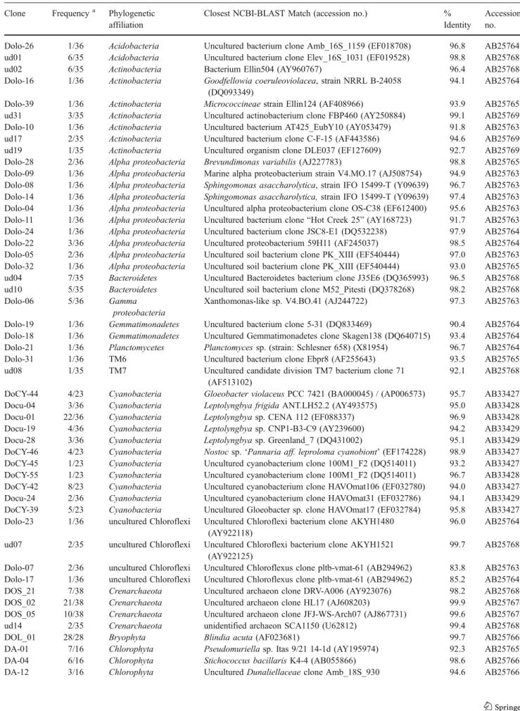
Table 2 Phylogenetic affinities of SSU rRNA gene sequences obtained from dolomite in the Piora Valley, Central Alps Clone Frequencya Phylogenetic
affiliation
Closest NCBI-BLAST Match (accession no.) %
Identity
Accession no.
Dolo-26 1/36 Acidobacteria Uncultured bacterium clone Amb_16S_1159 (EF018708) 96.8 AB257649
ud01 6/35 Acidobacteria Uncultured bacterium clone Elev_16S_1031 (EF019528) 98.8 AB257683
ud02 6/35 Actinobacteria Bacterium Ellin504 (AY960767) 96.4 AB257684
Dolo-16 1/36 Actinobacteria Goodfellowia coeruleoviolacea, strain NRRL B-24058 (DQ093349)
94.1 AB257641
Dolo-39 1/36 Actinobacteria Micrococcineae strain Ellin124 (AF408966) 93.9 AB257657
ud31 3/35 Actinobacteria Uncultured actinobacterium clone FBP460 (AY250884) 99.1 AB257697
Dolo-10 1/36 Actinobacteria Uncultured bacterium AT425_EubY10 (AY053479) 91.8 AB257636
ud17 2/35 Actinobacteria Uncultured bacterium clone C-F-15 (AF443586) 94.6 AB257690
ud19 1/35 Actinobacteria Uncultured organism clone DLE037 (EF127609) 92.7 AB257692
Dolo-28 2/36 Alpha proteobacteria Brevundimonas variabilis (AJ227783) 98.8 AB257650
Dolo-09 1/36 Alpha proteobacteria Marine alpha proteobacterium strain V4.MO.17 (AJ508754) 94.9 AB257635 Dolo-08 1/36 Alpha proteobacteria Sphingomonas asaccharolytica, strain IFO 15499-T (Y09639) 96.7 AB257634 Dolo-14 1/36 Alpha proteobacteria Sphingomonas asaccharolytica, strain IFO 15499-T (Y09639) 97.4 AB257639 Dolo-04 1/36 Alpha proteobacteria Uncultured alpha proteobacterium clone OS-C38 (EF612400) 95.6 AB257630 Dolo-11 1/36 Alpha proteobacteria Uncultured bacterium clone“Hot Creek 25” (AY168723) 91.7 AB257637
Dolo-24 1/36 Alpha proteobacteria Uncultured bacterium clone JSC8-E1 (DQ532238) 97.9 AB257648
Dolo-22 3/36 Alpha proteobacteria Uncultured proteobacterium 59H11 (AF245037) 98.5 AB257646
Dolo-05 2/36 Alpha proteobacteria Uncultured soil bacterium clone PK_XIII (EF540444) 97.0 AB257631
Dolo-32 1/36 Alpha proteobacteria Uncultured soil bacterium clone PK_XIII (EF540444) 93.0 AB257653
ud04 7/35 Bacteroidetes Uncultured Bacteroidetes bacterium clone J35E6 (DQ365993) 96.5 AB257685
ud10 5/35 Bacteroidetes Uncultured soil bacterium clone M52_Pitesti (DQ378268) 98.2 AB257688
Dolo-06 5/36 Gamma
proteobacteria
Xanthomonas-like sp. V4.BO.41 (AJ244722) 97.3 AB257632
Dolo-19 1/36 Gemmatimonadetes Uncultured bacterium clone 5-31 (DQ833469) 90.4 AB257644
Dolo-18 1/36 Gemmatimonadetes Uncultured Gemmatimonadetes clone Skagen138 (DQ640715) 93.4 AB257643
Dolo-21 1/36 Planctomycetes Planctomyces sp. (strain: Schlesner 658) (X81954) 96.7 AB257645
Dolo-31 1/36 TM6 Uncultured bacterium clone Ebpr8 (AF255643) 93.5 AB257652
ud08 1/35 TM7 Uncultured candidate division TM7 bacterium clone 71
(AF513102)
92.1 AB257687
DoCY-44 4/23 Cyanobacteria Gloeobacter violaceus PCC 7421 (BA000045) / (AP006573) 95.7 AB334275
Docu-04 3/36 Cyanobacteria Leptolyngbya frigida ANT.LH52.2 (AY493575) 95.0 AB334284
Docu-01 22/36 Cyanobacteria Leptolyngbya sp. CENA 112 (EF088337) 96.9 AB334282
Docu-19 4/36 Cyanobacteria Leptolyngbya sp. CNP1-B3-C9 (AY239600) 94.2 AB334292
Docu-28 3/36 Cyanobacteria Leptolyngbya sp. Greenland_7 (DQ431002) 95.1 AB334294
DoCY-46 4/23 Cyanobacteria Nostoc sp.‘Pannaria aff. leproloma cyanobiont’ (EF174228) 98.9 AB334277
DoCY-45 1/23 Cyanobacteria Uncultured cyanobacterium clone 100M1_F2 (DQ514011) 93.2 AB334276
DoCY-55 1/23 Cyanobacteria Uncultured cyanobacterium clone 100M1_F2 (DQ514011) 96.7 AB334280
DoCY-42 8/23 Cyanobacteria Uncultured cyanobacterium clone HAVOmat106 (EF032780) 94.0 AB334274
Docu-24 2/36 Cyanobacteria Uncultured cyanobacterium clone HAVOmat31 (EF032786) 94.1 AB334293
DoCY-39 5/23 Cyanobacteria Uncultured Gloeobacter sp. clone HAVOmat17 (EF032784) 95.8 AB334273
Dolo-23 1/36 uncultured Chloroflexi Uncultured Chloroflexi bacterium clone AKYH1480 (AY922118)
96.0 AB257647
ud07 2/35 uncultured Chloroflexi Uncultured Chloroflexi bacterium clone AKYH1521 (AY922125)
99.7 AB257686
Dolo-07 2/36 uncultured Chloroflexi Uncultured Chloroflexus clone pltb-vmat-61 (AB294962) 83.8 AB257633 Dolo-17 1/36 uncultured Chloroflexi Uncultured Chloroflexus clone pltb-vmat-61 (AB294962) 85.2 AB257642
DOS_21 7/38 Crenarchaeota Uncultured archaeon clone DRV-A006 (AY923076) 98.2 AB257680
DOS_02 21/38 Crenarchaeota Uncultured archaeon clone HL17 (AJ608203) 99.9 AB257674
DOS_05 10/38 Crenarchaeota Uncultured archaeon clone JFJ-WS-Arch07 (AJ867731) 99.6 AB257676
ud14 2/35 Crenarchaeota unidentified archaeon SCA1150 (U62812) 99.4 AB257689
DOL_01 28/28 Bryophyta Blindia acuta (AF023681) 99.7 AB257668
DA-01 7/16 Chlorophyta Pseudomuriella sp. Itas 9/21 14-1d (AY195974) 92.3 AB257659
DA-04 6/16 Chlorophyta Stichococcus bacillaris K4-4 (AB055866) 98.6 AB257661
in the range between 95% and 97% sequence identity, showing genus level relation [84], while 13 phylotypes (29%) were within the species level (more than 97% sequence identity).
Bacterial Community
The quantitative distribution of the different mostly heterotrophic phylotypes in the bacterial clone libraries “ud” and “Dolo” (excluding chloroplasts and the specific
cyanobacterial libraries “Docu” and “DoCY”) is
dia-grammed in Fig. 2. There are four predominant groups accounting for more than 80% of 64 clones: the Actino-bacteria together with ProteoActino-bacteria (alpha and gamma), Bacteroidetes, and Acidobacteria are the most numerous. Looking separately at individual bacterial phylotypes, the five clones ud01, ud02, ud04, ud10, and Dolo-06 are the most numerous ones, all in all accounting for 45% of the non-oxigenic “ud” and “Dolo” clones. Based on NCBI-BLAST [1], these phylotypes represent Bacteroidetes (ud04=10.9%, Table 2 (continued)
Clone Frequencya Phylogenetic
affiliation
Closest NCBI-BLAST Match (accession no.) %
Identity
Accession no. (EF023670)
Docu-30 2/36 Chloroplast Chloroplast of Hymenostylium recurvirostre (DQ629553) 99.7 AB334295
Dolo-34 1/36 Chloroplast Uncultured chlorophyte clone FQSS008 (EF522228) 96.9 AB257654
Dolo-01 4/36 Chloroplast Uncultured chlorophyte clone FQSS008 (EF522228) 97.5 AB257629
DoAr-09 39/39 Euamoebida Saccamoeba limax (AF293903) 99.5 AB257667
a
The frequency of the clones is given as the number of clones of one sort of phylotype divided by the total number of clones in that library
Sum of all eight libraries
0 10 20 30 40 50 0 50 100 150 200 250 Dolo 0 10 20 30 0 10 20 30 40 0 1 2 3 4 5 ud 0 4 8 12 0 10 20 30 40 0 10 20 30 40 0 10 20 30 40 0 10 20 30 40 DA 0 1 2 3 4 5 0 4 8 12 16 DOS 0 1 2 3 4 5 DoCY 0 2 4 6 0 5 10 15 20 25 Docu 0 2 4 6 number of clones Operational T
axonomic Units (OTUs) DoAr or DOL
Figure 1 Rarefaction curves for the different libraries and for the sum of all clones obtained. The threshold is set at 3% sequence difference to distinguish be-tween different OTUs. For clone names see, Table1
ud10=7.8%, each percentage referring to the sum of non-oxigenic “ud” and “Dolo” clones), Acidobacteria (ud01= 9.4%), Actinobacteria (ud02=9.4%), and Gamma Proteo-bacteria (Dolo-06=7.8%; Table2, and Fig.2). None of these five most numerous sequences show a similarity to known SSU rRNA gene sequences of less than 95%. Several bacterial groups collectively account for a significant fraction of the total number of clones, while individual phylotypes are not particularly numerous. Nine phylotypes belong to the class Alpha Proteobacteria representing 22% of bacterial clones. Seven phylotypes belong to the phylum Actinobacteria and represent 23% of bacterial clones. Four phylotypes affiliate with uncultured Chloro-flexi, accounting for 9.4% of the clones. Two phylotypes fall into the category of the phylum Gemmatimonadetes and consist of one clone each (3.1%). Phylotypes of Planctomycetes, of TM6 and of TM7 appear only once, each representing 1.6% of the bacterial clones. The phylogenetic position of the bacterial phylotypes is depicted in the trees in Fig.3a and b.
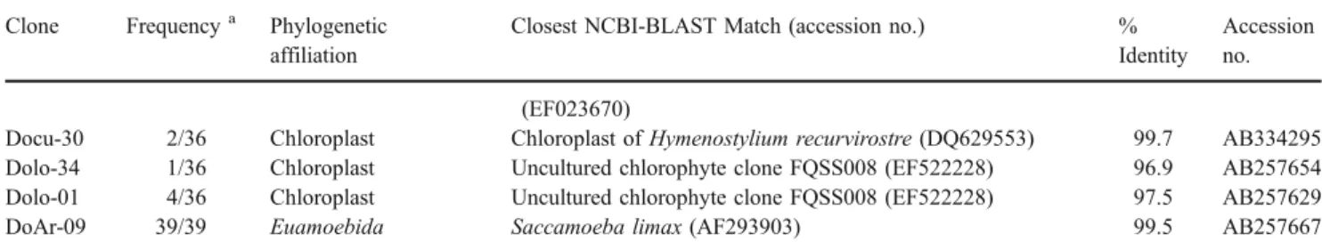
Archaea
The archaeal library generated with the primer pairs 519f/1392r and 89Fb/915R (Table 1) resulted in three phylotypes—or four if ud14 and DOS_02 are counted as two separate phylotypes. They are 99.8% identical within their 420 bp fragment between positions 519 and 934 (Escherichia coli numbering). All the archaeal phylotypes found belong to the phylum Crenarchaeota and therein to the uncultured Crenarchaeota (Fig.4). The phylotype of the clone DOS_02 amounts for the largest part of the crenarchaeal clones with 21 of 40 representatives (52.5%). It is followed by DOS_05 with ten clones (25%), DOS_21 with seven (17.5%), and ud14 with two (5%) out of 40 clones. All these clones show similarities of more than 98% with SSU rRNA gene
sequences from the public database, but for the time being, these are all uncultured archaeons. The closest named organism is Cenarchaeum symbiosum, an uncultured marine sponge symbiote [37], with similarities of 86% to ud14 and 81% to DOS_02, according to the EMBOSS Pairwise Alignment Tool provided by the EBI.
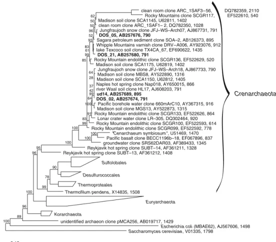
Eukaryotic Microorganisms
The primer combinations 8aF/1512uR and 8aF/1517r resulted in several eukaryotic sequences of SSU rRNA (Table2). As the clones DA-04 and DA-15 are quite similar (97.2%), they are counted as one phylotype, likewise the clones DA-01 and DA-11, with 98.1% similarity. Hence, there are five different phylotypes, three of which belong to the class Chlorophyta: one to the order Euamoebida in the class Lobosea, and one to a moss in the division of the Bryophyta (Fig. 5). The phylotype of DoAr09 is 99.4% identical to Saccamoeba limax and the most numerous, with 39 out of a total of 83 eukaryotic clones (47%). Nevertheless, these numbers should not be overestimated, since they come from three combined clone libraries which were obtained under different conditions (chloroplasts not included). The moss represented by DOL_01 forms one third (33.7%) of the eukaryotic clones and is followed by the clones DA-01 (8.4%), DA-04 (7.2%), and DA-12 (3.6%), all belonging to the Chlorophyta. Two phylotypes, DA-01 and DA-12, have similarities of less than 95% to other sequences in public databases. DOL_01, DA-04, and DoAr-09 have NCBI-BLAST matches of more than 98%. Interestingly, DOL_01 (AB257668) is 99.2% identical to the Hymenostylium recurvirostre 18S rRNA (DQ629394), and Docu-30 (AB334295) is 99.7% identical to the H. recurvirostre chloroplast 16S rRNA (DQ629553), which suggests that protonemata of Hymenostylium prosper in the interstices of dolomite rock.
Figure 2 Distribution of phyla among the bacterial libraries “Dolo” and “ud”. The five groups Actinobacteria, Proteo-bacteria (mainly Alphaproteo-bacteria), Bacteroidetes, Acidobacteria, and Chloroflexi are predominant in terms of the number of OTUs with 3% level distinction
Betaproteobacteria Lysobacter antibioticus, AB019582, 1494
Dolo_12, AB257638, 1498 Dolo_06, AB257632, 1498 Xanthomonas – like sp, AJ244722, 1501
Aquimonas voraii GPTSA 20, AY544768, 1433 Escherichia coli, U00096, 1542 Gammaproteobacteria
Aminobacter aminovorans (DSM 6450T), AJ011762, 1466
Dolo_05, AB257631, 1442
uncultured soil bacterium PK_XIII, EF540444, 1410 Dolo_32, AB257653, 1565
alpha proteobacterium PI_GH2.1.D5, AY162047, 1345 marine alpha proteobacterium V4.MO.17, AJ508754, 1442
Fulvimarina litoralis, AY178863, 1437
Dolo_09, AB257635, 1439
Rhodobacter veldkampii, D16421, 1386
Dolo_11, AB257637, 1423
uncultured bacterium clone "Hot Creek 25", AY168723, 1428 Dolo_28, AB257650, 1417
Brevundimonas variabilis, AJ227783, 1416
uncultured alpha proteobacterium KCM – C 79, AJ581615, 1401 Dolo_04, AB257630, 1457
uncultured alpha proteobacterium clone OS–C38, EF612400, 1443 uncultured bacterium, DQ532207, 1444
Erythrobacter flavus, AF500005, 1442
uncultured proteobacterium, AF245037, 1469 Dolo_22, AB257646, 1441
Dolo_24, AB257648, 1494
uncultured bacterium clone JSC8–E1, DQ532238, 1453
Sphingomonas asaccharolytica, Y09639, 1441
Dolo_08, AB257634, 1443 Dolo_14, AB257639, 1441
Rhodospirillum rubrum, D30778, 1406
Mitochondria
Rickettsia sibirica, U12462, 1436 Epsilonproteobacteria
Deltaproteobacteria
uncultured soil bacterium M52_Pitesti, DQ378268, 1487 ud10, AB257688, 886
Flavobacterium ferrugineum, M62798, 1492
uncultured Bacteroidetes bacterium clone J35E6, DQ365993, 1430 ud04, AB257685, 885
uncultured Bacteroidetes bacterium BDC2_H01, AY690304, 834
Flexibacter litoralis, AB078056, 1482 Chlorobium limicola, Y10113, 1358
Dolo_21, AB257645, 1461
Planctomyces sp., X81954, 1490
Planctomyces brasiliensis, AJ231190, 1447
agricultural soil bacterium SC–I–2, AJ252607, 1046 Dolo_18, AB257643, 1492
uncultured Gemmatimonadetes, DQ640715, 1400 uncultured bacterium clone 5–31, DQ833469, 1519 Dolo_19, AB257644, 1483
Acidobacterium capsulatum, D26171, 1422
uncultured bacterium clone Amb_16S_1159, EF018708, 1343 Dolo_26, AB257649, 1449
ud01, AB257683, 890
uncultured Acidobacterium clone 39p18, AY281355, 1377 uncultured bacterium clone Elev_16S_1031, EF019528, 1407
Geothrix fermentans, U41563, 1447
Dolo_31, AB257652, 1496
uncultured soil bacterium, AY989229, 716
uncultured cand. div. TM6 bacterium clone Ebpr8, AF255643, 1471
Bacillus subtilis, AY172514, 1469 Anabaena cylindrica, AF091150, 1457 Deinococcus radiodurans, AE001871, 1500 Thermotoga maritima, AJ401017, 1504
Aquifex pyrophilus, M83548, 1563
unidentified Yellowstone bacterium clone OctSpA1–106, AF015925, 1548
Pyrococcus furiosus, U20163, 1495
Saccharomyces cerevisiae, V01335, 1798 0.10 Gammaproteobacteria Alphaproteobacteria Bactero-idetes Planctomycetes Gemmatimonadetes Acidobacteria Division TM6 100 98 100 97 71 100 54 31 41 56 52 100 100 98 55 52 96 64 100 23 21 97 99 61 31 99 88 79 96 57 100 15 36 100 36 25 17 28 50 99 84 100 52 61 66 100 100 100 100 100 100 100 49 45 69 5 6 22 31 29 92 100 89 1 32 56 87 46 8
a
Figure 3 Phylogenetic tree with bacterial endolithic SSU rRNA gene sequences from alpine dolomite rock of the Piora Valley (in bold type) together with the closest relatives according to NCBI and ARB (tree calculated with ARB, Maximum Parsimony Method). The figures of
Bootstrap values are given in percent. Saccharomyces cerevisiae is used to root the tree. Accession numbers and the length of the sequences (nucleotides) are indicated after the names. a part 1, b part 2
Proteobacteria Acidobacteria
Dolo_16, AB257641, 1475
Goodfellowia coeruleoviolacea, DQ093349, 1497 Dermatophilus chelonae, AJ243919, 1495
Dolo_39, AB257657, 1468
Micrococcineae str. Ellin124, AF408966, 1383 ud31, AB257697, 886
uncultured actinobacterium clone FBP460, AY250884, 1336
Cryptosporangium minutisporangium, AB048220, 1438 Microthrix parvicella, X89560, 1508
ud19, AB257692, 888
uncultured organism clone DLE037, EF127609, 1408 Dolo_10, AB257636, 1474
uncultured actinobacterium GWS–K39, AY370631, 1146 uncultured bacterium AT425_EubY10, AY053479, 1437 bacterium Ellin504, AY960767, 1469
Solirubrobacter pauli, AY039806, 1360
ud02, AB257684, 885 ud17, AB257690, 883
uncultured bacterium clone C–F–15, AF443586, 1402
uncultured chlorophyte clone FQSS008, EF522228, 1365 Dolo_01, AB257629, 1474
Dolo_34, AB257654, 1493
Chloroplast of uncultured eukaryote (DGGE gel band B13), AY153457, 380 Chloroplast of Chlorella kessleri, D11346, 1543
Chloroplast of uncultured eukaryote (DGGE gel band C2), AY153449, 423 Docu–30, AB334295, 978
Chloroplast of Hymenostylium recurvirostre, DQ629553, 1238 Docu–01, AB334282, 979
Leptolyngbya sp. CCMEE6111, AY790858, 380 Leptolyngbya sp. CENA 112, EF088337, 1414 Leptolyngbya frigida strain ANT.LH70.1, AY493574, 1464
Leptolyngbya sp. Greenland_7, DQ431002, 1435
Docu–28, AB334294, 978 Docu–04, AB334284, 977
Leptolyngbya sp. CENA 103, EF088339, 1415 Leptolyngbya frigida strain ANT.LH52.2, AY493575, 1462 Leptolyngbya sp. CNP1–B3–C9, AY239600, 1009 Leptolyngbya sp. LLi18 , DQ786166, 1468
Docu–19, AB334292, 978
Leptolyngbya antarctica ANT.LH18.1, AY493607, 1374
uncultured cyanobacterium clone FQSS039, EF522259, 1316 Docu–24, AB334293, 978
uncultured cyanobacterium (DGGE gel band C1), AY153448, 424 uncultured antarctic cyanobacterium Fr297, AY151733, 1402
uncultured cyanobacterium clone HAVOmat31, EF032786, 1407
Gloeothece membranacea, X78680, 1460 Gloeocapsa sp, AB039000, 1440
Chroococcidiopsis thermalis, AB039005, 1443 Microcoleus steenstrupii, AF355396, 990
Nostoc commune, strain M–13, AB088405, 1444
Nostoc sp.’Pannaria aff. leproloma cyanobiont’, EF174228, 1481
DoCY–46, AB334277, 979
Nostoc sp. CCMEE6108, AY790855, 380
DoCY–55, AB334280, 278 DoCY–45, AB334276, 237
uncultured cyanobacterium clone GLT4, AY790391, 380 uncultured cyanobacterium clone 100M1_F2, DQ514011, 1001 uncultured cyanobacterium clone EPLS028, EF522217, 960
uncultured cyanobacterium (clone 3–10), AY153461, 815
Spirirestis rafaelensis SRS6, AF334690, 1113 Scytonema hyalinum, AF334699, 1107
DoCY–47, AB334278, 979
uncultured cyanobacterium (DGGE gel band B15), AY153458, 425 DoCY–42, AB334274, 978
uncultured cyanobacterium (clone 5–33), AY153463, 827 uncultured cyanobacterium clone HAVOmat106, EF032780, 1411
Gloeobacter violaceus PCC 7421, AP006573, 1485
uncultured Gloeobacter HAVOmat17, EF032784, 1407 DoCY–44, AB334275, 977
DoCY–39, AB334273, 977
Dolo_07, AB257633, 1429
uncultured bacterium clone LEB2, AF392760, 474 Dolo_17, AB257642, 1442
uncultured Chloroflexi clone pItb–vmat–61, AB294962, 1427 Dolo_23, AB257647, 1444
uncultured Chloroflexus bacterium clone AKYH1480, AY922118, 1342 ud07, AB257686, 876
uncultured Chloroflexus bacterium clone AKYH1521, AY922125, 1353 Chloroflexi
Thermomicrobium roseum, ATCC 27502, M34115, 1519 uncultured cand. div. TM7 bacterium clone 71, AF513102, 1399
ud08, AB257687, 876
uncultured soil bacterium clone C026, AF507686, 1417 Thermotogae
Aquificae
Pyrococcus furiosus, U20163, 1495
Saccharomyces cerevisiae, V01335, 1798 0.10 36 100 95 99 100 100 100 100 100 100 99 50 47 94 66 40 100 57 58 64 100 Chloroplasts Actinobacteria (high GC Gram positive bacteria) Cyanobacteria uncultured Chloroflexi Division TM7 100 100 100 100 100 67 86 69 49 63 59 54 68 59 96 98 49 58 12 99 29 48 100 53 40 48 91 65 63 52 28 91 34 80 53 74 100 71 92 92 99 65 84 85 36 88 71 44 11 66 100 100 98 79 100 100 100 100 100 70 44 30 35 51 97 30 42
b
Figure 3 (continued)Cyanobacterial Libraries
Two libraries were constructed with the specific primers CYA359F/CYA1342R. One came from a direct extraction of DNA from dolomite rock (DoCY) as described before, the other was obtained from an enrichment in a ten times diluted cyanobacterial BG11 medium seeded with rock material containing an endolithic band (Docu). 16S rRNA gene amplification, cloning, and sequencing yielded five different Leptolyngbya species (01, 04, Docu-19, Docu-24, Docu-28) as well as a chloroplast of the moss H. recurvirostre (Docu-30, 99.7%). The DoCY cloning yielded six different phylotypes related to Nostoc (DoCY-46), Gloeobacter (DoCY-39 and DoCY-44), uncultured Spirirestis (DoCY-45 and DoCY-55), and an uncultured cyanobacterium (DoCY-42). The cyanobacterial sequences are included in the phylogenetic tree depicted in Fig.3b.
Discussion
Many endolithic ecosystems were studied in the past century, focusing mainly on algal and cyanobacterial diversity, by use of culture techniques and microscopic morphotypes for identification [96]. As the various stress
factors present in endolithic sites may induce variations in size, color, and morphology, one cannot rely on morpho-logical properties in situ or after cultivation. Gloeocapsa sanguinea/alpina changes its color from red (G. sanguinea) to blue (G. alpina), depending on the environmental pH level [45]. Morphological information alone may substan-tially mislead taxonomic identification [65]. Neither can pure culture techniques cover the full biodiversity, since in such a community culture, replication times of different species vary considerably, and mutualistic relations between species may get lost. Furthermore, it is questionable whether the better known epilithic microorganisms differ from the endolithic ones, which are thought to be restricted to the subsurface only. As it has, so far, hardly been possible to culture most environmental microorganisms, culture-independent molecular methods are suitable to obtain more information on the bacterial diversity. Walker and Pace suggest that, compared to other terrestrial ecosystems such as soil, endolithic communi-ties in the Rocky Mountains, the Antarctica or the ones described here, are relatively simple systems with a rather restricted diversity. However, they also admit that molecular surveys do not completely sample the genetic diversity of a community [90].
Diels [19] and Jaag [45] found cyanobacteria in European Dolomite sites, Bell [7] in semi-arid regions and
Crenarchaeota
clean room clone ARC_1SAF3–56, DQ782359, 2110 Rocky Mountains clone SCGR117, EF522610, 540 Madison soil clone SCA1145, U62811, 1402
clean room clone ARC_1SAF1– 2, DQ782350, 1028 Jungfraujoch snow clone JFJ–WS–Arch07, AJ867731, 791 DOS_05, AB257676, 790
Sagara petroleum sediment clone SOA–2, AB126373, 895 Whipple Mountains varnish clone DRV–A006, AY923076, 912 lake Texcoco soil clone TX4CA_67, EF690622, 1435
DOS_21, AB257680, 791
Rocky Mountain endolithic clone SCGR136, EF522629, 520 Madison soil clone SCA1175, U62819, 1402
Jungfraujoch snow clone JFJ–WS–Arch18, AJ867733, 790 Madison soil clone MBS8, AY522890, 1316
Madison soil clone SCA1150, U62812, 1405 Naples hot spring clone Nap018, AY650015, 866 river Waal soil clone HL17, AJ608203, 791 ud14, AB257689, 895
DOS_02, AB257674, 791
Pacific borehole water clone 660mArC10, AY367315, 916 Madison soil clone MGS13, AY522873, 1315
Rocky Mountain endolithic clone SCGR133, EF522626, 864 Lonar crater water clone LR–305, DQ302464, 920 Rocky Mountain endolithic clone SCGR100, EF522593, 614 Rocky Mountain endolithic clone SCGR099, EF522592, 778
"Cenarchaeum symbiosum", U51469, 1470 Pacific basalt clone BECC1196b–18, EF067896, 837 groundwater clone SRS62DAR03, AF389433, 1345 Reykjavik hot spring clone SUBT–14, AF361211, 1328 Reykjavik hot spring clone SUBT–13, AF361212, 1408
Sulfolobales Desulfurococcales Thermoproteales Thermofilum pendens, X14835, 1508 Euryarchaeota Korarchaeota
unidentified archaeon clone pMCA256, AB019717, 1429
Escherichia coli (MBAE62), AJ567606, 1498 Saccharomyces cerevisiae, V01335, 1798 0.10 100 89 96 100 100 100 100 100 99 78 90 96 95 99 99 83 91 64 93 100 99 85 64 28 37 61 83 96 50 50 62 52 42 Figure 4 Phylogenetic tree of
archaeal endolithic SSU rRNA gene sequences obtained from alpine dolomite rock of the Piora Valley (in bold type) to-gether with other sequences of Archaea (tree calculated with ARB, Maximum Parsimony Method). All sequences found fall into the group of uncultured Crenarchaeota. E. coli and S. cerevisiae are used as the out-group. The figures of Bootstrap values are given in percent. Accession numbers and the length of the sequences (nucleotides) are indicated after the names
deserts in the southwest of the United States, Nienow and Friedmann [64] in the Antarctica, and Ferris and Lowson [22] as well as Gerrath et al. [31,32] in limestone of the Niagara escarpment, all of which were classified by microscopy and culture techniques. Only a few of those genera have been confirmed with molecular methods. In endolithic habitats, cyanobacterial species related to Plec-tonema [17] and Acaryochloris [18] have been found as well as species related to Anabaena, Chroococcidiopsis, Microcoleus, Nostoc, and Scytonema [82]. The relationship between most of these sequences and the cultured strains is less than 96%. Up to now, Walker and Pace [89] have only found phylotypes “considerably different” from cultivated cyanobacteria. They have discovered two novel clades of specific endolithic cyanobacteria which are related to
cultivated strains with less than about 94% sequence similarity {Owl Canyon Sandstone clone OCSS038 (EF522486) as compared with Spirirestis rafaelensis (AF334690)}. Lists of cultivated species and those of sequenced SSU rRNA genes hardly ever overlap, suggest-ing that species easy to cultivate may be the rare ones in nature. Norris and Castenholz [65] isolated endolithic phototrophs from rock material by culture techniques. Their list contains Gloeocapsa, very common in dolomite rock, as well as Schizothrix, Nostoc, and Leptolyngbya; all these genera were already mentioned by Jaag [45] or found with molecular methods by Sigler et al. [82]. However, about one third of the cultures listed by Norris and Castenholz have a similarity of less than 97% to the closest relatives known, and according to currently used criteria [84] may be
Spermatophyta
Marchantia polymorpha, X75521, 1818 Sphagnum cuspidatum, X80213, 1815 Bryum alpinum, AF023700, 1802 Leptobryum pyriforme, X80980, 1827
Dicksoniaceae clone Amb_18S_1226, EF023785, 1809
Hymenostylium recurvirostre, DQ629394, 1622 Dicranum scoparium, X89874, 1823 Tortella tortuosa, AJ239056, 1824
DOL_01, AB257668, 1797
Blindia acuta, AF023681, 1771 Tracheophyta Chara vulgaris, U81271, 1752
Stichococcus bacillaris K4–4, AB055866, 1803
trebouxiophyte sp. UR47/4, AY762604, 1751 DA–04, AB257661, 1770
DA–15, AB257666, 1772
Stichococcus sp. MBIC10465, AB183601, 1784
uncultured eukaryote clone rtCF18sti, EF591011, 1784
Chlorella mirabilis (Andreyeva 748–I), X74000, 1770 Chlorella saccharophila SAG 211–9b, X73991, 2159 Chlorella sorokiniana UTEX 2805, AM423162, 1793 Trebouxia asymmetrica SAG 48.88, Z21553, 1796 Coenocystis inconstans, AB017435, 1786
Hydrodictyon reticulatum, M74497, 1788
Pseudomuriella sp. Itas 9/21 14–1d, AY195974, 1793
DA–12, AB257663, 1778 DA–11, AB257662, 1775 DA–01, AB257659, 1778
Chlamydomonas nivalis UTEX LB 1969, U57696, 1695 Heterochlamydomonas rugosa, AF367859, 1785
Volvox carteri HK10 (UTEX 1885), X53904, 1788 Fungi
Acanthamoeba polyphaga 5SU, AF260725, 2245 Acanthamoeba tubiashi OC–15C, AF019065, 2517
Hartmanella vermiformis, M95168, 1837
Rhizamoeba saxonica ATCC 50742, AY121847, 1794 Hartmannellidae sp. LOS7N/I, AY145442, 1865
DoAr–09, AB257667, 1245
Saccamoeba limax F–13, AF293902, 1908
Rhodophyta .
Dinophyceae Euglenozoa Foraminifera Thermococcus hydrothermalis, Z70244, 1486
Escherichia coli (MBAE62), AJ567606, 1498
0.10 92 58 100 96 62 44 61 46 47 24 25 52 39 55 97 95 73 31 100 100 53 34 17 45 25 100 66 98 87 27 51 79 27 100 100 100 100 36 30 30 58 87 84 56 82 100 100 Bryophyta (mosses) Chlorophyta (green algae) Hartmannellidae Acanthamoebidae
uncultured choanoflagellate dfmo4345.026, AY969241, 731 uncultured Dunaliellaceae clone Amb_18S_930, EF023670, 1786 uncultured eukaryote clone Elev_18S_6291, EF025026, 1809
Figure 5 Phylogenetic tree of eukaryotic endolithic SSU rRNA gene sequences obtained from alpine dolomite rock of the Piora Valley (in bold type) together with other sequences of Eukarya (tree calculated with
ARB, Maximum Parsimony Method). Accession numbers and the length of the sequences are indicated after the names. E. coli is used to root the tree. The figures of Bootstrap values are given in percent
considered to be new species. This indicates that the bacterial diversity in most ecosystems must be larger than what has so far been detected by microscopy or cultivation as well as by sequencing.
By using specific cyanobacterial primers (CYA359F and CYA1342R), we found 11 phylotypes of cyanobacteria and three different sequences of chloroplasts of two green algae and one moss (Table 2). The cyanobacterial sequences indicated as closest cultivated relatives Gloeobacter violaceus, Spirirestis rafaelensis, several Leptolyngbya sp., Nostoc edaphicum, and Nostoc commune. Microcoleus steenstrupii was found to be related to the clones 45 and DoCY-55, which were difficult to sequence and are only available as short sequences of about 200 bp. Sequences from the same sampling site, obtained earlier, suggest that M. steenstrupii as well as relatives of Nostoc PCC7120, of several Chroococci-diopsis sp., and of Chlorella sp. are also present there [82]. Sigler’s DGGE band C1 obtained from an enrichment culture (AY153448) is now seen as the closest relative of our clone Docu-24. Both of them represent so far uncultivated cyanobacteria with 99.8% similarity between each other.
The closest known cultivated strain to “band C1” is
Leptolyngbya sp. PCC 9221 (94%), which confirms that there is still a gap in our knowledge as far as cultivated strains and collected environmental sequences are concerned. Sigler’s sequence of band 15 (AY153458) now shows the closest similarity to clone DoCY-47 (AB334278) while bands 3 and 14 come closest to clone 46C-WNS (AB374402), which was gathered from a very similar environment in the Grisons, Switzerland. Interestingly, we also found a single chloroplast sequence, Docu-30 (AB334295), which corre-sponds 99.7% with a known chloroplast sequence of the moss Hymenostylium recurvirostre (DQ629553). This is affirmed by the presence of the 18S rRNA gene sequence of clone DOL_01 (AB257668) which is similar to the 18S rRNA gene sequence of Blindia acuta (AF023681) and of H. recurvir-ostre (DQ629394) by 99.5% and 99.2%, respectively.
Most environmental information on endolithic micro-organisms is available on cyanobacteria. Clusters of Leptolyngbya are widely present in broad variations in all investigated ecosystems, in endolithic communities in the Rocky Mountains, in travertine of the Yellowstone National Park, in deep-sea basalt, and in alpine Piora dolomite [55,
65,82,89, and this paper]. Nostoc type filamentous organisms have been found in Piora and the Yellowstone, while relatives of coccoid Gloeobacter were observed in Piora and the Antarctica. Gloeocapsa, Synechococcus, Synechocystis, and Chroococcidiopsis are also present in all the above-mentioned systems but have not been detected in this study.
Little is known about the biodiversity of the heterotro-phic bacterial communities accompanying the phototrophs. They were not dealt with in older studies for technical reasons. Sigler et al. [82] mentioned a large number of
“non-cyanobacterial” clones without giving details. The phylogenetic tree (Fig.3a and b) shows that in spite of the hostile environment, the heterotrophic endolithic population is quite diverse and consists of many different species. The cloning yielded 31 different chemotrophic bacterial clones with only a few doublets. This and the rarefaction curves of the clone libraries“Dolo” and “ud” indicate that the inventory of new sequences is far from complete (Fig. 1). It contrasts with the organismic composition found in antarctic endolithic communities, where in communities with cyanobacteria as primary producers only two heterotrophic groups, the α-proteobacteria and the Thermus-Deinococcus group, were predominant besides the Cyanobacteria. The three groups together contributed to over 80% of the communities [17]. It remains to be tested whether it is possible to find more phylotypes in the McMurdo Dry Valleys or in the Piora dolomite by using different DNA extraction methods and different primers for the SSU rRNA gene. Using primer 1524r, for instance, instead of primer 1525r, with a difference of one base at the 3-prime end, already results in a strongly decreased number of detected cyanobacteria. Most Piora sequences did not closely match with known sequences, and none of them were fully identical with a known sequence. The phylogenetic composition of the endolithic communities in Swiss dolomite was broader than the one in the Rocky mountains [89] with many phylotypes in the group of Actinobacteria, of Alphaproteobacteria, of Bacteriodetes, and of Acidobacteria. The group of Actino-bacteria make up 23% of all phylotypes found in Piora dolomite, with a similar occurrence in the Rocky Moun-tains [89], on a wall in Fairy Cave, Glenwood Springs, CO, USA [3], and in rock varnish of the Whipple Mountains [48], but with 44%, they are more frequent in limestone of Ek Balam, Yucatan, Mexico [62] and with 65% predomi-nant in rocks of the geothermal environment of the Yellowstone Park [88]. An explanation for the high fraction of Actinobacteria could be their strong cell wall and the capability of forming spores. Their high GC-content is also an advantage in extreme environments. In the dolomite of Central Switzerland, the overall sequence similarity of non-phototrophic prokaryotes was 94.9%; 40% of the bacterial clones and 45% of the chemotrophic ones showed a similarity of less than 95% to known SSU rRNA gene sequences. The highest similarity to cultured strains has been found in clones Dolo-40 and Dolo-28 with similarities of 99.4% and 98.8%, respectively, they are related to
Brevundimonas variabilis, an α-proteobacterium. The
lowest degree of similarity as compared with known 16S rRNA genes showed the clones Dolo-07, Dolo-17, and Dolo-29 with similarities of around 84%.
The observation of an in vivo absorption peak at about 720 nm in the pigments of the endolithic populations [42] suggests the presence of organisms from the branch of green
nonsulfur phototrophs. These organisms were originally thought to live only in extreme environments such as hot springs [9,38,39,70,71], but some time ago, they were also found in temperate and even cold environments, such as wastewater treatment systems [4,8,80], the deep ocean [33], endolithic systems [17, 67, 89], as well as subsurface soil (paleosol) at a depth of 188 m [14]. Our sequence data confirm the presence of several green nonsulfur strains in the dolomite rock of the Piora Valley.
As in Antarctic endolithic communities [17,83], except for Cyanobacteria and Actinobacteria, many phylotypes appeared in low numbers or even just as one, suggesting that the diversity must be substantially larger than presented by the clone libraries. This contrasts with some of the rarefaction curves obtained (Fig.1), which level off rapidly. We assume that this rapid flattening of some curves in Fig. 1 is due to technical limitations such as biased DNA extractions and/or insufficiently fitting amplification primers for the communities in question.
While Smith et al. [83], de la Torre et al. [17], and Sigler et al. [82] did not describe any Archaea in endolithic communities, Crenarchaeota phylotypes were found in the Rocky Mountains and in deep-sea basalt [55, 89]. In the phylogenetic tree with the archaeal branch (Fig.4) the three sampling sites show a different distribution. Together with sequences from Australian marine stromatolites [67] and other uncultured Crenarchaea, samples from marine basalt {clone BECC1196b-18 (EF067896) as representative} group closely around Cenarchaeum symbiosum. On the other hand, the archaeal clones from the Rocky Mountains partially group around our clones ud14 and DOS_02 or form a slightly different group clustered around the clone “ARC_1-SAF3-56” (DQ782359) from a clean assembly room for NASA spacecraft [61] but are still closer to ud14 than to the basalt group. Interestingly, many other locations all over the world harbor Crenarchaea similar to the ones in the Piora dolomite, such as snow from Jungfraujoch in the Swiss Alps (AJ867733), for example, or soil from a rarely flooded plain by the river Waal in The Netherlands (AJ608203), or slit from a hot spring near Naples (Italy; AY650015), or the ODP 892 b borehole in the Pacific (AY367315), or soil in an agricultural research station in Madison (USA; U62812), or soil in the former Lake Texcoco close to Mexico City (EF690622), or in excaved material from a borehole, 200 m deep, of an oil drilling project in Japan (AB126373), or in the sediment of the Lonar Crater Lake in India (DQ302464). On the whole, the archaeal sequences from the arid endolithic sites [present study and89] are more related to each other than to endolithic organisms from aquatic sites [55]. A similar clustering has been observed in the group of the Cyanobacteria. The phylogenetic cluster formation of clones in similar habitats is more common than that of clones which live in different environments and are geographically
further apart from each other. This indicates that both, geographical distances of the habitats and site-specific environmental factors have an influence on the biogeography of the organisms.
Among the heterotrophs, phagotrophic protists, mainly ciliates and flagellates, play an important role in the nutrient cycle as consumers of bacteria in aquatic environments. It has recently been discovered that Amoeba feed on cyanobacteria [97]. It is, thus, of special interest to find such consumers also in dry endolithic environments, where cyanobacteria form a large part of the biomass.
Conclusion
The results presented in this paper demonstrate that the bacterial diversity in endolithic habitats, especially of chemotrophic, nonpigmented organisms, is considerable but has been hidden and, therefore, underestimated previ-ously. As most of the sequences have only been found once or in low numbers, a much greater diversity than the one described here may be expected. The finding of some ribosomal sequences of the crenarchaeal branch demands for a more detailed study of the Archaea.
Acknowledgements We are grateful to Steven M. Holland for providing his program Analytic Rarefaction as well as to John Marti for some revisions of the manuscript. And last but not least, we would like to thank the reviewers for their helpful comments and corrections.
References
1. Altschul SF, Madden TL, Schäffer AA, Zhang J, Zhang Z, Miller W, Lipman DJ (1997) Gapped BLAST and PSI-BLAST: a new generation of protein database search programs. Nucleic Acids Res 25:3389–3402
2. Ascaso C, Wierzchos J (2002) New approaches to the study of Antarctic lithobiontic microorganisms and their inorganic traces, and their application in the detection of life in Martian rocks. Int Microbiol 5:215–222
3. Barton HA, Taylor MR, Pace NR (2004) Molecular phylogenetic analysis of a bacterial community in an oligotrophic cave environment. Geomicrobiol J 21:11–20
4. Beer M, Seviour EM, Kong Y, Cunningham M, Blackall LL, Seviour RJ (2002) Phylogeny of the filamentous bacterium Eikelboom Type 1851, and design and application of a 16S rRNA targeted oligonucleotide probe for its fluorescence in situ identification in activated sludge. FEMS Microbiol Lett 207:179–183
5. Bell RA, Athey PV, Sommerfeld MR (1986) Cryptoendolithic algal communities of the Colorado Plateau. J Phycol 22:429–435 6. Bell RA, Athey PV, Sommerfeld MR (1988) Distribution of endolithic algae on the Colorado Plateau of Northern Arizona. Southwest Nat 33:315–322
7. Bell RA (1993) Cryptoendolithic algae of hot semiarid lands and deserts. J Phycol 29:133–139
8. Björnsson L, Hugenholtz P, Tyson GW, Blackall LL (2002) Filamentous Chloroflexi (green non-sulfur bacteria) are abundant
in wastewater treatment processes with biological nutrient removal. Microbiol 148:2309–2318
9. Boomer SM, Lodge DP, Dutton BE, Pierson B (2002) Molecular characterization of novel red green nonsulfur bacteria from five distinct hot spring communities in Yellowstone National Park. Appl Environ Microbiol 68:346–355
10. Buckley DH, Graber JR, Schmidt TM (1998) Phylogenetic analysis of nonthermophilic members of the kingdom Crenarch-aeota and their diversity and abundance in soils. Appl Environ Microbiol 64:4333–4339
11. Burggraf S, Stetter KO, Rouviere P, Woese CR (1991) Methano-pyrus kandleri: an archaeal methanogen unrelated to all other known methanogens. Syst Appl Microbiol 14:346–351
12. Cappitelli F, Principi P, Pedrazzani R, Toniolo L, Sorlini C (2007) Bacterial and fungal deterioration of the Milan Cathedral marble treated with protective synthetic resins. Science Total Environ 385:172–181
13. Cavender JA (1978) Taxonomy with confidence. Math Biosci 40:271–280
14. Chandler DP, Brockman FJ, Bailey TJ, Fredrickson JK (1998) Phylogenetic diversity of Archaea and Bacteria in a deep subsurface paleosol. Microb Ecol 36:37–50
15. Cockell CS, Lee P, Osinski G, Horneck G, Broady P (2002) Impact-induced microbial endolithic habitats. Meteoritics Plane-tary Science 37:1287–1298
16. Cockell ChS, Lee P, Broady P, Lim DSS, Osinski GR, Parnell J, Koeberl Ch, Pesonen L, Salminen J (2005) Effects of asteroid and comet impacts on habitats for lithophytic organisms– A synthesis. Meteoritics Planetary Science 40:1901–1914
17. de la Torre JR, Goebel BM, Friedmann EI, Pace NR (2003) Microbial diversity of cryptoendolithic communities from the McMurdo Dry Valleys, Antarctica. Appl Environ Microbiol 69:3858–3867 18. de los Rios A, Grube M, Sancho LG, Ascaso C (2007) Ultrastructural
and genetic characteristics of endolithic cyanobacterial biofilms colonizing Antarctic granite rocks. FEMS Microbiol Ecol 59:386–395 19. Diels L (1914) Die Algen-Vegetation der Südtiroler Dolomitriffe.
Ber Dtsch Bot Ges 32:502–526
20. Edwards U, Rogall T, Blöcker H, Emde M, Böttger EC (1989) Isolation and direct complete nucleotide determination of entire genes. Characterization of a gene coding for 16S ribosomal RNA. Nucleic Acids Res 17:7843–7853
21. Felsenstein J (1985) Confidence limits on phylogenies: An approach using the bootstrap. Evolution 39:783–791
22. Ferris FG, Lowson EA (1997) Ultrastructure and geochemistry of endolithic microorganisms in limestone of the Niagara escarp-ment. Can J Microbiol 43:211–219
23. Friedmann EI (1971) Light and scanning electron microscopy of the endolithic desert algal habitat. Phycologia 10:411–428 24. Friedmann EI, Ocampo R (1976) Endolithic blue-green algae in
the Dry Valleys: primary producers in the Antarctic desert ecosystem. Science 193:1247–1249
25. Friedmann EI (1980) Endolithic microbial life in hot and cold deserts. Orig Life 10:223–235
26. Friedmann EI (1982) Endolithic microorganisms in the Antarctic cold desert. Science 215:1045–1053
27. Friedmann EI, Kappen L, Meyer MA, Nienow JA (1993) Long-term productivity in the cryptoendolithic microbial community of the Ross Desert, Antarctica. Microb Ecol 25:51–69
28. Garbary DJ, Van Thielen N, Miller A (1996) Endolithic algae from gypsum in Nova Scotia. J Phycol 32(Suppl):17
29. Garcia-Pichel F, Lopez-Cortes A, Nübel U (2001) Phylogenetic and morphological diversity of cyanobacteria in soil desert crusts from the Colorado Plateau. Appl Environ Microbiol 67:1902–1910 30. Garty J (2000) Lithobionts in the eastern mediterranian. In:
Seckbach J (ed) Journey to diverse microbial worlds, adaptation to exotic environments. Kluwer Academic, Norwell, pp 257–277
31. Gerrath JF, Gerrath JA, Larson DW (1995) A preliminary account of endolithic algae of limestone cliffs of the Niagara Escarpment. Can J Bot 73:788–793
32. Gerrath JF, Gerrath JA, Matthes U, Larson DW (2000) Endolithic algae and cyanobacteria from cliffs of the Niagara Escarpment, Ontario, Canada. Can J Bot 78:807–815
33. Giovannoni SJ, Rappé MS, Vergin KL, Adair NL (1996) 16S rRNA genes reveal stratified open ocean bacterioplankton populations related to the Green Non-Sulfur bacteria. Proc Natl Acad Sci USA 93:7979–7984
34. Golubic S, Friedmann EI, Schneider J (1981) The lithobiotic ecological niche, with special reference to microorganisms. J Sediment Res 51:475–478
35. Gorbushina AA (2007) Life on the rocks. Environ Microbiol 9:1613–1631
36. Grossmann AR, Schaefer MR, Chiang GG, Collier JL (1994) The responses of cyanobacteria to environmental conditions: light and nutrients. In: Briant DA (ed) The molecular biology of cyanobac-teria. Kluwer Academic, Dordrecht, pp 641–675
37. Hallam SJ, Konstantinidis KT, Putnam N, Schleper C, Watanabe Y, Sugahara J, Preston C, de la Torre J, Richardson PM, DeLong EF (2006) Genomic analysis of the uncultivated marine crenarch-aeote Cenarchaeum symbiosum. Proc Natl Acad Sci U S A 103:18296–18301
38. Hanada S, Hiraishi A, Shimada K, Matsuura K (1995) Chloro-flexus aggregans sp. nov., a filamentous phototrophic bacterium which forms dense cell aggregates by active gliding movement. Int J Syst Bacteriol 45:676–681
39. Hanada S, Takaichi S, Matsuura K, Nakamura K (2002) Rose-iflexus castenholzii gen. nov., sp. nov., a thermophilic filamentous, photosynthetic bacterium that lacks chlorosomes. Int J Syst Evol Bacteriol 52:187–193
40. Hofmann BA, Farmer JD (2000) Filamentous fabrics in low-temperature mineral assemblages: are they fossil biomarkers? Implications for the search for a subsurface fossil record on the early Earth and Mars. Planet Space Sci 48:1077–1086
41. Horath Th, Neu ThR, Bachofen R (2004) Endolithic populations in dolomite rock. 63rd Annual Assembly of the Swiss Society of Microbiology, Lugano
42. Horath Th, Neu ThR, Bachofen R (2006) An endolithic microbial community in dolomite rock in Central Switzerland: characterization by reflection spectroscopy, pigment analyses, scanning electron microscopy, and laser scanning microscopy. Microb Ecol 51:353–364 43. Horowitz NH, Cameron RE, Hubbard JS (1972) Microbiology of
the Dry Valleys of Antarctica. Science 176:242–245
44. Hughes KA, Lawley B (2003) A novel Antarctic microbial endolithic community within gypsum crusts. Environ Microbiol 5:555–565 45. Jaag O (1945) Untersuchungen über die Vegetation und Biologie
der Algen des nackten Gesteins in den Alpen, im Jura und im schweizerischen Mittelland. Beitr Kryptogamenflora Schweiz 9:1–560
46. Judson O (2004) Some Things Are Better Left on Mars. The New York Times. April 19, 2004 [http://www.nytimes.com/2004/ 04/19/opinion/19JUDS.html?ex=1083442884&ei=1&en=b0629 b4e2e7f63ea]
47. Komarek J (2003) Coccoid and colonial cyanobacteria. In: Wehr JD, Sheath RG, Thorp JH (eds) Freshwater algae of North America. Elsevier Science, Amsterdam, pp 59–116
48. Kuhlmann KR, Fusco WG, La Duc MT, Allenbach LB, Ball CL, Kuhlman GM, Anderson RC, Erickson IK, Stuecker T, Benardini J, Strap JL, Crawford RL (2006) Diversity of microorganisms within rock varnish in the Whipple Mountains, California. Appl Environ Microbiol 72:1708–1715
49. Lane DJ, Pace B, Olsen GJ, Stahl DA, Sogin ML, Pace NR (1985) Rapid determination of 16S ribosomal RNA sequences for phylogenetic analyses. Proc Natl Acad Sci 82:6955–6959