Ehab M. Kamel Stefano Binaghi Daniel Guntern Elyazid Mouhsine Pierre Schnyder Nicolas Theumann Received: 19 January 2009 Accepted: 6 May 2009 Published online: 16 June 2009
# European Society of Radiology 2009
Outcome of long-axis percutaneous
sacroplasty for the treatment of sacral
insufficiency fractures
Abstract Our aim was to assess the clinical outcome of patients who were subjected to long-axis sacroplasty for the treatment of sacral insufficiency fractures. Nineteen patients with uni-lateral (n=3) or biuni-lateral (n=16) sacral fractures were involved. Under local anaesthesia, each patient was sub-jected to CT-guided sacroplasty using the long-axis approach through a sin-gle entry point. An average of 6 ml of polymethylmethacrylate (PMMA) was delivered along the path of each sacral fracture. For each individual patient, the Visual Analogue pain Scale (VAS) before sacroplasty and at 1, 4, 24 and 48 weeks after the procedure was obtained. Furthermore, the use of analgesics (narcotic/non-narcotic) along with the evolution of post-interventional patient mobility before and after sacroplasty was also recorded. The mean pre-procedure VAS was 8±1.9 (range, 2 to 10). This rapidly and significantly (P<0.001) declined in the first week after the procedure (mean 4±1.4; range, 1 to 7) followed by a gradual and significant (P<0.001) decrease along the rest of
the follow-up period at 4 weeks (mean 3±1.1; range, 1 to 5), 24 weeks (mean 2.2±1.1; range, 1 to 5) and 48 weeks (mean 1.6±1.1; range, 1 to 5). Eleven (58%) patients were under narcotic analgesia before sacroplasty, whereas 8 (42%) patients were using non-narcotics. Corresponding values after the procedure were 2/19 (10%; nar-cotic, one of them was on reserve) and 10/19 (53%; non-narcotic). The re-maining 7 (37%) patients did not address post-procedure analgesic use. The evolution of post-interventional mobility was favourable in the study group as they revealed a significant improvement in their mobility point scale (P<0.001). Long-axis percuta-neous sacroplasty is a suitable, mini-mally invasive treatment option for patients who present with sacral in-sufficiency fractures. More studies with larger patient numbers are needed to explore any unrecognised limita-tions of this therapeutic approach. Keywords Sacroplasty . Sacral fracture . Long-axis approach
Introduction
Sacral insufficiency fracture is a common clinical problem in elderly osteoporotic individuals. This debilitating patho-logical condition may subject the patients to the known complications of long-term immobilisation like respiratory and urinary tract infections, bed sores and deep venous thrombosis with or without subsequent pulmonary
embo-lism [1]. Treatment of sacral insufficiency fractures is primarily based upon bed rest along with analgesic coverage and physical therapy/rehabilitation as soon as tolerated. This conservative approach, however, is known to be time-consuming, as several months must have elapsed before obtaining significant recovery [2]. Further-more, pre-existing osteoporosis may progress secondary to such a relatively long period of inactivity. Percutaneous
E. M. Kamel . S. Binaghi . D. Guntern . P. Schnyder . N. Theumann
Department of Diagnostic and Interventional Radiology, Centre Hospitalier Universitaire Vaudois (CHUV),
CH-1011 Lausanne, Switzerland E. Mouhsine
Department of Diagnostic and Orthopaedic, Centre Hospitalier Universitaire Vaudois (CHUV), 1011 Lausanne, Switzerland E. M. Kamel (*)
Department of Radiology,
Lausanne University Hospital-CHUV, 1011 Lausanne, Switzerland
e-mail: Mohamed-ehab.kamel@chuv.ch Tel.: +41-21-3144569
placement of iliosacral screws is a relatively new minimally invasive alternative [2–5]. It is noteworthy, however, that besides the recorded technical difficulty of this approach, stable sacroiliac osteosynthesis may not be fully achieved in bone chronically weakened by osteoporosis [5].
With the successful introduction of cement augmenta-tion of cancellous bone in the treatment of osteoporotic vertebral compression fractures, there has been rising interest in applying the same concept to treat sacral insufficiency fractures [6–9]. Currently, there are some encouraging reports on the favourable outcome of patients subjected to short-axis sacroplasty. As we have previously introduced the advantageous CT-guided long-axis ap-proach that reduces the number of needles used and provides precision, safety and speed [9], we were interested in resuming our experience, along with the clinical outcome of the first cohort of patients treated with this new technique.
Materials and methods
Institutional review board approval was obtained for the present analysis, which included 19 patients (16 women and 3 men; mean age, 78 years; age range, 58 to 97 years) who were treated with percutaneous sacroplasty for sacral insufficiency fractures. In our institute, all patients must give their written informed consent after having the procedures explained to them. Using the long-axis ap-proach, as previously described by Binaghi et al. [9], standard prone positioning of the patients in the CT unit under conscious sedation with midazolam was chosen. CT data were obtained with an eight-row CT unit (Lightspeed Ultra; GE Medical Systems, Milwaukee, WI). The area to be treated was prepared in a strictly sterile manner, and then local anaesthesia was applied. The first part of the CT guidance dedicated to the placement of the needle is set in incremental mode, with the tilting of the CT gantry in a plane as parallel as possible to that of the body of the sacrum, in order to visualise the complete intraosseous path along which the needle has to be advanced. When needed, some specific positioning pillows were placed under the patient’s thighs or knees to adjust the vertical position of the sacral bone in order to fit the tilting limits of the CT gantry. After a small skin incision, an 11-gauge beveled vertebroplasty needle (OsteoSite Bone Biopsy Needle Set, Cook, Bjaeverskov, Denmark) was positioned at the entry point to the sacrum. Then, the needle was inclined, directed and advanced according to the laser alignment light of the CT unit in order to achieve precise control of the access between the sacroiliac joints and the sacral foramina, going from the inferior aspect to the superior aspect of the fracture. Accordingly, complete visualisation of the needle on the same CT slice could be achieved until its final destination. The successive injection of polymethylmetha-crylate (PMMA, Vertebroplastic, DePuy Acromed Inc.,
Raynham, MA) was performed under helical CT control, with the CT gantry in a vertical position. Two 10-ml high-pressure injector sets (OsteoForce, Cook, Bjaeverskov, Denmark) were simultaneously used by two operators on both sides of the fracture for the injection of PMMA. An average of 6 ml of PMMA was delivered on each side of the fracture; the injection was carried out very slowly with constant checking for any cement leakage towards the sacral foramina or the sacroiliac joints. If leakage was going to appear, the bevel of the needle had to be immediately directed to the opposite direction of the leakage or the needle had to be withdrawn slightly. During PMMA administration, the needles were progressively withdrawn from the first sacral level towards the entry point located on the third or fourth sacral level, under intermittent observation by successive helical CT series, in order to fill the entire length of the fracture.
To obtain the follow-up data, all subjects were first contacted by telephone to obtain their oral agreement to participation after explaining the aim of the study. In the case of agreement, each patient was required to score his pain (1, none; 10, worst) before and after sacroplasty (at 1, 4, 24 and 48 weeks) using the Visual Analogue Scale (VAS). Using a 5-point scale (1 normal, no pain; 2 normal with pain; 3, limited, with pain; 4, wheel chair; 5, bedridden) all patients were asked about the evolution of their post-interventional mobility. Furthermore, the use of analgesics (narcotic/non-narcotic) after discharge until the day of interrogation was also recorded. The pre-sacroplasty state of mobility and analgesic use were both obtained from the inpatient hospital charts of each individual subject. The telephone interview was then terminated by a question about the general post-procedure satisfaction. Besides any potential treatment failure, peri-procedural complications like penetration of the sacral cortex, haemorrhage or cement extrusion into the sacral neuroforamina were recorded. 0 1 2 3 4 5 6 7 8 9 Baseline* 1w* 4w* 24w* 48w*
VAS Pain Score (mean values)
Fig. 1 Plot shows the evolution of the VAS pain score in the study cohort throughout a follow-up period of 48 weeks. *P<0.001, 95% confidence interval of VAS values at baseline, 1 week, 4 weeks, 24 weeks and 48 weeks were 7.03–8.96, 3.29–4.70, 2.43–3.56, 1.64–2.80 and 1.10–2.23, respectively
Results
The most likely provocative factors for sacral insufficiency fracture in our series were osteoporosis (n=17) and post-radiotherapy osteopenia (n=2). Sixteen patients had bilat-eral sacral alar fractures and underwent percutaneous sacroplasty on either side, whereas three patients had unilateral fractures. Among the entire study population, 18 (95%) patients were available at all follow-up intervals, whereas 1 patient died 4 months after the procedure of unrelated causes. The mean pre-procedure VAS score was 8±1.9 (range, 2 to 10). This rapidly and significantly (P< 0.001, ANOVA analysis) declined in the first week after the procedure (mean 4±1.4, range, 1 to 7) followed by a gradual but still significant (P<0.001, ANOVA analysis) decrease throughout the rest of the follow-up period at 4 weeks (mean 3±1.1, range, 1 to 5), 24 weeks (mean 2.2± 1.1, range, 1 to 5) and 48 weeks (mean 1.6±1.1, range, 1 to 5) (Fig. 1). Eleven (58%) patients were under narcotic analgesia before sacroplasty, whereas eight (42%) patients
were using non-narcotics (Fig. 2). Corresponding values after the procedure were 2/19 (10%) (narcotic, one of them was on reserve) and 10/19 (53%) (non-narcotic). The remaining 7 (37%) patients did not address post-procedure analgesic use as shown in Fig. 2. The evolution of post-interventional mobility was favourable in the study group as they revealed a significant improvement in their mobility point scale (pre-procedure mean value, 4.3±1; range, 3 to 5 vs. 2.3±1.2; range, 1 to 5 for post-procedure, P<0.001, paired t test). It is worth noting that 11 (58%) patients revealed a full return to normal mobility (Fig.3).
Fifteen (79%) patients were generally satisfied with their treatment and recorded a significant improvement in their lifestyle (Fig. 4). On the other hand, four patients with persisting sacralgia (n=1) and low backache (n=3) were not generally satisfied. The origin of sacralgia in the first patient was inadequate cement distribution along the transverse component of an oblique sacral fracture (Fig. 5). In the other three patients with persisting low backache, despite adequate and homogeneous cement distribution along the sacral fracture lines, two had non-consolidating fractures of the transverse processes of the
0 1 2 3 4 5 6 7 8 9 10 n=2 n=10 n=7 0 2 4 6 8 10 12 12 n=11 n=8
n=0
Narcotics Non-narcotics No analgesics
Narcotics Non-narcotics No analgesics Pre-sacroplasty
Post-sacroplasty
a
b
Fig. 2 Two plots show the distribution of analgesic use in the study cohort (a) before and (b) after sacroplasty
0 2 4 6 8 10 12 14 0 2 4 6 8 10 12 n=2* n=6 n=11 n=13 n=6 n=0 Pre-sacroplasty Post-sacroplasty
Bedridden Reduced mobility Normal mobility
Bedridden Reduced mobility Normal mobility
a
b
Fig. 3 Two plots show the evolution of post-interventional mobility in the study cohort (a) before and (b) after sacroplasty (*because of general weakness and equilibrium disturbances)
fifth lumbar vertebra and of the iliac bone, whereas the third was known to have an untreated lower lumbar disc herniation. During the hospitalisation period, no major complications were encountered, and most patients could be discharged within 24 h of the procedure.
Discussion
The incidence of sacral insufficiency fracture is increasing within the rapidly growing population of elderly people where osteoporosis prevails. Other, less frequent risk factors include rheumatoid arthritis, prolonged corticoste-roid treatment and pelvic irradiation [10]. Given that the overall 1-year mortality rate associated with pelvic insuf-ficiency fractures is ~14% [11], every effort must be made
to diagnose early and efficiently treat this pathological condition. Unlike the well-established clinical benefit of percutaneous vertebroplasty, sacroplasty does not represent the standard care for patients who present with sacral insufficiency fracture. This is primarily related to the obvious lack of studies that consider its outcome and safety profile. In the present report, we observed a dramatic response to this therapeutic option in almost all patients with sacral insufficiency fracture. In fact, most patients addressed a significant reduction in their pain score along with remarkable improvement in their daily mobility (Figs.1,2and3). This is not surprising as the consolidating effect of the PMMA has the potential to reduce or even suppress the motion within the fracture line that provokes pain through stimulating the periosteal nerve endings along the fracture interface. Second, its thermogenic effect has been claimed to be somewhat analgesic and is thought to be the main contributor to short-term pain relief [10].
Since the first report on sacroplasty for the treatment of sacral insufficiency fractures was published, five dedicated studies have evaluated the clinical outcomes of 120 patients who were subjected to this new treatment approach [1, 12–15]. These studies invariably reported rapid and substantial pain relief, which led to significant improve-ment in the overall quality of life. Our results lend support to these findings; it has to be noted, however, that some
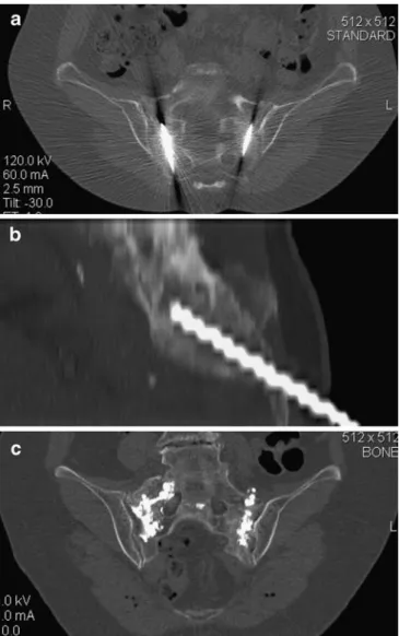
Fig. 4 CT of a 76-year-old female patient with bilateral sacral fractures successfully treated with long-axis sacroplasty (a and b) shows adequate and homogeneous cement distribution on both sides (c)
a
b
Fig. 5 CT of a 62-year-old male patient with persisting sacralgia after treatment shows lack of cement filling in the transverse component of his fracture (arrows in a and b)
complications like transient radiculitis and cement extra-vasation into the central canal, sacral foramina or sacroiliac joint were previously encountered. These events could be efficiently contained by immediate measures to minimise the propagation of any hazardous effect. In our series, however, we did not observe any of the aforementioned complications, potentially because of applying the CT-guided long-axis approach, which facilitates cement deliv-ery along the fracture line using a single entry point. Furthermore, this approach allows better determination of the needle depth and trajectory into the sacral mass and consequently facilitates its manipulation during the proce-dure. We believe that the CT-guided long-axis sacroplasty is advantageous over the same fluoroscopy-guided ap-proach, especially in avoiding the risk of penetrating the cephalad cortex of the S1 sacral ala that usually leads to cement extrusion into the adjacent soft tissues [16]. Additionally, its application in obese patients may also facilitate the procedures and reduce the overall time required for cement installation. It has to be recognised, however, that real-time monitoring of cement installation using fluoroscopy is a vital advantage that is not currently provided by the CT-guided approach. Whether long-axis sacroplasty is also feasible for traumatic, non-displaced sacral fractures in patients with senile osteoporosis needs to be established.
Incomplete treatment success in the form of persisting sacralgia was observed in 1/19 patients (0.5%) in the present series. The reason why sacroplasty failed in this patient was inadequate cement distribution along the transverse component of an oblique sacral fracture (Fig. 5). This may imply that for sacral fractures with mixed longitudinal and transverse components, a tradi-tional short-axis sacroplasty should be taken into con-sideration besides the long-axis approach. Three patients with successfully treated unilateral (n=1) and bilateral (n=2) sacral fractures addressed persisting low backache after the procedure. In fact, two of these three patients had
non-consolidating fractures of the transverse processes of the fifth lumbar vertebra and of the iliac bone, respectively. We believe that these unhealed fractures were responsible for the patients’ symptomatology as they had adequate and homogeneous cement distribution along their sacral frac-ture lines. The third patient was known to have an untreated disc hernia of the lower lumbar segment.
A limitation of the present study is its low number of patients (n=19). Despite this fact, our series still represents the largest one yet published that selectively presents the outcome of patients subjected to the long-axis sacroplasty approach. A second limitation is that we did not randomise a control group with a conservative treatment algorithm in our study design. It has to be recognised, however, that the remarkable improvement in the post-procedure mobility of the patients, along with the significant short-term reduction in pain score and analgesic dependence, are obvious arguments in favour of sacroplasty being the limiting factor in patient improvement in the present series, different from the spontaneous healing phase of conservative treatment, which needs several months to evoke a similar effect.
Conclusion
Long-axis percutaneous sacroplasty is a suitable, mini-mally invasive treatment option for patients who present with sacral insufficiency fractures. Future studies with larger patient numbers are warranted to determine any unrecognised limitations of this therapeutic approach, along with estimating its utility for traumatic, non-displaced sacral injuries.
Acknowledgements The authors gratefully acknowledge the technical assistance of Martine Bernasconi, and Martial Narbel. Part of this work was presented at the 96th Annual Meeting of the Swiss Society of Radiology, Geneva, Switzerland, June 04-06, 2009.
References
1. Whitlow CT, Mussat-Whitlow BJ, Mattern CW, Baker MD, Morris PP (2007) Sacroplasty versus vertebro-plasty: comparable clinical outcomes for the treatment of fracture-related pain. AJNR Am J Neuroradiol 28:1266–1270
2. Tjardes T, Paffrath T, Baethis H, Shafizadeh S, Steinhausen E, Steinbuechel T et al (2008) Computer assisted percutaneous placement of augmented iliosacral screws: a reasonable alternative to sacroplasty. Spine 33:1497–1500
3. Hinsche AF, Giannoudis PV, Smith RM (2002) Fluoroscopy-based multiplanar image guidance for insertion of sacro-iliac screws. Clin Orthop Relat Res 395:135–144
4. Templeman D, Schmidt A, Freese J et al (1996) Proximity of iliosacral screws to neurovascular structures after internal fixation. Clin Orthop Relat Res 329:194–198
5. Sciubba DM, Wolinsky JP, Than KD, Gokaslan ZL, Witham TF, Murphy KP (2007) CT fluoroscopically guided percutaneous placement of transiliosa-cral rod for satransiliosa-cral insufficiency fracture: case report and technique. AJNR Am J Neuroradiol 28:1451– 1454
6. Masala S, Mastrangeli R, Petrella MC, Massari F, Ursone A, Simonetti G (2009) Percutaneous vertebroplasty in 1,253 levels: results and long-term effectiveness in a single centre. Eur Radiol 19(1):165–171
7. Pitton MB, Herber S, Koch U, Oberholzer K, Drees P, Düber C (2008) CT-guided vertebroplasty: analysis of technical results, extraosseous cement leakages, and complications in 500 procedures. Eur Radiol 18:2568–2578 8. Pitton MB, Morgen N, Herber S, Drees
P, Böhm B, Düber C (2008) Height gain of vertebral bodies and stabiliza-tion of vertebral geometry over one year after vertebroplasty of osteoporotic vertebral fractures. Eur Radiol 18:608– 615
9. Binaghi S, Guntern D, Schnyder P, Theumann N (2006) A new, easy, fast, and safe method for CT-guided sacro-plasty. Eur Radiol 16:2875–2878
10. Heron J, Connell DA, James SL (2007) CT-guided sacroplasty for the treatment of sacral insufficiency fractures. Clin Radiol 62:1094–1003
11. Taillandier J, Langue F, Alemanni M, Taillandier-Heriche E (2003) Mortality and functional outcomes of pelvic insufficiency fractures in older patients. Joint Bone Spine 70:287–289
12. Frey ME, Depalma MJ, Cifu DX, Bhagia SM, Carne W, Daitch JS (2008) Percutaneous sacroplasty for osteopo-rotic sacral insufficiency fractures: a prospective, multicenter, observational pilot study. Spine J 8:367–373 13. Frey ME, DePalma MJ, Cifu DX,
Bhagia SM, Daitch JS (2007) Efficacy and safety of percutaneous sacroplasty for painful osteoporotic sacral insuffi-ciency fractures: a prospective, multi-center trial. Spine 32:1635–1640
14. Strub WM, Hoffmann M, Ernst RJ, Bulas RV (2007) Sacroplasty by CT and fluoroscopic guidance: is the procedure right for your patient? AJNR Am J Neuroradiol 28:38–41
15. Butler CL, Given CA 2nd, Michel SJ, Tibbs PA (2005) Percutaneous sacro-plasty for the treatment of sacral in-sufficiency fractures. AJR Am J Roentgenol 184:1956–1959
16. Smith DK, Dix JE (2006) Percutaneous sacroplasty: long-axis injection technique. AJR Am J Roentgenol 186:1252–1255