Infection 33 · 2005 · No. 1 © URBAN & VOGEL
39
Deadly Carousel or Difficult Interpretation of
New Diagnostic Tools for Whipple’s Disease:
Case Report and Review of the Literature
S.A. Müller, P. Vogt, M. Altwegg, J.D. Seebach
Abstract
Whipple‘s disease is a rare systemic disorder classically
presenting with weight loss, arthralgias, and diarrhea,
which was first described in 1907. The causative bacterium
Tropheryma whipplei, is a fastidious organism not growing on
conventional media. Before the introduction of polymerase
chain reaction (PCR)-based methods, the diagnostic gold
standard was histological detection of diastase-resistant
periodic acid Schiff (PAS)-positive macrophages or electron
microscopy. As in the present case, contradictory results
between the former and new diagnostic methods may
obscure the correct diagnosis. We critically summarize the
performance of the different diagnostic methods and discuss
their impact on the clinical management of patients with
suspected Whipple‘s disease.
Infection 2005; 33: 39–42 DOI 10.1007/s15010-005-4067-7
Introduction
Intestinal lipodystrophy was first recognized as a new
disor-der in 1907 by the pathologist George Hoyt Whipple [1]. The
histological criteria for Whipple‘s disease as summarized in
1949 were periodic acid Schiff (PAS)-positive inclusions
detectable in macrophages of the intestines and mesenteric
lymph nodes [2]. In 1991, the bacterium of Whipple’s
dis-ease was partially characterized at the molecular level by
broad-range bacterial 16S rDNA PCR and sequencing [3].
Isolation of the bacterium was achieved in the late 1990s
in long-term culture systems with interleukin-4-deactivated
human primary macrophages [4] and fibroblasts [5]
provid-ing a basis for further characterization of the organism [6].
Since then the organism is officially named Tropheryma
whipplei. It is a small, uniform, rod-shaped, gram-positive,
not acid-fast bacterium measuring 0.2
X1.5–2.0 µm in size
[7, 8]. By transmission electron microscopy, the bacterial
cell wall appears as a trilamellar structure. The analysis of
its small (925 kb) single circular chromosome points to a
host-restricted lifestyle and immune evasion as an
impor-tant role in the pathogenesis of the chronic course of
Whip-ple’s disease [9]. The modern molecular-based techniques
greatly improved the diagnostic methods to recognize
Whipple’s disease which is characterized by a great
varia-tion in clinical presentavaria-tion [7, 8]. Untreated Whipple’s
dis-ease has a chronic progressive and potentially fatal course
due to cardiac or central nervous system failure, wasting
syndrome or septic shock [7, 8, 10, 11]. More than 90%
of the patients respond to antibiotic therapy, but about 5
to 30% relapse despite prolonged treatment [7, 8, 12, 13].
Difficulties occur for the clinicians when the results of the
different methods are contradictory. Such conflicting
re-sults may lead to a false diagnosis and death of the patient,
as the present case demonstrates.
Case Report
A 66-year-old man was admitted to the hospital because of recur-rent fever, arthralgias, and exanthema. The patient had been well until 7 years earlier when polymyalgia rheumatica was diagnosed and was treated with prednisone and methotrexate. Two years before admission, intermittent episodes of fever with leukocyto-sis and elevated C-reactive protein (CRP) levels occurred, which were successfully treated with amoxicillin. On admission the pa-tient complained of weight loss, irregular bowel movements with constipation and diarrhea, polyarthralgias, pain and stiffness of the proximal limbs, sicca symptoms, pleuritic pain, and a pale patchy rash. Laboratory analysis showed anemia (hemoglobin 11.8 g/dl), leukocytosis (22.8 X 10⁹/l) with neutrophilia (97%) and lymphopenia (1.8%), and elevated inflammatory markers, i.e. blood sedimentation rate of 82 mm/h and CRP 76 mg/l. Cul-tures from blood, urine, stool, and knee joint fluid did not reveal
Infection
Case Report
S.A. Müller
Dept. of Internal Medicine, University Hospital of Zürich, Zürich, Switzerland
P. Vogt
Dept. of Pathology, University Hospital of Zürich, Zürich, Switzerland
M. Altwegg
Institute of Medical Microbiology, University of Zürich, Zürich, Switzerland
J.D. Seebach (corresponding author)
Dept. of Internal Medicine, University Hospital of Zürich, Rämistrasse 100, C HOER 31, CH–8091 Zürich, Switzerland; Phone: (+41/44) 255- 4134, Fax: -4445, E-mail: [email protected]
a causative pathogen. Serum protein electrophoresis, immuno-globulins, and serological tests for infections and autoantibodies were negative. Additional diagnostic procedures including bone marrow and skin biopsy, MR-angiography, echocardiography, positron emission tomography, and endoscopy were unremark-able. PCR from the knee joint fluid and a duodenal biopsy by semi-nested amplification using the primer pairs TW1/TW2 and TW4/TW2 were positive for T. whipplei (Figure 1). However, broad-spectrum bacterial PCR using a 16S rRNA gene fragment as well as the confirmation by another T. whipplei-specific PCR using a different technique [14] performed on the same specimens were negative. Further investigation by histological examination of duodenal biopsies did not reveal PAS-positive macrophages. Therefore, Whipple’s disease was ruled out and a systemic in-flammatory disorder of unknown origin was assumed. During the following 3 months, the patient was treated with indometacin and prednisone, but the clinical situation worsened and he died of multiorgan failure. Examination at autopsy revealed foamy mac-rophages filled with diastase-resistant PAS-positive particles in the lamina propria of the small and large intestines, the myo- and
pericardium (Figure 2), the skeletal muscles, the bone marrow, and the retroperitoneal soft tissue. Scanty PAS-positive granular inclusions were also detected in hippocampal ganglion cells, but not in the liver, spleen, and lymph nodes. The joints were not examined at autopsy. Reevaluation of the duodenal biopsies also showed a small number of PAS-positive macrophages. Based on these pathological findings a final postmortem diagnosis of Whip-ple’s disease was made.
Discussion
Whipple’s disease is a systemic infection that may involve
any major organ system. The leading symptoms of weight
loss, arthropathy, and diarrhea are not specific. Thus, the
clinical suspicion has to be confirmed by further diagnostic
tests on specimens obtained from the distal duodenum, the
jejunum or the site of clinical manifestation such as heart
valves, lymph nodes, synovial tissue and cerebrospinal fluid
(CSF). Even stool specimens may be used to demonstrate
the presence of DNA for T. whipplei [15]. The histological
hallmark is the presence of foamy macrophages staining
purple with diastase-resistant PAS, whereas PAS
stain-ing alone is not completely specific. PAS-positive
macro-phages are also found in patients with infections caused
by Mycobacterium avium-intracellulare, Rhodococcus equi,
Bacillus cereus, Corynebacterium sp., Histoplasma
capsula-tum, or other fungi. Some of the histopathological features
of Whipple’s disease are known also to occur in
melano-sis coli, histiocytomelano-sis, Crohn’s disease, and Waldenström’s
macroglobulinemia [7, 8]. A further histological finding in
lymphatic tissue, liver and the gastrointestinal tract
asso-ciated with Whipple’s disease are non-caseating,
epithe-loid-cell granulomas (sarcoid-like) [15]. Confusingly, the
reactive macrophages present in these unspecific lesions
are PAS-negative indicating that they do not contain T.
whipplei.
S.A. Müller et al. Deadly Diagnostic Carousel for Whipple’s Disease
40
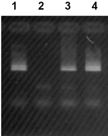
Infection 33 · 2005 · No. 1 © URBAN & VOGELFigure 1. PCR from the knee joint fluid. Agarose gel electrophoresis of PCR products after semi-nested amplification using primer pairs TW1/TW2 (upper band, amplicon size 267bp) followed by TW4/TW2 (lower band, amplicon size 229bp). Lane 1, positive control with a constructed plasmid; lane 2, negative control with a strain of
Esche-richia coli, passing the whole procedure of DNA extraction and PCR
reaction; lane 3, joint fluid of patient, undiluted DNA extract; lane 4, joint fluid of patient, DNA extract diluted 1:5. The relative intensities of bands in lanes 3 and 4 indicate that the amplification was slightly inhibited when undiluted DNA extract was tested.
Figure 2. Pathological finding at autopsy: specimen of the myocar-dium stained with periodic acid Schiff (PAS) and the specific diastase PAS (inset). Microscopic examination demonstrates multiple macro-phages engorged with PAS-positive material (arrows) between the myocytes and in the interfibrillar connective tissue.
In addition, T. whipplei can be identified by electron
microscopy in tissue samples from infected organs due to
its unusual and highly specific trilamellar cell wall.
How-ever, electron microscopy is not a convenient method for
rapid clinical diagnosis and data comparing its performance
with other diagnostic methods in Whipple’s disease are not
available. Therefore, electron microscopy is primarily used
in questionable cases [7, 8].
Since the sequencing of the 16S rDNA gene and the
description of specific primers for T. whipplei, gene
am-plification with PCR has been introduced as a diagnostic
tool. However, clinicians have to be aware of several
dif-ferent factors influencing the performance of PCR.
Na-tive clinical specimens give better results as compared to
formalin-fixation tissue due to partial degradation of the
DNA [7]. Moreover, DNA extraction which is one of the
crucial steps of all PCR techniques has to be adapted for
particular clinical samples especially for those
contain-ing inhibitors of the Taq polymerase (e.g. feces) [7, 16].
Amplification with semi-nested and nested methods is
as-sociated with a higher risk for contamination, which can
be reduced by using at least two independent PCR tests
based on different target genes [8]. The sensitivity of these
molecular tests also depends on the target gene and the
length of the amplified fragments [7]. Specimens suitable
for PCR are duodenal-biopsy tissue, lymph nodes, heart
valves, vitreous humor, stool, and synovial or
cerebrospi-nal fluid [8]. PCR may also be positive in samples from
the sites of clinical manifestations of Whipple’s disease,
e.g. from a disc biopsy in a patient with
spondylodisci-tis or from joint fluid as in the present case [7, 12, 17].
However, it is currently impossible to detect T. whipplei
DNA reproducibly from peripheral blood samples in
pa-tients with proven disease [7, 18]. On the other hand, T.
whipplei was amplified from saliva, dental plaque, gastric
juice, duodenal-biopsy samples, and feces in 4 to 35 %
of healthy persons and patients without Whipple’s
dis-ease [14, 16, 19–22], indicating that the diagnostic value
of these specimens is limited in patients with a low clinical
pretest probability.
Due to the rareness of Whipple’s disease, quantitative
comparative assessment of the different diagnostic
meth-ods is limited by the lack of studies directly addressing
this question in a sufficient number of patients. Currently,
there is no diagnostic gold standard conclusively
defin-ing Whipple’s disease. Therefore, analyzdefin-ing all published
cases of Whipple’s disease collected by Dutly and Altwegg
[7], as well as data reported in more recent studies [14,
16, 19–23], we calculated the sensitivity and specificity
of PCR and histology to detect Whipple’s disease (Table
1). In gastrointestinal samples the sensitivity of PCR and
histology were similar. In contrast, PCR techniques had a
higher sensitivity than the presence of PAS-positive
mac-rophages in histological evaluations of specimens from
involved organs. The specificity of PCR was limited by
false-positive results on saliva, dental plaque, and
gastro-intestinal samples in patients without Whipple’s disease.
As mentioned above, histological results showing the
presence of macrophages with PAS-positive inclusions
are not specific for Whipple’s disease; however
quantita-tive data on this issue are not available.
In conclusion, untreated Whipple’s disease has a
chronically progressive and potentially fatal course.
However, most patients respond to antibiotic treatment
with ceftriaxone, trimethoprim-sulfomethoxazole, or
tet-racycline resulting in rapid improvement of the clinical
status and lasting remissions [7, 8, 12, 13]. Therefore, in
cases of contradictory results between the former gold
standard, PAS staining of duodenal biopsies, and recently
introduced, highly sensitive PCR techniques, antibiotic
treatment is warranted. In addition, critically reviewing
the diagnostic results including meticulous reevaluation
of all specimens and repeated sampling may help to find
the correct diagnosis.
S.A. Müller et al. Deadly Diagnostic Carousel for Whipple’s Disease
Infection 33 · 2005 · No. 1 © URBAN & VOGEL
41
Samples from Gastrointestinal tract Involved organs
Histology PCR Histology PCR
Sensitivity (95% CI) 78% (71–85)a 84% (71–92)b 79% (64–90)c 100% (87–100)d
Specificity (95% CI) NA 94% (92–95)e NA NA
Histology: detection of periodic acid Schiff (PAS)-positive macrophages; PCR: polymerase chain reaction with different primers and tech-niques; CI: confidence interval; NA: not available. Sensitivity and specificity including exact 95% binominal confidence intervals were calculated using the patients published in the references [14, 16, 19–23]. Patients redundantly described in more than one reference were counted only once. a Biopsies from the gastrointestinal tract were diagnostic in 123 of 157 patients with intestinal or extraintestinal mani-festations of Whipple‘s disease. b PCR from gastrointestinal samples (biopsies, gastric juice, stool, saliva) was positive in 46 of 55 patients with intestinal or extraintestinal manifestations of Whipple‘s disease. c Biopsies from involved extraintestinal organs were diagnostic in 34 of 43 patients with Whipple‘s disease. d PCR from involved extraintestinal organs was positive in 27 of 27 patients with Whipple‘s dis-ease. e PCR from gastrointestinal samples (biopsies, gastric juice, stool, saliva, dental plaque) was positive in 60 of 970 patients without Whipple‘s disease
Table 1
42
Infection 33 · 2005 · No. 1 © URBAN & VOGELAcknoledgments
We are indebted to Dr. J. Muntwyler for his aid in statistical analy-sis of the data and to Dr. M. Schneemann for helpful discussions and critical reading of the manuscript.
References
1. Whipple GH: A hitherto undescribed disease characterized ana-tomically by deposits of fat and fatty acids in the intestinal and mesenteric lymphatic tissues. Johns Hopkins Hosp Bull 1907; 18: 382–391.
2. Black-Schaffer B: The tinctorial demonstration of a glycoprotein in Whipple’s disease. Soc Exp Biol Med 1949;72:225–227. 3. Wilson KH, Blitchington R, Frothingham R, Wilson JA: Phylogeny
of the Whipple’s-disease-associated bacterium. Lancet 1991; 338: 474–475.
4. Schoedon G, Goldenberger D, Forrer R, Gunz A, Dutly F, Hochli M et al.: Deactivation of macrophages with interleukin-4 is the key to the isolation of Tropheryma whippelii. J Infect Dis 1997; 176: 672–677.
5. Raoult D, Birg ML, La Scola B, Fournier PE, Enea M, Lepidi H et al.: Cultivation of the bacillus of Whipple‘s disease. N Engl J Med 2000; 342: 620–625.
6. La Scola B, Fenollar F, Fournier PE, Altwegg M, Mallet MN, Raoult D: Description of Tropheryma whipplei gen. nov., sp. nov., the Whipple‘s disease bacillus. Int J Syst Evol Microbiol 2001; 51: 1471–1479.
7. Dutly F, Altwegg M: Whipple’s disease and “Tropheryma
whip-pelii”. Clin Microbiol Rev 2001; 14: 561–583.
8. Marth T, Raoult D: Whipple’s disease. Lancet 2003; 361: 239–246. 9. Bentley SD, Maiwald M, Murphy LD, Pallen MJ, Yeats CA, Dover
LG et al.: Sequencing and analysis of the genome of the Whip-ple‘s disease bacterium Tropheryma whipplei. Lancet 2003; 361: 637–644.
10. Maizel H, Ruffin JM, Dobbins WO, III: Whipple‘s disease: a review of 19 patients from one hospital and a review of the literature since 1950. Medicine (Baltimore) 1970; 49: 175–205.
11. Durand DV, Lecomte C, Cathebras P, Rousset H, Godeau P: Whipple disease. Clinical review of 52 cases. The SNFMI Research
Group on Whipple Disease. Société Nationale Francaise de Médecine Interne. Medicine (Baltimore) 1997; 76: 170–184. 12. Misbah SA, Mapstone NP: Whipple‘s disease revisited. J Clin
Pathol 2000; 53: 750–755.
13. von Herbay A, Ditton HJ, Schuhmacher F, Maiwald M: Whipple‘s disease: staging and monitoring by cytology and polymerase chain reaction analysis of cerebrospinal fluid. Gastroenterology 1997; 113: 434–441.
14. Zinkernagel AS, Gmur R, Fenner L, Schaffner A, Schoedon G, Schneemann M: Marginal and subgingival plaque – a natural habitat of Tropheryma whipplei? Infection 2003; 31: 86–91. 15. Marth T, Strober W: Whipple‘s disease. Semin Gastrointest Dis
1996; 7: 41–48.
16. Maibach RC, Dutly F, Altwegg M: Detection of Tropheryma
whip-plei DNA in feces by PCR using a target capture method. J Clin
Microbiol 2002; 40: 2466–2471.
17. Altwegg M, Fleisch-Marx A, Goldenberger D, Hailemariam S, Schaffner A, Kissling R: Spondylodiscitis caused by Tropheryma
whippelii. Schweiz Med Wochenschr 1996; 126: 1495–1499.
18. Marth T, Fredericks D, Strober W, Relman DA: Limited role for PCR-based diagnosis of Whipple‘s disease from peripheral blood mononuclear cells. Lancet 1996; 348: 66–67.
19. Dutly F, Hinrikson HP, Seidel T, Morgenegg S, Altwegg M, Bauer-feind P: Tropheryma whippelii DNA in saliva of patients without Whipple’s disease. Infection 2000; 28: 219–222.
20. Amsler L, Bauerfeind P, Nigg C, Maibach RC, Steffen R, Altwegg M: Prevalence of Tropheryma whipplei DNA in patients with various gastrointestinal diseases and in healthy controls. Infec-tion 2003; 31: 81–85.
21. Street S, Donoghue HD, Neild GH: Tropheryma whippelii DNA in saliva of healthy people. Lancet 1999; 354: 1178–1179.
22. Ehrbar HU, Bauerfeind P, Dutly F, Koelz HR, Altwegg M: PCR-positive tests for Tropheryma whippelii in patients without Whipple’s disease. Lancet 1999; 353: 2214.
23. Maiwald M, von Herbay A, Persing DH, Mitchell PP, Abdelmalek MF, Thorvilson JN et al.: Tropheryma whippelii DNA is rare in the intestinal mucosa of patients without other evidence of Whipple disease. Ann Intern Med 2001; 134: 115–119. S.A. Müller et al. Deadly Diagnostic Carousel for Whipple’s Disease