Abstract Transcranial magnetic stimulation (TMS) was used to study visuospatial attention processing in ten healthy volunteers. In a forced choice recognition task the subjects were confronted with two symbols simulta-neously presented during 120 ms at random positions, one in the left and the other in the right visual field. The subject had to identify the presented pattern out of four possible combinations and to press the corresponding re-sponse key within 2 s. Double-pulse TMS (dTMS) with a 100-ms interstimulus interval (ISI) and an intensity of 80% of the stimulator output (corresponding to 110–120% of the motor threshold) was applied by a non-focal coil over the right or left posterior parietal cortex (PPC, corresponding to P3/P4 of the international 10-20 system) at different time intervals after onset of the visu-al stimulus (starting at 120 ms, 270 ms and 520 ms). Double-pulse TMS over the right PPC starting at 270 ms led to a significant increase in percentage of errors in the contralateral, left visual field (median: 23% with TMS vs 13% without TMS, P=0.0025). TMS applied earlier or later showed no effect. Furthermore, no significant in-crease in contra- or ipsilateral percentage of errors was found when the left parietal cortex was stimulated with the same timing. These data indicate that: (1) parietal in-fluence on visuospatial attention is mainly controlled by the right lobe since the same stimulation over the left pa-rietal cortex had no significant effect, and (2) there is a vulnerable time window to disturb this cortical process, since dTMS had a significant effect on the percentage of errors in the contralateral visual hemifield only when ap-plied 270 ms after visual stimulus presentation.
Keywords TMS · Human · Attention · Parietal lobe
Introduction
It is generally accepted that the parietal lobes play a ma-jor role in the network of visuospatial attention. Patients with unilateral lesions may show a large spectrum of neuropsychological deficits, e.g. impairment of percep-tion, of directing visual and covert attention to the con-tralateral space. Furthermore, they may have impair-ments of mental representation of space (for a review, see, e.g. Rafal 1994; Lezak 1997; Heilman and Valen-stein 1993). Moreover, asymmetries between right and left parietal lobe function are described: preferential right parietal activation is found in arousal, enhanced vigilance (Paus et al. 1997), global processing, as well as in visuospatial attention for the global orientation in space (Corbetta et al. 1993). The activation of the left parietal lobe seems to be more important to local pro-cessing, e.g. subsequent analysis of local features (Driver et al. 1992), for directing attention to spatial locations on the contralateral side of space, and for temporal atten-tional processes (Coull and Nobre 1998).
Despite this functional specialisation, visual extinc-tion, i.e. the unawareness of contralesional visual stimuli during simultaneous presentation, seems to occur with equal frequency after right or left hemisphere lesions (Rafal 1994). The complete neglect syndrome, on the other hand (Bisiach and Luzzatti 1978; Mesulam 2000), is mainly found after right hemisphere lesions involving the temporoparietal junction, and is thought to be caused by a disruption of the network of primary attentional functions as well as of visuospatial functions.
However, it remains unclear how the parietal lobes are involved in visuospatial attention processing, and particularly the temporal dimension is not well under-stood. Event related potential studies show that there is evidence for early and late processing in visual attention (for a review see Luck et al. 2000). Moreover, the timing of attention processes seems to be task dependent. Many
R.M. Müri (
✉
)Department of Neurology, University of Bern, Inselspital, 3010 Bern, Switzerland
e-mail: rene.mueri@insel.ch Fax: +41-31-6329679
R.M. Müri · R. Bühler · D. Heinemann · U.P. Mosimann J. Felblinger · T.E. Schlaepfer · C.W. Hess
Eye Movement Research Laboratory, Department of Neurology, Department of Psychology, and Psychiatric Neuroimaging Group, Inselspital, University of Bern, Bern, Switzerland
DOI 10.1007/s00221-002-1009-9
R E S E A R C H A R T I C L E
R.M. Müri · R. Bühler · D. Heinemann
U.P. Mosimann · J. Felblinger · T.E. Schlaepfer C.W. Hess
Hemispheric asymmetry in visuospatial attention assessed
with transcranial magnetic stimulation
Received: 11 November 2001 / Accepted: 10 December 2001 / Published online: 8 February 2002 © Springer-Verlag 2002
studies showed that early attention selection might start as soon as 60–100 ms after stimulus appearance (e.g. Hillyard and Münte 1984; Mangun and Hillyard 1988). Such early sensory-evoked components are typically found to be larger when a stimulus is presented at an at-tended location as compared with an unatat-tended loca-tion.
However, it seems that attention operates also in dif-ferent cognitive subsystems for difdif-ferent tasks (Luck et al. 2000), and that attention operates at a postperceptual stage. In the real world, a typical visual scene contains many different objects, not all of which can fully pro-ceeded with by the visual system at any given time. The late component N2pc of event-related potentials seems to be a good indicator for attentional selectivity in visual search (Eimer 1996; Woodman and Luck 1999; Hopf et al. 2000). This negative component occurs typically be-tween 200 and 300 ms after the presentation of a visual search array. Several studies showed that the N2pc com-ponent is related to the covert orienting of visual atten-tion before completing object recogniatten-tion (Luck and Hillyard 1994a, 1994b).
Transcranial magnetic stimulation (TMS) is a non-invasive technique, which allows interference with corti-cal processing by briefly disrupting a corticorti-cal region. It is an ideal tool with which to study the temporal organi-sation of complex processes (Cracco et al. 1999; Walsh and Rushworth 1999), and to determine functional rele-vance in time of a cortical region. It has been shown (Amassian et al. 1989; Miller et al. 1996; Epstein and Zangaladze 1996; Epstein et al. 1996; Kamitani and Shimojo 1999) that single-pulse TMS over the occipital cortex induces visual suppression if applied at critical time intervals after target presentation, and repetitive TMS (Pascual-Leone et al. 1994) induced contralateral visual extinction by stimulating the parietal lobes. Fur-thermore, TMS over the parietal cortex disturbs the per-formance in a visual search task (Walsh et al. 1999).
The aim of the present study was to perform a tempo-ral mapping of parietal lobe function in a complex visuo-spatial attention and recognition task. By using the double-pulse (dTMS) technique with an interstimulus interval (ISI) of 100 ms, the right or left parietal lobes were stim-ulated at corresponding coordinates during a forced-choice recognition task. The subjects had to identify a pattern of two simultaneously flashed symbols during the short presentation time of 120 ms, out of four possi-ble arrangements. The symbols were always presented in both visual hemifields.
Materials and methods
Subjects
Ten healthy subjects, four women and six men, were examined. Their mean age was 32 years (range 22–40 years). None of them took psychoactive medicaments. The local ethics committee ap-proval had been obtained for the study and subjects gave their in-formed consent to participate in the study. Handedness was
as-sessed by the Oldfield questionnaire (Oldfield 1971). Nine sub-jects were right handed and one subject was ambidextrous.
Subjects were seated in front of a 17-inch screen at a distance of 60 cm. The eye level was aligned to the height and mid-width of the screen. The chin was set on a chinrest and the head was fixed during the experiment by using a Velcro band across the forehead.
Preliminary experiments (results not shown in detail)
Three preliminary experiments were performed in three out of the ten subjects before all subjects were tested in the main experiment. In the first preliminary experiment, we used the same experimen-tal design as was used in the study by Pascual-Leone et al. (1994). In brief, a small asterisk was presented either in the left, right or both visual hemifields. Repetitive TMS trains of five pulses at 25 Hz were applied with the beginning of target presentation. In contrast, we applied dTMS instead of repetitive TMS. The ISI was always 100 ms, and stimulation started 120 ms, 270 ms, or 520 ms after simultaneous presentation of the visual targets in both hemi-fields. Double-pulse TMS in this experiment had no effect on per-formance; in particular, no visual extinction of the contralateral vi-sual target was induced.
In the second preliminary experiment, we used the symbol combination paradigm as described in the main experiment (see below). In this experiment, symbol combinations appeared with-out additional gridlines, and the symbols were always presented at the same spatial location in the right and left hemifield. Double-pulse TMS applied at the same time intervals as in the first prelim-inary experiment had no effect on symbol recognition at any inter-val.
The third preliminary experiment differed from the previous one by overlaying horizontal gridlines of 8×8 cm to the symbol combination. Double-pulse TMS at the mentioned timing had no effect on symbol recognition at any stimulated interval.
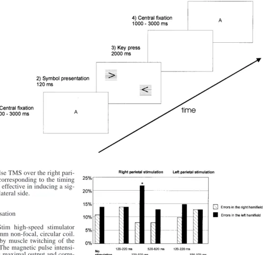
Main experiment
In the main experiment (Fig. 1), the task started with a central fix-ation of randomly changing letters for a variable time interval of 1000–3000 ms. Simultaneously with the extinction of this central fixation point, a combination of the two symbols with the overlaid horizontal gridlines was simultaneously presented in both visual hemifields for 120 ms. In contrast to the preliminary experiments, the symbol combination was displayed now at various and random positions on the left and right side of the screen. A 6-cm large, vertical zone in the centre of the screen, which corresponded to 5.9° of visual angle, was spared. Four combinations of simulta-neously presented symbols were possible, each of 2×2 cm dimen-sions (1.9° of visual angle): (1) >>; (2) <<; (3) <>; and (4) ><. Ac-cording to the four possible combinations of the two simulta-neously presented symbols, four key responses were attributed, and the subjects were instructed to respond by pressing the corre-sponding key press within 2000 ms after the extinction of the cen-tral fixation point. The next trial started with the appearance of the central fixation point. Each subject was individually trained with-out stimulation until 70–80% of correct responses was achieved (generally between 20 and 40 trials). All trials were performed in blocks of ten symbol presentations, and the subject decided indi-vidually when to continue the experiment after performing a block. In each subject, four to six blocks for each interval and stimulation side were acquired for each experiment. The subjects were examined in three sessions each lasting about 45 min, and in each session trials with and without stimulation were performed.
Control experiment
Finally, after obtaining significant results in the main experiment with dTMS starting at 270 ms, we tested the subjects again in a control experiment with single-pulse TMS. The issue of this
ex-periment was to test whether single-pulse TMS over the right pari-etal cortex at 270 ms or 370 ms (i.e. corresponding to the timing where dTMS pulses were applied) was effective in inducing a sig-nificant increase in errors of the contralateral side.
TMS parameters and stimulation localisation
TMS was applied by using a MagStim high-speed stimulator (Magstim Co., Wales, UK) with a 90-mm non-focal, circular coil. The motor threshold was determined by muscle twitching of the subject’s relaxed small hand muscles. The magnetic pulse intensi-ty was fixed to 80% of the stimulator’s maximal output and corre-sponded to 110–120% of the observed motor threshold. The coil position to stimulate the parietal cortex was determined according to the international 10-20 system of electrode placement (P3, P4). The inducing current in the coil segment overlaying the parietal target region flowed in the posterior direction, and the handle of the coil was posteriorly oriented. Double-pulse TMS was applied with 100 ms ISI starting at 120 ms, 270 ms, and 520 ms after the onset of the presentation of the symbols during right parietal tex stimulation and at 120 ms and 270 ms during left parietal cor-tex stimulation.
Data analysis
The percentage of response errors was analysed and the percent-age of response errors on the left or right side of the visual field (i.e. in which hemifield the symbol had been misinterpreted: left, right or both) was calculated. The absence of a key response was counted as an error in both hemifields.
Statistical analysis of the influence of the different stimulation intervals on percentage of response errors was performed by the Friedmann test. The Wilcoxon signed ranks test was used to com-pare the percentage of response errors with and without stimula-tion. Furthermore, the Bonferroni correction was made for multi-ple testing.
Results
Figure 2 shows the effect of dTMS over the right and left parietal cortex. The Friedmann test revealed a significant difference in the percentage of response errors during the different stimulus conditions when dTMS over the right
parietal cortex (P<0.001), but not when the left parietal cortex, was stimulated. Without stimulation, the median percentage of response errors in the right hemifield was 11% (range: 6–20%), and in the left hemifield 14% (range: 4–20%). Response errors in both hemifields oc-curred rarely, with a median of 6% (range: 0–10%).
A significant contralateral increase in percentage of response errors was found with dTMS over the right pa-rietal cortex at 270 ms (median 22.2%, range 5–26%, P=0.0025, Wilcoxon signed rank test, Bonferroni’s cor-rection, P<0.05) compared to without stimulation. Double-pulse stimulation over the right parietal cortex at 120 ms [median (range) for errors in the right hemifield: 14% (3–28%) and for errors in the left hemifield: 14% (8–21%) respectively] or 520 ms [8% (5–23%) for errors in the right hemifield and 13% (5–23%) for errors in the left hemifield] had no significant effect on the percent-age of response errors. The percentpercent-age of response errors in both hemifields always remained low during dTMS Fig. 1 The main paradigm.
Af-ter presentation of the central fixation point, a combination of two symbols was shown for 120 ms. During a 2-s period, the subject had to respond by pressing the corresponding key
Fig. 2 Bar graphs showing the median percentage of errors for the different stimulation intervals and stimulated sides. Percentage of errors contralateral to the stimulated right parietal cortex was sig-nificantly increased after double-pulse TMS applied starting at 270 ms (*P=0.0025). Double-pulse TMS over the left parietal cor-tex had no significant effect. Finally, single-pulse TMS over the right parietal cortex at 270 ms or 370 ms had no significant effect on the percentage of errors
(median for 120 ms: 5%; for 271 ms: 8%; and for 520 ms: 5%).
In the control experiment, single-pulse TMS had no significant effect on ipsi- or contralateral percentage of response errors [median for TMS at 270 ms: 18% (14–23%) in the left hemifield and 12% (7–23%) in the right hemifield; for TMS at 270 ms: 16% (10–20%) in the left hemifield and 10% (0–15%) in the right hemi-field].
Discussion
In this study we performed a temporal mapping of visuo-spatial attention processing in the posterior parietal lobes. The main results were: firstly, there is a distinct time interval during which TMS interferes with parietal attentional processing, since dTMS with an ISI of 100 ms applied at 270 ms over the right PPC significant-ly increased the percentage of response errors in the con-tralateral visual hemifield. Earlier or later dTMS applica-tion had no significant effect, suggesting a specific effect in time. Secondly, the observed effect was hemispheric-specific since stimulation at 270 ms over the left parietal cortex had no significant effect on contralateral percent-age of response errors.
These results suggest that dTMS interfered with vis-uospatial attention processing in the parietal cortex, but did not induce visual suppression or extinction. Previous studies (Amassian et al. 1989, 1998) have shown that in-duction of visual suppression crucially depends on TMS application early after visual presentation. Amassian et al. (1989) found that single-pulse TMS over the occipital cortex 80–100 ms after stimulus presentation induced contralateral suppression, and, by combining two or three pulses (Amassian et al. 1993), the effective interval for visual suppression was delayed. However, when the first pulse was later than 100 ms, i.e. after 120 ms ap-plied, contralateral visual suppression did not occur. In our experiments, early TMS application had no effect on contralateral percentage of response errors. Moreover, visual extinction can be induced by early repetitive TMS over the parietal cortex. Pascual-Leone et al. (1994) in-duced visual extinction of contralaterally presented visu-al targets at a constant position by repetitive TMS over right and left parietal cortex starting the stimulation si-multaneously with the visual presentation. By using the same experimental design, we were not able with dTMS in our preliminary experiments to induce visual extinc-tion.
The effect of dTMS was hemispheric specific, since only stimulation of the right parietal cortex but not over the left parietal cortex had a significant effect on per-centage of response errors in the contralateral hemifield. Such a right hemispheric dominance of visuospatial abil-ities is also found in patients with permanent lesion of the parietal cortex (e.g. von Cramon and Kerkhoff 1993). Only few studies used TMS to explore visuospatial attention mechanisms (Sabatino et al. 1996; Ashbridge et
al. 1997; Walsh et al. 1999), and they were mainly inves-tigating the interference with attention processes of visu-al search. However, the results were not unequivocvisu-al. Sabatino et al. (1996), e.g. stimulated continuously over the temporal lobes with 0.5 Hz during a verbal and a vis-uospatial cancellation task and was not able to influence performance of a search task using low-frequency TMS. However, during prefrontal stimulation, task perfor-mance was even better. Ashbridge et al. (1997) used a vi-sual search array, which was presented for 750 ms, and stimulated the parietal cortex 100 ms or 160 ms after stimulus onset. Reaction time was increased when the right parietal cortex was stimulated at 100 ms, and stim-ulation at 160 ms increased reaction time when the target was absent. In a second study (Walsh et al. 1999), they showed that magnetic stimulation over the left parietal cortex decreased reaction time when the target was pre-sented in the left hemifield. However, in both studies TMS did not affect the error rate.
Finally, a recent rTMS study by Rushworth et al. (2001) found a similar right hemisphere dominance of parietal lobe stimulation during an attentional paradigm.
A conspicuous although somewhat expected feature of this study is the relatively late vulnerable time win-dow for the TMS interference that we found. In contrast to classical visual attention tasks where early sensory at-tention selection mechanisms are expected, the task we used included different cognitive components such as object recognition, working memory and response selec-tion.
Therefore, late attention mechanisms may be much more important in our experiment. There is evidence from event related potential (ERP) studies during more complex tasks showing similar critical time intervals to what we found: The N2pc component, an indicator of at-tention selectivity, is typically observed between 200 and 300 ms after the array presentation. The component is also related to covert orienting of visual attention before the completion of object recognition (Luck and Hillyard 1994a, 1994b), and becomes more activated in attention-al filtering processes such as in visuattention-al discrimination tasks (Eimer 1996). Woodman and Luck (1999) found in a visual search task the most important attention-related changes in ERP components were observed between 200 and 300 ms after visual presentation. In a target detection task (Menon et al. 1997), significant activation of the pa-rietotemporal cortex was found even 285–610 ms after stimulus onset. For object recognition, a critical time in-terval of about 250 ms after target presentation was de-termined by EEG coherence analysis (Mima et al. 1999). Finally, Yamaguchi et al. (2000) showed that right pari-etotemporal region and left posterior temporal region were differentially activated during acttentional alloca-tion to global and local features of a visual scene. These effects started around 240 ms after presentation. Taking all these studies together, the vulnerable time window for disrupting attention processing as observed in our exper-iment would correspond well with the timing found in ERP studies in complex visual search tasks.
Luck SJ, Hillyard SA (1994b) Spatial filtering during visual search: evidence from human electrophysiology. J Exp Psy-chol Hum Percept Perform 20:1000–1014
Luck SJ, Woodman GF, Vogel EK (2000) Event-related potential studies of attention. Trends Cogn Sci 4:432–440
Mangun GR, Hillyard SA (1988) Spatial gradients of visual atten-tion: behavioral and electrophysiological evidence. Electro-encephalogr Clin Neurophysiol 70:417–428
Menon V, Ford J, Lim K, Glover G, Pfefferbaum A (1997) Com-bined event-related fMRI and EEG evidence for temporal-pa-rietal cortex activation during target detection. Neuroreport 8:3029–3037
Mesulam MM (2000) Principles of behavioral and cognitive neu-rology, 2nd edn. Oxford University Press, Oxford
Miller MB, Fendrich R, Eliassen JC, Demirel S, Gazzaniga MS (1996) Transcranial magnetic stimulation: delays in visual suppression due to luminance changes. Neuroreport 7:1740– 1744
Mima T, Oluwatimilehin T, Hiraoka T, Simpkins N, Hallett M (1999) Transient interhemispheric coherence reflects visual awareness. Soc Neurosci Abstr 25:355
Nobre AC, Sebestyen GN, Gitelman DR, Mesulam MM, Frackowiak RS, Frith CD (1997) Functional localisation of the system for visuospatial attention using positron emission to-mography. Brain 120:515–533
Oldfield R (1971) The assessment and analysis of handedness: the Edinburgh inventory. Neuropsychologia 9:97–113
Pascual-Leone A, Gomez-Tortosa E, Grafman J, Always D, Nichelli P, Hallett M (1994) Induction of visual extinction by rapid-rate transcranial magnetic stimulation of parietal lobe. Neurology 44:494–498
Paus T, Zatorre RJ, Hofle N, Caramanos Z, Gotman J, Petrides M, Evans AC (1997) Time-related changes in neural systems un-derlying attention and arousal during the performance of an auditory vigilance task. J Cogn Neurosci 9:392–408
Posner MI, Cohen Y, Rafal RD (1982) Neural system control of spatial orienting. Phil Trans R Soc Lond B 298:187–198 Posner MI, Walker JA, Freidrich FA, Rafal RD (1984) Effects of
parietal injury on covert orienting of attention. J Neurosci 4: 1863–1874
Rafal RD (1994) Neglect. Curr Opin Neurobiol 4:231–236 Rushworth MF, Ellison A, Walsh V (2001) Complementary
local-ization and laterallocal-ization of orienting and motor attention. Nat Neurosci 4:656–661
Sabatino M, Di Nuovo S, Sardo P, Abbate CS, La Grutta V (1996) Neuropsychology of selective attention and magnetic cortical stimulation. Int J Psychophysiol 21:83–89
von Cramon DY, Kerkhoff G (1993) On the cerebral organization of elementary visuo-spatial perception. In: Bulyas B, Ottoson D, Roland P (eds) Functional organization of the human visual cortex. Pergamon Press, Oxford
Walsh V, Rushworth MFS (1999) The use of transcranial magnetic stimulation in neuropsychological testing. Neuropsychologia 37:125–135
Walsh V, Ellison A, Ashbridge E, Cowey A (1999) The role of the parietal cortex in visual attention – hemispheric asymmetries and the effects of learning: a magnetic stimulation study. Neuropsychologia 37:245–251
Woodman GF, Luck SJ (1999) Electrophysiological measurement of rapid shifts of attention during visual search. Nature 400: 867–869
Yamaguchi S, Yamagata S, Kobayashi S (2000) Cerebral asymme-try of the “top-down” allocation of attention to global and local features. J Neurosci 20:RC72
In conclusion, the current results of the TMS interfer-ence suggest a critical visuospatial attention process of a recognition task controlled by the right parietal cortex 270 ms after visual presentation. Furthermore, a hemi-spheric asymmetry was found, since only stimulation over the right parietal cortex significantly disrupted the performance.
Acknowledgement This study was supported by Swiss National
Science Foundation grant no. 3100-050866.97.
References
Amassian VE, Cracco JB, Maccabee PM, Cracco RQ, Rudell AP, Eberle L (1989) Suppression of visual perception with the magnetic coil stimulation of human occipital cortex. Electro-encephalogr Clin Neurophysiol 74:458–462
Amassian VE, Maccabee PJ, Cracco RQ, Cracco JB, Rudell AP, Eberle L (1993) Measurement of information processing de-lays in human visual cortex with repetitive magnetic coil stim-ulation. Brain Res 605:317–21
Amassian VE, Cracco RQ, Maccabee PJ, Cracco JB, Rudell AP, Eberle L (1998) Transcranial magnetic stimulation in study of the visual pathway. J Clin Neurophysiol 15:288–304
Ashbridge E, Walsh V, Cowey A (1997) Temporal aspects of visu-al search studied by transcranivisu-al magnetic stimulation. Neuro-psychologia 35:1121–1131
Bisiach E, Luzzatti C (1978) Unilateral neglect of representational space. Cortex 14:129–133
Corbetta M, Miezin FM, Shulman GL, Petersen SE (1993) A PET study of visuospatial attention. J Neurosci 13:1202–1226 Coull JT, Nobre AC (1998) Where and when to pay attention: the
neural systems for directing attention to spatial locations and to time intervals as revealed by both PET and fMRI. J Neuro-sci 18:7426–7435
Cracco RQ, Cracco JB, Maccabee PJ, Amassian VE (1999) Cere-bral function revealed by transcranial magnetic stimulation. J Neurosci Methods 86:209–219
Driver J, Baylis GC, Rafal RD (1992) Preserved figure-ground segregation and symmetry perception in visual neglect. Neuro-psychologia 30:989–1000
Eimer M (1996) The N2pc component as an indicator of attention-al selectivity. Electroencephattention-alogr Clin Neurophysiol 99:225– 234
Epstein CM, Zangaladze A (1996) Magnetic coil suppression of extrafoveal visual perception using disappearance targets. J Clin Neurophysiol 13:242–246
Epstein CM, Verson R, Zangaladze A (1996) Magnetic coil sup-pression of visual perception at an extracalcarine site. J Clin Neurophysiol 13:247–252
Heilman KM, Valenstein E (1993) Clinical neuropsychology, 3rd edn. Oxford University Press, Oxford
Hillyard SA, Münte TF (1984) Selective attention to color and lo-cation: an analysis with event-related potentials. Percept Psy-chophys 36:185–198
Hopf JM, Luck SJ, et al. (2000) Neural sources of focused atten-tion in visual search. Cereb Cortex 10:1233–1241
Kamitani Y, Shimojo S (1999) Manifestation of scotomas created by transcranial magnetic stimulation of human visual cortex. Nat Neurosci 2:767–771
Lezak M (1997) Neuropsychological assessment, 3rd edn. Oxford University Press, Oxford
Luck SJ, Hillyard SA (1994a) Electrophysiological correlates of feature analysis during visual search. Psychophysiology 31: 291–308