CASE REPORT
Multiple bilateral asymmetrical deficiency of trunk muscles
Gertrude M. Beer&Hans-Hilmar Goebel&
Daniela Mihic-Probst&Peter Groscurth&
Mirjana Manestar
Received: 20 July 2008 / Accepted: 19 November 2008 / Published online: 16 December 2008 # Springer-Verlag 2008
Abstract Trunk muscles are an important source for pedicled and free flaps in reconstructive surgery. Unilateral deficiencies of trunk muscles are well known, either isolated or as part of Poland’s syndrome. Bilateral muscular deficien-cies and a“bilateral Poland anomaly” have also been spora-dically reported, but this is rare. We report on an 82-year-old male cadaver with clinically obscure, asymmetric bilateral deficiencies of the majority of trunk muscles. There was a history of acute poliomyelitis in childhood. Histological examination of representative muscle samples of the trunk showed extensive muscle atrophy with fat and connective tissue replacement. This was compatible with the prior diagnosis of poliomyelitis. However, representative sections of the spinal cord failed to reveal the antecedent poliomyeli-tis. The possibility of subclinical bilateral deficiencies of trunk muscles has to be taken into account in patients with a
history of poliomyelitis when planning reconstructions in cases of regional pedicled muscle transfers or free micro-vascular tissue transfers in reconstructive surgery.
Keywords Acquired muscle deficiency . Poliomyelitis . Reconstructive surgery . Trunk muscles
Introduction
Trunk muscles are an important source for pedicled and free flaps in reconstructive surgery. Deficiencies of the trunk muscles are rather rare. Unilateral deficiencies occur comparatively frequently, whereas bilateral deficiencies are virtually unknown. Poland in 1841 [1] described such muscular deformities including a completely absent pectoralis minor muscle, deficiency of the sternocostal portions of the pectoralis major muscle, absence of the inferior digitations of the serratus anterior muscle, and a tendinous appearance of the anterior and inferior parts of the obliquus externus abdominis muscle. He was not the first to appreciate this, but his description was the most complete. Despite many reports on Poland’s syndrome, neither the etiology nor the spectrum of this complex syndrome has yet been clearly analyzed. The same lack of knowledge about the etiology has been noted in the few reports on bilateral deficiencies of trunk muscles. In 1926, Jones [2] reported the occurrence of bilateral deficiency of the pectoralis major muscles en passant. In his clinical description of a 13-year-old girl with a“congenital” deficiency of the right pectoral muscles accompanied by a rudimentary nipple and a missing breast (apart from wasting of the left thigh and calf), he mentioned that the girl had suffered from an attack of acute anterior poliomyelitis when 2 years of age. In 1932, Sheehan [3] reported on the bilateral complete absence of the trapezius muscle in a 70-year-old male cadaver, and in
DOI 10.1007/s00238-008-0308-1
G. M. Beer
:
P. Groscurth:
M. Manestar Institute of Anatomy, University Zurich-Irchel, Winterthurerstrasse 190,8057 Zurich, Switzerland H.-H. Goebel
Institute of Neuropathology, Department of Pathology, University Hospital Zurich,
Rämistrasse 100, 8091 Zurich, Switzerland D. Mihic-Probst
Institute of Surgical Pathology, Department of Pathology, University Hospital Zurich,
Rämistrasse 100, 8091 Zurich, Switzerland G. M. Beer (*)
Division of Plastic and Aesthetic Surgery, Bodenseeklinik Swiss, Neuseeland 14,
CH-9404 Rorschacherberg, Switzerland e-mail: [email protected]
1972, Horan and Bonafede [4] presented a case of a young man with a bilateral absence of the trapezius and the sternal head of the pectoralis major muscle. These authors did not comment on a possible etiology.
Recently, Karnak and Tanyel [5] reported on a young girl with bilateral absence of the pectoralis major muscle, symmetrical chest wall deformities, and hand involvement as “the first case of a bilateral Poland anomaly.” Another report in 2003 [6] addresses a bilateral asymmetric deficiency of the pectoralis major muscle in a 72-year-old female cadaver. There were no obvious abnormalities on the outward appearance of the anterior thorax, including well-developed breasts. The authors speculated on the etiology being a congenital deficiency rather than a Poland’s syndrome or the sequel to acute poliomyelitis, although the patient was diagnosed with poliomyelitis at the age of 26 years and even had a reduced size of the left and right anterior horns of the cervical spine.
A case of bilateral asymmetric deficiencies of the majority of trunk muscles with a known history of acute poliomyelitis in childhood is presented.
Case report
In 2004 in the medical student gross anatomy dissection course, an 82-year-old male cadaver had an external appea-rance which appeared to be normal in all anatomical areas. The cause of death was a metastasized rectal carcinoma which had been diagnosed and operated on in 1997. A polio-myelitis infection had occurred in childhood. His relatives and internist revealed that he had survived the poliomyelitis without visible physical sequelae. As he became older, he developed a humpback, and after a knee replacement, he used a walking stick. He slowly lost muscle strength until finally, he could not stand without help. How much this physical deterioration was the sequela of the carcinoma, the advanced age, or the expression of a possible postpolio syndrome [7,8] remains unknown. In his final hospitalization in 1997, there was a short note in the discharge letter, which stated that there was visible atrophy of the upper and lower extremities and the paravertebral musculature.
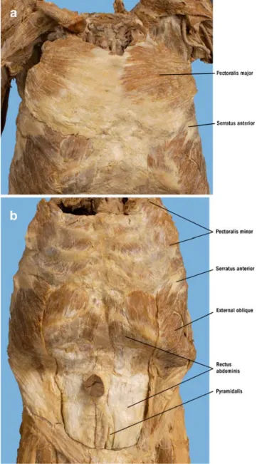
During his anatomical dissection, a bilateral sternocostal deficiency of the pectoralis major muscle was noted. The right complete sternocostal portion was absent and replaced by a tough fascial membrane. On the left, the caudal sternocostal portion alone was replaced by connective tissue and fat. In the anterior axillary fold, both muscles were fleshy and inserted into the lateral lip of the bicipital groove by a thin broad tendon. On the left side, the posterior lamina of the tendon was smaller than that on the contralateral right side (Fig. 1a). The pectoralis minor muscle showed similar deformities. On the right side, the
muscle was smaller and arose from the third to the fourth rib, but part of its cranial and caudal origins were replaced by connective tissue. On the left side, a well-developed, normal pectoralis minor muscle was present arising from the second to the fourth rib (Fig. 1b). These findings prompted us to look for further muscular deformities.
On the ventral side of the trunk, the obliquus externus abdominis, the rectus abdominis, the pyramidalis muscles, and the transversus abdominis and thoracic muscles showed a similar picture of spotted fatty and fibrous degeneration (Fig. 1b).
On the dorsal side of the trunk, the trapezius muscles were grossly replaced by connective tissue and fat with
Fig. 1 Overview of the anterior trunk. a Superficial muscular layer with view to the pectoralis major and serratus anterior muscles. b Deep layer showing deficiencies of the pectoralis minor, the obliquus externus abdominis, the rectus muscles, and the pyramidalis muscle
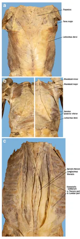
only few muscle fibers intermingled. The deficiency was more pronounced on the left than on the right side (Fig.2a). In addition, the latissimus dorsi, the teres major, the serratus anterior and posterior, both rhomboids, and both serratus posterior muscles had deficiencies of a differing intensity (Fig.2b). Additionally, the paraspinal muscles (iliocostalis, longissimus, and spinalis muscles) were significantly affected by the same deformities (Fig.2c).
The surface appearance of the muscles of the lower extremities appeared to be normal. However, on cross-section, the adductor longus magnus and brevis, the tensor fasciae latae, the vastus intermedius, all flexor muscles of the thighs, and the soleus revealed spotted areas of connective tissue and fat within their muscle bellies. The muscles of the upper extremities were normal, both at the surface and on cross-sections.
Immunohistochemistry for fast and slow muscle myosin in representative muscle biopsies demonstrated a diffuse (and not a checkered) appearance of atrophic and normal muscle fibers and an overall extensive muscle atrophy with fat and connective tissue substitution compatible with the diagnosis of prior poliomyelitis (Fig. 3). The histological examination of representative biopsies of the thoracic spine demonstrated normal content and distribution of motor neurons. Polymerase chain reaction for detection of polio virus in extracted DNA from paraffin-embedded biopsy of thoracic spine was negative.
Discussion
The anatomical dissection of an 82-year-old man revealed a bilateral asymmetrical deficiency of the majority of the trunk muscles. In addition, the typical deficiencies of muscles encountered in the lower extremities in poliomyeli-tis was observed. Some muscles of the trunk (trapezius and teres major) were almost completely deficient; others were macroscopically replaced by connective tissue and fat in a more or less pronounced manner. Histological examination of trunk muscles confirmed these findings, which were compatible with the diagnosis of very advanced remote neurogenic atrophy following poliomyelitis. Although rep-resentative sections of the spinal cord failed to reveal the antecedent poliomyelitis, as in other reports [6], there is little doubt that the etiology of the muscle deformities of the trunk, in addition to the muscle defects of the lower extremities, must be attributed to prior poliomyelitis.
Poliomyelitis was a common disease until the 1960s when mass immunization with oral poliovirus vaccine was begun in developed countries. The last natural outbreak of poliomyelitis in the USA occurred in 1972. There are now millions of adults who suffered from a more or less aggressive poliomyelitis infection in childhood and are left
Fig. 2 Overview of the posterior trunk. a Superficial muscular layer of the trapezius, the teres major, and both latissimus dorsi muscles. The end of the indicator line for the latissimus dorsi simultaneously indicates the sites of histological examination (bilaterally mirror-inverted). b Deep layer showing the rhomboids and the serratus posterior inferior muscles. c Intrinsic muscles of the back, including the iliocostalis, longissimus, and spinalis muscles
with a postpolio syndrome. In this, there is limitation in the range and power of movements and pain in the affected areas [9,10]. At the moment, poliomyelitis has been largely eradicated in industrialized countries, but since immuniza-tion programs have failed to reach the global populaimmuniza-tion, it has not been possible to eradicate the disease worldwide. On the other hand, poliomyelitis is still endemic in some developing countries [11,12], and any decrease in intensity of immunization could lead to large outbreaks of poliomy-elitis spread by travelers in this present world of modern global mobility.
Although poliomyelitis is predominantly associated with muscle defects in the lower extremities, theoretically, all muscles may be affected. When muscles of the trunk are affected only partially, surprisingly little disability may result, and the defects may pass mainly unnoticed by the patient. When examining such patients, denervation by electromyographic evidence is often far more extensive than the subjects realize [13].
In the case of muscles, which are visibly deficient, as in the case of the pectoralis major muscle, females especially often seek correction of the disfiguring appearance. This involves reconstruction of the asymmetric chests or the missing anterior axillary fold. The most successful recon-struction of such unilateral deformities of the pectoralis major muscle has been the use of the ipsilateral pedicled latissimus dorsi muscle. This is transferred to the anterior chest wall to simulate the pectoralis major muscle and to create an anterior axillary fold [14]. When the latissimus dorsi muscle is deficient [15,16], or the reconstruction with
the latissimus muscle is unsatisfactory, another favorite option has been the use of a transverse rectus abdominis flap. The ipsilateral rectus abdominis muscle may be deficient and thus not available for reconstruction [17]. In such cases, the safe microvascular transfer of contralateral muscles has been recommended; unfortunately, the contra-lateral muscles can be deficient, as has been sporadically reported [6] and our case clearly revealed.
For the clinicians, knowledge of trunk muscle deficiencies, especially in bilateral cases, is of major importance. This knowledge will promote a search for further unapparent abnormalities, both unilateral and bilateral.
This knowledge of unilateral muscular deficiencies, as in the classical “Poland’s syndrome,” is important where the relevant ipsilateral muscles for reconstruction are absent, and a contralateral muscle has to be considered for a free tissue transfer. This knowledge is even more important when bilateral muscular deficiencies are suspected, as in bilateral “Poland’s syndrome” or in cases with remote poliomyelitis.
This knowledge is mainly important in the era of advanced microsurgery; nearly all muscles can be used as donors.
In cases of clinically unrecognized trunk muscle defi-ciencies, such as in patients with a history of poliomyelitis, to prevent an unfavorable surprise intraoperatively, at any hint of muscle defects, the severity of the defect should be established and the surgery well planned before undertaking the operative procedure. Although poliomyelitis is seldom encountered in its acute form currently in our industrialized countries and the general knowledge of this infection fades,
Fig. 3 Histological appearance of the deficient muscles showing parts of the latissimus dorsi muscle on both sides. The specimens were taken from both sides within the latissimus dorsi muscles as indicated by the indicator line in Fig.2a. On the right side (a), the latissimus part is largely replaced by fat tissue, and only few muscle fibers are left (arrows, H & E, ×100); on the left side (b), the muscle fibers are microscopically present, though very small (H & E, ×50). Immunohisto-chemistry with diffuse evidence of fast (c, ×50) and slow muscle myosin (d, ×50) in atrophic and normal muscle fibers (arrows)
it should not be forgotten that many adults with a prior poliomyelitis infection in childhood and with possible muscular trunk defects are still alive.
Acknowledgment We thank Professor Kristian Borg from the Karolinska University in Stockholm for his helpful advice concerning the histological assessment. Furthermore, we are grateful to PD Dr. C. T. Bock from the University in Tübingen for polio PCR analysis and to N. Wey from the University Zürich for photographic reproductions.
References
1. Poland A (1841) Deficiency of the pectoral muscles. Guys Hosp Rep 6:191–192
2. Jones H (1926) Congenital absence of the pectoral muscles. Br Med J 6:59–60
3. Sheehan D (1932) Bilateral absence of trapezius. J Anat 67:180– 181
4. Horan F, Bonafede R (1972) Bilateral absence of the trapezius and sternal head of the pectoralis major muscles. J Bone Joint Surg 59:133
5. Karnak I, Tanyel FC (2003) Reply to correspondence from Shipkov and Anastassov—“bilateral Poland anomaly: does it exist?”. Am J Med Genet A 117(3):310–311
6. Mosconi T, Kamath S (2003) Bilateral asymmetric deficiency of the pectoralis major muscle. Clin Anat 16(4):346–349
7. Chang CW, Huang SF (2001) Varied clinical patterns, physical activities, muscle enzymes, electromyographic and histologic
findings in patients with post-polio syndrome in Taiwan. Spinal Cord 39(10):526–531
8. Luciano CA, Sivakumar K, Spector SA et al (1996) Electrophys-iologic and histologic studies in clinically unaffected muscles of patients with prior paralytic poliomyelitis. Muscle Nerve 19 (11):1413–1420
9. Bartels MN, Omura A (2005) Aging in polio. Phys Med Rehabil Clin N Am 16(1):197–218
10. Nathanson N (1982) Eradication of poliomyelitis in the United States. Rev Infect Dis 4(5):940–950
11. Sabin AB (1991) Perspectives on rapid elimination and ultimate global eradication of paralytic poliomyelitis caused by polio-viruses. Eur J Epidemiol 7(2):95–120
12. Thompson KM, Tebbens RJ (2007) Eradication versus control for poliomyelitis: an economic analysis. Lancet 369(9570):1363– 1371
13. Hayward M, Seaton D (1979) Late sequelae of paralytic poliomyelitis: a clinical and electromyographic study. J Neurol Neurosurg Psychiatry 42(2):117–122
14. Hester TR Jr., Bostwick J 3rd (1982) Poland’s syndrome: correction with latissimus muscle transposition. Plast Reconstr Surg 69(2):226–233
15. Cochran JH Jr., Pauly TJ, Edstrom LE et al (1981) Hypoplasia of the latissimus dorsi muscle complicating breast reconstruction in Poland’s syndrome. Ann Plast Surg 6(5):402–404
16. Beer GM, Kompatscher P, Hergan K (1996) Poland’s syndrome and vascular malformations. Br J Plast Surg 49(7):482–484 17. Drever JM, Zavala J (2002) Unilateral absence of the external
oblique muscle with hypoplasia of the rectus abdominis muscle in a patient with Poland syndrome. Plast Reconstr Surg 110 (7):1802–1803