DOI 10.1007/s00405-009-1129-y
L A R Y N G O L O G Y
Three-dimensional imaging of the larynx for pre-operative
planning of laryngeal framework surgery
Claudio Storck · Philipp Juergens · Claude Fischer · Olivia Haenni · Franz Ebner · Markus Wolfensberger · Erich Sorantin ·
Gerhard Friedrich · Markus Gugatschka
Received: 23 April 2009 / Accepted: 8 October 2009 / Published online: 28 October 2009 © Springer-Verlag 2009
Abstract Modern laryngeal framework surgery (LFS) requires an exact understanding of the laryngeal biome-chanics and precise pre-operative planning, for which bi-planar imaging is not suYcient. The aim of the study was to test whether MIMICS®, a commercially available software package for three-dimensional (3D) rendering of high-reso-lution computerised tomography (HRCT), is suitable for 3D imaging of the larynx, analysis of laryngeal biomechan-ics and pre-operative planning. We examined four cadaver larynx and one patient larynx. In the Wve larynges, all relevant structures and landmarks could be 3D visualised.
Superimposing of two HRCT scans shows that when the arytenoids move from ‘respiration’ to ‘phonation’, they perform a rotating, translating and tilting motion. More-over, we could demonstrate that the vocal fold elongates by 7% with cricothyroid approximation. We conclude that MIMCS® is well suited for 3D imaging of the larynx, anal-ysis of laryngeal biomechanics and pre-operative planning of LFS procedures.
Keywords 3D visualisation · Larynx · High-resolution computer tomography · Biomechanics · MIMICS
Introduction
Modern laryngeal framework surgery (LFS) aims at vocal fold medialisation (in cases of vocal fold palsy), thyroid expansion (for the treatment of spastic dysphonia), vocal fold relaxation (in cases of incomplete mutation) or vocal fold tensioning (after sex change procedures) [1]. Much more so than ablative surgery (be it for cancer resection or removal of benign vocal fold lesions), LFS requires an exact understanding of the laryngeal biomechanics as well as anatomically precise planning of the intervention [2]. Routine pre-operative workup in these cases usually includes imaging by high-resolution computerised tomog-raphy (HRCT) and/or magnetic resonance tomogtomog-raphy (MRT) [3]. However, we consider the standard, bi-planar imaging insuYcient in the context of LFS, since only three-dimensional imaging is able to depict the precise anatomy of the laryngeal cartilages, both at rest and in diVerent positions (e.g. respiration, phonation) [4]. So, what we are looking for is software, which is robust and suYciently fast to be useful for research purposes as well as for clinical work, and which, to avoid repeat radiological C. Storck (&) · C. Fischer · O. Haenni · M. Wolfensberger
Department of Otorhinolaryngology, Head and Neck Surgery and Phoniatrics, University Hospital Basel, Petersgraben 4, 4031 Basel, Switzerland
e-mail: [email protected]
P. Juergens
Hightech Research Centre of Cranio-Maxillofacial Surgery, University Hospital of Basel, Schanzenstrasse 46,
4031 Basel, Switzerland
F. Ebner
Section of Neuroradiology, Department of Radiology, Medical University of Graz, Auenbruggerplatz 9, 8036 Graz, Austria
E. Sorantin
Section of Paediatric Radiology, Department of Radiology, Medical University Graz, Auenbruggerplatz 34,
8036 Graz, Austria
G. Friedrich · M. Gugatschka
Department of Phoniatrics, ENT University Hospital, Medical University Graz, Auenbruggerplatz 26, 8036 Graz, Austria
examinations, is able to handle already existing imaging data. These data should preferably be HRCT data, since HRCT is more readily available and better suited to the examination of cartilage (and less expensive) than MRT. So far, we have not been aware of any 3D imaging soft-ware in clinical use that fulWls these criteria. MIMICS® Version 12.0 (Materialise™ Interactive Medical Image Control System, Leuven, Belgium) is an interactive soft-ware package, which is used in reconstructive maxillofa-cial surgery and in heart surgery for precise pre-operative planning [5, 6].
The purpose of this pilot study was to answer the follow-ing three questions: (1) Is MIMICS® suitable (with regard to image quality, software robustness and time consump-tion) for 3D imaging of both, hard and soft tissues of the larynx? (2) Are 3D images obtained by HRCT and MIM-ICS® adequate for biomechanical studies of laryngeal func-tion? (3) Are these images adequate and helpful for pre-operative planning of LFS?
In a Wrst step, we examined four cadaver larynges to answer the question, whether all relevant anatomical struc-tures and landmarks can be three-dimensionally visualised reliably, with suYcient precision and within an acceptable time frame, irrespective of laryngeal calciWcation (i.e. age and sex of the patient).
Two of us (C.S. and G.F.) routinely perform vocal fold medialisation and vocal fold tensioning procedures. Both procedures aVect the movements of the arytenoid carti-lages and/or the form of the glottic chink. As mentioned below, there exists no consensus on the subject of the functioning of the cricoarytenoid joint despite all the research that has been done in the past. So, in a second step (again performed on a cadaver larynx), we tried to Wnd out whether it was possible to visualise the movement of the arytenoid cartilages in three dimensions, when they moved from the respiratory to the phonatory position. Moreover, we checked whether it was possible to measure
the lengthening of the vocal folds on cricothyroid approx-imation.
Finally, having answered these questions in the aYrma-tive, we examined the normal larynx of a patient to make sure that the limitations inherent in a routine clinical exam (e.g. movement artefacts) did not jeopardise the results.
Materials and methods
We examined four cadaver larynges (2 male, 32 and 78 years, and 2 female, 49 and 71 years) and one patient larynx (male, 74 years old with no laryngeal pathology). The study was approved by the medical ethics committees of the University of Basel (Switzerland) and the University of Graz (Austria).
Preparation of the larynges
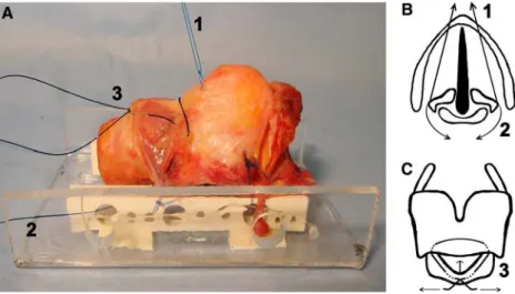
The fresh cadaver larynges were excised and examined within less than 24 h after death. Inclusion criteria were intact larynx without pathology, no previous laryngeal radi-ation or surgery and no infectious or rheumatologic disease. The pre-laryngeal strap muscles, as well as the pharyn-geal mucosa of the piriform sinus and the post-cricoid area, were removed. The conical ligament was exposed by removing the fat tissue between the cricothyroid muscles. The trachea was transected below the second cartilage ring. The larynges were mounted on a custom-made device, which consists of two perforated straight acrylic bars mea-suring 80 £ 10 £ 5 mm (Fig.1a).
To simulate the movement of the arytenoid cartilages in the cricoarytenoid joints, two non-absorbable threads were placed on each side, one mimicking the function of the thyroarytenoid muscle (Fig.1b, thread 1) and the other mimicking the function of the posterior cricoarytenoid muscle (Fig.2b, thread 2). Pulling on thread 1 closed the
Fig. 1 a Side view of a cadaver
larynx Wxed on a mounting device. b Top view draft of a larynx on the level of the glottis.
c Front view draft of a larynx. 1, 2, and 3: threads denote the
simulation of the positions ‘phonation’ (1), ‘respiration’ (2) and ‘cricothyroidopexy’ (3)
membranous part of the glottis by adduction of the aryte-noid cartilages, whereas pulling on thread 2 opened the glottis by abducting the arytenoid cartilages. We scanned three distinct positions, which we labelled ‘neutral’, ‘pho-natory’ and ‘respiratory’.
Finally, to simulate cricothyroid approximation, a third thread was placed mimicking the action of the two crico-thyroid muscles (Fig.1c, thread 3). Pulling on the two ends of this thread, the cricoid and the thyroid cartilage were approximated. We labelled this the ‘cricothyroidopexy’ position.
HRCT examination
HRCT examination was performed with an Aquilion ONE scanner (Toshiba®). The optimal setting was found to be 120 kV and 150 mA. The slice thickness was 1.0 mm. The rotation time was 1 s for cadaver larynges and 0.5 s for the patient’s larynx. The radiation dosage for one exam was 1.2 mSv. Of course, this number has to be multiplied by the
number of laryngeal positions examined. The HRCT DICOM data were then imported and converted to MIM-ICS® for post-process imaging.
Post-process imaging
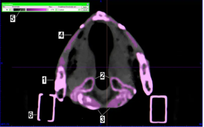
MIMICS® is a general-purpose segmentation programme for grey value images. It oVers an interactive tool for the visualisation, segmentation and 3D rendering of objects with HRCT as well as MRT data. MIMICS® interfaces with all common scanner formats. In a Wrst step, one can select the interested range of grey values. This can be done automatically by choosing a pre-deWned range (e.g. for cartilage) or manually by setting the range on a scale of grey values. In addition, manual editing functions make it possible to erase or restore parts of the images as well as to Wll internal gaps (Fig.2). This is typically used for smoothing surfaces and eliminating artefacts. Whereas three windows on the screen show axial, coronal and fron-tal planes, the fourth window shows the 3D image. Visual-isation functions include real-time rotation (e.g. to obtain front, side, top or any oblique view) and zoom. In addition, part of the larynx can be virtually cut away or made trans-parent to allow a look inside the larynx (Fig.3). Point-to-point measurements on the 3D reconstruction may also be performed. Finally, it is possible to superimpose two (or more) 3D reconstructions by selecting and marking three clearly deWned points (e.g. the thyroid notch, the lower border and the superior horns of the thyroid) on both 3D images.
To answer the question whether all relevant anatomical structures and (surgical) landmarks were visible on the 3D images, we identiWed the superior and inferior horns of the thyroid cartilage, the thyroid notch and laminae, the vocal and muscular processes of the arytenoid cartilages, the accessory cartilages, the epiglottis, the cricoid cartilage, the cricoarytenoid and cricothyroid joints, and the hyoid bone, as well as the thyroid notch, the inferior border and the Fig. 2 Screen of MIMICS® (axial view). The ossiWed areas of thyroid
cartilage (1), arytenoid cartilages (2) and cricoid cartilage (3) are detected automatically. The non-ossiWed thyroidal laminae (4) can be detected by adjusting the threshold (5). Mounting device (6)
Fig. 3 3D-rendered male
cadaver larynx (a) and patient’s larynx (b). All relevant anatomi-cal structures and landmarks can be visualised in both 3D images (1: thyroid cartilage; 2: cricoid cartilage; 3: arytenoid cartilages;
oblique line of the thyroid cartilage, and the annular border of the cricoid cartilage.
To depict the movement of the arytenoid cartilages in the cricoarytenoid joint, we overlaid the 3D images of the posi-tions ‘phonation’ and ‘respiration’ (Fig.4). To measure the lengthening of the vocal fold during cricothyroid approxi-mation, we overlaid the 3D images of the positions ‘neu-tral’ and ‘cricothyroidopexy’ (Fig.5).
Finally, by setting the grey range (automatically or manually) for mucosa, it is possible to visualise the muco-sal surface, thereby performing a ‘virtual endoscopy’ (Fig.6a, b).
Results
Time necessary for post-processing
Whereas in the research laboratory, time may be of sec-ondary importance, in clinical use it is of paramount
Visualisation of the laryngeal cartilages in the cadaver larynges
On all four larynges, all anatomically relevant structures and landmarks could be visualised by MIMICS® (Fig.3a). However, whereas ossiWed cartilages (or parts thereof) were adequately segmented (and hence 3D visualised) using the automatic detection threshold of MIMICS®, non-ossiWed parts of the cartilages could only be segmented adequately by setting the threshold manually.
Visualisation of the movement of the arytenoid cartilages Figure4 shows that by superimposing the 3D images of the ‘respiratory’ and ‘phonatory’ scans, the complex three-dimensional movement of the arytenoid cartilages can be visualised. If the arytenoid cartilages move from the respi-ratory to the phonatory position, the cartilages rotate down-ward around an axis that runs parallel to the cricoarytenoid joint surface (front view, Fig.4b) and inward around an Fig. 4 Side view (a), front view
(b), and top view (c) of the superimposed and 3D-rendered HRCT scans from the positions ‘phonation’ (yellow) and ‘respi-ration’ (blue). Side view (a): Arytenoid cartilages sliding along the axis of the cricoaryte-noid joint (red line). Front view (b): rotation of the arytenoid cartilages around the axis of the cricoarytenoid joint (red line). Top view (c): Tilting movement of the arytenoid cartilages around a virtual axis (blue line). MPR: Muscular process, ‘respi-ration’; MPP: Muscular process, ‘phonation’; VPR: Vocal pro-cess, ‘respiration’; VPP: Vocal process, ‘phonation’
that this movement has on the conWguration of the glottic chink can be seen in the surface (or virtual endoscopy type) images (Fig.6a, b).
Visualisation of the cricothyroid approximation
To analyse the eVect of the cricothyroid approximation on the vocal folds, we superimposed the positions ‘neutral’ and ‘cricothyroidopexy’. Figure5 shows the right half of a larynx at the two positions. The cricoid plate tilts back-wards. Consequently, the arytenoid cartilage, sitting on top of the cricoid plate, moves backwards and thereby elongates
the vocal fold. In our larynx, the distance from the anterior commissure (AC) to the vocal process (VP) measured 25 mm in the neutral position, and 27 mm in the ‘cri-cothyroidopexy” position. This corresponds to a vocal fold elongation of 7%.
Visualisation of the laryngeal cartilages in a patient’s larynx
This is primarily an in vitro study. However, as Fig.3b demonstrates, data from a patient HRCT are perfectly suit-able for 3D imaging by MIMICS®.
Discussion
The term ‘laryngeal framework surgery’ (LFS) refers to surgical interventions on the laryngeal cartilages aimed at improving, changing or restoring a patient’s voice [1]. LFS requires an exact understanding of the laryngeal biome-chanics as well as anatomically precise planning of the intervention. Pre-operative planning is routinely done by bi-planar (CT or MRT) imaging [3, 7]. Since it is very
diY-cult to mentally form a 3D image of the laryngeal struc-tures, let alone to visualise the movements of the cartilages in three dimensions on the basis of bi-planar scans, we con-sider bi-planar imaging insuYcient in the context of LFS. What we need are good 3D reconstructions. So far, we are not aware of any software in clinical use for 3D reconstruc-tion of laryngeal structures. However, since maxillofacial surgeons and vascular surgeons are successfully using MIMICS® for the planning of three-dimensional recon-structions of both, hard and soft tissues [8, 9], it seems rea-sonable to test the suitability of MIMICS® for 3D imaging of the larynx. Since HRCT is the imaging technique com-monly used in the context of LFS [4], we decided to use HRCT data.
Our pilot study on four cadaver larynges and on one patient larynx shows that with the help of MIMICS®, seg-menting and three-dimensional visualisation of all relevant Fig. 5 Right half of a larynx. Superimposed and 3D-rendered HRCT
scans of the positions ‘neutral’ (grey) and ‘cricothyroidopexy’ (gold). The cricoid plate and the arytenoid cartilage tilt backwards. Conse-quently, the distance between the anterior commissure (AC) and the vocal process lengthens from the neutral ‘position’ (VP1) to the ‘cri-cothyroidopexy’ position (VP2)
Fig. 6 Top view of a larynx
shows diVerent conWgurations of the glottic chink in the positions ‘respiration’ (a) and ‘phonation’ (b). Vocal folds (yellow)
laryngeal structures and landmarks is possible. We believe that apart from Hiramatsu et al. [10], no one has done this before.
MIMICS® provides pre-programmed segmentation thresholds for various tissues such as bone, muscle, fat, mucosa or implant materials. Because of the irregular calci-Wcation [11–15] and hence inhomogeneity of the laryngeal cartilages, automatic segmentation using pro-programmed thresholds does not work equally well in all larynges. Non-calciWed cartilages (or less Non-calciWed parts of cartilages) can only be segmented by adjusting the threshold manually. Of course, this takes more time than automatic segmenting. Whereas automatic segmenting takes about 20 min for an entire larynx, manual segmenting may take up to 60 min. We think that this is acceptable not only for research pur-poses, but also in a clinical setting. Moreover, we are con W-dent that with more experience, we will be able to manually segment a larynx in considerably less time, and that, once the method is established, segmenting can also be done by technical personnel. On setting the threshold for mucosa (either automatically or manually), we were able to produce 3D images of the mucosal surface, comparable to a virtual endoscopy. Of course, virtual endoscopy of the larynx is less interesting than virtual endoscopy of a less easily endo-scopically accessible organ such as the colon. Nevertheless, it allows a correlation of mucosal surface and exact carti-lage position. Unfortunately, although MIMICS® provides pre-set segmentation thresholds for muscles, we could not visualise individual muscles because the tissue densities of muscle and fat on HRCT were too close.
The exact trajectory of the arytenoid cartilages when they move from an extreme respiratory position through an intermediary position to the phonatory position has been the subject of countless studies [4, 16–21], and yet it is not completely understood. Similarly, what precisely happens during diVerent types of phonation (e.g. singing, whisper-ing) is not completely understood [22–24]. So far, imaging techniques have not added to our understanding of endola-ryngeal movements [17–20]. It is our long-term goal to examine these movements in volunteers and patients with diVerent pathologies using 3D imaging. Even if pulling on threads may seem (and indeed is) a primitive way of simu-lating laryngeal movements, our pilot study shows (1) that the arytenoid cartilages move in all three dimensions (and not just on a plane) and (2) that MIMICS® does allow us to superimpose two (or more) HRCT scans, thereby
visualis-on the same plane as the normal vocal fold, in cases of vagal nerve palsy it is excavated, more lateralised and on a slightly deeper plane than the normal vocal fold [25]. Both the excavation and the downward shift are probably due to the reduced tension of the cricothyroid muscle [25]. This diVerence has surgical consequences. Whereas simple vocal fold medialisation is suYcient for the treatment of recurrent nerve palsy, the vocal fold has to be medialised, tensioned, and smoothly pushed upwards in cases of vagal nerve palsy, Of course, these (subtle) diVerences can be ‘seen’ on laryngoscopy. However, for pre-operative plan-ning, precise imaging allowing us to quantify the necessary amount of displacement is more helpful. We hope that by demonstrating the exact position (in space) of the arytenoid cartilages in diVerent cases of laryngeal palsies, we will be better able to plan medialisation procedures. We also hope to produce a custom-made medialisation implant one day.
Another laryngeal framework procedure that we believe will beneWt from exact pre-operative planning is cri-cothyroidopexy, performed to raise the pitch of the voice in man-to-female transsexuals. The relationship between the length (and hence tension) of the vocal folds and the pitch of the voice in a given individual is not known. Pre-opera-tively measuring the amount of lengthening of the vocal fold achievable by maximal cricothyroid adduction and correlating this with the observed pitch will (hopefully) help us better plan the surgery (and to predict the outcome). Previous in vivo measurements of vocal fold length were all performed with conventional X-ray images or bi-planar CT scans [26–31]. Again, we believe that this is not ade-quate. Because the vocal fold plane changes with cricoid– thyroid approximation, only a three-dimensional image allows precise measuring of the vocal fold elongation. Our pilot study shows that precise measurement of vocal fold elongation achieved with cricothyroidopexy is possible in cadavers. We are conWdent that it will also be possible in patients. Of course, it would be most fascinating to study the voice changes resulting from vocal fold elongation in professional actors or singers with the help of 3D imaging [26].
We conclude that MIMICS®, in conjunction with HRCT scans, is well suited for 3D imaging of the larynx. It is help-ful both for the analysis of laryngeal biomechanics and for pre-operative planning of LFS procedures. This feasibility study will serve as a basis for further biomechanical studies on cadaver larynges and for the examination of patients with diVerent pathologies.
References
1. Friedrich G, De Jong FICRS, Mahieu HF, Benninger MS, Isshiki N (2001) Laryngeal framework surgery: a proposal for classiWca-tion and nomenclature by the Phonosurgery Committee of the European Laryngological Society. Eur Arch Otorhinolaryngol 375:1–8
2. Friedrich G, Lichtenegger R (1997) Surgical anatomy of the lar-ynx. J Voice 11:345–355
3. Friedrich G, Kainz J (1988) Morphometry of the larynx in horizon-tal sections. Normal data for the quantitative evaluation of current imaging technics. Laryngol Rhinol Otol (Stuttg) 67:269–274 4. Hunter EJ, Titze IR, Alipour F (2004) A three-dimensional model
of vocal fold abduction/adduction. J Acoust Soc Am 115:1747– 1759
5. Jacobs S, Grundert R, Mohr FW, Falk V (2008) 3D-Imaging of cardiac structures using 3D heart models for planning in heart sur-gery: a preliminary study. Interact Cardiovasc Thorac Surg 7:6–9 6. Mavili ME, Canter HI, Saglam-Aydinatay B, Kocadereli I (2007)
Tridimensional evaluation of maxillary and mandibular move-ments in orthognathic surgery. J Craniofac Surg 18:792–799 7. Friedrich G, Kainz J, Schneider GH, Anderhuber F (1989)
Computed tomography of the larynx in the diagnosis of dysphonia. Folia Phoniatr (Basel) 41:283–291
8. Doyle BJ, Grace PA, Kavanagh EG, Burke PE, Wallis F, Walsh MT, McGloughlin TM (2009) Improved assessment and treatment of abdominal aortic aneurysms: the use of 3D reconstructions as a surgical guidance tool in endovascular repair. Ir J Med Sci 178:321–328
9. Pham AM, RaWi AA, Metzger MC, Jamali A, Strong EB (2007) Computer modeling and intraoperative navigation in maxillofacial surgery. Otolaryngol Head Neck Surg 137:624–631
10. Hiramatsu H, Tokasjiki R, Suzuki M (2008) Usefulness of three-dimensional computed tomography of the larynx for evaluation of unilateral vocal fold paralysis before and after treatment: tech-nique and clinical applications. Eur Arch Otorhinolaryngol 265:725–730
11. Nemec SF, Krestan CR, Noebauer-Huhmann IM, Formanek M, Fruhwald J, Peloschek P et al (2009) Radiological normal anat-omy of the larynx and pharynx and imaging techniques. Radiologe 49:8–16
12. RuYng S, StruVert T, Grgic A, Reith W (2005) Imaging diagnos-tics of the pharynx and larynx. Radiologe 45:828–836
13. Castelijns JA, Doornbos J, Verbeeten B Jr, Vielvoye GJ, Bloem JL (1985) MR imaging of the normal larynx. J Comput Assist Tomogr 9:919–925
14. Castelijns JA, Golding RP, van Schaik SC, Valk J, Snow GB (1990) MR Wndings of cartilage invasion by laryngeal cancer:
value in predicting outcome of radiation therapy. Radiology 174:669–673
15. Steinkamp HJ, Heim T, Zwicker C, Mathe F, Schorner W, Felix R (1992) The value of nuclear magnetic resonance tomography in tumor staging of laryngeal-/hypopharyngeal cancer. HNO 40:339– 345
16. Frable MA (1961) Computation of motion at the cricoarytenoid joint. Arch Otolaryngol 73:73–78
17. von Leden H, Moore P (1961) The mechanics of the cricoaryte-noid joint. Arch Otolaryngol 73:541–550
18. Ardran GM, Kemp FH (1966) The mechanism of the larynx: part I. The movements of the arytenoid and cricoid cartilages. Br J Radiol 39:641–654
19. Wang R (1998) Three-dimensional analysis of cricoarytenoid joint motion. Laryngoscope 108(Suppl 86):1–17
20. Probst KX, Schon Ybarra MA, Kashima H, Crosby RW (2004) Topography and interactions of the arytenoid and cricoid articular surfaces: implications for vocal process positional shifts. Clin Anat 17:206–213
21. Sellars IE, Keen EN (1978) The anatomy and movements of the cricoarytenoid joint. Laryngoscope 88:667–674
22. Sonninen A, Hurme P, Laukkanen AM (1999) The external frame function in the control of pitch, register and singing mode: radio-graphic observations of a female singer. J Voice 13:319–340 23. Rubin AD, Praneetvatakul V, Gherson S, Moyer CA, SataloV RT
(2006) Laryngeal hyperfunction during whispering: reality or myth? J Voice 20:121–127
24. Solomon NP, McCall GN, Trosset MW, Gray WC (1989) Laryn-geal conWguration and constriction during two types of whisper-ing. J Speech Hear Res 32:161–174
25. Kruse E, OlthoV A, Schiel R (2006) Functional anatomy of the recurrent and superior laryngeal nerve. Langenbecks Arch Surg 391:4–8
26. Roers F, Mürbe D, Sundberg J (2009) Predicted singers’ vocal fold lengths and voice classiWcation—a study of X-ray morphological measures. J Voice 23:408–413
27. Sonninen A (1954) Is the length of the vocal cords the same at all diVerent levels of singing? Acta Otol Laryngol Suppl 118:219– 231
28. Damsté PH, Hollien H, Moore GP, Murry T (1968) An X-ray study of vocal fold length. Folia Phoniatr 20:349–359
29. Hollien H, Damsté PH, Murry T (1969) Vocal fold length during vocal fry phonation. Folia Phoniatr 21:257–265
30. Hollien H, Moore GP (1960) Measurements of the vocal folds dur-ing changes in pitch. J Speech Hear Res 3:157–165
31. Luchsinger R, PWster K (1961) Die Messung der Stim-mlippenverlängerung beim Steigern der Tonhöhe. Folia Phoniatr 13:1–12