HAL Id: inserm-01313147
https://www.hal.inserm.fr/inserm-01313147
Submitted on 9 May 2016
HAL is a multi-disciplinary open access
archive for the deposit and dissemination of sci-entific research documents, whether they are pub-lished or not. The documents may come from teaching and research institutions in France or abroad, or from public or private research centers.
L’archive ouverte pluridisciplinaire HAL, est destinée au dépôt et à la diffusion de documents scientifiques de niveau recherche, publiés ou non, émanant des établissements d’enseignement et de recherche français ou étrangers, des laboratoires publics ou privés.
Tavitian, Thomas Viel, Hideo Utsumi, Suha Yalçın, Marco de Spirito
To cite this version:
Giuseppe Maulucci, Goran Bačić, Lori Bridal, Harald Schmidt, Bertrand Tavitian, et al.. Imaging ROS-induced modifications in living systems 4 5. Antioxidants and Redox Signaling, Mary Ann Liebert, 2016, 24 (16), pp.939-58. �inserm-01313147�
ARS Forum Review Article 1
Editors: Harald HHHW Schmidt, Fabio Di Lisa 2
3
Imaging ROS-induced modifications in living systems 4
5
Giuseppe Maulucci,1 Goran Bačić,2 Lori Bridal,3 Harald HHW Schmidt,4 Bertrand Tavitian,5 6
Thomas Viel,5 Hideo Utsumi,6 A. Suha Yalçın,7 and Marco De Spirito1* 7
8
1 Institute of Physics, Catholic University of Sacred Heart, Roma, Italy 9
2 Faculty of Physical Chemistry, University of Belgrade, Belgrade, Serbia 10
3 Laboratoire d’Imagerie Biomédicale, Sorbonne Universités and UPMC Univ Paris 06 and 11
CNRS and INSERM, Paris, France 12
4 Dept. of Pharmacology and Personalised Medicine, CARIM, Faculty of Health, Medicine 13
& Life Science, Maastricht University, P.O. Box 616, 6200 MD Maastricht, The Nether-14
lands 15
5 Université Paris Descartes - Hôpital européen Georges Pompidou, Service de Radiolo-16
gie - Laboratoire de Recherche en Imagerie, 75015 Paris, France 17
6 Innovation Center for Medical Redox Navigation, Kyushu University 3-1-1 Maidashi, Hi-18
gashi-ku, Fukuoka 812-8582, Japan 19
7 Department of Biochemistry, School of Medicine, Marmara University, 34854 Maltepe, 20 İstanbul, Turkey 21 22 *Corresponding author: 23
Prof. Marco De Spirito 24
Faculty of Medicine and Surgery - Institute of Physics 25
Antioxidants & Redox Signaling
L.go F. Vito 1, 00168 Roma, Italy. 1
E-mail: [email protected] 2
3
Running title: ROS imaging 4
Word counts: 8692 5
Reference numbers: 143 6
Number of greyscale illustrations: 2 7
Number of color illustrations: 8. 8
Antioxidants & Redox Signaling
Abstract 1
Significance: Reactive Oxygen Species (ROS) may regulate signaling, ion channels, 2
transcription factors and biosynthetic processes. ROS-related diseases can be either due 3
to a shortage or to an excess of ROS. 4
Recent advances: Since biological activity of ROS depends on not only concentration but 5
also spatial and temporal distribution, real-time imaging of ROS, possibly “in vivo”, has be-6
come a need for scientists, with potential for clinical translation. New imaging techniques, 7
as well as new contrast agents in clinically established modalities, was developed in the 8
last decade. 9
Critical issues: An ideal imaging technique should determine ROS changes with high 10
spatio-temporal resolution, detect physiologically relevant variations in ROS concentration 11
and provide specificity towards different redox couples. Furthermore, for in vivo applica-12
tions, bioavailability of sensors, tissue penetration and a high signal-to-noise ratio are addi-13
tional requirements to be satisfied. 14
Future directions: None of the presented techniques fulfill all requirements for clinical 15
translation. The obvious way forward is to incorporate anatomical and functional imaging 16
into a common hybrid-imaging platform. 17
18
Antioxidants & Redox Signaling
Introduction 1
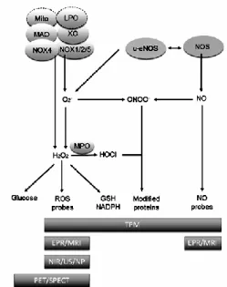
Reactive oxygen species (ROS) such as superoxide, hydrogen peroxide and perox-2
ynitrite are highly reactive in terms of oxidative modifications of biomacromolecules. Exog-3
enous ROS can be produced from pollutants, tobacco, smoke, drugs, xenobiotics, or ra-4
diation whereas endogenous ROS are produced intracellularly through multiple mecha-5
nisms. Depending on the cell and tissue types, the major sources are NADPH oxidase 6
(NOX) complexes (7 distinct isoforms) in cell membranes, mitochondria, peroxisomes, and 7
endoplasmic reticulum (30). Mitochondria produce superoxide radical (O2−), when oxygen
8
is prematurely and incompletely reduced. Superoxide can initiate lipid peroxidation in its 9
protonated form, hydroperoxyl HO2·, and can be converted to hydrogen peroxide (H2O2).
10
Myeloperoxidase (MPO), which is released from cytoplasmic granules of activated phago-11
cytes by a degranulation process, reacts with H2O2 and chloride ions to generate
hypo-12
chlorous acid/hypochlorite (HOCl/OCl(-)). HOCl, a strong oxidant, in turn reacts with pro-13
teins to form HOCl-modified proteins. Reactive nitrogen species (RNS) derive from nitric 14
oxide (NO) and superoxide (O2−) via the enzymatic activity of inducible nitric oxide
syn-15
thase (NOS) and NADPH oxidase, respectively. The reaction of nitric oxide (NO) with su-16
peroxide (O2−) leads to the the formation of peroxynitrite (ONOO−) (Fig. 1)(30).
17
These reactive species are essential regulators of several physiological processes, 18
ranging from intermediary metabolism to the inflammatory response. Their altered spatio-19
temporal distribution plays a central role in the physiopathology of disease (21). 20
Understanding complexity of ROS signaling requires the determination of their spatial and 21
temporal distribution with high resolution, specificity and sensitivity. Toward this aim, signif-22
icant progress in ROS imaging at the level of intact cells, tissues and whole organs, as 23
well as living organisms was achieved in the last decade. Among these advancements, an 24
important role was played by the development of novel synthetic or genetically encoded 25
Antioxidants & Redox Signaling
In particular, the possibility of detecting ROS dynamics in vivo has stimulated re-1
search in medical imaging with the aim of providing new information that will be beneficial 2
for disease management. This area of medical imaging research covers a wide domain of 3
different imaging modalities, each with its own sensitivity and resolution. Modalities include 4
Magnetic Resonance Imaging (MRI), Ultrasound (US), Positron Emission Tomography 5
(PET), Single Photon Emission Computed Tomography (SPECT) and other optical imag-6
ing methods (2, 51, 103, 108, 109, 138) (Fig. 1). In this context, improvements in detection 7
efficiency as well as new contrast agents for these well-established modalities was devel-8
oped in the last decade. However, low levels of intracellular ROS require new and more 9
sensitive methods. Here, we will review the methods emerging to image the complexity of 10
the ROS dynamics in vivo with a focus on those that have potential for clinical application. 11
12
Redox-Sensitive Two-Photon Microscopy (TPM) 13
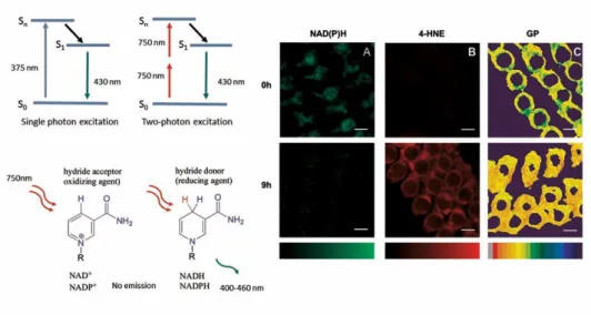
Two-photon microscopy (TPM) is a well-established sub-micron resolution imaging 14
technique characterized by low phototoxicity and deep tissue penetration. In the two-15
photon process, the probe absorbs two photons whose individual energy is only half of the 16
energy needed to excite that molecule (109). TPM excitation via near infrared (NIR) laser 17
light reduces tissue and water absorption. Penetration depths can reach 1 mm into biologi-18
cal tissues, and the reduction of photo-bleaching, photo-damage and phototoxicity is 19
achieved by the spatial confinement of excitation (109). With these advantages, TPM has 20
yielded novel and unique structural and functional information on cells and tissues. 21
In this context, determination of the spatial distribution of redox active compounds 22
and their time evolution is an important issue to address, since redox homeostasis, playing 23
a crucial role in many pathologies, can be a decisive target for pharmaceutical intervention 24
(30). 25
Antioxidants & Redox Signaling
Several fluorescence approaches to imageROS and redox potentials with high reso-1
lution have been attempted to address this task. Two of them are the most promising: Two 2
photon Fluorescence Ratio Imaging Microscopy (TP-FRIM) and Two Photon Fluorescence 3
Lifetime Imaging Microscopy (TP-FLIM) (94, 137, 143). These techniques have allowed 4
quantifying specific concentrations of intracellular redox species by canceling out the pos-5
sible perturbations due to instrument efficiency and dye concentration. 6
In TP-FRIM, the absorption or emission spectrum is differently sensitive to the redox 7
state of the compound. One wavelength range of the emission or excitation spectrum may 8
be less sensitive, or sensitive in the opposite direction with respect to another selected 9
range. Because absorption or emission originates from the same volume, the ratio of fluo-10
rescence measured in the two ranges is independent of optical path-length, probe concen-11
tration and excitation intensity. 12
TP-FLIM allows for the detection of the redox state of compounds by measuring dif-13
ferences in the exponential decay rate of the fluorescence (lifetime) of the probe by single-14
wavelength excitation. A quantitative determination of the redox state independent of 15
probe concentration could be obtained. 16
17
Two-photon redox sensitive probes
18
Determination of the spatial distribution of different redox active compounds (GSH, 19
NAD(P)H, H2O2, NO etc.) is an important aim for diagnosis and treatment. Although a
20
number of probes that are able to detect fluorescence in cultured cells are available, re-21
cent efforts have aimed at developing specific and highly sensitive TP-FRIM and TP-FLIM 22
based probes to improve quantitative analysis of ROS in deep tissue and for intra-vital mi-23
croscopy(17, 19, 27, 79, 126, 145). 24
25
Antioxidants & Redox Signaling
Two-Photon sensitive probes for assessment of glutathione redox state
1
The redox state of the reduced and oxidized glutathione couple (GSH:GSSG), the 2
most abundant redox couple in a cell, is an informative readout of the cellular redox envi-3
ronment (30). Glutathione specific redox-sensitive variants of the Yellow Fluorescent Pro-4
tein (rxYFP) and the Green Fluorescent Protein (roGFP1 and roGFP2) allowed FRIM real-5
time monitoring in the intracellular GSH:GSSG redox ratio (92–94, 123). The specificity of 6
these probes for glutathione was enhanced by linking them to human glutaredoxin 1 7
(Grx1) (12). To extend in vivo use of these probes, Wolf et al. (145) generated transgenic 8
mice expressing roGFP in several tissues to ratiometrically monitor oxidative stress in skin 9
epidermal keratinocytes. However, visible excitation and emission light do not permit a 10
deeper penetration in tissues. As a result, measurement of the cellular glutathione redox 11
potential (EG) is affected by non-negligible systematic errors (95). Furthermore these re-12
dox probes, when linked to enzymatically active redox proteins (i.e. Grx1), may alter cellu-13
lar redox homeostasis. Guzman and coworkers reported measurements of mitochondrial 14
oxidative stress on dopaminergic neurons in transgenic mice expressing mito-roGFP, a 15
roGFP that selectively tags targeted to mitochondria, with TPM (42). However, the suita-16
bility of this probe for potential TP-FRIM application has yet to be tested. To overcome 17
these issues, several GSH-sensitive, TP-excitable non-encoded chemoselective probes, 18
were engineered for in vivo applications (79). However, even in these cases , TP-19
FRIM/TP-FLIM potentials have yet to be characterized. Besides all these profuse efforts, 20
further improvements in the development of glutathione-specific redox probes are still 21
needed. 22
23
Two Photon NADPH redox state sensitive probes
24
The intracellular metabolic substrates NADH and NADPH (NAD(P)H) have been 25
Antioxidants & Redox Signaling
oxygen supply allowing label-free in vivo imaging of tissues (17, 97, 124, 125, 130). 1
NAD(P)/NAD(P)H auto-fluorescence can therefore be detected and related to other differ-2
ent physical quantities to gain further details on insight into the processes regulating redox 3
homeostasis. For example, the mechanism by which noise-induced ROS mediates an im-4
pairment in acoustic recovery capacity was elucidated by relating intracellular distribution 5
of NAD(P)H in a noise-stressed mammalian cochlea to the generation of lipid peroxides 6
and to the spatial organization of lipids inside membranes. This allowed to disclose the 7
mechanism of noise induced ROS production and impairment in the acoustic recovery ca-8
pacity (Fig. 2) (97). Skala et al. (127) combined cellular redox ratio, NAD(P)H and FAD life-9
time, and subcellular morphology to quantitatively detect NAD(P)H. With this approach 10
metabolic and structural modifications at the earliest stages of cancer development have 11
been identified in several epithelial tissues in vivo. Moreover, cell Phasor, a label-free, fit-12
free, and sensitive innovative method that allows classification of metabolic states of cells 13
during differentiation has been developed from TP-FLIM data (129). Zhuo et al. (17) used 14
two-photon autofluorescence and second harmonic generation (SHG) microscopy to 15
monitor cancer progression and to classify normal and dysplastic human colonic tissues. 16
Overall, these findings demonstrate that auto-fluorescence can provide structural and 17
functional information for the diagnosis and therapy of pathologic epithelial tissues. 18
19
Two-photon H2O2 sensitive probes
20
Hydrogen peroxide plays a key role as a cellular second messenger in a variety of 21
signal transduction processes (30). Genetically encoded fluorescent proteins HyPer, Hy-22
Per-3, roGFP2-Orp1 enabled transient live-cell imaging and allow high-resolution H2O2
im-23
aging with high specificity(41). However, these probes are applicable only to single photon 24
FRIM or FLIM and have limited application in vivo since they may alter redox homeostasis 25
Antioxidants & Redox Signaling
production of intracellular H2O2 (41), but their potential TP-FRIM application have yet to be
1
tested. For TP-FRIM imaging approach, a promising probe is Peroxy Naphthalene 1 2
(PN1). This probe can be excited at 750 nm, has high photostability and negligible toxicity. 3
It also allows determination of H2O2 distribution in live cells and tissue by TPM (19).
4 5
Two-Photon Nitric Oxide (NO) sensitive probes
6
Many of the NO sensitive probes are not reversible sensors as they form covalent 7
bonds with NO. Genetically encoded FRET-based proteins allow high resolution NO im-8
aging in cell-based experiments (122). However, in vivo applications of these probes are 9
very limited. For NO detection in an in vivo context, a TPM probe (QNO) with high selectiv-10
ity, low cytotoxicity, pH insensitivity and long-wavelength emission has been designed 11
(27). QNO is composed of a quinoline derivative as the fluorophore and an o-12
phenylenediamine moiety as the receptor for NO, linked with glycinamide. The probe re-13
sponded to NO over a linear range from 0.4 to 3.4 μM with a detection limit of 0.084 μM. 14
QNO detects NO in living cells and tissues at a depth of 180 μm. However, TP-FRIM/FLIM 15
properties are still not tested and further improvements in the development of NO-specific 16
redox probes are needed. 17
Chemiluminescent imaging of ROS in-vivo 18
NIR fluorescence and chemiluminescence
19
Over the last decade, substantial progress has been made in the non-invasive real-20
time assessment of reactive oxygen and nitrogen species in biological systems. Bioimag-21
ing methods based on fluorescence and reaction-based approaches have received most 22
attention, due to their ease of use, sensitivity and selectivity to different reactive species, 23
including reactive oxygen, nitrogen and sulfur species. A key interest in this rapidly grow-24
Antioxidants & Redox Signaling
single reactive species. A great number of different reaction schemes have been exploited 1
towards achieving this goal (reviewed in ref. (16)) as summarized in Table 1. Moreover, 2
the reaction-based monitoring of selective species can be combined with targeting of the 3
probe to specific cellular organelles, as exemplified by the boronate MitoPY1 for the imag-4
ing of mitochondrial H2O2 (24).
5
While fluorophores have been used widely for cellular imaging of reactive species, 6
they have a number of limitations that restricts their successful application to tissues and 7
animals. For the latter, fluorophores with absorption and emission maxima in the near-8
infrared region (650-900 nm) are required to maximize tissue penetration and, at the same 9
time, minimize interference from auto-fluorescence and hemoglobin absorption. Nagano 10
and co-workers recently synthesized the near-infrared fluorescent probe FOSCY-1 to mon-11
itor reactive species in a mouse model of peritonitis (105). As this probe reacts with sever-12
al biologically relevant reactive species, it provides general information about the presence 13
of oxidative events rather than the participation of specific reactive species in vivo. In a fur-14
ther development, the same group designed and synthesized a novel far-red to near-15
infrared probes based on Si-rhodamine to selectively and non-invasively monitor HOCl in 16
real-time in mice suffering from peritonitis (70). Judged by the advances over the last dec-17
ade, it can be reasonably expected that the development of additional reactive species 18
specific and non-specific far-red to near-infrared fluorescent probes will progress rapidly. 19
Reaction-based methods to detect reactive species are also applicable to in vivo 20
imaging modalities other than fluorescence. Chemiluminescence, for example, was used 21
to monitor oxidative events in vivo. Perhaps the most commonly used probe is L-012, an 22
analog of luminol (5-amino-2,3-dihydro-1,4-phthalazinedione) that produces much stronger 23
signals than either luminol, lucigenin, or MCLA (59). L-012 has been used successfully to 24
non-invasively image different inflammatory processes in mice [26-28]. The probe reacts 25
Antioxidants & Redox Signaling
L-012-derived luminescence was abolished in mice lacking phagocyte NADPH oxidase ac-1
tivity (66). 2
While much emphasis is placed on chemoselective bioimaging, reactive species in biologi-3
cal systems likely exist as mixtures of rapidly interconverting species. As a result, the bio-4
logical relevance of a single imaged species is difficult to assess, even if its presence cor-5
relates with the biological process studied, as exemplified above with L-012 in NOX2-6
deficient mice (66). In this context, non-specific probes that react with several different re-7
active species can have the advantage that they may give more general information on a 8
biological process (e.g., inflammation) than do selective probes. Indeed, when combined 9
with LC/MS/MS-based analytical analysis of non-reacted probe as well as its different 10
types of reaction products, non-selective probes may be seen as multi-purpose probes 11
that provide quantitative information about different reactive species, as exemplified re-12
cently with hydroethidine (88). 13
Chemiluminescent nanoparticles and ROS imaging
14
Nanoparticles (NPs) are particles with at least one dimension less than 100 nm. They have 15
different shapes, unique physico-chemical properties and a high ratio of surface area to 16
volume. NPs have several advantages over small molecule probes used in cellular sens-17
ing and imaging (142). Firstly, NPs have stronger luminescent emission due to the large 18
number of molecular probes that can be loaded into each particle. Additionally, their high 19
surface area to volume ratio provides a higher probability for analyte detection. The NPs 20
may also protect the sensory contents from external interference, such as undesirable en-21
zymatic reactions and nonspecific uptake by proteins. Moreover, it is also possible to tar-22
get NPs to cells and subcellular compartments by conjugating appropriate ligand moieties 23
onto their surface, which will allow for enhanced targeting to cells and subcellular com-24
partments. Encapsulation and conjugation of different molecules, such as luminescent 25
Antioxidants & Redox Signaling
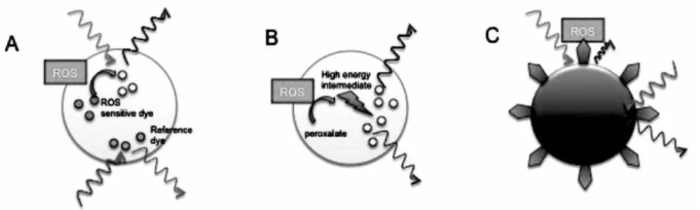
probes, proteins or DNA, provides infinite possibilities in NP design for specific functions. 1
In view of all of the above mentioned properties, NPs are becoming widely used tools in 2
the field of sensing and imaging (Fig. 3) (115, 119). 3
Recent advances in developing various luminescence probes have enabled moni-4
toring of ROS in cells and in animals. NP-based luminescent ROS sensors and their appli-5
cations are summarized in Table 1. Lee et al. (77) developed peroxalate-based NPs 6
formulated from peroxalate esters and fluorescent dyes to image H2O2 in vivo with high
7
specificity and sensitivity. Peroxalate NPs are capable of imaging H2O2 in the peritoneal
8
cavity of mice during a lipopolysaccharide-induced inflammatory response. The same 9
group has improved the method by reducing the size of the NPs and modifying their con-10
tent to detect H2O2 at physiological concentrations (22, 78). Luminescent NPs have also
11
been exploited for in vivo targeting and imaging of tumor tissues. In a recent study chemi-12
luminescent NPs were successfully developed to image H2O2 as a tumor signal molecule
13
(18). Such probes improve the stability of peroxalates in aqueous systems and are sensi-14
tive to low, physiologically relevant concentrations of H2O2 within the physiological range.
15
This way of monitoring H2O2 should be helpful for clinical diagnosis of other ROS related
16
diseases. 17
18
Ultrasound in ROS imaging 19
Ultrasonic imaging has been applied in many studies to detect changes in functional 20
blood flow and atherosclerotic plaque associated with oxidative stress (84). Although radi-21
cal oxidants cannot currently be directly detected in a clinical setting with ultrasound, a va-22
riety of original methods are being developed to enable such detection. Proposed ap-23
proaches vary widely, but all rely on the central principle behind clinical contrast ultraso-24
nography - the high sensitivity of ultrasound to echoes from gas bodies. 25
Antioxidants & Redox Signaling
Contrast enhanced ultrasound detects strong acoustic echoes when the ultrasonic 1
pulse encounters micrometric gas-bubble contrast agents. A very specific acoustic 2
signature can be obtained from microbubbles when they are acoustically-driven at levels 3
resulting in nonlinear response during the compression and expansion phases of the 4
microbubble. Several, very different solutions for detection of radical oxidants have been 5
proposed based on the ultrasonic detection of microbubbles that are targeted to specific 6
ligands, generated by chemical reactions or produced by micromotors. 7
Feasibility to detect inhibition of NADPH oxidase in advanced atherosclerosis has 8
been shown in mice using targeted contrast microbubbles bearing ligands for endothelial 9
cell adhesion molecules involved in monocyte recruitment (84). Lipid-shelled 10
decafluorobutane microbubbles were targeted to P-selectin or VCAM-1 and detected with 11
a clinical ultrasound system (7 MHz), eight minutes after injection at regions of 12
atherosclerotic plaque in the aortic arch of mice. Inhibition of NADPH oxidase was 13
associated with decreased targeted-detection of P-selectin and VCAM-1. This targeted 14
ligand approach is the basis for a large amount of research in ultrasonic molecular imaging 15
but it remains to be a relatively indirect approach to assess oxidative stress. A more direct, 16
bio-sensing ultrasound contrast agent for ROS detection has been proposed based on 17
chemical reactions that generate gas-forming molecules in the presence of radical 18
oxidants (108). In the presence of radical oxidants, allylhydrazine oxidizes into 2-propenyl-19
diazene that spontaneously undergo a retro-ene reaction to generate gas-forming nitrogen 20
and propene molecules. Allylhydrazine encapsulated in phospholipid liposomes (APLs) 21
were produced (60 to 110 nm in diameter) and injected intravenously in mice. Images of 22
the liver obtained 10 minutes after APL injection with a 14 MHz Siemens Acuson Sequoia 23
512 clinical ultrasound system were shown to present 40% higher video intensity in mice 24
with inflammation as compared to mice without inflammation. APLs were specific to the 25
Antioxidants & Redox Signaling
sensitive to radical oxidant concentrations as low as 10 M. Even more recently, 1
micromotor converters (MMCs) have been designed to produce microbubbles when H2O2
2
is present (104). Tubular MMCs with platinum coated inner surface were constructed to 3
break down H2O2 as fuel while expelling an oxygen-microbubble trail. When injected in an
4
in vivo, model for abscess in rats, contrast-specific imaging revealed increased image
5
brightness. 6
PET/SPECT in vivo imaging of oxidative stress using radiotracers 7
The nuclear medicine imaging techniques Positron Emission Tomography (PET) and 8
Single Photon Emission Tomography (SPECT) are based on non-invasive detection of the 9
distribution of radioactively labeled molecules (radiotracers), and combine an exquisite 10
sensitivity (down to the femtomolar range) with a relatively low spatial resolution (one to a 11
few millimeters). After it has been injected intravenously, the radiotracer circulates in body 12
fluids and interacts with molecules such as membrane receptors, transporters, enzymes, 13
structural proteins etc., and/or is transformed by local tissue conditions, e.g. blood flow, 14
pH, redox potential, etc. Over time, the distribution of the radiotracer is modified according 15
to the molecular composition of different parts of the body, creating the contrast in PET 16
and SPECT images. With an ideal, i.e. diffusible / high-affinity / low non-specificity radio-17
tracer, the laws of molecular interactions that govern reversible binding or irreversible 18
trapping apply and allow deriving truly quantitative information from the images, such as 19
the concentration of a target protein or the activity of a target enzyme. Unfortunately, any 20
PET or SPECT radiotracer that binds directly to ROS species has not been described so 21
far. However, radiotracers that can image events correlating more or less with oxidative 22
stress are available, i.e. in increasing relevance order (i) glucose consumption, (ii) cellular 23
retention depending on the cytoplasmic redox potential and (iii) radiotracers targeting ROS 24
scavengers and the mitochondrial complex I-IV. 25
Antioxidants & Redox Signaling
Imaging glucose consumption as a surrogate of oxidative stress
1
The radiotracer that is most widely in use is [18F]Fluorodeoxyglucose (FDG), a glu-2
cose analog transported into the cells principally by GLUT-1 and GLUT-3. FDG is trapped 3
in the cell cytoplasm following its phosphorylation by hexokinase to FDG-6-phosphate. The 4
rate of radioactivity accumulation reflects local glucose consumption and PET imaging with 5
FDG is used universally for imaging glucose-avid tissues such as the brain or tumors. 6
Jung et al. reported an indirect link between FDG uptake and ROS concentrations in can-7
cer cell lines and tumor-bearing mice (61). They observed a parallel reduction of 30-50% 8
of FDG uptake and ROS concentration after administration of resveratrol at doses of 50-9
150µM in vitro and 100mg kg-1 in vivo. The ROS scavenger N-acetylcysteine had the 10
same effect while ROS inducers had an opposite effect (20-40% increase) on FDG uptake 11
in vitro. Resveratrol treatment decreased the expression of the membrane glucose
trans-12
porter GLUT-1. 13
The report by Jung et al. suggesting a relationship between FDG uptake and oxidative 14
stress remains to be confirmed by other studies. In fact, a number of separate studies tend 15
to indicate that increased oxidative stress is associated with glucose hypometabolism in 16
neurodegenerative disorders, (99). Thus, it is likely that FDG uptake and ROS production 17
are indirectly linked to other co-occurring factors. Further studies are necessary to deter-18
mine whether the possibility to image changes in ROS production using PET imaging is 19
relevant to specific diseases and/or to particular pharmacological challenges. 20
Radiotracers with redox potential-dependent cellular retention
21
Popular SPECT radiotracers for imaging tissue perfusion, such as [99mTc]-22
HMPAO, [99mTc]-HL-91 and [99mTc]-MIBI, are redox couples that, depending on the redox 23
potential of the medium, can switch from a reduced, lipophilic, membrane-permeable form 24
to an oxidized, hydrophilic, non-membrane-permeable form. These radiotracers have high 25
Antioxidants & Redox Signaling
octanol-water coefficients and cross cell membranes freely in a few seconds. Once in the 1
intracellular space they are oxidized in the cytosol by glutathione or reduced proteins and 2
the radioactive signal builds up through trapping of the membrane-impermeable oxidized 3
form, leading to radioactivity concentrations proportional to perfusion in the normally per-4
fused brain or myocardium for[99mTc]-HMPAO and [99mTc]-MIBI, respectively [43-44]. Con-5
versely, defects in tissue perfusion following stroke or myocardial ischemia appear as 6
negative contrast on scintigraphic or SPECT images. Interestingly, the trapping of these 7
radiotracers is also impaired following oxidative stress, suggesting that [99mTc]-HMPAO 8
and [99mTc]-MIBI can negatively image changes in the cellular redox state, although it is 9
not clear whether the cause is a drop in glutathione concentration or a modification of the 10
redox status (101). Sasaki et al. examined the redox potential in the brains of young and 11
old male DBF1 mice using [99mTc]-HMPAO, glucose transport and metabolism using
[1-12
14
C]2-deoxy-D-glucose (2-DG), and mitochondrial electron transport function using [15O]O2
13
(121). They found a decrease of [99mTc]-HMPAO brain uptake at 24 and 30 months of age, 14
a late decrease of [15O]O2 uptake at 30 months, and a trend towards increased 2-DG
up-15
take with aging. Blankenberg and colleagues (13) used [99mTc]-HMPAO to evaluate the ef-16
ficacy of a novel redox modulating agent in patients with rare and fatal mitochondrial brain 17
diseases, including Leigh syndrome, polymerase γ deficiency, MELAS, Friedreich ataxia, 18
Kearns–Sayre syndrome, Pearson syndrome, and mtDNA depletion syndrome. Although 19
no control group could be included for obvious ethical reasons and the number of patients 20
was limited, they observed a significant correlation between clinical improvement after 21
treatment and reduced 99mTc-HMPAO brain uptake, suggesting that [99mTc]-HMPAO may 22
be a useful marker of redox state in brain regions under conditions of chronic oxidative 23
stress. 24
Antioxidants & Redox Signaling
Radiotracers with hypoxia-dependent cellular retention
1
Radiotracer imaging of hypoxia is based on the principle of free diffusion according 2
to plasma flow followed by specific trapping of the radiotracer in hypoxic tissues. Several 3
radiotracers are based on nitroimidazole derivatives such as the fluorine-18-labeled fluo-4
romisonidazole ([18F]FMISO). Once inside the cell, the nitro group of [18F]FMISO is re-5
duced to a nitro radical anion that is immediately reoxidized by oxygen in normoxic condi-6
tions. Conversely, under low oxygen pressure, [18F]FMISO is not re-oxidized but under-7
goes further reduction by electron transfer, leading to reactive species that form adducts 8
with proteins and nucleic acids. Since radioactivity is trapped in hypoxic conditions, 9
[18F]FMISO administration produces positive images of tissue hypoxia, i.e., the lower the 10
oxygen pressure the higher the radioactivity concentration. However, the relationship be-11
tween uptake and hypoxia is not straightforward in all tissues because of the complex me-12
tabolism of [18F]FMISO and of its slow clearance from normoxic tissue (102), whereas 18F 13
has a half-life of less than 2 h. In attempts to obtain more suitable radiotracers, other ni-14
troimidazole derivatives such as [18F]-, [124I]- and [123I]-azomycin derivatives (IAZA, IAZGP, 15
FAZA, respectively) (103) have been developed for PET and SPECT imaging, as well as 16
non nitroimidazole compounds including [62Cu]- and [64Cu]-PTSM, [99mTc]-ATSM, [99m Tc]-17
HL-91, etc. (5). Several of these compounds are commercially available and are in clinical 18
use for the staging of tumors according to their hypoxic status, and/or to assess radiother-19
apy- or chemotherapy-induced hypoxia. Consensus on the utilization of hypoxia tracers 20
and on the correlation between their capacity to image hypoxia and ROS production re-21
mains to be defined. 22
23
Antioxidants & Redox Signaling
Radiotracers targeting ROS scavengers or mitochondrial complex I-IV
1
An “old” radiotracer that recently regained interest is [99m Tc]-DTPA–glutathione ([99m
2
Tc]-GSH), a labeled derivative of the intracellular tripeptide glutathione present in all tis-3
sues where its physiological function is to neutralize ROS (31). The transporter of GSH is 4
over expressed in cancer cells, leading to higher concentrations of GSH in tumors, in par-5
ticular during multidrug and radiation resistance and in metastatic cancers. It was recently 6
reported that the uptake of [99m Tc]-GSH is high in CT-26 colon cancer xenografted in mice 7
with tumor-to-muscle ratios reaching 4.3 at 4 hours, compared to 2.0 in inflammatory tis-8
sue with lower ROS levels (67). 9
There have been continuous efforts by Japanese groups to develop radiotracers di-10
rectly targeting the mitochondrial complex I-IV (MC I-IV) of the respiratory electron 11
transport chain. Sasaki et al. have reported the labeling of [11C]idebenone, a coenzyme Q 12
(CoQ)-related compound, and compared its biodistribution with that of [11C]CoQ0 (120).
13
Although [11C]CoQ0 was better retained in cerebral tissue than [11C]idebenone, its
clear-14
ance from the blood circulation was too slow for in vivo imaging of the brain given the half-15
life of carbon-11 (20.4 min). The authors concluded that further modifications of the iso-16
prenoid side chain in [11C]CoQ would be necessary to obtain more suitable radiopharma-17
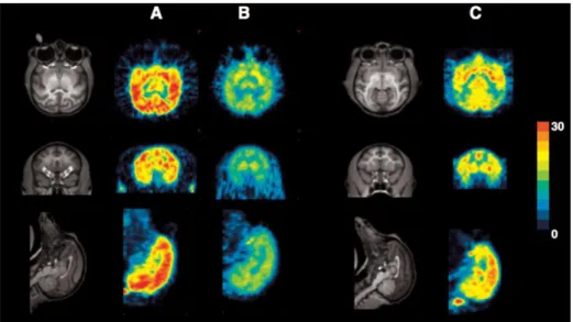
ceuticals. Recently, Tsukada et al. developed fluorine-18 derivatives of BMS-747158-01, 18
an inhibitor of the PSST subunit of MC I, among which [18F]F-BCPP-EF showed interesting 19
pharmacokinetics in rats and monkeys, with rapid uptake into the brain and heart followed 20
by gradual elimination (138). Specificity of the uptake was demonstrated using predosing 21
with rotenone as a specific MC-I inhibitor. [18F]F-BCPP-EF was used to image the extent 22
of neuronal damage in a rat model of brain ischemia, and the age-associated neuronal im-23
pairment of MC I activity in the brain of living monkeys (Fig. 4). 24
25
Antioxidants & Redox Signaling
Magnetic Resonance modalities 1
Basic principles and technical considerations
2
Electron paramagnetic resonance, EPR (or equivalently electron spin resonance, ESR) 3
is a spectroscopic technique that can directly detect paramagnetic species (species having 4
an electronic spin due to the unpaired electron). However, there is very little to be ob-5
served by EPR in biological systems apart from some stable carbon centered radicals, 6
melanin or transition metals. Reactive oxygen species such as superoxide or the hydroxyl 7
radical are much too short lived to be detected by conventional EPR. Therefore, EPR de-8
tection of ROS can be accomplished by techniques which are not always direct. The 9
scheme in Figure 5 explains the basic principles and strategies in ROS imaging. Injection 10
of an EPR visible nitroxide allows its detection in various organs in vivo. Endogenous ROS 11
react with nitroxide reducing it to an EPR silent hydroxylamine thus diminishing the EPR 12
signal. The rate of reduction is the measure of the redox status of the tissue. But one has 13
to be careful when interpreting such data, since the signal decay rate depends on several 14
kinetic factors such as the distribution of the spin probe from the blood to the tissue and 15
vice versa, urinary excretion through kidneys, fecal excretion through liver and bile. Never-16
theless, EPR monitoring of the decay rate of the injected nitroxide is the most efficient way 17
to assess the redox metabolism in vivo since one can use various nitroxides to unravel dif-18
ferent processes. The alternative to this approach is to use acyl-protected hydroxylamine 19
which, introduced in the tissue, can be easily deprotected inside cells by intracellular es-20
terases and then converted to the EPR visible species by ROS induced oxidation [58]. An 21
entirely different approach to ROS imaging is EPR spin trapping, which is the ‘true’ ROS 22
imaging. The method relies on introducing a compound that will trap short-lived radicals 23
and convert them to the more stable paramagnetic compound (Fig. 5). 24
Antioxidants & Redox Signaling
Unfortunately, trapped radicals usually have rather complex EPR spectra that
1
are not suitable for imaging. However, since multiple EPR lines do not affect overall
2
paramagnetic properties of the compound, MRI has been successfully used in
im-3
muno spin trapping(136).
4
Most of the basic principles of in vivo EPRI/EPRS have been established in the 80’s 5
(4, 7, 9, 26, 29, 60, 100, 111, 133). Much of this work has been stimulated by the discov-6
ery that nitroxides can report on the redox metabolism in cells and tissues and that the rate 7
of reduction is highly dependent on the concentration of oxygen (see e.g. (131). Since 8
then, several research groups have been developing specific spin probes with adequate in 9
vivo life time and other desirable properties, as well as instruments suitable for in vivo
10
EPR. A standard commercial EPR spectrometer operating at 9.5 GHz (X-band) can at best 11
accommodate a mouse tail due to non-resonant absorption of the electromagnetic radia-12
tion by the dielectric liquids in biological systems. Imaging of small animals thus has been 13
performed at L-band (1.2 GHz) or even lower frequencies (around 700 or 300 MHz)(10). 14
Commercial EPRI machines suitable for in vivo applications were not available until re-15
cently, hence most of researchers used and still are using home-made apparatus or modi-16
fication of commercial ones. 17
The realization that one can introduce metabolically responsive and relatively stable 18
paramagnetic free radicals in the body and detect these processes, promptly stimulated 19
the introduction of MR in the area. MRI detects paramagnetic species indirectly, since they 20
increase the relaxation rate of water molecules which can be seen by the enhanced signal 21
on T1 weighted images. At the beginning, nitroxides were studied as potential clinical con-22
trast agents, primarily for tumors, but recently they are more often used to study the redox 23
state (14). MRI has no problems in imaging subjects of any size, including humans, since it 24
operates in the frequencies of few hundreds of MHz, but detection of ROS is indirect. 25
Antioxidants & Redox Signaling
Both techniques have their advantages and drawbacks in in vivo ROS detection/imaging 1
but the sensible simultaneous use of both is a way to employ the potential of these tech-2
niques, which has been demonstrated even for solutions (8). Namely, EPRI does not pro-3
vide images of anatomy, it just shows the distribution of injected nitroxide within the body, 4
and it does not have good spatial resolution. Conversely, MRI has excellent spatial resolu-5
tion, but gives little or no information on the paramagnetic species involved. Hence, using 6
MR as imaging modality and EPRS in combination can provide unique information (35, 7
37). It is also possible to use both techniques in imaging modality and overlay EPRI 8
providing redox information on top of MRI providing anatomic information (15, 44, 57). 9
Numerous examples of combining these two techniques in oxymetry imaging can 10
be found elsewhere (2, 81, 82). There are also constructions of dual EPR/MR imaging ma-11
chines (32, 39, 116). Probably the best way to fuse EPR and MRI into a single machine is 12
to use the dynamic nuclear polarization (DNP or Overhauser effect) which uses a unique 13
method for radical detection (see below). An entirely different approach has been the 14
combination of X-ray CT with EPRI in studying a mouse knee (11). 15
Perhaps the most powerful application of in vivo EPRI is measurement of oxygen 16
(EPR oxymetry). This subject will not be covered per se due to limited space although it is 17
closely connected with the scope of this review. In addition, this subject has been exten-18
sively and regularly reviewed. What follows are characteristic examples which illustrate 19
applications of magnetic resonance techniques in imaging ROS, particularly emphasizing 20
how fruitful a combination of EPR and MRI can be in achieving optimal analysis of the in-21
vestigated subject. A more comprehensive list of examples and literature overview on EPR 22
imaging of the oxidative stress can be found in the recent review (28), and more technical 23
aspects of various EPR and MRI approaches with examples can be found in (56, 90). 24
25
Antioxidants & Redox Signaling
Examples of EPRI/MRI of ROS/RNS
1
Early EPRI images were rather crude (same as first MRI) and it took some 5-6 min 2
to make crude 2D images using filtered back-projection with only 8 projections resulting in 3
low spatial resolution. (3, 111). It took full 45 min to obtain a complete 3D data set (60), 4
which certainly limits temporal studies. Yet, this research stimulated further development 5
and today’s machines are capable of producing 3D EPR images in around 1 minute with 6
up to 80 projections, where the actual performance depends on a selected task (34, 55, 7
155). Most research using EPRI and MRI was conducted using derivatives of TEMPO and 8
PROXYL. In the beginning, carboxyl-PROXYL (3CxP or then termed PCA) has been used 9
(3, 4, 111), but later carbamoyl-PROXYL (3CP) became almost the universal choice for 10
imaging, although different derivatives, such as hydroxylmethyl (HM-P) and others, have 11
been used especially in brain imaging (118, 151, 155). The proper selection of these 12
probes with a different properties such as in vivo half-life, membrane-permeability, lipid 13
solubility etc., enables clarification of the location of in vivo ROS generation and redox sta-14
tus. As a rule, piperidine nitroxides have an in vivo half-life of a few minutes while half-life 15
of pyrrolidine is typically around 15 minutes or more. This is why pyrrolidines are generally 16
used to image metabolism while piperidines are useful for probe circulation. 17
18
Brain imaging (without tumors)
19
The brain, due to its complex structure and function, has been a natural target for 20
ROS EPR/MR imaging since the beginning of development of EPRI (60). This research 21
has been accelerated by synthesis of the blood-brain-barrier (BBB) permeable nitroxides 22
(117, 140) and instrumental developments. Yokoyama et al. published a nice series of ar-23
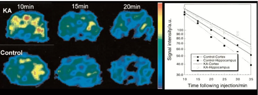
ticles on various conditions induced in experimental animals (149, 150, 152–154). Figure 6 24
illustrates the basic concept of time resolved brain EPRI (153). In rats with kainic-acid (KA) 25
Antioxidants & Redox Signaling
was significantly prolonged; indicating impaired reducing ability, whereas the prolongation 1
of the cortical half-life was not significant. These findings were confirmed by using an acyl-2
protected hydroxylamine which undergoes intracellular oxidation to nitroxides (150) show-3
ing that oxidative stress in the hippocampus and striatum in KA-treated animals is en-4
hanced, but not in the cortex. Another set of studies, performed on the effect of various 5
neuroleptics that are known to induce oxidative stress on the brain, revealed diminished 6
ability of various brain areas in treated animals to reduce injected nitroxide (149, 152, 154). 7
The study on intracerebral reducing ability after acute stress in adult rats showed dimin-8
ished reducing ability in rats that were subjected to neonatal isolation (154). Studies em-9
ploying ischemia-reperfusion (I/R) injury induced by mid-carotid-artery-occlusion using ei-10
ther only MRI (13) or EPRI/MRI combination (52) revealed slower reduction rates in brains 11
that have undergone I/R. Another common way of altering the redox state is to induce sep-12
tic shock, and it was shown that reduction rates of injected nitroxides are accelerated in 13
brains of septic mice (36). Radiation is a certain way to induce vast changes in redox sta-14
tus and various nitroxides have been successfully tested as potential radioprotectors (see 15
(23) and references cited therein). In that study, nitroxides were used both as radioprotec-16
tors and indicators of redox status, and pharmacokinetics of nitroxides in brain, salivary 17
gland, tongue and oral muscle have been determined using MRI. 18
Tumor imaging
19
Due to their heterogeneous structure, tumors have been studied since the introduc-20
tion of EPRI (9, 26). Redox status and oxygenation are important in designing therapy (es-21
pecially radiotherapy) and/or assessing tumor response to therapy. Tumors are heteroge-22
neous in both aspects hence it is desirable to obtain spatially resolved images of nitroxide 23
distribution and clearance simultaneously within the tumor volume as well as oxygenation, 24
if possible. Various approaches employing the EPR/MRI combination, or individual tech-25
Antioxidants & Redox Signaling
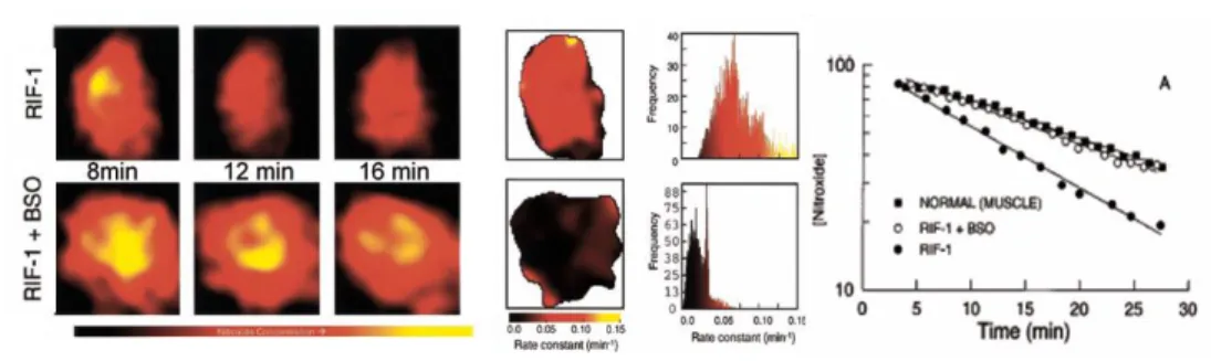
An example of tumor heterogeneity in reduction rates of nitroxide is given in Figure 7. It 1
has been generally concluded that reduction of nitroxides in tumors is faster than in normal 2
tissue, irrespective of whether the reduction in tumors implanted in the muscle is com-3
pared to the muscle (54, 58, 73, 74, 146) or when gastric cancer is compared to normal 4
mucosa (98). Faster bioreduction in tumors can be a consequence of an increased amount 5
of endogenous reducing agents such as thiols (reduction was slower in both normal tissue 6
and tumor in animals depleted with thiols (74, 146)), ascorbate, enzymes (see Fig. 7). 7
Chemically, nitroxides do not react with thiols, but altering the concentration of the thiol or 8
changing the ratio of redox pairs have an impact on the clearance of the nitroxide, there-9
fore in-vivo reduction of nitroxides depends also on the oxygen content and on the levels 10
of GSH (74). Lack of oxygen, reflecting the well known fact that reduction is faster in oxy-11
gen depleted tissues may also be responsible and tumors tend to have large hypoxic re-12
gions. Study of tumors in animals breathing carbogen showed decreased reduction of ni-13
troxides and decreased reduction heterogeneity with increased oxygenation (58), but a 14
simultaneous study on reduction of nitroxides and direct oxymetry showed rather poor cor-15
relation between these in normal air-breathing animals (132). 16
17
Other organs
18
Skin is an ideal target organ for EPRI for several reasons. Imaging of ROS does not 19
require a large penetration depth, so one can use the S-band (2.2 - 3.0 MHz) for in vivo or 20
even X-band for in vitro specimens, which results in improved sensitivity. Imaging does not 21
require full 2D or 3D; once nitroxides are applied topically a simple spectral-spatial 1D im-22
aging with one gradient orthogonal to the skin surface is sufficient to obtain distribution of 23
nitroxides and redox status in different skin layers. Surface loop coils are sufficient, i.e. the 24
whole objects need not to be within the resonator, which allows EPRI of objects of any size 25
Antioxidants & Redox Signaling
vivo study of human skin (46) which opens the possibilities of studying various skin
pathol-1
ogies, ageing or photo-damage. The effect of UV exposure on free radical production and 2
redox status of the skin has been studied both in vivo and in vitro (45–47). 3
Pharmacokinetics of nitroxides in abdominal organs (liver, kidneys, bladder) was 4
first studied by in vivo EPRS (4) and EPRI (3, 111). The distribution and reduc-5
tion/clearance of nitroxides demonstrated the feasibility of EPRI studies. But apart from 6
having low spatial and temporal resolution, it has revealed difficulties in anatomical locali-7
zation of different organs on EPR images. A decade later, it has been shown that this 8
problem can be overcome by combining EPRI and MRI (44, 57) and that whole body sim-9
ultaneous measurements of pharmacokinetics and distribution of nitroxides can be per-10
formed on ten different locations within the body (57). These studies were performed to il-11
lustrate technical developments, and were not aimed at investigating any particular pathol-12
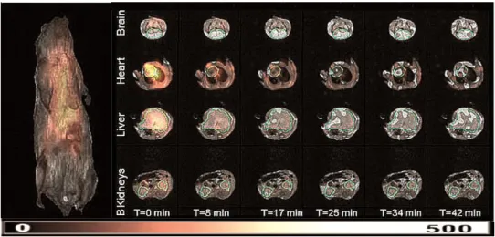
ogy. An excellent application of a hybrid EPR/MRI machine has studied the redox status of 13
different organs in mice exposed to cigarette smoke (Fig. 8). On the other hand, different 14
important pathologies were studied using less technically demanding direct time resolved 15
EPRI. The study of mice liver showed much slower reduction of 3CP in carbon-16
tetrachloride damaged liver than in the control (135). Another study of mice with hepatic 17
ischemia-reperfusion injury showed that CV159-Ca2+/calmodulin blockade inhibiting Ca2+ 18
overloading has a profound effect on the liver reducing ability (69). The ischemia-19
reperfusion acute renal failure produced prolonged reduction of 3CP in kidneys (48), while 20
it was much faster in the kidneys of diabetic mice (128). The latter study also showed that 21
treatment with angiotensin returns the reduction to the control level, confirming the antioxi-22
dant properties of this drug. Another drug (Azelnidipine) has been studied in the murine 23
hypertension model and it has been found that it improves renal reducing ability of free 24
radicals thus ameliorating the renal redox status (49). A somewhat different model of the 25
Antioxidants & Redox Signaling
transcriptional factor-deficient mice. The combination of deficiency and ageing resulted in 1
four times longer half life of 3CP in the upper abdomen than in juvenile wild-type mice indi-2
cating that low reducing ability may play a role in the onset of autoimmune nephritis (50). A 3
set of hydroxylamine spin probes detecting site-specific production of the superoxide radi-4
cal allowing subcellular resolution and organelle specificity was developed and used in-5
vivo on several organs. The detection mechanism is based on rapid reaction of cyclic hy-6
droxylamines with superoxide, producing stable nitroxides (25). These probes were ap-7
plied in-vivo on old rats, showed how ROS generation was significantly increased com-8
pared to their young counterparts in blood,skeletal muscle,lung and heart, but did not 9
change in intestine, brain, liver, and kidney. (71) 10
11
Imaging of trapped radicals
12
This attractive modality offers a possibility to image specific ROS as opposed to 13
previous examples where the overall redox state was imaged. However, EPRI of trapped 14
ROS is extremely difficult. First, the concentration of radicals is very low and it requires 15
very high amounts of spin trapping agent to be injected (up to 100 mmol/kg), which raises 16
the question of toxicity. Second, EPR spectra of trapped radicals usually contain numerous 17
closely spaced lines of multiple adducts (Fig. 5) and it is almost impossible to isolate spe-18
cific lines for imaging the selected adduct. Third, trapped products are not very stable 19
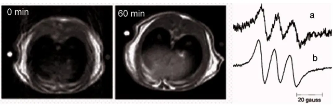
which narrows the time window for imaging. Nevertheless, imaging of trapped NO (Fig. 9) 20
is a good example of how to combine in vivo EPR and MRI. The role of in vivo EPR is not 21
to image radicals but to add the unique information on the nature of the radical species 22
(fingerprinting) which actually enhances the tissue signal on T1W MRI and a similar ap-23
proach has been employed in brain imaging (36). Although there are no true images of 24
trapped radicals, useful in vivo studies have been performed employing specific EPR coils 25
Antioxidants & Redox Signaling
traps in detecting radicals (134), NO generation in mice following cardiopulmonary arrest 1
(75), studies of simultaneous detection of oxygen and NO in the induced septic shock (33). 2
Another example of successful in vivo detecting of trapped radical is on irradiated mouse, 3
since irradiation produces a large amount of free radicals (43). Imaging of spin trapped su-4
peroxide or hydroxyl radical could be improved by developing more resistant spin traps 5
and using 15N-substituted probes, which will improve sensitivity and resolution by decreas-6
ing the number of EPR lines (see the nice review on in-vivo trapping (64)). 7
8
Dynamic nuclear polarization DNP-MRI (OMRI, PEDRI)
9
This technique deserves to be treated separately due to the unique detection mecha-10
nism and high potential for in vivo measurements although the aim of these studies is the 11
same as outlined above. DNP is not a simple overlaying of separate EPR and MR images 12
obtained in a hybrid apparatus but a technique that includes parts of EPRI and MRI. De-13
tection of radicals (unpaired electron spin) is based on a different principle. There are sev-14
eral DNP mechanisms (89), but most of the biological applications have been performed 15
using classical Overhauser effect, hence Overhauser MRI or OMRI. The first experiment 16
using nitroxides and transfer to protons has been performed almost 30 years ago (86) and 17
was referred to as PEDRI (proton electron double resonance imaging). Briefly, two-spin 18
system (e.g. nitroxide/water protons) in magnetic field is irradiated by RF at an EPR fre-19
quency of nitroxides (unpaired electron), magnetization is transferred to protons enhancing 20
water proton NMR signal intensities and the overall effect is detected by conventional pro-21
ton MRI (86). This effect is completely different from classical enhancement of proton re-22
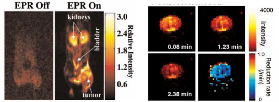
laxation by nitroxides (theoretically 330 times higher). This is illustrated in Fig. 10 on MRI 23
of nitroxide infused mice (72). Without EPR RF irradiation nitroxides are invisible since 24
classical enhancement is weak, while they can be clearly seen in the ‘EPR on’ mode. 25
Antioxidants & Redox Signaling
redox status have been published (147, 148). The OMRI combines the sensitivity of EPR 1
with the advantages of MRI thus presenting an ideal machine for ROS imaging. However, 2
OMRI apparatus has to be home-built, which requires substantial skill and resources. The 3
impetus for further development may come from the fact that in vivo DNP-MRI has an in-4
trinsic capacity for molecular imaging of multiple species, similar to MR chemical shift im-5
aging. By changing the frequency of EPR irradiation in DNP-MRI distinct images of differ-6
ent radicals having different EPR spectra can be obtained, which has been demonstrated 7
in an experiment where nitroxides labeled with 14N or 15N were simultaneously imaged 8
(141). This approach was further extended, albeit in test tubes, to simultaneous imaging of 9
free radical intermediates involved in the mitochondrial electron transport chain and radi-10
cals derived from vitamins E and K1 (53). Being able to simultaneously image species with
11
a heterogeneous broad line having poor hyperfine splitting or species with complicated hy-12
perfine splitting lines, which is impossible for standard EPRI, opens a host of possibilities 13
including metabolic imaging in various pathologies and imaging of spin-trapped radicals. 14
15
Conclusions 16
The relevance of ROS in human physiopathology is now a well-established clinical no-17
tion (30). Reactive species are essential regulators in the physiopathology of disease, the 18
knowledge of their concentration and local distribution with subcellular resolution is there-19
fore a necessary clinical tool. This can be achieved by using different approaches based 20
on the detection of redox couples, biomarkers that specifically bind to a redox species or 21
that can modify their properties in the presence of ROS or some by-product of the oxida-22
tion (Fig. 1). The most investigated solution is the “photonic” one. A wide variety of fluores-23
cent probes and fluorescent nanoparticles in the visible range, specific for each redox 24
couples, allow redox mapping with 200 nm resolution, according to the Abbe 25
Antioxidants & Redox Signaling
law. Recently, new optical techniques (Stimulated emission-depletion fluorescence mi-1
croscopy (STED), photoactivated localization microscopy (PALM), etc.) (80, 144) have 2
been developed to break down the resolution limit up to 20 nm, but specific probes are 3
needed and, at the moment, these are unavailable for redox detection. Instead, the devel-4
opment of redox sensitive fluorescent probes, allowed a quantitative detection of each 5
component of each redox couples, representing a further step-ahead towards the compre-6
hension of ROS involvement in the human physiopathology. 7
For these reasons, fluorescence microscopy has become very popular in biomedi-8
cal research activities (i.e. on cell lines), but also has found relevant translation applica-9
tions in the histo-pathology of tissues from biopsies and in the investigation of dermal inju-10
ries (i.e melanoma detection) (109). 11
These techniques, however, suffer from two main drawbacks that hampered their 12
potential translational applications: i) the low penetration of the visible light into tissues 13
(roughly 300 nm) due to tissues’ optical absorbance (mainly due to hemoglobin) and multi-14
ple scattering, and ii) the toxicity of fluorescent probes. Two Photon Microscopy with en-15
dogenous or chemo-selective probes offers an attractive approach to in vivo ROS detec-16
tion, due to probes’ general compatibility with many biological systems without external ac-17
tivating enzymes and genetic manipulation. TPM for in vivo and internal tissue imaging by 18
using endogenous probes is a very attractive option and have stimulated the development 19
of TPM microendoscopes using a gradient-index (GRIN) rod lens, miniature compound 20
lens (68, 114). Otherwise, to avoid the low signal to noise ratio provided by endogenous or 21
chemo-selective probes, and to increase the penetration depth, the use of TP-excited IR 22
and chemiluminescent probes have been proposed. 23
Microscopy in the NIR-VIS region of the electromagnetic spectrum is therefore very 24
promising, although may suffer of a limited clinical applications, when large spatial areas 25
Antioxidants & Redox Signaling
the whole organ of a human being) we need different approaches. These can be furnished 1
by intriguing applications of techniques commonly adopted in clinical investigation. 2
Ultrasound based techniques, as demonstrated for the APL bio-sensors, allow to 3
detect physiological concentrations of ROS, with a contrast and a spatial resolution that 4
can exceed those provided by fluorescence and chemiluminescence based contrast 5
agents. Evaluation is possible on a rapid time-scale (minutes) and imaging systems are in 6
widespread clinical use. However, while toxicity of APLs and MMCs remains a concern, 7
functionalizing these agents allow a selective destruction of target tissues. 8
Toxicity is a minor feature for PET and SPECT that are non-invasive, but they do 9
involve exposure to ionizing radiation. Besides its established role as a diagnostic tech-10
nique, PET has an expanding role as a method to assess the response to therapy, in par-11
ticular, cancer therapy, where the risk to the patient from lack of knowledge about disease 12
progress is much greater than the risk from the test radiation. The principal concern in PET 13
and SPECT redox imaging is the lack of radiolabeled molecules that bind to ROS, and that 14
has limited success of nuclear medicine in the direct imaging of ROS. Nevertheless, indi-15
rect methods for imaging of glucose consumption, redox potential, hypoxia, as well as di-16
rect imaging of ROS scavengers and mitochondrial complexes have undisputable clinical 17
interest. Considering the sustained efforts in development of new isotopes and labeling 18
methods, PET and SPECT are poised to make significant contributions to the field in the 19
future. Another limit is the resolution of clinical and pre-clinical PET cameras (roughly 1 20
mm). 21
The EPR and MR imaging in vivo has become a powerful tool in experimental and 22
preclinical studies of ROS/RNS or redox status on animals. Basic concepts are well un-23
derstood and directions for future developments are clear. On instrumental side, further 24
development of hybrid machines is an obvious goal. The problem remains that these ma-25
Antioxidants & Redox Signaling
traps, development of those that show specificity towards certain ROS, specificity toward 1
certain organs (e.g. tumors) and showing longer in vivo life time is required; some studies 2
along these lines are already underway (1, 85, 107, 113). The major obstacle in the trans-3
lation of these techniques to the clinic is the scarcity of centers possessing the equipment, 4
and the lack of a focused concerted effort on certain clusters of widely relevant pathologies 5
in which ROS may play a key role (e.g. Amyotrophic Lateral Sclerosis, Parkinson, Alz-6
heimer and other neurodegenarative diseases). Clinical application of EPR spectroscopy 7
have been summarized recently (65), stating that the best perspectives are in oximetry 8
and dosimetry ionizing irradiation. One can add certain potential in investigating skin pa-9
thologies (including) melanoma to the list, but, due to problems with penetration depth of 10
microwaves, EPRI of human body analogous to MRI will never be possible. On the other 11
hand, it has been successfully demonstrated that OMRI machines accommodating large 12
subjects including humans can be built (72, 87), opening possibilities to combine all MRI 13
capabilities with molecular specificity of EPR in diagnosis and treatment follow-up. 14
15
Outlook 16
Potential clinical translation of ROS imaging is straightforward. Each of the present-17
ed techniques possess an attractive potential, but none of them can fulfill all the require-18
ments in terms of sensitivity, spatial resolution, temporal resolution, probe availability, tox-19
icity and cost. The obvious solution is to perform parallel studies with two or more tech-20
niques or even better to integrate imaging modalities that may offer synergistic advantages 21
over any single modality alone. Some hybrid imaging systems such as PET/CT, 22
SPECT/CT, PET/MRI, EPR/MRI have already been developed. Anatomical imaging tech-23
niques such as CT and MRI provide structural details; whereas functional modalities such 24
as PET, SPECT, TP-fluorescence, EPR and others provide insight into functional and 25
Antioxidants & Redox Signaling
metabolic aspects. Incorporating anatomical and functional imaging in a common hybrid 1
imaging platform should allow improved diagnosis, therapeutic planning and follow-up 2
studies. 3
4
Antioxidants & Redox Signaling
ACKNOWLEDGEMENTS 1
This work was supported by the Ministry of Education, Culture, Sports, Science and Tech-2
nology, Japan, Japan Science and Technology Agency, KAKENHI (Grant Numbers 3
22249003, 25253005 and 25713004), Japan Society for the Promotion of Science, Minis-4
try of Education, Science and Technological, Serbia (project III-41005), Fondi di Ateneo, 5
UCSC Rome, Italy (Linea D1). Some of the two photon acquisitions and analysis de-6
scribed in the article were performed at LabCeMi, UCSC, Rome. Several authors of this 7
review were supported by the European Cooperation in Science and Technology (COST 8
Action BM1203/EU-ROS). 9
10
AUTHOR DISCLOSURE STATEMENTS 11
The authors declare no conflict of interest. 12
13 14
Antioxidants & Redox Signaling
LIST OF ABBREVIATIONS 1
TPM: Two photon microscopy 2
SHG: Second harmonic generation 3
TP-FRIM: Two photon Fluorescence Ratio Imaging Microscopy 4
TP-FLIM: Two photon Fluorescence Lifetime Imaging Microscopy 5
NIR: Near-infrared 6
rxYFP: GSH-sensitive Yellow Fluorescent Protein 7
roGFP: GSH-sensitive Green Fluorescent Protein 8
HyPer: H2O2 fluorescent sensor
9
ROS: Reactive oxygen species 10
RNS: Reactive nitrogen species 11 NP: Nanoparticles 12 GSH: Glutathione 13 GSSG: Glutathionedisulfide 14 MPO: Myeloperoxidase 15
NAD: Nicotinamide adenine dinucleotide 16
NADP: Nicotinamide adenine dinucleotide phosphate 17
NADPH: Nicotinamide adenine dinucleotide phosphate NOX: Nicotinamide adenine dinu-18
cleotide phosphate oxidase 19
NOS: Nitric Oxide Synthase 20
O2−: Superoxide Radical
21
ONOO−: Peroxynitrite radical 22
FAD: Flavin adenine dinucleotide 23
H2O2: Hydrogen peroxide
24
HO2· : Hydroperoxyl radical
25
Antioxidants & Redox Signaling
NO: nitric oxide 1
Grx1: glutaredoxin 1 2
APL: Allylhydrazine encapsulated in phospholipid liposomes 3
MMC: micromotor converters 4
PET: Positron Emission Tomography 5
SPECT: Single Photon Emission Computed Tomography 6
FDG: [18F]Fluorodeoxyglucose 7
FMN: flavin mononucleotide 8
EG: glutathione intracellular redox potential 9
PN1: Peroxy Naphthalene 1 10
TEMPO: 2,2,6,6-tetramethylpiperidine 1-oxyl 11
PROXYL: 2,2,5,5-tetramethylpyrrolidine 1-oxyl 12
Table 13
14
Reactive Species
subclassifi-cation Structure
Biological Half-life(s)
Reference
Hydrogen peroxide ROS H2O2 10-5
(38)
Hydroxyl radical ROS HO• 10-9
(20, 38)
Hypochlorous acid ROS HOCl ?
?
Nitric oxide RNS NO 10-3÷1
(63, 106, 112, 158)
Peroxyl radical, including al-kylperoxyl and hydroperoxyl
radi-cals (wherein R = H)
ROS ROO• 10-1÷1
(20)
Peroxynitrite anion RNS ONOO– 10-2÷1
(6, 106)
•O – -6 (38, 62)
Antioxidants & Redox Signaling