HAL Id: inserm-01152594
https://www.hal.inserm.fr/inserm-01152594
Submitted on 18 May 2015HAL is a multi-disciplinary open access archive for the deposit and dissemination of sci-entific research documents, whether they are pub-lished or not. The documents may come from teaching and research institutions in France or abroad, or from public or private research centers.
L’archive ouverte pluridisciplinaire HAL, est destinée au dépôt et à la diffusion de documents scientifiques de niveau recherche, publiés ou non, émanant des établissements d’enseignement et de recherche français ou étrangers, des laboratoires publics ou privés.
Interleukin-6 and C-reactive protein as predictors of
cognitive decline in late midlife.
Archana Singh-Manoux, Aline Dugravot, Eric Brunner, Meena Kumari,
Martin Shipley, Alexis Elbaz, Mika Kivimaki
To cite this version:
Archana Singh-Manoux, Aline Dugravot, Eric Brunner, Meena Kumari, Martin Shipley, et al.. Interleukin-6 and C-reactive protein as predictors of cognitive decline in late midlife.. Neurol-ogy, American Academy of NeurolNeurol-ogy, 2014, 83 (6), pp.486-93. �10.1212/WNL.0000000000000665�. �inserm-01152594�
MS ID#: NEUROLOGY/2013/560177 Interleukin-6 and C-reactive protein as predictors of cognitive decline in late midlife
Archana Singh-Manoux, PhD*1,2,3 ; Aline Dugravot, MSc1,4 ; Eric Brunner, PhD2 ; Meena Kumari,PhD2 ; Martin Shipley, MSc2 ; Alexis Elbaz, MD, PhD1,2 ; Mika Kivimaki, PhD2
* Corresponding author & address:
1
INSERM, U1018, Centre for Research in Epidemiology and Population Health Hôpital Paul Brousse, Bât 15/16, 16 Avenue Paul Vaillant Couturier
94807 VILLEJUIF CEDEX, France
Telephone: +33 (0)1 77 74 74 10 Fax: +33 (0)1 77 74 74 03
Email: Archana.Singh-Manoux@inserm.fr
2
Department of Epidemiology and Public Health, University College London, UK
3
Centre de Gérontologie, Hôpital Ste Périne, AP-HP, France
4
University Versailles St-Quentin en Yvelines, France
Supplemental data
[25] All Cognitive Disorders/Dementia, [ 36 ] Cognitive aging, [54] Cohort studies, [59] risk factors in epidemiology
Word count: text: 2998; abstract: 248. Character count title : 87
References: 40
Study funding: US National Institutes of Health (R01AG013196 to ASM; R01AG034454 to ASM & MKi, R01HL036310 to MKi), the UK Medical Research Council (K013351 to MKi), the Economic and Social Research Council (to MKi) and the British Heart Foundation (to EB).
Authors Contributions
ASM formulated the research question, supervised the analysis, and wrote first and successive drafts of the paper. AD undertook the statistical analyses and commented on drafts. EB, MKu, MS, AE and MKi interpreted the results and commented on drafts.
Disclosures
Dr. Singh-Manoux receives research support from the US National Institutes of Health (R01AG013196; R01AG034454 ).
Ms Dugravot reports no disclosures.
Prof. Brunner receives research support from the British Heart Foundation.
Dr Kumari receives support from the Economic and Social Research Council and National Institutes of Health.
Mr. Shipley reports no disclosures. Dr. Elbaz reports no disclosures.
Prof Kivimaki receives research support from US National Institutes of Health (R01AG034454, R01HL036310), the UK Medical Research Council (K013351) and the Economic and Social Research Council.
Abstract
Objective: Peripheral inflammatory markers are elevated in those with dementia. In order to assess their etiological role we examined whether interleukin-6 (IL-6) and C-reactive protein (CRP), measured in midlife predict concurrently assessed cognition and subsequent cognitive decline.
Methods: Mean value of IL-6 and CRP, assessed on 5217 persons (27.9% women) in 1991-1993 and 1997-1999 in the Whitehall II longitudinal cohort study, were categorized into tertiles to examine 10-year decline (assessments in 1997-1999, 2002-2004 and 2007-2009) in standardized scores (mean=0, standard deviation=1) of memory, reasoning, and verbal fluency using mixed models. Mini Mental State Examination (MMSE) was administered in 2002-2004 and 2007-2009, decline ≥3 points was modeled with logistic regression. Analyses were adjusted for baseline age, sex, education, and ethnicity; further analyses were also adjusted for smoking, obesity,
Framingham cardiovascular risk score, and chronic diseases (cancer, coronary heart disease, stroke, diabetes, and depression).
Results: In cross-sectional analysis, reasoning was 0.08 standard deviation (95% CI: -0.14, -0.03) lower in participants with high compared to low IL-6. In longitudinal analysis, 10-year decline in reasoning was greater (ptrend=0.01) among participants with highIL6 (0.35; 95% CI: 0.37, -0.33) than those with low 6 (-0.29; 95% CI: -0.31, -0.27). In addition, participants with high IL-6 had 1.81 times greater odds ratio of decline in MMSE (95% CI: 1.20, 2.71). CRP was not associated with decline in any test.
Conclusions: Elevated IL-6 but not CRP in midlife predicts cognitive decline; the combined cross-sectional and longitudinal effects over the 10-year observation period corresponded to an age effect of 3.9 years.
Introduction
Inflammatory molecules, including acute-phase inflammatory reactants and pro-inflammatory cytokines, have been found in the cerebral spinal fluid and beta amyloid plaques in patients with dementia.(1-3) Inflammation appears to be a feature of dementia; amyloid deposition
stimulates neuro-inflammatory processes with neurotoxic effects that exacerbate neuronal damage. There is also evidence that circulating levels of inflammatory markers are raised before clinical onset of dementia; for instance, in nondemented persons, interleukin-6 (IL-6) and C-reactive protein (CRP) have been shown to predict dementia.(4-6) However, given the long preclinical phase of dementia(7) it is unclear whether peripheral inflammation is a by-product of neuropathology or whether it contributes to neuronal damage.
Evidence of an association between peripheral inflammatory markers and cognitive decline rather than dementia, which represents the extreme clinical presentation of the pathologic process, would support an aetiological role for inflammation in cognitive ageing. To date, however, few prospective studies have examined inflammatory markers in midlife as predictors of cognitive decline. Here, we examine this association using two markers, IL-6 and CRP, expecting IL-6 to have a stronger association with cognition as it drives the production of downstream markers such as CRP. As single measures cannot reliably separate acute and chronic inflammation and may therefore underestimate associations with disease outcomes,(8) we use the mean of two measures taken five years apart to capture long-term inflammation. Our objective is to examine the association of CRP and IL-6 in midlife and cognitive decline over 10 years in a large community-dwelling sample of men and women.
Methods
The Whitehall II study is an ongoing cohort study of men and women, originally employed by the British civil service. The target population was all London-based office staff, aged 35-55. A total of 10,308 persons (6895 men and 3413 women), response rate 73%, were recruited to the study over the years 1985 to 1988.(9) Since the baseline medical examination, follow-up examinations have taken place approximately every 5 years: 1991-1993 (n=8815); 1997-1999 (n=7870); 2002-2004 (n=6967) and 2007-2009 (n=6761)
Standard Protocol Approvals, Registrations, and Patient Consents
Ethical approval for the Whitehall II study was obtained from the University College London Medical School committee on the ethics of human research (reference number 85/0938); all participants provided written informed consent.
Assessment of Interleukin-6 and CRP
Fasting serum was collected in 1991-1993 and 1997-1999 between 8 am and 1 pm and stored at −80°C. CRP was measured with a high-sensitivity immunonephelometric assay in a BN ProSpec nephelometer (Dade Behring, Milton Keynes, UK). IL-6 was measured with a high-sensitivity ELISA (R&D Systems, Oxford, UK). Values lower than the detection limit (0.15 mg/L for CRP, 0.08 pg/mL for IL-6) were assigned a value equal to half the detection limit. To measure short-term biological variation and laboratory error, a repeat sample was taken from 150 participants for CRP and 241 participants for IL-6 at the 1991-1993 measure (average elapsed time between samples, 32 days [SD=10.5 days]). Intra-assay and inter-assay coefficients of variation were 4.7% and 8.3% for CRP and 7.5% and 8.9% for IL-6.
Cognitive Function
The cognitive test-battery, administered at three clinical examinations over 10 years (1997-1999, 2002-2004, 2007-2009) was chosen to provide a comprehensive assessment of cognitive function and be appropriate for this population composed of individuals younger than in most studies on cognitive ageing.(10) The tests included in the analysis had high test-retest reliability, range 0.60 to 0.89, assessed on 556 participants who were invited back to the clinic within 3 months of having taken the test in 1997-1999.
Short-term verbal memory was assessed with a 20-word free recall test. Participants were presented a list of 20 one or two syllable words at two second intervals and were then asked to recall in writing as many of the words in any order with two minutes to do so.
The Alice Heim 4-I (AH4-I) is composed of a series of 65 verbal and mathematical
reasoning items of increasing difficulty.(11) It tests inductive reasoning, measuring the ability to identify patterns and infer principles and rules. Participants had 10 minutes to do this section.
We used two measures of verbal fluency, phonemic and semantic, combined for the purposes of the analysis.(12) Participants were asked to recall in writing as many words beginning with “S” (phonemic fluency) and as many animal names (semantic fluency) as they could. One minute was allowed for each test; the observed range on these tests was 0-35.
The Mini-Mental State Examination,(13) a brief 30-point measure of global cognitive function was administered at the last two waves of the cognitive data collection.
Covariates
Covariates included age, sex, ethnicity (white, south-Asian, Black, other), and education, the highest qualification on leaving full-time education (lower secondary school or less, higher secondary school (usually achieved at age 18), university or higher degree). Smoking status was
reported by participants and classified as never, ex- or current-smoker. Obesity was defined as body mass index (BMI) ≥30 kg/m2.(14) The Framingham general cardiovascular disease (CVD)
risk score was used to assess subclinical disease, calculated using data on age, HDL cholesterol, total cholesterol, systolic blood pressure, cigarette smoking, and diabetes.(15)
The chronic diseases considered as covariates were coronary heart disease, stroke, identified using hospital records database; cancer, using the National Health Service cancer registry; and diabetes, determined by oral glucose tolerance test (fasting glucose ≥ 7.0 mmol/l, 2-h postload glucose ≥ 11.1 mmol/l), reported doctor-diagnosed diabetes, or use of diabetes medication).(16) Antidepressant use was assessed using questions on medication. All
participants were followed for mortality using linkage to the national mortality database.
Statistical Analysis
All analyses were conducted using STATA 12. Participant characteristics were described as percentages or mean (standard deviation, SD) when appropriate. Inflammatory markers were categorised into tertiles using mean values from 1991-1993 and 1997-1999. We also ran
analyses using continuous scores, modelled as an increment of 1 standard deviation, after log transforming the inflammatory measures owing to skewed distributions. The association of inflammatory markers with mortality over the follow-up was examined using Cox proportional hazards models.
In order to allow comparison across cognitive tests, we standardized the raw scores of each test to z-scores (mean=0; standard deviation (SD)=1). In the longitudinal analysis, the standardization to the z-score was carried out using the mean and SD from 1997-1999. Linear mixed models(17) were used to estimate the cross-sectional and longitudinal association of
inflammatory markers with cognition (three assessments, 1997-1999, 2002-2004, 2007-2009). Mixed models use all available data over the follow-up, take into account the fact that repeated measures on the same individual are correlated with each other, and can handle missing data. In these analyses both the intercept and the slope were fitted as random effects, allowing individuals to have different cognitive scores at baseline and different rates of cognitive decline over the follow-up. The intercept in these models yields the cross-sectional association and the slope an estimate of the 10-year decline in cognitive scores and the associated 95% confidence interval (CI). These analyses were adjusted for sociodemographic characteristics (age, sex, ethnicity, and education). In a further step, analyses were additionally adjusted for covariates associated with higher levels of inflammatory markers (smoking, obesity, Framingham CVD risk, CHD, stroke, cancer, diabetes, and antidepressant use).
The interaction term between sex, time and inflammatory markers suggested similar cognitive decline in men and women (all p between 0.11 and 0.41) leading us to combine men and women in the analysis. For both the baseline and the longitudinal analysis, we tested for a linear trend as a function of tertiles of the inflammatory markers. To allow interpretation of the regression estimates of top vs. bottom tertile of the inflammatory marker, we compared them with the effect of age on cognition. The regression estimates were divided by the effect of one year increase in age on cognition, the latter was calculated by regressing the standardised 1997–1999 cognitive score on age.
In further analyses we examined the association of the inflammatory markers (mean value of measures taken in 1991-1993 and 1997-1999) and decline in MMSE between 2002-2004 and 2007-2009, modelled as a decline of 3 points or more. These analyses were
undertaken using logistic regression as the distribution was highly skewed and unsuitable for linear regression. We compared the C-statistics of two predictive models for decline in MMSE,
one containing age and sex only and the other additionally containing the inflammatory marker to assess its incremental discriminative ability.
In sensitivity analyses, we used a Bonferroni corrected critical p-value of 0.0125 (0.05/4) based on the fact that 4 cognitive tests were used in the analysis (memory, reasoning, fluency, MMSE).
Results
A total of 7666 (74.4%) of the original 10308 participants of the Whitehall II study
participated in 1991-1993 and 1997-1999 waves of data collection, when inflammatory markers were assessed. Those with CRP values ≥10 mg/L (N=159 in 1991-1993 and N=197 in 1997-1999), an indicator of acute infection, were removed from the analysis. 5316 had valid data at both waves, allowing mean values of IL-6 and CRP to be calculated (see Figure 1). 212 deaths were recorded over the cognitive follow-up and 1 SD higher CRP (1.25, 95% CI: 1.07, 1.47) and IL-6 (1.27, 95% CI: 1.11, 1.45) were associated with greater hazard ratio of mortality. Subsequent analyses were undertaken on 5217 participants with cognitive data over the follow-up.
Approximately 72% had cognitive data at all three waves and 18% at two waves. Compared to individuals not included in these analyses, the analytic samples consisted of younger (55.7 vs. 56.4 yrs, p<0.001) participants, more men (72.1% vs. 61.6%, p<0.001), and more educated individuals (30.4% vs. 23.8% had a university degree, p<0.001).
The median for IL-6 was 1.43 pg/mL (IQR: 1.07, 1.99), based on assessments in 1991-1993 (1.41pg/mL) and 1997-1999 (1.42pg/mL, correlation r=0.47); the corresponding median was 0.98 mg/L for CRP (IQR: 0.52, 1.94, first and second assessments 0.89mg/mL and 1.02mg/L, respectively, correlation r=0.61). Changes in inflammatory marker levels between the two assessments were not associated with cognitive tests (all rs≤0.03), leading us not to examine
them further. The inflammatory markers, averaged over two assessments, were moderately correlated with each other (rs=0.49, p<0.001). One year greater age was associated with -0.040
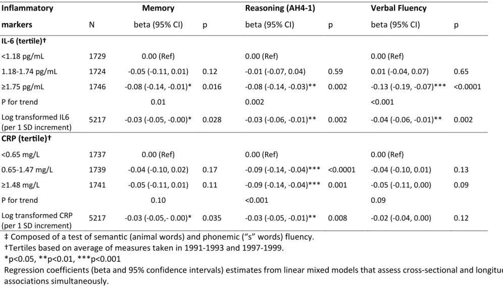
SD lower memory, -0.036 lower reasoning and -0.043 lower verbal fluency, all p<0.0001. Table 1 presents sample characteristics as a function of the tertiles of IL-6 and CRP. Both markers were associated with all baseline covariates, except stroke; CRP was also not associated with cancer. In cross-sectional analyses (Table 2), adjusted for sociodemograhic measures, those in the highest IL-6 tertile had poorer cognitive scores on all tests, the difference compared to those in the lowest tertile ranging from -0.08 to -0.13 of a standard deviation – corresponding to an age effect of 2.0 years for memory, 2.2 years for reasoning and 3.0 years for verbal fluency. Those in the intermediate and high tertile of CRP had lower scores in reasoning (0.09; 95% CI: -0.14, -0.04). Adjustment for the full set of covariates (Table e-1) attenuated associations with memory but higher IL-6 remained associated with poorer scores on reasoning and fluency and higher CRP levels with poorer scores on reasoning.
In longitudinal analyses, a decline over 10-years of approximately one third of a baseline standard deviation was observed on all tests (Table 3). Compared to the lowest IL-6 tertile (-0.29, 95% CI:-0.31, -0.27), those in the intermediate (-0.32, 95% CI:-0.35, -0.30; p<0.05) and highest (-0.35, 95% CI:-0.37, -0.33, p<0.001) tertiles experienced faster decline in reasoning. The difference in decline in reasoning between the highest and lowest IL-6 tertile corresponded to an age effect of 1.7 years. The decline in the highest tertile remained statistically significant when adjusted for the full range of covariates (Table e-2). CRP in the highest tertile was also associated with faster decline in reasoning (Table 3) but adjustment for all covariates rendered the difference non-significant (Table e-2).
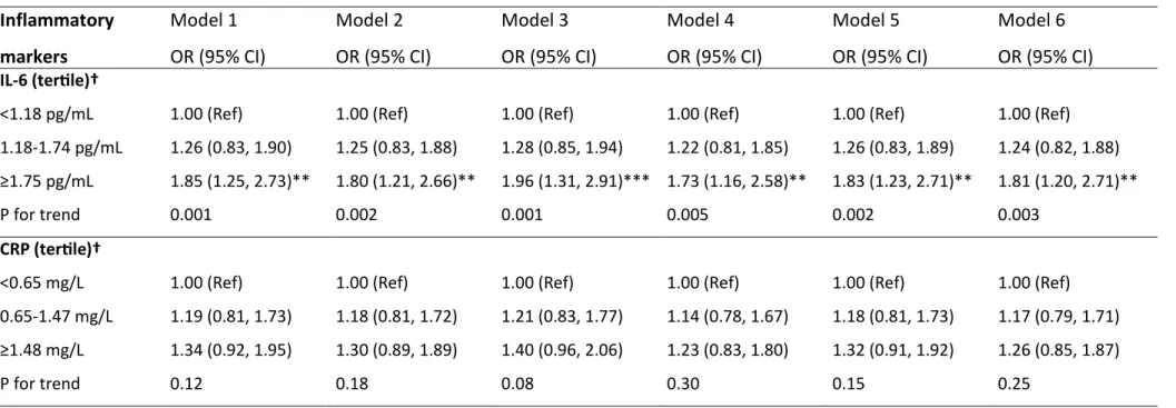
Table 4 shows the association of the inflammatory markers with decline of ≥3 points (N=191, 4.7%) in MMSE scores between 2002-2004 and 2007-2009. Those in the highest IL-6
tertile were 1.85 (95% CI: 1.25, 2.73) times more likely to register decline in MMSE than those in the lowest tertile. This greater likelihood of decline in the highest IL-6 category remained in the fully adjusted model (odds ratio (OR) 1.81; 95% CI: 1.20, 2.71; Model 6). The associations of CRP with decline in MMSE did not reach statistical significance. The change in C-statistics suggested that IL-6 (from 0.666 to 0.684, p=0.05) but not CRP (from 0.666 to 0.671, p=0.32) improve discriminative ability in prediction of decline in MMSE.
The cross-sectional and longitudinal associations between IL-6 and reasoning (tables 2-4) remained statistically significant after Bonferroni correction for 4 tests, this was also the case for IL-6 and decline in MMSE.
Discussion
Our results, based on participants aged 56 years at the start of cognitive testing suggest that inflammatory markers are associated with cognition, in both cross-sectional and
longitudinal associations. Three features qualify these findings. One, memory had no strong association with the inflammatory markers. Two, of the two markers examined, IL-6 had a more robust association with cognition. Three, although the cross-sectional effects were stronger the longitudinal effects were nevertheless present, suggesting that midlife inflammation is related to cognitive decline. The effects may appear small but over the 10-year observation period the combined effect using both cross-sectional and longitudinal effects corresponds to an age effect of 3.9 years for reasoning.
Previous studies on the association between inflammation and cognitive decline are inconsistent. Some studies show no association (18-20) and others show greater decline in those with higher inflammation.(21-24) However, much of the evidence is based on older
used repeated measures of inflammatory markers to correct for within-person fluctuations in inflammatory markers in analysis with cognitive outcomes. Memory appears not to be one of the domains affected by long-term inflammation in our analysis. Previous cross-sectional studies report associations with multiple cognitive domains,(25) the longitudinal studies used either a global measure, such as the MMSE(21) or combined tests to produce global scores.(24) All the tests used in the present analyses have been shown previously to be sensitive to age-related changes in cognition in midlife.(10) Timed tests of reasoning, such as the Alice Heim test, are good measures of processing speed or mental efficiency.(26) Further support for the lack of association with memory comes from studies showing greater effects of inflammatory factors on vascular rather than Alzheimer’s type dementia.(6, 27)
Previous studies on the association between inflammatory markers and cognitive
outcomes have not been conclusive on the specific marker that may be of special interest. In the Rotterdam study, IL-6 but not CRP was associated with an increased risk of dementia.(4) In relation to cognition, some studies report associations with CRP but not IL6,(28) others with IL-6 but not CRP, (23) and some find both to be associated with cognition.12 However, neuro-imaging
studies show consistently stronger associations with IL-6 than with CRP.(29-31) IL-6 is a major proinflammatory cytokine, produced in a variety of tissues, and CRP is a downstream product of the acute phase response, derived via IL-6 dependent hepatic biosynthesis and secretion into the systemic circulation.(32) Thus, IL-6 might be a more sensitive and appropriate marker for a study of inflammation and cognition.
Brains of dementia patients have activated microglia, inflammatory factors and high circulating inflammatory markers. However, at least one previous study suggests that reverse causation is an unlikely explanation of the association between peripheral inflammatory markers and dementia. CRP was assessed in the Honolulu ageing study 25 years prior to the
assessment of dementia and those with higher levels of CRP had greater risk of dementia.(33) Elevated levels of peripheral inflammatory markers are not simply a consequence of dementia related processes; infection, trauma, ischemia or accumulation of lipids can trigger
inflammatory responses.(34) Inflammatory markers might influence cognition through their prothrombotic effects,(35) they are thought to be involved in atherothrombotic vascular events as suggested by their association with silent brain infarction.(36)
Our findings need to be considered in light of study strengths and limitations. A key strength of the study is the use of two measures of each inflammatory marker taken five years apart, prior to cognitive testing in order to better account for within person variability and measurement error. The age range of the participants (45-69 at the start of cognitive testing) is a strength as the results are unlikely to be minimally if at all affected by preclinical dementia. A further strength is the use of appropriate statistical methodology to model change. Many studies (21, 23, 24) showing inflammatory markers to predict cognitive decline adjusted inappropriately for the baseline cognitive score.
Limitations of our study include the lack of socioeconomic and ethnic diversity in the population; all participants were in stable employment at study inception, thus not fully representative of the general population. A further limitation is the use of only two serologic markers of systemic inflammation. Although CRP and IL-6 are widely measured, it remains unclear whether there are more relevant markers of peripheral inflammation in relation to cognitive function. Finally, the categorization of the inflammatory markers and decline in MMSE is arbitrary and further research is needed to find clinically relevant cut-offs in non-elderly populations.
Taken together, although much recent effort has been directed at understanding the role of abnormal amyloid metabolism for dementia, multiple factors are likely to be involved in
the cognitive ageing. Inflammation is likely to play a role because of its impact on cerebral small vessel disease which could lead to changes that affect cognitive ageing.(37) The use of non-steroidal anti-inflammatory drugs (naproxen or celecoxib) in the Alzheimer's Disease Anti-Inflammatory Prevention Trial (ADAPT) was found not to improve cognitive function in older adults(38) but recent reanalysis of those data suggests a beneficial role of anti-inflammatory drugs in the early, asymptomatic, phases of the dementia.(39) The finding of elevations in proinflammatory cytokine production during early middle-age in persons with a family history of Alzheimer’s disease(40)also suggests a role for inflammation in cognition. Our findings do not establish causality but they suggest that peripheral inflammation in midlife might influence cognitive ageing.
Acknowledgements
We thank all of the participating civil service departments and their welfare, personnel, and establishment officers; the British Occupational Health and Safety Agency; the British Council of Civil Service Unions; all participating civil servants in the Whitehall II study; and all members of the Whitehall II study team. The Whitehall II Study team comprises research scientists,
statisticians, study coordinators, nurses, data managers, administrative assistants and data entry staff, who make the study possible.
Reference List
(1) Duong T, Nikolaeva M, Acton PJ. C-reactive protein-like immunoreactivity in the neurofibrillary tangles of Alzheimer's disease. Brain Res 1997;749:152-156.
(2) Iwamoto N, Nishiyama E, Ohwada J, Arai H. Demonstration of CRP immunoreactivity in brains of Alzheimer's disease: immunohistochemical study using formic acid pretreatment of tissue sections. Neurosci Lett 1994;177:23-26.
(3) Gorelick PB. Role of inflammation in cognitive impairment: results of observational epidemiological studies and clinical trials. Ann N Y Acad Sci 2010;1207:155-162.
(4) Engelhart MJ, Geerlings MI, Meijer J, et al. Inflammatory proteins in plasma and the risk of dementia: the rotterdam study. Arch Neurol 2004;61:668-672.
(5) Tan ZS, Beiser AS, Vasan RS, et al. Inflammatory markers and the risk of Alzheimer disease: the Framingham Study. Neurology 2007;68:1902-1908.
(6) Sundelof J, Kilander L, Helmersson J, et al. Systemic inflammation and the risk of Alzheimer's disease and dementia: a prospective population-based study. J Alzheimers Dis 2009;18:79-87. (7) Jack CR, Jr., Knopman DS, Jagust WJ, et al. Hypothetical model of dynamic biomarkers of the
Alzheimer's pathological cascade. Lancet Neurol 2010;9:119-128.
(8) Danesh J, Kaptoge S, Mann AG, et al. Long-term interleukin-6 levels and subsequent risk of coronary heart disease: two new prospective studies and a systematic review. PLoS Med 2008;5:e78.
(9) Marmot M, Brunner E. Cohort Profile: the Whitehall II study. Int J Epidemiol 2005;34:251-256. (10) Singh-Manoux A, Kivimaki M, Glymour MM, et al. Timing of onset of cognitive decline: results
from Whitehall II prospective cohort study. BMJ (Clinical research ed 2012;344:d7622. (11) Heim AW. AH 4 group test of general Intelligence. Windsor, UK: NFER-Nelson Publishing
Company Ltd., 1970.
(12) Borkowski JG, Benton AL, Spreen O. Word fluency and brain damage. Neuropsychologica 1967;5:135-140.
(13) Folstein MF, Folstein SE, McHugh PR. "Mini-mental state". A practical method for grading the cognitive state of patients for the clinician. J Psychiatr Res 1975;12:189-198.
(14) WHO. Obesity: preventing and managing the global epidemic. Report of a WHO consultation. World Health Organ Tech Rep Ser 2000;894:i-253.
(15) D'Agostino RB, Sr., Vasan RS, Pencina MJ, et al. General cardiovascular risk profile for use in primary care: the Framingham Heart Study. Circulation 2008;117:743-753.
(16) Alberti KG, Zimmet PZ. Definition, diagnosis and classification of diabetes mellitus and its complications. Part 1: diagnosis and classification of diabetes mellitus provisional report of a WHO consultation. Diabet Med 1998;15:539-553.
(18) Dik MG, Jonker C, Hack CE, Smit JH, Comijs HC, Eikelenboom P. Serum inflammatory proteins and cognitive decline in older persons. Neurology 2005;64:1371-1377.
(19) Alley DE, Crimmins EM, Karlamangla A, Hu P, Seeman TE. Inflammation and rate of cognitive change in high-functioning older adults. J Gerontol A Biol Sci Med Sci 2008;63:50-55.
(20) Weuve J, Ridker PM, Cook NR, Buring JE, Grodstein F. High-sensitivity C-reactive protein and cognitive function in older women. Epidemiology (Cambridge, Mass 2006;17:183-189.
(21) Yaffe K, Lindquist K, Penninx BW, et al. Inflammatory markers and cognition in well-functioning African-American and white elders. Neurology 2003;61:76-80.
(22) Wilson CJ, Cohen HJ, Pieper CF. Cross-linked fibrin degradation products (D-dimer), plasma cytokines, and cognitive decline in community-dwelling elderly persons. J Am Geriatr Soc 2003;51:1374-1381.
(23) Rafnsson SB, Deary IJ, Smith FB, et al. Cognitive decline and markers of inflammation and hemostasis: the Edinburgh Artery Study. J Am Geriatr Soc 2007;55:700-707.
(24) Weaver JD, Huang MH, Albert M, Harris T, Rowe JW, Seeman TE. Interleukin-6 and risk of cognitive decline: MacArthur studies of successful aging. Neurology 2002;59:371-378. (25) Noble JM, Manly JJ, Schupf N, Tang MX, Mayeux R, Luchsinger JA. Association of C-reactive
protein with cognitive impairment. Arch Neurol 2010;67:87-92.
(26) Salthouse TA. The processing-speed theory of adult age differences in cognition. Psychol Rev 1996;103:403-428.
(27) Schmidt R, Schmidt H, Curb JD, Masaki K, White LR, Launer LJ. Early inflammation and dementia: a 25-year follow-up of the Honolulu-Asia Aging Study. Ann Neurol 2002;52:168-174.
(28) Teunissen CE, van Boxtel MP, Bosma H, et al. Inflammation markers in relation to cognition in a healthy aging population. J Neuroimmunol 2003;134:142-150.
(29) Jefferson AL, Massaro JM, Wolf PA, et al. Inflammatory biomarkers are associated with total brain volume: the Framingham Heart Study. Neurology 2007;68:1032-1038.
(30) Satizabal CL, Zhu YC, Mazoyer B, Dufouil C, Tzourio C. Circulating IL-6 and CRP are associated with MRI findings in the elderly: the 3C-Dijon Study. Neurology 2012;78:720-727.
(31) Fornage M, Chiang YA, O'Meara ES, et al. Biomarkers of Inflammation and MRI-Defined Small Vessel Disease of the Brain: The Cardiovascular Health Study. Stroke 2008;39:1952-1959. (32) Rattazzi M, Puato M, Faggin E, Bertipaglia B, Zambon A, Pauletto P. C-reactive protein and
interleukin-6 in vascular disease: culprits or passive bystanders? J Hypertens 2003;21:1787-1803. (33) Schmidt R, Schmidt H, Curb JD, Masaki K, White LR, Launer LJ. Early inflammation and dementia:
a 25-year follow-up of the Honolulu-Asia Aging Study. Ann Neurol 2002;52:168-174.
(34) Sullivan GW, Sarembock IJ, Linden J. The role of inflammation in vascular diseases. J Leukoc Biol 2000;67:591-602.
(36) Hoshi T, Kitagawa K, Yamagami H, Furukado S, Hougaku H, Hori M. Relations of serum high-sensitivity C-reactive protein and interleukin-6 levels with silent brain infarction. Stroke 2005;36:768-772.
(37) Marchesi VT. Alzheimer's dementia begins as a disease of small blood vessels, damaged by oxidative-induced inflammation and dysregulated amyloid metabolism: implications for early detection and therapy. FASEB J 2011;25:5-13.
(38) Martin BK, Szekely C, Brandt J, et al. Cognitive function over time in the Alzheimer's Disease Anti-inflammatory Prevention Trial (ADAPT): results of a randomized, controlled trial of naproxen and celecoxib. Arch Neurol 2008;65:896-905.
(39) Breitner JC, Baker LD, Montine TJ, et al. Extended results of the Alzheimer's disease anti-inflammatory prevention trial. Alzheimers Dement 2011;7:402-411.
(40) van EE, Eikelenboom P, Comijs H, et al. Vascular factors and markers of inflammation in offspring with a parental history of late-onset Alzheimer disease. Arch Gen Psychiatry 2009;66:1263-1270.
Table1. Baseline (1997-1999) correlates of IL-6 and CRP.†
IL-6 (ter;le)† CRP (tertile)†
Low (<1.18 pg/mL) Intermediate (1.18-1.75 pg/mL) High (≥ 1.75 pg/mL) P‡ Low (<0.65 mg/L) Intermediate (0.65-1.48 mg/L) High (≥ 1.48 mg/L) P‡ N 1729 1742 1746 1737 1739 1741 Age, years, M(SD) 54.0 ( 5.6) 55.9 (6.0) 57.2 (6.1) <0.001 54.8 (5.9) 55.8 (6.0) 56.5 (6.2) <0.001 Male, % 75.0 73.2 68.2 <0.001 74.5 74.4 67.4 <0.001 High education, % 36.2 29.7 24.2 <0.001 35.5 28.6 26.0 <0.001 White, % 95.8 92.7 87.3 <0.001 93.9 92.3 89.5 <0.001 Current smoker, % 5.3 8.5 13.6 <0.001 5.8 8.0 13.7 <0.001 Obesity, % 4.3 10.9 22.9 <0.001 3.1 11.0 24.0 <0.001 Antidepressants, % 2.7 2.5 3.3 0.02 2.1 2.8 3.6 <0.001 Framingham CVD risk, M(SD) 9.2 (6.2) 12.1 (8.2) 14.8 (10.2) <0.001 9.7 (6.8) 12.1 (8.2) 14.3 (10.0) <0.001
Coronary heart disease, % 3.9 5.7 8.3 <0.001 3.3 6.3 8.3 <0.001
Stroke, % 0.4 0.3 0.6 0.37 0.5 0.4 0.4 0.87
Diabetes, % 2.5 4.6 7.6 <0.001 2.4 4.5 7.9 <0.001
Cancer, % 2.2 3.3 4.1 0.01 3.2 2.8 3.8 0.27
†TerƟles based on average of measures taken in 1991-1993 and 1997-1999. ‡ P for trend.
Table 2. Cross-sectional association between IL-6 and CRP (mean of measures in 1991-1993 and 1997-1999) and cognitive function (1997-1999). Inflammatory Memory Reasoning (AH4-1) Verbal Fluency
markers N beta (95% CI) p beta (95% CI) p beta (95% CI) p
IL-6 (ter;le)†
<1.18 pg/mL 1729 0.00 (Ref) 0.00 (Ref) 0.00 (Ref)
1.18-1.74 pg/mL 1724 -0.05 (-0.11, 0.01) 0.12 -0.01 (-0.07, 0.04) 0.59 0.01 (-0.04, 0.07) 0.65 ≥1.75 pg/mL 1746 -0.08 (-0.14, -0.01)* 0.016 -0.08 (-0.14, -0.03)** 0.002 -0.13 (-0.19, -0.07)*** <0.0001
P for trend 0.01 0.002 <0.001
Log transformed IL6
(per 1 SD increment) 5217 -0.03 (-0.05, -0.00)* 0.028 -0.03 (-0.06, -0.01)** 0.002 -0.04 (-0.06, -0.01)** 0.002
CRP (ter;le)†
<0.65 mg/L 1737 0.00 (Ref) 0.00 (Ref) 0.00 (Ref)
0.65-1.47 mg/L 1739 -0.04 (-0.10, 0.02) 0.17 -0.09 (-0.14, -0.04)*** <0.0001 -0.04 (-0.10, 0.01) 0.13 ≥1.48 mg/L 1741 -0.05 (-0.11, 0.01) 0.11 -0.09 (-0.14, -0.04)*** 0.001 -0.05 (-0.11, 0.00) 0.09 P for trend 0.10 <0.001 0.09 Log transformed CRP (per 1 SD increment) 5217 -0.03 (-0.05,- 0.00)* 0.035 -0.03 (-0.05, -0.01)** 0.008 -0.02 (-0.04, 0.00) 0.12
‡ Composed of a test of semanƟc (animal words) and phonemic (“s” words) fluency. †Tertiles based on average of measures taken in 1991-1993 and 1997-1999.
*p<0.05, **p<0.01, ***p<0.001
Regression coefficients (beta and 95% confidence intervals) estimates from linear mixed models that assess cross-sectional and longitudinal associations simultaneously.
Table 3. Association between IL-6 and CRP (mean of measures in 1991-1993 and 1997-1999) and cognitive decline (1997-1999, 2002-2004, 2007-2009).
Inflammatory Memory Reasoning (AH4-1) Verbal Fluency‡
markers N beta (95% CI) p beta (95% CI) p beta (95% CI) p
IL-6 (ter;le)†
<1.18 pg/mL 1729 -0.30 (-0.34, -0.26) -0.29 (-0.31, -0.27) -0.38 (-0.41, -0.35)
1.18-1.74 pg/mL 1724 -0.27 (-0.31, -0.23) 0.26 -0.32 (-0.35, -0.30)* 0.047 -0.39 (-0.42, -0.36) 0.71 ≥1.75 pg/mL 1746 -0.30 (-0.34, -0.25) 0.96 -0.35 (-0.37, -0.33)*** 0.001 -0.37 (-0.40, -0.33) 0.48
P for trend 0.99 0.001 0.59
Log transformed IL6
(per 1 SD increment) 5217 -0.01 (-0.03, 0.01) 0.44 -0.02 (-0.03, -0.00)* 0.037 -0.00 (-0.02, 0.02) 0.83 CRP (ter;le)† <0.65 mg/L 1737 -0.28 (-0.32, -0.24) -0.32 (-0.34, -0.29) -0.36 (-0.39, -0.33) 0.65-1.47 mg/L 1739 -0.29 (-0.33, -0.24) 0.84 -0.30 (-0.32, -0.27) 0.25 -0.38 (-0.41, -0.35) 0.49 ≥1.48 mg/L 1741 -0.29 (-0.34, -0.25) 0.61 -0.35 (-0.37, -0.33)* 0.044 -0.40 (-0.43, -0.37) 0.08 P for trend 0.64 0.05 0.09 Log transformed CRP (per 1 SD increment) 5217 0.00 (-0.02, 0.02) 0.82 -0.02 (-0.03, -0.00)* 0.020 -0.01 (-0.03, 0.00) 0.09
Analysis adjusted for age, sex, ethnicity, and education
‡ Composed of a test of semanac (animal words) and phonemic (“s” words) fluency. †Terales based on average of measures taken in 1991-1993 and 1997-1999.
*p<0.05, **p<0.01, ***p<0.001
Regression coefficients (beta and 95% confidence intervals) estimates from linear mixed models that assess cross-sectional and longitudinal associations simultaneously.
Table 4. Association between IL-6 and CRP(mean of measures in 1991-1993 and 1997-1999) and decline of 3-points or more in MMSE (2002-2004, 2007-2009). Inflammatory markers Model 1 OR (95% CI) Model 2 OR (95% CI) Model 3 OR (95% CI) Model 4 OR (95% CI) Model 5 OR (95% CI) Model 6 OR (95% CI) IL-6 (ter;le)†
<1.18 pg/mL 1.00 (Ref) 1.00 (Ref) 1.00 (Ref) 1.00 (Ref) 1.00 (Ref) 1.00 (Ref) 1.18-1.74 pg/mL 1.26 (0.83, 1.90) 1.25 (0.83, 1.88) 1.28 (0.85, 1.94) 1.22 (0.81, 1.85) 1.26 (0.83, 1.89) 1.24 (0.82, 1.88) ≥1.75 pg/mL 1.85 (1.25, 2.73)** 1.80 (1.21, 2.66)** 1.96 (1.31, 2.91)*** 1.73 (1.16, 2.58)** 1.83 (1.23, 2.71)** 1.81 (1.20, 2.71)**
P for trend 0.001 0.002 0.001 0.005 0.002 0.003
CRP (ter;le)†
<0.65 mg/L 1.00 (Ref) 1.00 (Ref) 1.00 (Ref) 1.00 (Ref) 1.00 (Ref) 1.00 (Ref) 0.65-1.47 mg/L 1.19 (0.81, 1.73) 1.18 (0.81, 1.72) 1.21 (0.83, 1.77) 1.14 (0.78, 1.67) 1.18 (0.81, 1.73) 1.17 (0.79, 1.71) ≥1.48 mg/L 1.34 (0.92, 1.95) 1.30 (0.89, 1.89) 1.40 (0.96, 2.06) 1.23 (0.83, 1.80) 1.32 (0.91, 1.92) 1.26 (0.85, 1.87)
P for trend 0.12 0.18 0.08 0.30 0.15 0.25
Decline ≥ 3 in MMSE score (between 2002-2004 and 2007-2009)
†Tertiles based on average at measures taken in 1991-1993 and 1997-1999. **p<0.01, ***p<0.001
Model 1: analysis adjusted for age, sex, ethnicity, and education; Model 2: Model 1+ smoking; Model3: Model 1 + obesity; Model 4: Model 1+ Framingham CVD risk score; Model 5: Model 1+CHD, stroke, cancer, diabetes, anti-depressant use: Model 6: all covariates.
Table e-1. Cross-sectional association between IL-6 and CRP (mean of measures in 1991-1993 and 1997-1999) and cognitive function (1997-1999). Inflammatory Memory Reasoning (AH4-1) Verbal Fluency
markers N beta (95% CI) p beta (95% CI) p beta (95% CI) p
IL-6 (ter;le)†
<1.18 pg/mL 1729 0.00 (Ref) 0.00 (Ref) 0.00 (Ref)
1.18-1.74 pg/mL 1724 -0.03 (-0.09, 0.03) 0.27 -0.01 (-0.06, 0.04) 0.74 0.02 (-0.04, 0.08) 0.53 ≥1.75 pg/mL 1746 -0.04 (-0.11, 0.02) 0.21 -0.06 (-0.12, -0.01)* 0.026 -0.11 (-0.17, -0.05)*** 0.001
P for trend 0.18 0.029 0.001
Log transformed IL6
(per 1 SD increment) 5217 -0.01 (-0.04, 0.01) 0.31 -0.03 (-0.05, -0.00)* 0.028 -0.03 (-0.06, -0.00)* 0.024
CRP (ter;le)†
<0.65 mg/L 1737 0.00 (Ref) 0.00 (Ref) 0.00 (Ref)
0.65-1.47 mg/L 1739 -0.03 (-0.09, 0.03) 0.39 -0.09 (-0.14, -0.04)*** 0.001 -0.04 (-0.09, 0.02) 0.23 ≥1.48 mg/L 1741 -0.01 (-0.08, 0.05) 0.67 -0.07 (-0.13, -0.02)** 0.009 -0.03 (-0.09, 0.04) 0.43 P for trend 0.65 0.007 0.42 Log transformed CRP (per 1 SD increment) 5217 -0.01 (-0.04, 0.01) 0.39 -0.02 (-0.04, 0.00) 0.08 -0.01 (-0.03, 0.02) 0.58
‡ Composed of a test of semanac (animal words) and phonemic (“s” words) fluency. †Tertiles based on average of measures taken in 1991-1993 and 1997-1999.
*p<0.05, **p<0.01, ***p<0.001
Regression coefficients (beta and 95% confidence intervals) estimates from linear mixed models that assess cross-sectional and longitudinal associations simultaneously.
Analysis adjusted for age, sex, ethnicity, education, smoking, obesity, Framingham CVD risk score, CHD, diabetes, stroke, cancer, and antidepressant use.
Table e-2. Association between IL-6 and CRP (mean of measures in 1991-1993 and 1997-1999) and cognitive decline (1997-1999, 2002-2004, 2007-2009).
Inflammatory Memory Reasoning (AH4-1) Verbal Fluency‡ markers N beta (95% CI) p beta (95% CI) p beta (95% CI) p
IL-6 (ter;le)†
<1.18 pg/mL 1729 -0.29 (-0.34, -0.25) -0.30 (-0.32, -0.27) -0.39 (-0.42, -0.36)
1.18-1.74 pg/mL 1724 -0.27 (-0.31, -0.23) 0.36 -0.33 (-0.35, -0.30) 0.08 -0.39 (-0.42, -0.36) 0.98 ≥1.75 pg/mL 1746 -0.30 (-0.35, -0.26) 0.75 -0.34 (-0.37, -0.32)** 0.008 -0.36 (-0.39, -0.32) 0.13
P for trend 0.74 0.007 0.17
Log transformed IL6
(per 1 SD increment) 5217 -0.01 (-0.04, 0.01) 0.28 -0.01 (-0.02, 0.01) 0.21 0.01 (-0.01, 0.02) 0.54 CRP (ter;le)† <0.65 mg/L 1737 -0.28 (-0.32, -0.23) -0.32 (-0.35, -0.30) -0.37 (-0.40, -0.34) 0.65-1.47 mg/L 1739 -0.29 (-0.33, -0.25) 0.66 -0.30 (-0.32, -0.28) 0.19 -0.38 (-0.41, -0.35) 0.65 ≥1.48 mg/L 1741 -0.30 (-0.34, -0.26) 0.44 -0.34 (-0.37, -0.32) 0.22 -0.39 (-0.42, -0.36) 0.32 P for trend 0.47 0.25 0.33 Log transformed CRP (per 1 SD increment) 5217 0.00 (-0.02, 0.02) 0.99 -0.01 (-0.03, 0.00) 0.15 -0.01 (-0.03, 0.01) 0.36
‡ Composed of a test of semanac (animal words) and phonemic (“s” words) fluency. †Terales based on average of measures taken in 1991-1993 and 1997-1999.
*p<0.05, **p<0.01, ***p<0.001
Regression coefficients (beta and 95% confidence intervals) estimates from linear mixed models that assess cross-sectional and longitudinal associations simultaneously.
Analysis adjusted for age, sex, ethnicity, education, smoking, obesity, Framingham CVD risk score, CHD, diabetes, stroke, cancer, and antidepressant use
Figure 1. Study Profile
Cognitive function
1997-1999, age 45-69 years
n=5858, 4813 with valid data on inflammation n=7666 participated in both 1991-1993 & 1997-1999 n=7522 with inflammatory data in 1991-1993 159 excluded as CRP ≥10mg/L
n=6289 with inflammatory data in 1997-1999 197excluded as CRP ≥10mg/L
n= 5316 with valid data at both waves 2449 excluded
144 because of lack of inflammation data in 1991-93 2206 because of lack of two measures of inflammation 99 because of missing data for cognitive function
Deaths between 1997-1999 and 2007-2009, n=212 of 5217
Cognitive function
2002-2004, age 50-74 years
n=6256, 4533 with valid data on inflammation
Cognitive function
2007-2009, age 55-79 years
n=5998, 4273 with valid data on inflammation
Longitudinal analyses n=5217
Cross-sectional analyses n=5217