HAL Id: hal-01270527
https://hal.archives-ouvertes.fr/hal-01270527
Submitted on 15 Jan 2021HAL is a multi-disciplinary open access archive for the deposit and dissemination of sci-entific research documents, whether they are pub-lished or not. The documents may come from teaching and research institutions in France or abroad, or from public or private research centers.
L’archive ouverte pluridisciplinaire HAL, est destinée au dépôt et à la diffusion de documents scientifiques de niveau recherche, publiés ou non, émanant des établissements d’enseignement et de recherche français ou étrangers, des laboratoires publics ou privés.
Photowritable Silver-Containing Phosphate Glass
Ribbon Fibers
Sylvain Danto, Frédéric Désévédavy, Yannick Petit, Jean-Charles Desmoulin,
Alain Abou Khalil, Clément Strutynski, Marc Dussauze, Frédéric Smektala,
Thierry Cardinal, Lionel Canioni
To cite this version:
Sylvain Danto, Frédéric Désévédavy, Yannick Petit, Jean-Charles Desmoulin, Alain Abou Khalil, et al.. Photowritable Silver-Containing Phosphate Glass Ribbon Fibers. Advanced Optical Materials, Wiley, 2016, 4 (1), pp.162-168. �10.1002/adom.201500459�. �hal-01270527�
1
DOI: 10.1002/((please add manuscript number))
Article type: Full paper
Photo-writable Silver-containing Phosphate Glass Ribbon Fibers
S. Danto, F. Désévédavy, Y. Petit, J-C. Desmoulin, A. Abou Khalil, C. Strutynski, M. Dussauze,
F. Smektala, T. Cardinal, L. Canioni
Dr. S. Danto, Prof. Y. Petit, J-C. Desmoulin, Dr. T. Cardinal
Institute of Chemistry of the Condensed Matter of Bordeaux (ICMCB)
University of Bordeaux
33608 Pessac – France
E-mail: [email protected]
Dr. F. Désévédavy, C. Strutynski, Prof. F. Smektala
Laboratoire Interdisciplinaire Carnot de Bourgogne, UMR 6303 CNRS
University of Bourgogne Franche-Comté
9 Avenue Alain Savary, BP 47870 - 21078 Dijon, France
Prof. Y. Petit, Prof. L. Canioni, A. Abou Khalil
Center for Intense Lasers and Applications (CELIA)
University of Bordeaux
33405 Talence – France
Dr. M. Dussauze
Institute of Molecular Science
University of Bordeaux
2
Abstract
Tailored silver-containing zinc-phosphate glasses possess excellent thermo-viscous ability and
optical properties. Beyond they have proven to form a favorable matrix for the direct Laser
writing of photo-luminescent and nonlinear patterns. Here, bringing together the merits of these
materials with fiber optic technology, we report on the first photosensitive, photo-writable
silver-containing glass ribbon fibers. These novel devices are thermally scaled-down in a homothetic
fashion from a macroscopic preform to produce tens-of-meters of continuous structure. We
demonstrate that luminescence properties of the native glass are preserved after the shaping
process. Furthermore we establish that the unique fiber's flat geometry allows for the convenient,
accurate Laser writing of complex luminescent silver clusters patterns within the glass matrix.
We believe the drawing of silver-containing zinc-phosphate glasses could lead to a decisive
breakthrough in the field of photosensitive fibers. They would offer a promising platform for the
design of highly efficient sensing devices based on amplified optical processes effects, for
metamaterials or as fundamental bricks for photonics.
3 1. Introduction
Photosensitive fibers represent a ubiquitous platform for Fiber Bragg gratings (FBGs). Their
discovery paved the way for the development of a multitude of in-fiber optical components
(wavelength-selective reflectors, gain-flattening filters, chirped FBGs for pulse compression and
dispersion compensation, interferometers…) ultimately forming the backbone for long-haul
modern telecommunication systems or for fiber-based sensors.[1] When it comes to industrial
applications, silica glass vastly dominates the field of photosensitive fibers. Tuning of their
photosensitivity usually relies either on pre-drawing co-doping (intrinsic photosensitivity), or on
post-drawing hydrogen-loading (extrinsic photosensitivity).[1] Beside silica, other materials may
be employed for the fabrication of photosensitive fibers, including polymers, fluorides or
chalcogenides glasses.[2-4] Polymers find applications too in fluorescent optical fibers for
high-energy particles detection, imaging, and sensors.[5-7]
Phosphate glasses however inherently lack photosensitivity, therefore strongly limiting their amenability for Bragg’s mirrors inscription into fibers. Various photo-sensitization approaches
have been developed, such as ion-exchange,[8] Germanium doping[9] or UV inscription combined
with photo-thermal annealing;[10] yet critical limitations (technical complexity, sensitivity,
scattering loss) persist to date. Best results were obtained with the use of IR-femtosecond laser
pulses and phase-masks, enabling for FBGs inscription into rare earth doped phosphate glass
fibers.[11, 12]
Here we report on a novel strategy for developing intrinsically photosensitive fibers, a strategy
that relies on the direct drawing of tailored silver-containing zinc-phosphate glass materials.
Zinc-phosphate glasses possess good thermo-chemical durability, excellent linear optical
properties and form a competing matrix for the heavy loading of homogeneously-dispersed silver
4
formation of locally distributed silver clusters with fluorescence and 2nd-order nonlinear optical
properties. While extensively studied in bulk form, attempts for the fabrication of luminescent
silver-containing fibers glass remain scarce to date,[16] including to our knowledge no report on
the use of phosphate glasses.
Then, going a step further in the shaping of silver-containing phosphate glasses, we introduce an
innovative approach for the design of flat-substrate, sharp corner-edges fibers. Due to surface energy minimization, fiber optics’ geometry has long remained narrowed down to cylindrical
shapes. Yet approaches have emerged recently to overcome this limitation, which involve the
drawing of stacked polymers sheets[17, 18] or of hollow silica preforms, subsequently flattened
under vacuum.[19] Our method consists in the direct, homothetic shaping of a rectangular
phosphate glass preform into tens-of-meters-long ribbon fibers. Key to this breakthrough is the
precise control of the viscosity-regime applied to the glass during the drawing. Building on this
result, we show that ribbon fibers represent a powerful platform for the Direct Laser Writing
(DLW) of silver clusters motifs with elaborated profiles within the glass matrix.
The manufacturing of intrinsically photosensitive silver-containing zinc-phosphate ribbon fibers
offers several important features as fully described below. To efficiently treat our research
endeavor we selected the most suitable materials for the targeted application, developed the
fiberization technique and adopted innovative geometry. Raman spectroscopy, together with
absorption and fluorescence spectroscopies were appropriately exploited to establish the
5 2. Results and discussion
2.1. Cylindrical silver-containing zinc-phosphate glass fibers
The selected tailored glass compositions with their glass transition temperatures (Tg), density and
refractive index are specified in Table 1. It consists of the glasses 40P2O5-55ZnO-1Ga2O3
-4Na2O (hereafter PZG-4N) and 40P2O5-55ZnO-1Ga2O3-2Na2O-2Ag2O (hereafter PZG-2N2A).
The substitution of Na2O with Ag2O (2% mol) aims to demonstrate the effect of the drawing on
the luminescence properties of the glass. The materials were fabricated using a standard
melt-quench technique in a platinum crucible (see Experimental section).
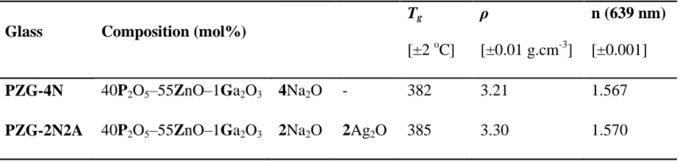
Table 1 Investigated glasses and related properties
Glass Composition (mol%)
Tg [±2 oC] ρ [±0.01 g.cm-3] n (639 nm) [±0.001] PZG-4N 40P2O5–55ZnO–1Ga2O3 4Na2O - 382 3.21 1.567
PZG-2N2A 40P2O5–55ZnO–1Ga2O3 2Na2O 2Ag2O 385 3.30 1.570
Thermal properties are the main indicator for evaluating the potential of the glasses for optical
fiber manufacturing. The Tg of the PZN-4N and PZN-2N2A glasses are Tg = 382 °C and Tg =385
°C respectively. Above all the absence of discernable crystallization events on the Differential
Scanning Calorimetry (DSC) scans up to 700 °C attests their suitability for thermal drawing.
Fibers were drawn from a macroscopic scaled-up preform, which is elongated through localized
thermal heating while being hold vertically (See Experimental section). Phosphate-based
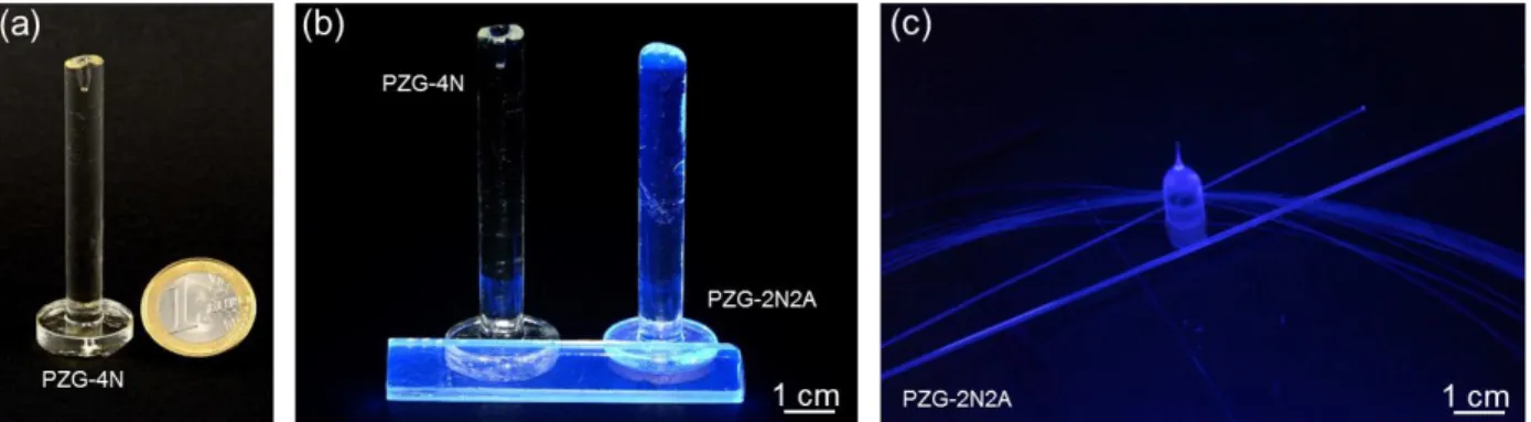
preforms are depicted on Figure 1 ((a) PZG-4N, (b) PZG-4N and PZG-2N2A). It consists of
cylindrical macroscopic glass rods of 8 mm in diameter and ~7 cm in length (note: the
6
drawing process). Alternatively rectangular preforms (Figure 1b) were designed, as will be
further discussed below. We assessed the drawing ability of both glass compositions. They were
brought to their softening temperature regime while temperature, preform-holder motion and
capstan velocity were continuously monitored to prevent catastrophic mechanical failure.
Figure 1 Phosphate-glass preforms and fibers (a) Undoped preform under white light (b)
Undoped and doped preforms under UV excitation light (c) PZG-2N2A glass (bottom-neck
preform, capillaries and fiber l ~40 m, ϕ ~150 µm) under UV excitation light
Following this procedure, each glass was drawn into ~40-meters-long fibers with diameters
ranging ϕ = 200 µm, down to 100 µm. Figure 1c depicts a bundle of PZG-2N2A fibers, as well
as a bottom-neck preform and two tens-of-centimeter-long capillaries under UV excitation (254
nm). No signs of heterogeneities or inclusions were visually apparent. In order to confirm the
optical transparency of the fibers, attenuation measurements were performed by the cut-back
7 Table 2 Fiber loss at λ = 1064 nm and λ = 1550 nm (measured by the cut-back method)
Glass Composition (mol%)
1064 nm [dB.m-1]
1550 nm [dB.m-1] PZG-4N 40P2O5–55ZnO–1Ga2O3 4Na2O - 1.8 9.7
PZG-2N2A 40P2O5–55ZnO–1Ga2O3 2Na2O 2Ag2O 1.6 8.5
Loss are similar with wavelengths for both compositions (Table 2). We find 1.8 dB.m-1 and 1.6
dB.m-1 at 1064 nm, and 9.7 dB.m-1 and 8.5 dB.m-1 at 1550 nm for the glasses 4N and
PZG-2N2A respectively. We believe the increase in loss observed at 1550 nm relates to the first
overtone of the hydroxyl absorption and to the multi-phonons cutting edge of the glass (centered
on 3 µm for a sample of 0.5 mm in thickness). Loss measurement proves that the resistance
toward devitrification of the selected materials effectively prevents the nucleation and growth of
parasitic crystallite centers during the drawing process. Although minimum loss remain
relatively high for practical applications, we must note that no purification protocol, other than
the use of high-purity precursors, was introduced at this stage. Furthermore measurements were
conducted on bare mono-material fiber, with no core/clad geometry or protective polymer
cladding involved.
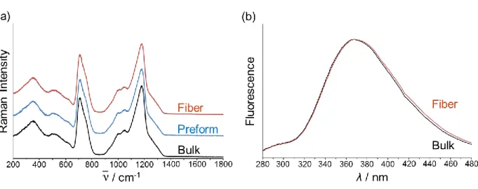
Raman spectroscopy was carried out on the PZG-2N2A bulk, preform and fiber samples in order
to assess an eventual structural deviation induced by the drawing process (λExcitation = 532 nm).
The three normalized Raman signatures precisely overlap (Figure 2a). The Raman spectra are
dominated by two broad envelopes centered on 705 and 1175 cm-1, assigned respectively to
symmetric P-O-P stretching modes and to symmetric stretching of PO2- in PØ2O2- tetrahedral
units (where Ø is a bridging oxygen) expected for a derived structure of metaphosphate.[20] Less
8
modes of PØO32- units forming the end of the phosphate chains.[21, 22] At lower frequencies, the
large envelopes peaking at 350 and 550 cm-1 are mainly due to bending modes of phosphate units
with probably weak contributions coming from gallium and zinc oxides vibrational modes.
Similarly, the fluorescence properties of the PZG-2N2A glass before and after drawing have
been compared (Figure 2b). Remarkably we observe that the fluorescence emission of the bulk
glass is fully preserved in the fiber sample. The PZG-2N2A glass exhibits an excitation band
centered on λ = 245 nm (due to the absorption band associated with Ag+ ions) and it emits
intrinsic fluorescence mainly around λ = 368 nm under UV excitation. This emission has been
attributed to the d10↔d9s1 transition of Ag+ ions homogenously dispersed throughout the glassy
matrix.[13]
Figure 2 Silver-doped phosphate glass PZG-2N2A (a) Normalized Raman spectra of the bulk,
preform and fiber glass (λExcitation = 532 nm) (b) Normalized fluorescence emission of the bulk
versus fiber glass (λExcitation = 245 nm)
The preservation of the luminescence properties of the PZG-2N2A glass is a crucial result as it
indicates that neither clustering nor reduction of Ag+ ions species has occurred during the
9
ensues from maintaining the material under oxidizing conditions (O2 atmosphere), which
prevents the reduction and diffusion of the Ag+ ions.
2.2. Silver-containing zinc-phosphate glass ribbon fibers
The thermo-viscous flowing ability of the PZG-2N2A glass, associated with the precise control
of the drawing conditions, allows for the fabrication of intrinsically photosensitive
phosphate-based fibers. Now we proceed to overtake a more sophisticated and original task, namely the
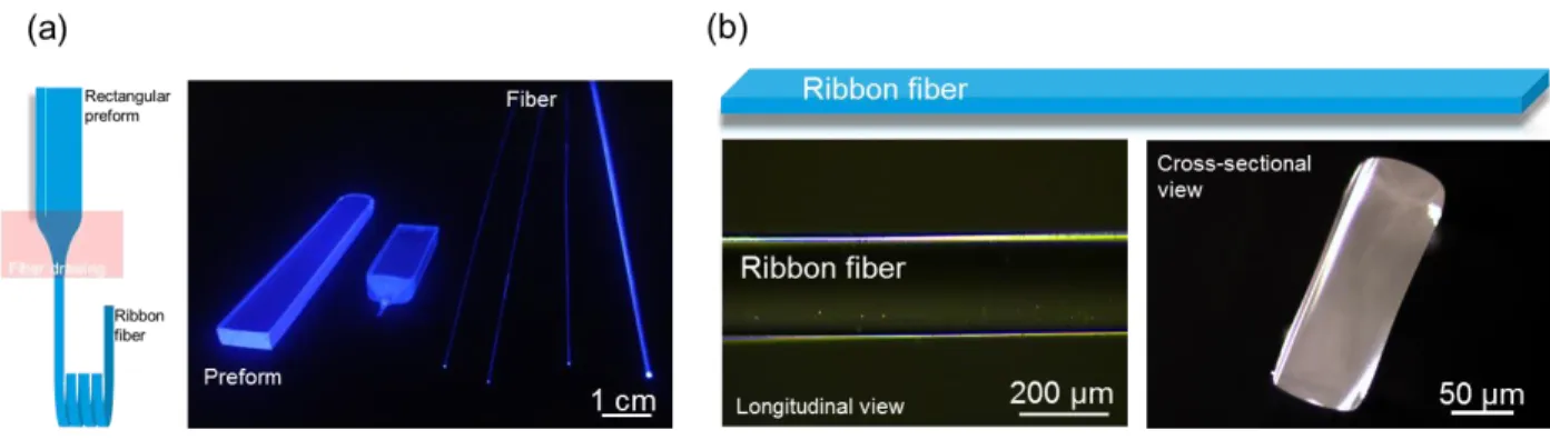
direct preform-to-fiber drawing of a planar, sharp corner-edges device. Figure 3 outlines our
approach to ribbon fiber processing. The procedure starts with the preparation of a macroscopic
scale rectangular preform (panel a). For this a glass slab is grinded and optically polished to the
proper dimensions (typically: 10-mm wide, 0.4-mm thick, and 80-mm long). The preform is then
thermally scaled down into tens-of-meters mechanically flexible flat fiber. Optical and SEM
micrographs in panel b depict the longitudinal and cross-sectional views of the fiber (left and
right panels respectively) and of its dimensions.
Figure 3 Thermal drawing of ribbon fibers (a) Preform, bottom-neck preform and fiber samples
10
Ribbon fibers fabrication occurs through homothetic dimensional reduction of the drawn preform.
To succeed a stringent viscosity-regime trade-off is imposed to the glass medium during its
shaping. Drawing parameters are finely tuned to allow the material to reach its necessary
low-viscous fluid state while, in the meantime, it is being maintained in a high-viscosity regime so as
to slow down the kinetics of surface energy mechanisms driven during the shaping process. This
operation allows preventing transverse features to evolve toward cylindrical shape.
2.3. DLW on silver-containing zinc-phosphate glass ribbon fibers
Direct femtosecond laser writing is a powerful method to implement active compounds into zinc
phosphate glasses.[14, 15] Herein, taking advantage of the flat, uniform geometry of the ribbon
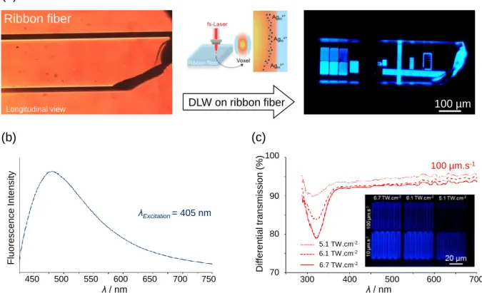
fibers, we generate silver ions clustering in photosensitive PZG-2N2A glass. A ribbon fiber was
truncated and exposed to irradiation (Figure 4a). DLW was performed typically 50 µm below
the surface by linearly moving the fiber sample at controlled speeds along its longitudinal axis
(see Experimental). Motifs were made of multiple series of lines parallel and perpendicular to the
fiber main axis. Laser structuring places stringent requirements on the surface and optical quality
of the substrate. Therefore, in this regard, successful DLW carried out in PZG-2N2A fiber first
stresses the flatness and homogeneity of the device. Silver ions clustering is triggered by
exposing the glass to a high-repetition-rate pulse train from a tightly focused NIR femtosecond
laser. It results from multiphoton ionization associated with cumulative thermal effects, allowing
for the reduction of Ag+ ions, their diffusion and aggregation into Agmx+ clusters (m: number of
atoms, m < 20; x: ionization degree).[23] The clusters are localized at the envelope of the focusing
voxel. Under exposure to UV radiations (λ = 405 nm), the Agmx+ clusters emit homogenous
fluorescence with a broad spectrum covering the visible range centered on λ = 480 nm (Figure
11 Figure 4 DLW in ribbon fibers (a) Glass PZG-2N2A fiber sample before (under white light) and
after irradiation (under UV light, λExcitation = 405 nm) and DLW schematic (b)
Micro-fluorescence emission spectrum of photo-inscribed Agmx+ clusters (c) Matrix of curvilinear
patterns with irradiances and sample motions, and related micro-transmission spectra for v = 100
μm.s-1.
In order to highlight the Laser-induced modifications, we have performed differential
micro-transmission measurements on structured zones with respect to the pristine glass as a function of
the irradiation dose. Six curvilinear patterns were processed for the three irradiance values I =
5.1 TW.cm-2, 6.1 TW.cm-2 and 6.7 TW.cm-2 and for the two sample velocities v = 10 µm.s-1 and
100 µm.s-1 (corresponding at each position to 2.0 106 to 2.0 105 deposited pulses respectively).
Figure 4c shows the wide-field fluorescence imaging of the Laser-induced silver clusters. We
observe that emitted luminescence, and then that the related concentration of Laser-induced
(a) 100 µm (b) λ / nm Fluo resce n ce In te n sity λExcitation = 405 nm 450 500 550 600 650 700 750 Longitudinal view Ribbon fiber DLW on ribbon fiber (c) λ / nm Di ff e ren tia l tran sm issi o n (%) 100 µm.s-1 300 400 500 600 700 70 80 90 100 6.7 TW.cm-2 6.1 TW.cm-2 5.1 TW.cm-2
12
clusters, increases with the cumulative deposited dose. For clarity we show the differential
micro-transmission spectra only for the velocity v = 100 µm.s-1. They exhibit an intense
absorption band centered at λ = 324 nm (E = 3.82 eV) (and a much weaker one around λ = 290
nm (E = 4.27 eV)). The amplitude of the multicomponent absorption band around λ = 324 nm
grows linearly with the deposited dose. These bands have been assigned to Agmx+ clusters and
are in good agreement with previous observations made on the silver zinc-phosphate glass.[23]
Finally we note a modification of the baseline of the differential micro-transmission spectra over
the whole spectral range, from the ideal 100% value down to 95%, typically. We believe the
observed decrease in transmitted light intensity which affects the whole spectral domain is due to
scattering effect.
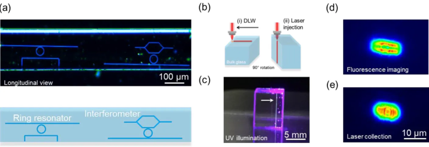
The in-fiber Laser-structuring represents an original method for implementing complex
light-manipulating architectures. To illustrate its potential we have schematically inscribed optical
micro-ring resonators directly in ribbon fibers (Figure 5a), without having confirmed and
characterized their optical functionalities yet, such demonstration being beyond the scope of this
article. The first structure (left-side) consists in a closed resonator ring side-coupled to two light
input/output bus waveguides, one of which being addressed from the side of the fiber.
Alternatively (right-side) a Mach–Zehnder interferometer made of two arms split from a single
source is coupled to one side of the resonator ring. To get confident about the potentiality of this
route to generate photonics structures, we aimed at verifying the waveguiding properties of the
photo-inscribed Agmx+ clusters features. To assess such guiding abilities, far field experiments
were conducted using a Yb:KGW Laser operating at 1030 nm in CW low-power regime as a
light source, hence preventing any glass structuring and multi-photon fluorescence excitation
(see Experimental). For the purpose of the demonstration, the propagation characteristics were
13
experiments highlight the fact that both physico-chemical properties and Laser-writing ability are
fully preserved from bulk samples to ribbon fibers. A 7-mm-long linear pattern was produced in
a silver zinc-phosphate bulk glass (with 4% of molar Ag2O), typically at 100 µm below its
surface. Then the sample was rotated 90° in order to allow for Laser injection from above and for
the collection of the received guided light from beneath (Figure 5b). Micrograph of the bulk
glass and of the fluorescent photo-written tube under broad field UV illumination at 405 nm is
depicted on Figure 5c.
Figure 5 Waveguiding evidence in silver zinc-phosphate bulk glass (a) Ring resonators and
Mach-Zehnder interferometer written in ribbon fibers (λExcitation = 405 nm) (b) Schematic of the
experiment: DLW in the linear x translation followed by 90° rotation of the sample for
subsequent Laser injection (c) Micrograph of the bulk glass under broad field illumination with
405 nm UV light (arrow: fluorescent photo-written tube) (d) Output fluorescence imaging at the
bottom plane of the bulk sample upon UV tightly-focused Laser injection at 405 nm (e) Injection
of the 1030 nm Laser in the written tube and output bottom imaging after its propagation along
14
The light from a tightly-focused Laser diode at 405 nm was injected from the top surface of the
structured bulk sample while imaging of the induced fluorescence was performed at the bottom
plane (Figure 5d). Note that spectral filtering was employed to cancel the incident Laser diode
light. The transversal dimensions of the collected spot of photo-luminescent signal from the
Laser-written tube are L ~ 8 µm and l ~ 4 µm. Furthermore it exhibits a double-line shape that is
typical of Agmx+ clusters features.[23] In order to get a clearer demonstration of waveguiding
abilities of the structure, the same operation was performed with a tightly-focused CW
low-power 1030 nm Laser (Figure 5e), for which no fluorescence is excited. Upon the latter injection,
we observe a transmitted Laser light localized on the photo-written features, which demonstrates
unambiguously their waveguiding properties. Indeed, since the top injection plane and the
bottom imaging plane are 7-mm distant, we claim that the observed light at the bottom imaging
plane cannot come from the collection of free propagating injected light. To support the guiding
observation we claim, the refractive index variation between the pristine glass and the Agmx+
clusters areas was estimated using a phase-contrast microscopy method (see Experimental),
which gives access to the local index modification (providing that the structure thickness had
been previously measured by high-resolution confocal microscopy). It showed an increase of the refractive index in the modified region of Δn ~5.10-3
over the visible spectral range, with a
spatial distribution corresponding to that of the silver clusters. Further effort is currently engaged
to characterize the beam modes, loss and the magnitude of the photo-induced index modification
to accurately regulate the propagation of light through the device. Laser-structuring allows for
the inscription of perennial silver cluster-based waveguides in fibers, thanks to the thermal and
optical stability of the photo-induced clusters.[14, 24] It provides unique opportunities for
applications in nonlinear optics, cavity-enhanced spectroscopic sensing, filtering or
15 3. Conclusion and perspectives
Here we have detailed the methodology for the manufacturing of silver-containing zinc
phosphate glass fibers. The fabrication of photosensitive, photo-writable silver-containing
phosphate-based ribbon fibers stem from judiciously adjusting both the materials composition
and its geometry. We validated in first place the aptitude of the glasses to be shaped into fibers.
Loss measurements verify the effective transparency of the fiber in the NIR region. Notably, as
evidenced by photo-luminescence measurements, Ag+ ions remain homogenously dispersed
upon drawing within the glass matrix. The intrinsically photosensitive fibers proposed here
authorizes for large amount of silver ions doping without clustering, hence reducing the need for
germanium doping or hydrogen-loading. Following we have presented a straightforward
manufacturing method of elongated ribbon structures. Cost-limited, photosensitive fibers in a flat
format is anticipated to ease the design of high-sensitivity fully-distributed fiber-based dosimeter
sensors. Already, silica-based optical flat fibers have been proposed as the basis for such
purpose[19] while high irradiation dose studies have been initiated on bulk silver zinc-phosphate
glasses.[25] More sophisticated interconnected fiber-based 2D screen may eventually be
envisioned for applications in the field of high-dose therapy dosimetry where image
reconstruction instruments are needed. The in-fiber Laser-structuring scheme enables the
integration in a single device of an entire array of unconventional device architectures on a
flexible substrate platform. Beyond, we reported recently on structured light-induced DLW of
nontrivial fluorescent Agmx+ topologies obtained using non-Gaussian beam profiles.[26] This
result could be exploited for the implementation of innovative photonic architectures relying on
the spatial distribution of photo-induced patterns with linear and nonlinear optical properties.
Additionally we have shown that, upon subsequent post-irradiation annealing of the glass above
16
for the in-fiber sub-micrometric 3D patterning of metal–dielectric composites within low-Tg
glass fiber, a promising approach for engineering highly-efficient plasmonic sensing devices or
increasingly complex electronic circuits.
Future work will consist in establishing improved glass purification and fiber manufacturing
protocols in order to satisfy technological specifications. Other work will consist in developing
core-clad fiber structures to propose innovative laser materials and geometry able to handle high
flux of photons without premature ageing and, by taking advantage of nonlinear optical
processes, to favor all-fiber short-length monolithic systems. Beyond, our approach could lead to
a powerful alternative to bottom-up synthesis methods for producing scalable in-fiber 3D
nano-structured materials, with functionalities spanning sensing, photonics, or even metamaterials
devices.
4. Experimental Section
Glasses and preforms synthesis The zinc-phosphate glasses were melted using the standard
melt-quench technique. High-purity precursors (ZnO, Zn(PO3)2, Ga2O3, NaPO3, AgNO3) were
weighted in powder forms and mixed together in a platinum crucible. The mix was ramped up
(1 °C.min−1) at 1100 °C and then kept at this temperature for 12 hours. Following the liquid was
poured on a brass plate to freeze the melt. Preforms were produced by casting the glass in a
specifically designed brass mold, pre-heated at ~ Tg-10 °C and annealed at Tg-40 °C for 12 hours.
Fiber drawing Thermal drawing was performed using a dedicated 3-meters-high optical fiber
draw tower composed of an annular electrical furnace with a sharp temperature profile, a
diameter monitor, a tension dancer and a collecting drum. The preform was slowly fed into the
furnace and the temperature was gradually increased at a rate of 10 °C.mn-1 up to ~700 °C under
17
were controlled in real-time to produce the targeted fiber diameter. Following this procedure,
meters of fibers were drawn with diameters ranging from 250 µm, down to 75 µm.
Direct Laser Writing DLW on ribbon fibers was performed with a Yb:KGW femtosecond
oscillator (T-Pulse 200, Amplitude Systemes, up to 2.6 W, 9.1 MHz, 390 fs at 1030 nm). The
irradiation duration and transmitted irradiance were controlled by an acousto-optic modulator,
enabling the accumulation of N = 105–106 pulses with energies from 50 to 150 nJ. The
positioning and displacement of the sample were performed with a high-precision 3D translation
stage (XPS-50 stages, Micro-Contrôle). Irradiations were carried out by focusing laser pulses
with a microscope objective (Mitutoyo, APO PLAN NIR, 20, NA 0.4). In order to highlight waveguiding abilities of the photo-written features, a CW low-power 1.03-µm Laser was
injected inside the tube using a microscope objective (Olympus, 20, NA 0.5). An identical bottom microscope objective was used to collect the transmitted (guided) light, and to conjugate
the imaged plane on a CCD camera. With the considered 7-mm long sample, the injection and
collection planes of these two objectives were optically distinct, ensuring that any collected light
at the bottom plane could only be guided light.
Characterization instruments Thermal analysis was performed by differential thermal analysis
(DSC 404 PC from Netzsch Inc.). About 20 mg of powdered glass was inserted into a Pt pan and
placed in the DSC chamber along with an empty reference pan. Characteristic temperatures were
measured as the inflection point of the endotherm at a heating rate of 10 °C/min (precision ±2°C).
Refractive indices were measured using Abbe refractometer. Micro-absorption measurements
were performed in bright-field using a ‘CRAIC Technologies’ microscope equipped with a
Xenon lamp and a condenser as a white light source. A conjugated 10 magnification microscope objective was used to collect the transmitted light through the laser-induced
18
a 532 nm excitation line with 10 mW of incident power. Raman spectra in the range of 200–2000
cm−1 were recorded with a resolution of 2.5 cm−1. The macroscopic luminescence spectra
(emission and excitation) were recorded at room temperature with a SPEX Fluorolog-2
spectrofluorimeter (Horiba Jobin-Yvon). The excitation source was a 450 W xenon lamp
enabling continuous excitation from 200 nm to 800 nm. The signal was detected and amplified
by a Hamamatsu R298 photomultiplier. The samples were grounded into powder to cancel
geometric effects. Refractive index variation between the pristine glass and the Agmx+ clusters
areas was estimated using a phase-contrast microscopy method with commercially available equipment’s from PHASICS Inc.
Acknowledgements
The authors wish to thank Frédéric Adamietz for micrographs of the preforms and Arnaud
Royon for the helpful discussions. Funding for this work has been provided from the French
Government, managed by the French National Research Agency (ANR Grant #40611), by the
Programme IdEx at the University of Bordeaux, the Cluster of excellence LAPHIA and by the
Aquitaine Region.
Received: ((will be filled in by the editorial staff))
Revised: ((will be filled in by the editorial staff))
19 REFERENCES
[1] A. Croteau, A. C. J. Poulin, “Photosensitive Fibers” in Specialty Optical Fibers Handbook,
Elsevier, London, UK, 2007.
[2] G. D. Peng, P. L. Chu, “Polymer optical fiber gratings” in Polymer optical fibers, American
Scientific Publishers, Valencia, CA, 2004.
[3] M. Bernier, D. Faucher, R. Vallée, A. Saliminia, G. Androz, Y. Sheng, S. L. Chin, Opt. Lett.
2007, 32, 454.
[4] A. F. Abouraddy, O. Shapira, M. Bayindir, J. Arnold, F. Sorin, D. S. Hinczewski, J. D.
Joannopoulos, Y. Fink, Nat. Mater., 2006, 5, 532.
[5] A. S. Beddar, Radiation Measurements, 2007, 41, 124.
[6] B. A. Flusberg, E. D. Cocker, W. Piyawattanametha, J. C. Jung, E. L. M. Cheung, M. J.
Schnitzer, Nat. Methods, 2005, 12, 941.
[7] P. Aiestaran, V. Dominguez, J. Arrue, J. Zubia, Opt. Mater., 2009, 31, 1101.
[8] S. Pissadakis, A. Ikiades, P. Hua, A. K. Sheridan, J. S. Wilkinson, Opt. Express 2004, 12,
3131.
[9] S. Suzuki, A. Schülzgen, S. Sabet, J. V. Moloney, N. Peyghambarian, Appl. Phys. Lett. 2006,
89, 171913.
[10] L. Xiong, P. Hofmann, A. Schülzgen, N. Peyghambarian, J. Albert, Opt. Mater. Express
2014, 4, 1427.
[11] J. Thomas, E. Wikszak, T. Clausnitzer, U. Fuchs, U. Zeitner, S. Nolte, A. Tünnermann,
Appl. Phys. A 2007, 86, 153.
[12] P. Hofmann, C. oigtl nder, S. olte, . Peyghambarian, A. Sch lzgen, J. Lightwave
20
[13] K. Bourhis, A. Royon, M. Bellec, J. Choi, A. Fargues, M. Treguer, J-J. Videau, D. Talaga,
M. Richardson, T. Cardinal, L. Canioni, J. Non-Cryst. Solids 356 (2010) 2658.
[14] A. Royon, K. Bourhis, M. Bellec, G. Papon, B. Bousquet, Y. Deshayes, T. Cardinal, L.
Canioni, Adv. Mater. 2010, 22, 5282.
[15] G. Papon, Y. Petit, N. Marquestaut, A. Royon, M. Dussauze, V. Rodriguez, T. Cardinal, L.
Canioni, Opt. Mater. Express 2013, 3, 1855.
[16] D. S. Agafonova, A. I. Sidorov, E. V. Kolobkova, A. I. Ignatiev; N. V. Nikonorov, Proc.
SPIE 9141 Optical Sensing and Detection III 2014 91411T.
[17] N. Chocat, G. Lestoquoy, Z. Wang, D. M. Rodgers, J. D. Joannopoulos, Y. Fink, Adv.
Mater. 2012, 24, 5327.
[18] G. Lestoquoy, N. Chocat, Z. Wang, J. D. Joannopoulos, Y. Fink, Appl. Phys. Lett. 2013, 102,
152908-5.
[19] A. Alawiah, S. Bauk, H. A. Abdul-Rashid, W. Gieszczyk, S. Hashim, G. A. Mahdiraji, N.
Tamchek, D. A. Bradley, Radiat. Phys. Chem. 2015, 106, 73.
[20] L. L. Velli, C. P. E. Varsamis, E. I. Kamitsos, D. Möncke, D. Ehrt, Phys. Chem. Glasses
2005, 46, 178.
[21] P. Hee, R. Christensen, Y. Ledemi, J. E. C. Wren, M. Dussauze, T. Cardinal, E. Fargin, S.
Kroeker, Y. Messaddeq, J. Mater. Chem. C 2014, 2, 7906.
[22] M. Dussauze, V. Rodriguez, L. Velli, C. P. E. Varsamis, E. I. Kamitsos, J. Appl. Phys. 2010,
107, 043505.
[23] N. Marquestaut, Y. Petit, A. Royon, P. Mounaix, T. Cardinal, L. Canioni, Adv. Funct.
Mater. 2014, 24, 5824.
[24] A. Royon, K. Bourhis, L. Béchou, T. Cardinal, L. Canioni, Y. Deshayes Microelectronics
21
[25] K. Bourhis, A. Royon, G. Papon, M. Bellec, Y. Petit, L. Canioni, M. Dussauze, V.
Rodriguez, L. Binet, D. Caurant, M. Treguer, J-J. Videau, T. Cardinal, Mater. Res. Bull. 2013, 48,
1637.
[26] K. Mishchik, Y. Petit, E. Brasselet, A. Royon, T. Cardinal, L. Canioni, Optics Lett. 2015, 40,
201.
Table of contents entry
The thermal drawing of photosensitive silver-containing zinc phosphate glass ribbon fibers is demonstrated. Structural and luminescence properties of the native glass are preserved. The
flat fiber geometry allows for the convenient, accurate Laser inscription of complex luminescent
silver clusters patterns within the glass matrix. These fibers offers a promising platform for the
design of highly-efficient sensing, photonics or metamaterials devices.
Keywords
phosphate, glasses, fiber, photosensitivity, laser inscription
C. Author 2, D. E. F. Author 3, A. B. Corresponding Author*
F. Désévédavy, Y. Petit, J-C. Desmoulin, A. Abou Khalil, C. Strutynski, M. Dussauze, F.
Smektala, T. Cardinal, L. Canioni, S. Danto*
Title
22 Table of Content figure (55 × 50 mm or 110 × 20 mm)
Copyright WILEY-VCH Verlag GmbH & Co. KGaA, 69469 Weinheim, Germany, 2013.
Title
Photo-writable Silver-containing Phosphate Glass Ribbon Fibers
Author(s), and Corresponding Author(s)*
Frédéric Désévédavy, Yannick Petit, Jean-Charles Desmoulin, Alain Abou Khalil, Clément
Strutynski, Marc Dussauze, Frédéric Smektala, Thierry Cardinal, Lionel Canioni and Sylvain