Differences in the Regulation
Expression Between Epithelial
of Thrombospondin-1
Cells and Fibroblasts
By
Roberto Karlo Rodriguez
B.S. Biochemistry
University of California, Davis, 1999
Submitted to the Department of Biology in Partial Fulfilment of the
Requirements for the Degree of Doctor of Philosophy in Biology
at
the
Massachusetts Institute of Technology
June 2007
©
2007 Massachusetts Institute of Technology
All
rights reserved
Signature of Author , - --.. /. ... D epartment of Biology
A
/l A/1
May 25, 2007 Certified by, S,,,,, - Randolph WatnickAssistant Professor of Vascular Biology Children's Hospital, Boston Thesis Supervisor Certified by_
/
Accepted by , , , MASSACHUStrS INs E OF TECHNOLOGYJUN 0 5 2007
ARCHIVES
LIBRARIES
Tyler Jacks Professor of Biology Thesis Supervisor Stephen P. Bell Chairman, Graduate Committee__
Differences in the Regulation of Thrombospondin-1
Expression Between Epithelial Cells and Fibroblasts
By
Roberto Karlo Rodriguez
B.S. Biochemistry
University of California, Davis, 1999
Submitted to the Department of Biology on May 25, 2007 in Partial
Fulfillment of the Requirements for the Degree of Doctor of Philosophy in
Biology
ABSTRACT
Induction of angiogenesis is a critical and rate-limiting step in the progression of
cancer. It is widely acknowledged that this induction requires the concomitant
stimulation of pro-angiogenic and repression of anti-angiogenic proteins. It has been
demonstrated that in human epithelial cells repression of the angiogenesis inhibitor
Thrombospondin-1 (Tsp-1) requires stimulation of Myc in combination with
hyper-physiologic levels of oncogenic Ras.
This work demonstrates that in human mammary epithelial cells, repression of Tsp-1
requires the activation of Myc by a Ras-induced pathway that activates the MAPK
p38. This work also demonstrates that repression of Tsp-1 in human fibroblasts
requires the combined inhibition of the tumor suppressors p53 and pRb. These
results suggest that the molecular requirements for the induction of angiogenesis
differ significantly between carcinomas and sarcomas.
Thesis Supervisor: Randolph S. Watnick
Title: Assistant Professor in the Vascular Biology Program at Children's Hospital, Boston
Table of Contents
Chapter 1. Introduction 9
Section I: The role of angiogenesis in tumorigenesis 9 Section II: The role of the tumor stroma and carcinoma-associated
fibroblasts in angiogenesis 17
Section III: The role of Thrombospondin-1 in angiogenesis 24 Section IV: The role of c-Myc in oncogenesis and angiogenesis 30 Section V: The role of p53 in tumor development and angiogenesis 37 Section VI: The role of pRb in tumor development and angiogenesis 42 Section VII: The role of SV40 large T antigen in cellular transformation 48
Section VIII: Summary 49
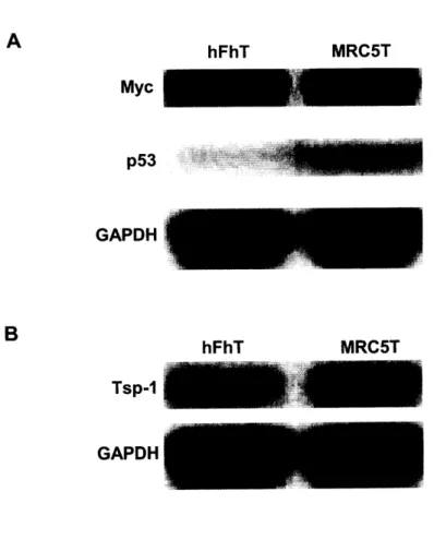
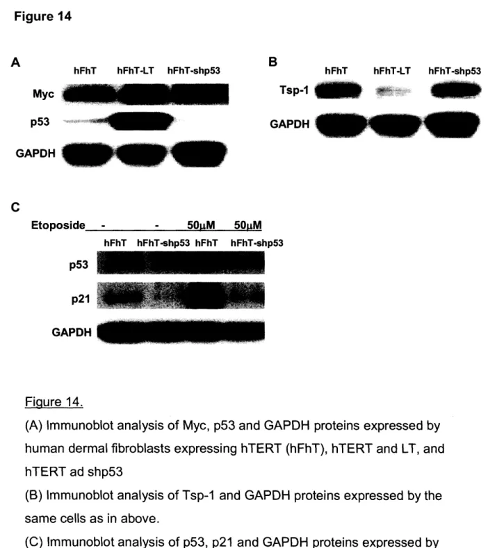
Chapter 2. Regulation of Tsp-1 expression in epithelial cells 51 Section I: Expression signatures in epithelial cells 52 Section II: The effects of SV40 large T antigen on epithelial cells 52 Section III: The effects of p53 deficiency on Myc and Tsp-1
expression in epithelial cells 54
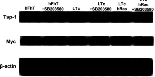
Section IV: Ras stimulates Myc expression through the MAPK p38
in epithelial cells 56
Section V: Preliminary experiments determining the role of p63
in Tsp-1 expression 59
Section VI: Summary of Results 62
Section VII: Materials and Methods 63
Figures: 67
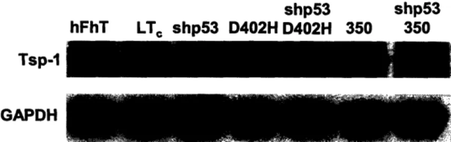
Chapter 3. Regulation of Tsp-1 expression in fibroblasts 77 Section I: Expression signatures in fibroblasts 78 Section II: Effects of SV40 large T antigen on fibroblasts 79 Section III: The effects of p53 deficiency on the Myc and
Section IV: The effects of SV40 large T antigen mutants on
p53, Myc, and Tsp-1 expression in fibroblasts 84
Section V: The combined loss of p53 and pRb activity represses
Tsp-1 expression in fibroblasts 85
Section VI: The role of E2F1 in the modulation of Tsp-1 expression
in fibroblasts 86
Section VII: Preliminary observations implicating topoisomerase
inhibitors in the regulation of Tsp-1 expression 89
Section VIII: Summary of Results 92
Section IX: Materials and Methods 94
Figures: 98
Chapter 4. Discussion 109
Section I: Summary 109
Section II: Future Work 113
Section III: Perspectives 125
Figures: 130
Chapter
1.
Introduction
I:
The role of angiogenesis in tumorigenesis
Angiogenesis is the process of new capillary blood vessels arising from preexisting blood vessels (Clark and Clark 1939). As such, it is an essential component of development, reproduction and wound healing (Dvorak et al. 1981). Angiogenesis also occurs in tissues where there is a change in mass or metabolism
(Dvorak et al. 1981). It is in these ways that physiologic, i.e. non-pathogenic, angiogenesis allows the remodeled tissue to maintain adequate levels of oxygen (Dvorak et al. 1981). The process of angiogenesis was first described in rabbit ear chambers in 1939 and has been extensively studied since (Clark and Clark 1939). One of the major breakthroughs occurred when, using cultured endothelial cells, Folkman et al were able to establish in vitro models of capillary network formation (Gimbrone et al. 1973; Folkman and Haudenschild 1980).
Induction of angiogenesis occurs through growth factor-dependent activation of endothelial cells. This activation is most often initiated in response to stimuli such as hypoxia, inflammation and mechanical stress (Ausprunk and Folkman 1977; Hobson and Denekamp 1984). These stimuli induce the production of growth factors, such as vascular endothelial growth factor (VEGF), fibroblast growth factor (FGF), hepatocyte
growth factor (HGF), and epidermal growth factor (EGF), from endothelial cells and pericytes, undifferentiated fibroblast-like cells (Arbiser 2004). These growth factors initiate signaling cascades in endothelial cells that lead to the activation of transcription factors, which enhance expression of cell cycle mediators, integrins, and proteases, that promote the progression of angiogenesis (Milkiewicz et al. 2006). The
activated transcription factors further stimulate production of angiogenic growth factors and receptors, which provides a positive feedback loop that results in a more robust response (Milkiewicz et al. 2006). Induced proteins such as matrix metalloproteinases (MMPs) can also serve as components of negative feedback loops downregulating the angiogenic response by activating angiogenesis inhibitors like endostatin and angiostatin (O'Reilly et al. 1994; O'Reilly et al. 1997). This regulatory network helps to coordinate the complex process of angiogenesis. Induction of angiogenesis can be broken down into five basic steps: endothelial cell infiltration, endothelial cell proliferation, breakdown of the basement membrane, endothelial cell migration, and endothelial tube formation and stabilization (Ausprunk and Folkman 1977; Hobson and Denekamp 1984).
Permeability, or endothelial cell infiltration, is the first step in angiogenesis, and is controlled through adherens junctions located on the surface of endothelial cells (Dejana 2004). Adherens junctions are formed through the interactions of adhesive molecules, the most prominent of which is vascular endothelial-cadherin (VE-cadherin) (Corada et al. 1999). Anchoring to the actin cytoskeleton is essential for optimal adhesion of VE-cadherin, and is mediated by binding in complex with a- and p-catenins (Lampugnani et al. 1995; Corada et al. 1999). Dissociation of adherens junctions leads to an increase in endothelial cell permeability (Dejana 2004). Growth factors like EGF and HGF can induce endothelial cell permeability by phosphorylating p-catenin via their receptor tyrosine kinases (Hoschuetzky et al. 1994).
Subsequent to increasing permeability, activated endothelial cells undergo cell proliferation. VEGF binding to VEGF receptor 2 (VEGFR2) stimulates endothelial cell
proliferation (Quinn et al. 1993; Bernatchez et al. 1999). VEGFR2 activates mitogen activated protein kinases (MAPKs) such as ERK-1, ERK-2 and p38, as well as phosphatidylinositol 3-kinase (PI3K) (Yu and Sato 1999). Activation of these signaling pathways leads to induction of endothelial cell proliferation by modulating cell cycle regulators. For example, VEGF-induced Akt signaling leads to the repression of the CDK inhibitor p27 (Potente et al. 2003). Activation of the Akt pathway is also involved in promoting endothelial cell survival (Gerber et al. 1998). This is accomplished through induction of anti-apoptotic molecules such as Bcl-2
(Flusberg et al. 2001).
Once endothelial cells are activated, the sheath-like covering of the existing blood vessel (also known as the basement membrane) needs to be broken down to create space for the new vessel that will sprout. The basement membrane is degraded by active MMPs (Birkedal-Hansen 1995). The two MMPS that are most critical in this process are MMP-2 and MT1-MMP (Nguyen et al. 2001; Ohno-Matsui et al. 2003; Langlois et al. 2004). One way in which production of active MMPs is stimulated is via the mechanical forces involved in reshaping the vasculature (Boyd et
al. 2005). Signaling through VEGFR2 induces degradation of the basement membrane at the leading edge of migrating endothelial cells (Pepper 2001). The VEGFR2-mediated degradation effect occurs via the activation of the trypsin protease
plasmin following proteolytic cleavage of plasminogen (Pepper 2001).
After the basement membrane is degraded at the site of the new budding vessel, migrating endothelial cells home to the bud in order to elongate the new blood vessel. Endothelial cell migration occurs through cellular attachment to matrix
proteins like integrins and through modulation of components of the cytoskeleton
(Ingber 2002). VEGF and bFGF are the primary mediators of endothelial cell motility (Shono et al. 2001; Lavenburg et al. 2003). VEGF can induce cell migration through activation of the Rho family of GTPases, which regulate the dynamic function of the actin cytoskeleton (Lavenburg et al. 2003). Basic-FGF can act as a chemoattractant to induce endothelial cell motility, which is mediated through MAPK signaling (Shono et al. 2001).
Once the endothelial cells have migrated to the site of vessel budding, they must form the lumen, which will become the new vessel, and stabilize the lumen to complete the process of angiogenesis. Lumen formation occurs through morphological changes in endothelial cells that involve cell elongation along with either intercellular or intracellular lumenization (Egginton et al. 2001). Integrins play an essential role in lumen formation. For example, endothelial cells can form the lumen by generating and fusing intracellular vacuoles, and the mechanism of vacuole formation is integrin a201-dependent (Davis and Camarillo 1996).
Endothelial cells not only undergo terminal differentiation during lumenization, but they also undergo replicative quiescence. At a molecular level, it has been shown that MAPKs like ERK aid in both lumen formation and replicative quiescence (Yang et
al. 2004). Angiopoietinl, through the activation of its cognate receptor Tie2, can induce quiescence in endothelial cells by promoting cell survival through the MAPK and P13K signaling pathways (Peters et al. 2004). Undifferentiated fibroblast-like cells called pericytes and smooth muscle cells aid in the stabilization of the lumen by providing structural support to the new vessel (Park et al. 1994; Green et al. 2001;
Ishida et al. 2001; Luttun et al. 2002). Pericytes and smooth muscle cells also stimulate an increase in the diameter of the new blood vessel via placental growth factor (PIGF) mediated signaling through the VEGF receptor-1 on the surface of these cells (Park et al. 1994; Green et al. 2001; Ishida et al. 2001; Luttun et al. 2002).
The complex process of angiogenesis is regulated at many different steps by a variety of cytokines and signaling molecules. As mentioned above, VEGF is one of the most potent angiogenic stimulators. VEGF can induce endothelial cell permeability, proliferation, survival, and migration. VEGF transcription is also induced in response to hypoxia, through the transcription factor HIF-ca (Forsythe et al. 1996). Additionally, VEGF can be induced in response to an increase in inflammatory mediators like interleukins and transforming growth factor P (TGF- P )(McColl et al. 2004). In fact, many of the commonly studied growth factors mediate their activities in part by stimulating production of VEGF (Dulak et al. 2000). For example, EGF binding to HER2 (human epidermal growth factor receptor 2) increases synthesis of
HIF-la, which leads to VEGF induction (Laughner et al. 2001).
Under normal physiological conditions, angiogenesis is a tightly regulated process that is dependent on the balance between endogenous angiogenic promoters and inhibitors. The first demonstrated endogenous molecule to show anti-angiogenic effects was interferon alpha (IFN-a), which inhibits endothelial cell migration (Brouty-Boye and Zetter 1980). IFN-a has also been shown to inhibit basement membrane degradation by inhibition of both urokinase-type plasminogen activator (uPA) and MMP-9 expression (Pepper et al. 1994; Ma et al. 2001a; Ma et al. 2001b). Cleavage of plasminogen not only releases the angiogenic stimulator
plasmin, but also releases the inhibitor angiostatin (Nelson and Kastan 1994). Angiostatin inhibits angiogenesis by blocking both endothelial cell proliferation and migration (O'Reilly et al. 1994). Endostatin, another endogenous angiogenesis inhibitor released by proteolysis, is produced when collagen XVIII is cleaved by the cysteine-protease cathepsin L (O'Reilly et al. 1997; Felbor et al. 2000; Maeshima et al. 2001). Endostatin has been shown to specifically inhibit the processes of endothelial cell proliferation, endothelial cell migration, VEGFR2-mediated signaling, and activation of both MT1-MMP and MMP-2 (Dhanabal et al. 1999; Yamaguchi et al. 1999; Kim et al. 2000; Kim et al. 2002b). Additionally, endostatin is known to induce cell apoptosis by repressing Bcl-2 and Bcl-XL (Dhanabal et al. 1999; Yamaguchi et al.
1999; Kim et al. 2000; Kim et al. 2002b).
Thrombospondins represent another class of matrix-derived angiogenesis inhibitors. Thrombospondin-1 (Tsp-1) was the first protein identified to be a naturally occurring inhibitor of angiogenesis (Good et al. 1990). Briefly, Tsp-1 has been shown to promote endothelial cell apoptosis and to inhibit endothelial cell proliferation and migration through activation of TGFP3 and CD36 (Baird and Durkin 1986; Frater-Schroder et al. 1986; Dawson et al. 1997; Crawford et al. 1998; Jimenez et al. 2000). Tsp-2 can also function as an angiogenesis inhibitor (Streit et al. 1999). Tsp-2 has been shown to promote endothelial cell apoptosis and inhibit VEGF-dependent endothelial cell migration and lumenization (Noh et al. 2003). These and other endogenous inhibitors help to maintain the balance of positive and negative angiogenic stimuli that regulate physiological angiogenesis.
Tumor growth also requires the induction of angiogenesis (Folkman et al.
1971). Tumor angiogenesis allows solid tumors to grow beyond a microscopic size of
approximately 2mm in diameter, the size constraint imposed by the diffusion limit of
oxygen and nutrients (Gimbrone et al. 1972). In much the same manner as
physiologic angiogenesis, tumor angiogenesis is achieved by altering the balance of
inducers and inhibitors towards the side of the inducers. In tumor angiogenesis this
shift towards angiogenic induction is accomplished by abrogating many of the
negative feedback loops, which results in unrestrained endothelial cell proliferation
and blood vessel formation (Dvorak 1986). Interestingly, it has been demonstrated
that many tumors also stimulate the production of inhibitors such as angiostatin, a
factor that suppresses the angiogenesis and growth of metastatic colonies
.
A wide variety of mechanisms can induce tumor angiogenesis. For example,
when a tumor grows to a size that limits the diffusion of oxygen, hypoxia induces the
production of HIF-la, which in turn induces production of VEGF (Forsythe et al.
1996). VEGF can then induce angiogenesis by stimulating the growth and migration
of endothelial cells. Tumors can also induce angiogenesis through production and
secretion of the cytokine interleukin 8 (IL-8). IL-8 has been shown to induce the
proliferation and migration of endothelial cells, and is thought to be the major
angiogenic factor produced in some tumors (Koch et al. 1992; Smith
et
al. 1994;
Lingen et al. 1998). Tumors can also promote angiogenesis and metastasis by
inhibiting the expression of angiogenesis inhibitors. For example Tsp-1 expression is
repressed in various tumors and fibroblasts, which are part of the tumor
microenvironment (Kalas et al. 2005).
Tumors are able to modulate expression of angiogenesis promoters and
inhibitors through the activation of oncogenes. For example, the oncogene ets-
I
is a
transcription factor that can induce expression of MMPs and urokinase-type
plasminogen activator in endothelial cells, thus promoting cell migration and lumen
formation (Iwasaka et al. 1996; Lavenburg et al. 2003). Ets-1 has also been found to
induce transcription of VEGF in endothelial cells (Hashiya et al. 2004).
Activated Ras is another oncogene that promotes angiogenesis. A recent
study has shown that in a model of melanoma, Ras was required for the maintenance
of angiogenesis and that inactivating Ras led to endothelial cell apoptosis (Tang et al.
2005). It has also been shown that Ras can lead to the repression of Tsp-1
expression in a genetically engineered tumor model system (Watnick et al. 2003).
Expression of the myc oncogene can promote tumor angiogenesis by inducing VEGF
production and repressing Tsp-1 expression (Janz et al. 2000; Watnick et al. 2003;
Knies-Bamforth et al. 2004). A recent study has also found that Myc can activate a
micro-RNA cluster that promotes angiogenesis through repression of Tsp-1 (Dews et
al.
2006). These studies clearly indicate the essential role of angiogenesis in the
progression of cancer, especially in the development of metastasis. Additionally,
these studies demonstrate that tumor cells can activate various signaling pathways
that lead to the induction of angiogenesis.
II1: The role of the tumor stroma and carcinoma-associated
fibroblasts in anqiogenesis
During the initial stages of cancer progression, tumor cells are confined as a
neoplastic legion within a given tissue (termed carcinoma in situ) that is separated
from neighboring tissue by the basement membrane (Hanahan and Weinberg 2000a).
The basement membrane, immune cells, blood vessels, ECM and fibroblasts
surrounding the tumor cells constitute the tumor stroma (Ronnov-Jessen et al. 1996).
Tumor progression, including angiogenesis, invasion, and metastasis has been
demonstrated to be promoted by fibroblasts located within the tumor stroma
(Elenbaas and
Weinberg
2001). Fibroblasts within the tumor stroma are known as
carcinoma-associated fibroblasts (CAFs) (Olumi et al. 1999). It is believed that CAFs
are very similar to, if not the same as, activated fibroblasts found in the stroma of
tissues undergoing wound repair (Durning et al. 1984; Tsukada et al. 1987; Schor et
al.
1988a; Schor et al. 1988b).
Fibroblasts are elongated cells of the connective tissue of organs that are
characterized as not being vascular, inflammatory, or epithelial cells (Tarin and Croft
1969). Though morphologically distinct, the molecular identity of fibroblasts is poorly
defined. Fibroblasts are embedded within the matrix of the connective tissue and
have many important functions, including synthesis and deposition of ECM, regulation
of epithelial cell differentiation, and regulation of inflammatory response (Parsonage
et al. 2005; Tomasek et al. 2005). Fibroblasts also play a major role in wound repair
(Gabbiani 2003). Specifically, during wound repair, fibroblasts become activated,
invade the wound, produce ECM to anchor other cells involved in the repair process, and facilitate healing wound contractions (Castor et al. 1979; Gabbiani 2003).
Activated fibroblasts proliferate at a greater rate, produce greater amounts of ECM, and express a-smooth-muscle actin (Gabbiani 2003). Injured epithelial cells release growth factors like TGFP3 and bFGF that can induce fibroblast activation, or direct cell-cell contacts with leukocytes can also lead to activation (Clayton et al. 1998; Choi and Tseng 2001). In order to accommodate production of greater amounts of ECM, activated fibroblasts contain an oval-sized euchromatic nucleus, rough endoplasmic reticulum, and prominent Golgi apparatus (Castor et al. 1979). Activated fibroblasts also produce proteases that degrade the ECM, such as MMPs, aid in the turnover and reorganization of the ECM, and secrete growth factors like
HGH and bFGF (Rodemann and Muller 1991). Once the wound is repaired, the number of activated fibroblasts is greatly decreased, though overall the number of fibroblasts in not significantly changed (Gabbiani 2003). It is not clear whether the decrease in the number of activated fibroblasts is due to apoptosis and repopulation of normal fibroblasts from neighboring tissue, or if the activated fibroblasts revert back to normal fibroblasts. However, it seems more likely that the activation is transient and once wound repair is complete, the fibroblasts revert back to a quiescent
phenotype.
Studies have shown that fibroblasts in the tumor stroma exist in a state similar to the activated fibroblasts associated with wound healing (Durning et al. 1984; Dvorak et al. 1984; Tsukada et al. 1987; Ronnov-Jessen et al. 1996). For example, in breast cancer approximately 80% of the fibroblasts within the tumor stroma are
thought to become activated (Sappino et al.
1988).
Though it is not clear how
fibroblasts become CAFs, in vitro studies have shown that TGFPI can induce CAF-like
properties in normal fibroblasts (Ronnov-Jessen and Petersen 1993). Unpublished
studies presented in a recent review suggest that human carcinoma cells can convert
normal fibroblasts into CAFs in a mouse xenograft model (Orimo and
Weinberg
2006). Once fibroblasts become CAFs, they can be cultured in the absence of
carcinoma cells and retain their CAF phenotype in culture until they undergo
senescence (Orimo et al. 2005). It is interesting to note that chickens infected with
Rous sarcoma virus develop invasive carcinomas when wounded, showing the
oncogenecity of tumor stroma (Dolberg et al.
1985).
The tumor stroma has also been
shown to be associated with increased blood vessel density (Kalluri and Zeisberg
2006). Taken together, these studies demonstrate the importance of the tumor
stroma, and CAFs in particular, to the process of tumor progression.
The transition of in situ carcinoma to invasive carcinoma has been well
characterized in breast cancer (Ronnov-Jessen et al. 1996). In normal breast tissue
mammary ducts are lined
with
ductal epithelium which is, in turn, surrounded by the
myoepithelial layer and sheathed by the basement membrane (Ronnov-Jessen et al.
1996). Connecting the mammary ducts is the stroma consisting of ECM, blood
capillaries, and fibroblasts (Ronnov-Jessen et al.
1996).
When oncogenic
transformation and proliferation of epithelial cells give rise to ductal carcinoma in situ,
the epithelial and myoepithelial linings become indistinct, the number of stromal
fibroblasts increases, the amount of ECM increases, and capillary density increases
(Hanahan and Weinberg 2000a). However, the basement membrane of the ducts
remains intact. When the carcinoma progresses to the invasive state the basement membrane is degraded and stromal cells, including CAFs, inflammatory response cells, and newly formed capillaries, come into contact with the tumor cells (Hanahan and Weinberg 2000a). CAFs in the stroma of invasive carcinoma continue depositing large amounts of ECM, including tenascin C in some cases (Chiquet-Ehrismann et al. 1986; Inaguma et al. 1988). It has been shown that in breast and bladder carcinomas expression of tenascin C correlates with increased tumor invasiveness (Mackie et al. 1987; Brunner et al. 2004). The accumulation of ECM in tumors contributes to increased interstitial fluid pressure that hinders oxygen and nutrient diffusion (Netti et al. 2000; Brown et al. 2004). This CAF-mediated hypoxia could lead to the expression of HIF-la and the induction of VEGF, thus providing a mechanism by which CAFs can promote angiogenesis in tumors.
As stated above CAFs are associated with tumor cells at most stages of cancer progression. Many studies have shown the ability of fibroblasts to promote cancer. For example patients genetically predisposed to breast cancer contained skin fibroblasts that proliferated more easily in vitro (Kopelovich 1982). It has also been demonstrated that CAFs can promote tumor growth in a mouse xenograft model whereas normal fibroblasts cannot (Olumi et al. 1999). In this study, SV40 large T antigen-immortalized prostate epithelial cells were mixed with either CAFs or normal fibroblasts and then implanted into immuno-compromised mice. Olumi et al. demonstrated that large tumors grew in the grafts containing CAFs, whereas no tumors grew in grafts containing normal fibroblasts. This demonstrates how CAFs aid
in the formation of tumors, probably through induction of tumorigenic changes in
epithelial cells.
Tumor progression is also mediated by CAFs. It has recently been shown that
mixing human breast carcinoma cells with CAFs in a mouse xenograft gave rise to
tumors that were larger and more angiogenic than when mixed with normal fibroblasts
(Orimo et al. 2005). Furthermore, it was shown that the increase in tumor cell
proliferation was mediated by stromal cell-derived factor
1 (SDF1) secreted by the
CAFs binding to the CXCR4 receptor on tumor cells. Additionally secreted SDF1 was
also shown to mediate angiogenesis by recruitment of endothelial progenitor cells
(EPCs), which are recruited into the carcinoma during tumor angiogenesis and
differentiate into vascular endothelial cells (Lyden et al. 2001). Another study
demonstrated the ability of CAFs to induce invasiveness in vivo with rat colon
carcinoma cells that were not invasive
in
vitro (Dimanche-Boitrel et al. 1994). CAFs
also secrete MMPs that help degrade the basement membrane and promote tumor
invasion.
For example, MMP3 secreted by CAFs can promote tumor cell
invasiveness (Lochter et al. 1997).
This is accomplished by MMP3-mediated
cleavage of the extra-cellular domain of the adhesive protein E-cadherin on the
surface of mammary epithelial cells. Cleavage of E-cadherin causes mammary
epithelial cells to disperse and undergo epithelial-to-mesenchymal transition, which
promotes tumor cell invasiveness.
CAFs have also been implicated in tumor metastasis, by promoting the
proliferation of tumor cells at the metastatic site.
For
example, a liver metastatic cell
line was shown to secrete factors that activate fibroblasts
in
vitro (Olaso et al. 1997).
These activated fibroblasts were shown to be within the tumor stroma of the
metastasis, and quiescent fibroblasts taken from the liver of mice were activated
when cultured with conditioned media (CM) from the melanoma metastasis. The
tumor CM induced fibroblast migration, proliferation and production of MMP2. This
suggests that CAFs help to create a niche for tumor cells at metastatic sites (Olaso
and Vidal-Vanaclocha 2003). A recent study has shown that mice deficient for the
Mtsl protein, which stimulates tumor metastasis, failed to grow metastases when
highly metastatic mammary carcinoma cells were grafted onto these mice
(Grum-Schwensen et al. 2005). Furthermore, there was a significant delay in tumor uptake
as well a decrease in tumor incidence as compared to wild-type mice injected with the
carcinoma cells. When the tumor cells were mixed with Mtsl-competent fibroblasts
and injected into the
mtsl
knockout mice, the ability of these tumors to metastasize
was partially restored. Additionally, recent work from the Watnick group (manuscript
in review) has shown that CM from metastatic human breast and prostate carcinoma
cell lines are able to repress the expression of Tsp-1 in fibroblasts from tissues where
the carcinoma is known to metastasize. This demonstrates the ability of tumors to
prime metastatic sites for angiogenesis by decreasing the levels of an endogenous
angiogenesis inhibitor.
It is clear that angiogenesis is an essential step in the progression and
metastasis of tumors. Fibroblasts play an important role in promoting not only tumor
progression but angiogenesis as well. Carcinoma-associated fibroblasts produce
growth factors like VEGF that aid in the recruitment and activation of endothelial cells
within the tumor stroma. During tumor invasion, CAFs produce not only angiogenic
growth factors, but also produce proteases that break down the basement membrane
of the tumor associated tissue as well as the basement membrane of stromal blood
vessels, an essential step in angiogenesis. Finally, during tumor metastasis CAFs
are able to create permissive environments for tumor growth and angiogenesis at
metastatic sites. Studies from Kalas et al and from the Watnick laboratory have
shown that tumors secrete factors that are able to repress the expression of Tsp-1 in
fibroblasts (Kalas et al. 2005). These studies underscore the importance of Tsp-1 in
the induction of angiogenesis. In order for tumor cells to induce angiogenesis, the
balance of angiogenic stimulators and inhibitors must be shifted towards induction of
angiogenic stimulators in the tumor stroma. It is becoming clear that repression of the
angiogenesis inhibitor Tsp-1 in the tumor stroma is an important step in shifting the
II: The role of Thrombospondin-1 in ancqiocqenesis
First described in 1978, Tsp-1 is a 180 kDa glycoprotein that functions as a homotrimer (Lawler et al. 1978). Apart from its anti-angiogenic activity, Tsp-1 is also involved in cell adhesion, migration, and apoptosis (Lawler 2002). Tsp-1 is a
matricellular protein that is secreted into the extracellular matrix (ECM) of cells (Lawler 2002). Tsp-1 interacts with various components of the ECM through its functional domains. At the N-terminus, Tsp-1 has a heparin binding domain, followed by a procollagen homology region (PHR), three distinct repeat sequence regions (termed type 1, type 2 and type 3), and a C-terminal globular region that contains a calcium binding domain as well as binding domains for various integrins (Lawler 2002). The major anti-angiogenic effects of Tsp-1 are mediated through its type 1 repeats (termed TSR). Within the TSR are binding domains for TGF-J1 and CD36, which can inhibit tumor angiogenesis (Crawford et al. 1998).
TGF-31 is a cytokine that functions as a tumor suppressor and a regulator of angiogenesis (Markowitz and Roberts 1996). TGF-P1 has been shown to inhibit tumor growth both in vitro and in vivo (Tessier and Hoang 1988; Gomella et al. 1989; Braun et al. 1992; Cui et al. 1996). TGF-pl is secreted in an auto-inhibitory state and becomes functional when its latency-associated peptide (LAP) is cleaved (Nunes et al. 1997; Crawford et al. 1998). Tsp-1 is one of only a few proteins that can activate TGF-31, by binding to the TGF-pI LAP, which causes a conformational change within TGF-31 (Crawford et al. 1998). TGF-P1 inhibits angiogenesis by inhibiting endothelial cell proliferation and migration (Baird and Durkin 1986; Frater-Schroder et al. 1986; Muller et al. 1987). Though TGF-31 acts as a tumor suppressor early in tumor
development, it has been shown previously that TGF-031 can also increase tumor invasiveness and metastasis by interacting with the stroma (Bhowmick et al. 2004; Tian et al. 2004; Kang et al. 2005).
CD36 is a transmembrane glycoprotein that serves as an adhesion receptor for Tsp-1 (Asch et al. 1987; Asch et al. 1992). CD36 is involved in cell adhesion and is expressed on the surface of platelets and endothelial cells (Barnwell et al. 1989). Through its TSR, Tsp-1 can bind to CD36 on endothelial cells (Dawson et al. 1997). Activation of CD36 by Tsp-1 in endothelial cells leads to apoptosis and inhibition of cell migration (Dawson et al. 1997; Jimenez et al. 2000). Tsp-1 binding to CD36 activates a signaling cascade, involving Fyn, Caspase-3, p38, and MAP kinase, that leads to increased expression of Fas ligand and apoptosis in endothelial cells (Jimenez et al. 2000; Volpert 2000).
Integrin activation through the C-terminal domain of Tsp-1 has also been shown to have anti-angiogenic effects. For example, Tsp-1 activation of the integrin associated protein CD47 has been shown to block angiogenesis by inhibiting endothelial cell tube formation (Kanda et al. 1999; Freyberg et al. 2000). Tsp-1 can also inhibit angiogenesis by inhibiting the activation of matrix metalloproteinase-9 (MMP-9) (Bein and Simons 2000; Rodriguez-Manzaneque et al. 2001). MMP-9 is a matricellular proteinase that degrades components of the ECM (Shapiro 1998). One of the TSRs in Tsp-1 has been demonstrated to mediate the inhibition of MMP-9 activation by inhibiting the MMP-3-mediated processing of proMMP-9 to the active form (Bein and Simons 2000; Rodriguez-Manzaneque et al. 2001). One of the functions of MMP-9 in tumor progression is to release VEGF from the ECM (Bergers
et al. 2000). Thus, inhibiting MMP-9 activation inhibits VEGF activity in the tumor stroma.
Studies utilizing Tsp-1 knockout mice have helped to further elucidate the role of Tsp-1 in development and adult angiogenesis (Lawler et al. 1998; Wang et al. 2003; Wang et al. 2005). Lawler et al. demonstrated that Tsp-1 knockout mice develop normally and are fertile, yet these mice display a mild lordotic curvature of the spine and develop extensive and acute pneumonia (Lawler et al. 1998). There is also an increase in epithelial cell proliferation in the lung airways in these mice, leading to the conclusion that Tsp-1 is involved in normal lung homeostasis (Lawler et al. 1998). In addition, they found that the levels of Tsp-2, Tsp-3, and Tsp-4 mRNA were not increased in the Tsp-1 knockout mice compared to wild-type controls (Lawler et al. 1998). This result suggests that at least no single thrombospondin protein is compensating for the loss of Tsp-1 in these mice.
Further studies of the Tsp-1 knockout mice revealed a role for Tsp-1 in vascular homeostasis (Wang et al. 2003). Studies of retinal vascular development demonstrated that Tsp-1 knockout mice have an increase in retinal vascular density due to an increased number of retinal endothelial cells (Wang et al. 2003). This study of retinal vascular development also concluded that while Tsp-1 is not required for primary development of retinal vasculature, Tsp-1 is essential for the remodeling and maturation of retinal vasculature (Wang et al. 2003). A later study by the same group demonstrated that Tsp-1 knockout mice retain an isoform of platelet-endothelial cell adhesion molecule-1 (PECAM-1) during the maturation stage of retinal development (Wang et al. 2005). PECAM-1 is a cell adhesion molecule expressed on the surface
of endothelial cells, and is thought to be a regulator of endothelial cell adhesion and migration (Almendro et al. 1996). This study suggests that the retention of this isoform in retinal endothelial cells prevents the cells from attaining a quiescent, differentiated phenotype (Wang et al. 2005). Wang et al also showed that Tsp-1 knockout retinal endothelial cells (RECs) proliferate and migrate at a higher rate than wild-type control RECs, and they suggest that this is mediated by the presence of this isoform of PECAM-1 (Wang et al. 2005; Wang and Sheibani 2006). Further studies elucidated how proliferation is increased in Tsp-1 knockout RECs (Wang et al. 2006). It was demonstrated that cell cycle regulators (cyclins and cyclin dependent kinases) are expressed at higher levels in Tsp-1 knockout RECs than in wild-type RECs (Wang et al. 2006). Also, Tsp-1 knockout RECs expressed lower levels of active Fyn and JNK2 (effectors of endothelial cell apoptosis), which increased REC survival (Wang et al. 2006). Furthermore, the increased migratory phenotype of Tsp-1 knockout RECs was attributed to increased signaling through the Src/P13-kinase signaling pathways (Wang et al. 2006). Taken together these Tsp-1 knockout studies support the role of Tsp-1 as an angiogenesis inhibitor and as a regulator of vascular maturation and homeostasis.
Despite the overwhelming evidence suggesting that Tsp-1 is an inhibitor of angiogenesis, there are some cases where Tsp-1 seems to induce angiogenesis (Varani et al. 1986; Qian et al. 1997). For example, Tsp-1 was found to induce activation of MMP-9 in gastric, pancreatic, and breast cancers (Qian et al. 2001; Albo et al. 2002; Wang et al. 2002; Zhang et al. 2003). One possible explanation could be that increased amount of Tsp-1 induces some tumor cells to produce more
pro-angiogenic factors, like VEGF, that counteract the inhibitory effects of Tsp-1 and creates a Tsp-1 resistant tumor (Filleur et al. 2001). Another explanation could be the outgrowth of tumor cells that are insensitive to Tsp-1. The Tsp-1 insensitive cells would have a growth advantage during tumor progression to the angiogenic phenotype. Furthermore, it could be that Tsp-1 is proteolytically cleaved in the matrix of these tumors rendering Tsp-1 nonfunctional.
In addition to their role in tumor cell-autonomous growth regulation, tumor suppressors and oncogenes also play a role in regulating tumor angiogenesis (Slack and Bornstein 1994; Grant et al. 1998; Kawahara et al. 1998; Fontanini et al. 1999;
Janz et al. 2000; Ravi et al. 2000; Watnick et al. 2003; Kalas et al. 2005). In fact, acquisition of the angiogenic phenotype requires not only the repression of Tsp-1, but also the inactivation of tumor suppressors (Grant et al. 1998; Kawahara et al. 1998; Fontanini et al. 1999; Ravi et al. 2000). For example, the tumor suppressor p53 has been shown to be a positive regulator of Tsp-1 expression (Dameron et al. 1994). Studies have also demonstrated that aerosol delivery of p53 into a mouse with lung tumors leads to inhibition of tumor metastasis and up-regulation of Tsp-1 (Gautam et al. 2002). Loss of p53 has been demonstrated to induce the repression of Tsp-1 expression (Dameron et al. 1994; Grant et al. 1998). For example, in fibroblast cell lines derived from patients containing a mutant allele of p53 (Li-Fraumeni syndrome), it was found that loss of functional p53 leads to a reduction of Tsp-1 protein (Dameron et al. 1994). Also, it has been shown that in malignant melanomas expression of mutant p53 correlates with reduced Tsp-1 expression and the metastatic phenotype (Grant et al. 1998). It should be noted, however, that in both of these cases
repression of Tsp-1 expression could have resulted from a collaborating event that occurred when p53 activity was lost or mutated. In support of this hypothesis there have been various reported cases where no correlation was found between p53 status and Tsp-1 expression in tumor progression (Kawahara et al. 1998; Fontanini et al. 1999; Reiher et al. 2001; Grossfeld et al. 2002).
Oncogenes have been shown to negatively regulate Tsp-1 expression (Slack and Bornstein 1994; Janz et al. 2000; Watnick et al. 2003; Kalas et al. 2005). For example, expression of v-Src in rodent fibroblasts results in the repression of Tsp-1 gene expression (Slack and Bornstein 1994). Also, overexpression of c-Jun in rat fibroblasts leads to the transcriptional repression of Tsp-1 through binding of the WT1 tumor suppressor to the Tsp-1 promoter (Mettouchi et al. 1994; Dejong et al. 1999). Studies involving Idl knockout mice also revealed that Tsp-1 transcription is inhibited by the Idl transcription factor during angiogenesis (Volpert et al. 2002). Other studies demonstrate that expression of the oncogene Myc leads to the repression of Tsp-1 expression through an increase in the rate of mRNA turnover, thus destabilizing Tsp-1 transcripts (Janz et al. 2000). Furthermore, it has been shown that in epithelial cells repression of Tsp-1 expression occurs through phosphorylation of Myc induced by the Ras-Rho-ROCK signaling pathway (Watnick et al. 2003). Others have also shown that expression of Ras leads to the repression of Tsp-1 expression in fibroblasts (Kalas et al. 2005). While these findings clearly show the role of oncogenes and tumor suppressors in the regulation of Tsp-1, it remains unclear if the same regulatory pathways are utilized in all cell types or if there are cell type specific effects. It is also unclear if repression of Tsp-1 expression is the primary mechanism by which tumors
induce an angiogenically permissive environment in the tumor stroma, and if the same signal transduction pathways affect these changes.
IV: The role of c-Myc in oncogenesis and anqiogenesis
The nuclear protein Myc was one of the first identified oncogenes (Sheiness et al. 1978). Like Src, Myc was first described as a viral oncogene from the avian carcinoma virus MC29 that caused various carcinomas in chickens and that could transform cells in vitro (Duesberg et al. 1977; Sheiness et al. 1978). Numerous studies have shown that Myc expression plays an oncogenic role in tumorigenesis, and that elevated levels of Myc are one of the most common characteristics in human cancers (Little et al. 1983; Nau et al. 1984; Trent et al. 1986; Seshadri et al. 1989; Spencer and Groudine 1991; Nesbit et al. 1999). The precise mechanism by which Myc exerts its oncogenic effects on cells remains unclear; however it is thought that Myc-mediated cell cycle induction plays a role (Evan et al. 1994). Myc has also been implicated in promoting tumor angiogenesis in carcinomas (Pelengaris et al. 1999; Brandvold et al. 2000; Watnick et al. 2003).
Myc is a short-lived nuclear protein that functions as a transcription factor when bound to the nuclear protein Max in a heterodimer (Kato et al. 1992a). Myc contains two nuclear localization domains near the C-terminus (Persson and Leder 1984). Myc also contains an N-terminal transactivation domain and a basic helix-loop-helix leucine zipper domain at its C-terminus (bHLHZ) (Kato et al. 1990). The
Myc bHLHZ domain resembles those of certain families of transcription factors, where the bHLHZ domain mediates dimerization and sequence specific DNA binding (Kato et al. 1990). Myc cannot function as a transcription factor on its own because it does
not form homodimers and is unable to bind DNA (Kato et al. 1992b). However, it was demonstrated that Max, through its own bHLHZ domain can bind to Myc and recognize a sequence-specific DNA-binding domain termed the E-box domain (Blackwell et al. 1990; Fisher et al. 1991a; Fisher et al. 1991b; Kerkhoff and Bister 1991; Kerkhoff et al. 1991). Furthermore it was shown that Myc/Max heterodimers are able to function as transcriptional activators (Amati et al. 1992). Studies have shown that the Myc/Max dimer mediates transcriptional activation, at least in part, by acetylating H4 histones in the area where it binds the promoter (Eisenman 2001). This acetylation activity is mediated through binding of TRRAP to the Myc/Max complex, since TRRAP can bind the histone acetyltransferase GCN5 (Bouchard et al. 2001; Frank et al. 2001).
Unlike Myc, Max is a stable protein that is constitutively expressed in both growing and resting cells (Berberich et al. 1992; Blackwood et al. 1992). One of the functions of Max dimers is the regulation of differentiation, which is mediated by the differential expression of Max binding partners (Ayer and Eisenman 1993; Larsson et al. 1994). The unstable proteins Mad and Mxi-1 compete with Myc for Max binding, and their level of expression increases during differentiation while the level of Myc decreases. If Myc binds Max then differentiation is inhibited; however, if Mad or Mxi-1
bind Max then cell differentiation is induced.
The oncogenic role of Myc is mediated primarily through its ability to promote cell proliferation, even in response to cell cycle arrest signals such as p53 activation (Hermeking et al. 1995). Myc induces proliferation through modulation of cell cycle regulators by promoting G1-S transition in the cell cycle (Santoni-Rugiu et al. 2000).
When cells pass the restriction point (R-point) in G1 they are committed to undergoing DNA synthesis (S-phase) (Aguda and Tang 1999). Inactivation of the protein pRb through phosphorylation is one of the best understood events in passage through the R-point (Resnitzky and Reed 1995). One way that Myc stimulates cell proliferation is by promoting activation of the kinase complex cyclin E/Cdk2 (Amati et al. 1998). The cyclin E/Cdk2 complex is one of the kinases responsible for pRb phosphorylation during passage through the R-point (Resnitzky and Reed 1995). Myc can also induce the activation of cyclin E/Cdk2 by suppressing the inhibitory action of the CDK inhibitor p27 (Amati et al. 1998). During G1 p27 is bound to the cyclin E/Cdk2 complex, which inhibits the complex from phosphorylating pRb (LaBaer et al. 1997). Myc can induce expression of cyclin D2, which leads to sequestration of p27 and targets it for ubiquitin mediated degradation (Pagano et al. 1995; Bouchard et al. 1999). Additionally, Myc can induce the expression of a component of the ubiquitin complex that targets p27 (O'Hagan et al. 2000). Furthermore, the Myc/Max complex is able to bind to the transcriptional activator Miz-1 and inhibit transcriptional expression of the CDKIs p151NK4b and p21 (Staller et al. 2001; Seoane et al. 2002; Wu et al. 2003).
Myc induction is highly dependent on mitogenic signals, and is suppressed by growth inhibiting signals (Grandori et al. 2000). For example, Myc is rapidly induced in quiescent cells when growth factors, or serum, are introduced (Grandori et al. 2000). Over-expression of Myc leads to a decrease in growth factor requirements (Keath et al. 1984; Eilers et al. 1991). Furthermore, ectopic expression of Myc can immortalize cultured fibroblasts (Keath et al. 1984). The immortalization of fibroblasts
by Myc is believed to be due in part to the ability of Myc to induce expression of
hTERT, the catalytic domain of telomerase (Greenberg et al. 1999). Though Myc is
unable to transform cells on its own, combined expression of activated Ras and Myc
in rat embryo fibroblasts does lead to full oncogenic transformation in rodent
fibroblasts (Land et al. 1986).
Co-expression
of Ras and Myc in vivo also leads to
tumorigenesis (Sinn et al. 1987; Langdon et al. 1989; Haupt et al. 1992).
Myc has also been shown to be involved in tumor maintenance (Jain et al.
2002). In an inducible Myc osteosarcoma mouse model, where tumors are induced
by overexpressing Myc, it has been shown that a brief inactivation of Myc expression
leads to sustained tumor regression by differentiation of the tumor cells into normal
bone (Jain et al. 2002). Furthermore, this study demonstrated that reactivation of
Myc after tumor regression did not lead to re-formation of the tumor but instead to
apoptosis of the Myc-expressing tumor cells. This and other studies have led to the
idea that tumors become physiologically dependent on oncogenic signaling for the
retention of their malignant phenotype, and inactivation of a critical oncogene leads to
the differentiation or apoptosis of the malignant cells (Weinstein 2002).
Along with stimulating proliferation and promoting cell immortalization and
transformation, Myc also induces apoptosis (Askew et al.
1991;
Evan et al. 1992). In
these studies, it was demonstrated that cells constitutively expressing Myc induced
apoptosis when growth factor concentrations were low and when cells were arrested
at various stages of the cell cycle (Evan et al. 1992). It has been shown that Myc can
induce apoptosis through a multitude of signaling pathways. For example, Myc can
induce p53 dependent apoptosis by inhibiting p21 (Seoane et al. 2002). In this case
Myc can determine the choice between cell cycle arrest and apoptosis in the p53 DNA damage response. Alternatively Myc can induce p53-independent apoptosis by indirectly stimulating the release of cytochrome C from the mitochondria (Juin et al. 1999). Cytochrome C release appears to be mediated through Myc-dependent activation of Bax (Narita et al. 1998; Mitchell et al. 2000). The apoptotic function of Myc is mediated through the same structural domains that mediate cell proliferation, growth, and transformation (Evan et al. 1992). Myc-induced apoptosis has been postulated to be an intrinsic safety mechanism that prevents unnatural levels of cell proliferation from occurring in cells overexpressing Myc (Askew et al. 1991; Evan et al. 1992; Harrington et al. 1994). Thus tumor cells constitutively expressing Myc must be able to inhibit the apoptotic effects of Myc. Indeed, one study suggests that activation of Myc in some cancers like Burkitt's lymphomas leads to the transactivation of mutant p53, which has been shown to inactivate p53-mediated apoptosis (Roy et al. 1994).
Recent studies have also implicated Myc in non-cell autonomous mediated apoptosis, which may explain the outgrowth of cells constitutively expressing Myc in tumors (de la Cova et al. 2004; Moreno and Basler 2004). Two studies in Drosophila found that cells expressing high levels of dMyc in the wing imaginal disk out-compete adjacent cells expressing lower level of dMyc (de la Cova et al. 2004; Moreno and Basler 2004). This process is thought to help regulate the size of adult appendages (de la Cova et al. 2004). Moreover both studies found that dMyc-mediated competition resulted in the induction of apoptosis in the cells expressing lower levels of dMyc. These experiments demonstrate that Myc-induced apoptosis is not
necessarily cell autonomous, and suggest a method by which Myc-expressing cells
might grow into a tumor. Cells expressing Myc and resistant to Myc-induced
apoptosis might elicit this competition response and induce apoptosis in adjacent
cells. This would lead to an outgrowth of preneoplastic cells expressing high levels of
Myc and resistant to apoptosis (de
la
Cova et al. 2004; Moreno and Basler 2004).
Myc not only promotes tumorigenesis through its effects on proliferation and
cell growth, but also on its effects on angiogenesis.
Various reports have
demonstrated that Myc-induced malignancies display increased angiogenesis
(Pelengaris et al.
1999;
Brandvold et al. 2000). However, these studies did not
explain how Myc induced angiogenesis in the tumors. One of the first reported
effects of Myc on angiogenesis regulation was the observation that in rat fibroblasts
over-expressing Myc, Tsp-1 mRNA levels were repressed (Tikhonenko et al.
1996).
It was also shown in a mouse model that Myc-induced tumorigenesis led to the
repression of Tsp-1 expression (Ngo et al. 2000). Additionally, a recent study has
shown that Myc can activate a micro-RNA cluster that is responsible for the
repression of Tsp-1 (Dews et al. 2006). In this study p53- null mouse colonocytes
expressing both K-Ras and Myc were shown to upregulate expression of the
microRNA cluster miR 17-92 and expression of miR
17-92
was shown to repress
expression of Tsp-1. It should be noted that K-Ras was not implicated in the
upregulation of miR 17-92 because colonocytes only expressing K-Ras did not
upregulate miR 17-92 and Tsp-1 expression was not repressed. However, it is
interesting to note that in these mouse colonocytes, repression of Tsp-1 required both
the repression of p53 as well as the Myc-mediated expression of the miR 17-92 cluster.
Myc can also induce production of VEGF in cells. It has been shown in cmyc knockout mouse embryonic stem cells that defects in growth and differentiation potential are due in part to the requirement for VEGF, and expression of VEGF in these cells can partially abrogate these effects (Baudino et al. 2002). Furthermore, a recent study has shown that Myc can induce the production and release of VEGF in hypoxic regions of Myc-induced neoplastic legions in a mouse model of melanoma (Knies-Bamforth et al. 2004). In this study Myc expression in vivo led to VEGF transcription in keratinocytes at sites of tissue hypoxia. In cultured keratinocytes,
expression of Myc promoted VEGF secretion and expression of Myc under hypoxic conditions led to an increase in VEGF protein levels.
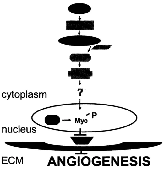
Watnick et al. have also shown that in epithelial cells Ras activates a ROCK-dependent signaling pathway that phosphorylates Myc. Myc phosphorylation results in an activated form of the protein and induces Tsp-1 repression (Watnick et al. 2003). Interestingly, a recent study has shown that hypoxia induces VEGF expression in colon cancer cells through the same Ras-ROCK-Myc signaling pathway that represses Tsp-1 in epithelial cells (Mizukami et al. 2006). This study demonstrates a HIF-l-independent pathway that induces hypoxia-mediated VEGF induction.
V: The role of
p53
in tumor development and anfqioqenesis
One of the most commonly mutated genes in human cancers is p53 (Levine
1993; Greenblatt et al. 1994). The protein p53 was first discovered as a cellular
protein that associated very tightly with Simian virus 40 (SV40) large T antigen (Lane
and Crawford 1979; Linzer and Levine 1979; Linzer et al. 1979). Early experiments
with cloned p53 described it as having oncogenic potential, though later studies
revealed that these p53 clones were obtained from mutant versions of the p53 gene
(Parada et al. 1984; Eliyahu et al. 1988; Finlay et al. 1988; Eliyahu et al. 1994).
These studies, however, demonstrated that mutation of p53 often leads to a
tumorigenic phenotype. Subsequent studies with wild-type p53 conclusively showed
that p53 functions as a tumor suppressor (Eliyahu et al. 1989; Finlay et al. 1989;
Baker et al. 1990; Chen et al. 1991).
The main function of p53 is in the response to cellular stress, particularly DNA
damage (Kastan et al. 1991). In response to DNA damage, p53 can either arrest the
cell cycle or induce apoptosis (Kastan et al. 1991; Yonish-Rouach et al. 1991;
Kuerbitz et al. 1992; Clarke et al. 1993; Lowe et al. 1993). The activity of p53 is
primarily a function of its ability to modulate transcriptional expression of target genes
(Luria and Horowitz 1986; Fields and Jang 1990). The N-terminal domain of p53
contains the transactivation domain, while the rest of the protein is composed of a
sequence-specific DNA-binding domain, a nuclear localization signal, a C-terminal
oligomerization domain, and a lysine-rich C-terminal ubiquitination domain (Kern et al.
the oligomerization domain, p53 forms a functional active tetramer (Sturzbecher et al. 1992).
Under normal conditions, p53 is maintained at low levels by ubiquitin-mediated proteolysis (Maki et al. 1996). This process is regulated by the ubiquitin ligase MDM-2 (Momand et al. 199MDM-2). MDM-MDM-2 binds p53 at the N-terminal transactivation domain and inhibits p53 function by ubiquitinating the p53 C-terminal lysines, thereby targeting it to the proteasome for degradation (Momand et al. 1992; Oliner et al. 1993; Honda et al. 1997; Fuchs et al. 1998). Activated p53 induces transcription of MDM-2, thus establishing an autoregulatory loop that ensures attenuation of p53 signaling (Momand et al. 1992). Mouse studies have shown the critical role that MDM-2 plays in regulating p53 expression to allow for proper cell cycle progression during development. Whereas the mdm2 knockout mouse is embryonic lethal, the p53
mdm2 double knockout is viable (Jones et al. 1995; Montes de Oca Luna et al. 1995).
Under conditions of cellular stress, such as DNA damage, p53 becomes stabilized through post-translational modifications, is relieved from MDM-2 binding, and becomes active (Canman et al. 1994). For example, gamma-irradiation activates the kinase ATM, which activates the CHK-2 kinase, and both kinases phosphorylate serines in the N-terminal transactivation domain of p53 (Canman et al. 1998; Chehab et al. 2000; Shieh et al. 2000). It is also worth noting that p53 can be activated in response to inappropriate cell proliferation due to oncogenic signaling, which is one mechanism by which p53 suppresses tumors. In response to oncogenic signaling,
pl19ARF is induced, which inactivates MDM-2 and releases p53 (Kurokawa et al. 1999).
The two main cellular responses arising from p53 activation are cell cycle
arrest and apoptosis. The p53 protein can arrest the cell cycle at both the G1 and G2
phases. Arrest at G1 can be mediated through activation of p21, which can inhibit
cyclinDl/Cdk4 and cyclinE/Cdk2 (Harper et al. 1993; Xiong et al. 1993). These two
CDK complexes promote progression through GI. Conversely, activation of 14-3-3
sigma by p53 causes cdc-25 and cyclinBl/Cdkl to be sequestered in the cytoplasm,
which helps maintain a G2 block (Hermeking et al. 1997). It has also been shown in
fibroblasts that constitutive expression of Myc can induce a p53-mediated G2 arrest
of the cell cycle (Felsher et al. 2000).
Apoptosis is induced by p53 through the transactivation of pro-apoptotic genes
like the mitochondrial pore protein Bax and the BH3 domain proteins PUMA and
NOXA, which mediate the release of cytochrome C from the mitochondria (Miyashita
et al. 1994; Miyashita and Reed 1995; Oda et al. 2000; Nakano and Vousden 2001;
Schuler and Green 2001). It has also been shown that p53 can transcriptionally
repress the cell survival gene bcl-2 (Miyashita et al. 1994).
Myc is one of the targets of p53-mediated transcriptional repression (Moberg et
al. 1992; Levy et al. 1993; Ragimov et al. 1993). Moberg et al. and Ragimov et al.
demonstrated the ability of p53 to block promoter activation of Myc by blocking
formation of the transcriptional pre-initiation complex on the promoter. Levy et al.
demonstrated that expression of p53 promotes a GO-like state in leukemic cells and
represses transcription of Myc. Furthermore, a recent study has indicated that p53
represses Myc expression by direct association with the Myc promoter and histone
H4 deacetylation (Ho et al. 2005). This study also demonstrates that Myc repression
leads to cell cycle arrest at G1 and highlights the interrelation between p53 and Myc. The interaction of Myc with p53 activation seems to play a critical role in determining the cellular response to stress, since overexpression of Myc can lead to apoptosis and repression of Myc can lead to G1 cell cycle arrest (Seoane et al. 2002; Ho et al. 2005).
Studies of murine cancer models have revealed that p53 does not play an essential role during development (Donehower et al. 1992; Jacks et al. 1994; Purdie et al. 1994). However, consistent with its role as a tumor suppressor, p53 knockout mice develop spontaneous tumors (Donehower et al. 1992; Jacks et al. 1994; Purdie et al. 1994). Mice heterozygous for p53 also develop spontaneous tumors, though the tumors develop later in life (Donehower et al. 1992; Jacks et al. 1994; Purdie et al. 1994). Interestingly heterozygous mice develop different types of tumors than the p53 null mice. Knockout mice develop mostly lymphomas and soft tissue sarcomas, while heterozygous mice have an equal distribution of lymphomas, soft tissue sarcomas and osteosarcomas (Jacks et al. 1994). Heterozygous mice also develop carcinomas at a much higher frequency than p53 knockout mice (Jacks et al. 1994).
Angiogenesis has also been shown to be regulated by p53. Many studies have demonstrated that expression of wild-type p53, in various types of carcinoma and sarcoma cell lines, leads to the repression of VEGF both in vitro, and in mouse
xenograft models (Mukhopadhyay et al. 1995; Bouvet et al. 1998; Zhang et al. 2000). Supporting studies have shown that in angiosarcomas expression of mutant p53 or overexpression of MDM-2 increases levels of VEGF expression (Zietz et al. 1998). p53-mediated VEGF repression has been demonstrated to be at the transcriptional