II. DEVELOPMENTAL ELECTRON OPTICS LABORATORY
Academic and Research Staff Dr. John W. Coleman Dr. Edward H. Jacobsen
Graduate Students Michael R. Graham
1. ULTRAHIGH-SENSITIVITY ELECTRON OPTICAL DETERMINATION AND LOCATION OF IMPURITY ATOMS IN Si AND GaAs
Joint Services Electronics Program (Contract DAAG29-78-C-0020) National Institutes of Health (Grant 1 RO1 GM23597)
John W. Coleman
The ultimate goal of the proposed research is the simultaneous species identification and spatial resolution of atoms of low atomic number in electron micrographs. The basis for the research is Auger electron spectroscopy coupled to high-resolution elec-tron optics. The apparatus to accomplish this is the fixed-beam Auger electron micro-scope, which is now being made operationally dependable. The goal for the current year, which has been met, included final instrumental buildup, with a correction in the optical system and reengineering of the vacuum system.
The correction in the optical system was in order to bypass the undesirable focus-ing effects due to the structure of the specimen support (Davisson-Calbick Effect). The optics were redesigned to allow the specimen to rest at the center of a cylindrical metal electrode. Preliminary indications are that this has made focusing far less critical.
The following vacuum problems necessitated complete redesign of the housing and pumping systems of the Auger microscope:
1. Oil residues in the old vacuum system (although cryotrapped) were sufficient
to cause continual microdischarges between electrodes several kV apart but spaced only millimeters away from each other.
2. These microdischarges constituted erratic changes in the optical properties of the electrostatic lenses, thus only by chance and fleetingly would all the lenses be at such value that designed-for trajectories would occur.
3. Further intolerable vacuum conditions (leaks in electrical feed-throughs and unclear diffusion pumping systems) limited vacuum to 10 minus 7 Torr, which precluded the use of the CEMA image intensifier (guarantee void for use at such values) and also made illogical the attempt to use Auger electrons to the exclusion of other secondaries (even the cleanest surface - necessary for Augers - contaminates too rapidly at 10 minus 7 Torr).
(II.
DEVELOPMENTAL ELECTRON OPTICS LABORATORY)
4. With the old system, the foil lens was de facto impracticable since the electron-transparent foil contaminated rapidly, i. e., within a matter of a few minutes became electron-opaque with the accelerating voltage (20 kV) used in the AEM. Without the foil lens, of course, correction of spherical aberration is impossible, and it is just
this correction which must be applied to realize the high spatial resolution which the design will allow.
A new vacuum system was designed, therefore, to enable the following: 1. Use of the CEMA image intensifier.
2. Use of the foil lens to correct spherical aberration.
3. Use of the magnetic prism energy analyzer without contaminating the faces. 4. Photography of the images.
5. Use of Augers mainly for imaging.
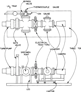
BYPASS VALVE
LN TRAP E e T HERMOCOUPLE GAUGE
O ION GAUGE
GATE ELECTRICAL
VALVE FEED-THRU
TURBOPUMP VIEWPORT TABLE TOP
SPECIMEN
PORT CAMERA
LEG
CASTOR
Fig. II-1. Auger electron-microscope vacuum-pumping and vacuum-housing systems.
The new vacuum system uses a copper-gasketed housing with a 100% oil-free air-suspended air-driven turbomolecular pump made by ALCATEL. This pump was nine months in delivery. A schematic of the instrument is shown in Fig. II-1.