HAL Id: pasteur-01574988
https://hal-pasteur.archives-ouvertes.fr/pasteur-01574988
Submitted on 17 Aug 2017
HAL is a multi-disciplinary open access
archive for the deposit and dissemination of
sci-entific research documents, whether they are
pub-lished or not. The documents may come from
teaching and research institutions in France or
abroad, or from public or private research centers.
L’archive ouverte pluridisciplinaire HAL, est
destinée au dépôt et à la diffusion de documents
scientifiques de niveau recherche, publiés ou non,
émanant des établissements d’enseignement et de
recherche français ou étrangers, des laboratoires
publics ou privés.
Distributed under a Creative Commons Attribution| 4.0 International License
Juan J Quereda, Marie A Nahori, Jazmín Meza-Torres, Martin Sachse,
Patricia Titos-Jiménez, Jaime Gomez-Laguna, Olivier Dussurget, Pascale
Cossart, Javier Pizarro-Cerdá
To cite this version:
Juan J Quereda, Marie A Nahori, Jazmín Meza-Torres, Martin Sachse, Patricia Titos-Jiménez, et al..
Listeriolysin S Is a Streptolysin S-Like Virulence Factor That Targets Exclusively Prokaryotic Cells
In Vivo.. mBio, American Society for Microbiology, 2017, 8 (2), �10.1128/mBio.00259-17�.
�pasteur-01574988�
Listeriolysin S Is a Streptolysin S-Like
Virulence Factor That Targets Exclusively
Prokaryotic Cells In Vivo
Juan J. Quereda,a,b,cMarie A. Nahori,a,b,cJazmín Meza-Torres,a,b,cMartin Sachse,d
Patricia Titos-Jiménez,eJaime Gomez-Laguna,eOlivier Dussurget,a,b,c,f
Pascale Cossart,a,b,cJavier Pizarro-Cerdáa,b,c
Institut Pasteur, Unité des Interactions Bactéries-Cellules, Paris, Francea; Institut National de la Santé et de la
Recherche Médicale, U604, Paris, Franceb; Institut National de la Recherche Agronomique, USC2020, Paris,
Francec; Institut Pasteur, Ultrapole, Paris, Franced; Anatomy and Comparative Pathology Department,
University of Cordoba, International Excellence Agrifood Campus CeiA3, Cordoba, Spaine; Cellule Pasteur,
Université Paris Diderot, Sorbonne Paris Cité, Paris, Francef
ABSTRACT Streptolysin S (SLS)-like virulence factors from clinically relevant Gram-positive pathogens have been proposed to behave as potent cytotoxins, playing key roles in tissue infection. Listeriolysin S (LLS) is an SLS-like hemolysin/bacteriocin pres-ent among Listeria monocytogenes strains responsible for human listeriosis out-breaks. As LLS cytotoxic activity has been associated with virulence, we investigated the LLS-specific contribution to host tissue infection. Surprisingly, we first show that LLS causes only weak red blood cell (RBC) hemolysis in vitro and neither confers re-sistance to phagocytic killing nor favors survival of L. monocytogenes within the blood cells or in the extracellular space (in the plasma). We reveal that LLS does not elicit specific immune responses, is not cytotoxic for eukaryotic cells, and does not impact cell infection by L. monocytogenes. Using in vitro cell infection systems and a murine intravenous infection model, we actually demonstrate that LLS expression is undetectable during infection of cells and murine inner organs. Importantly, upon intravenous animal inoculation, L. monocytogenes is found in the gastrointestinal sys-tem, and only in this environment LLS expression is detected in vivo. Finally, we confirm that LLS production is associated with destruction of target bacteria. Our re-sults demonstrate therefore that LLS does not contribute to L. monocytogenes tissue injury and virulence in inner host organs as previously reported. Moreover, we de-scribe that LlsB, a putative posttranslational modification enzyme encoded in the LLS operon, is necessary for murine inner organ colonization. Overall, we demon-strate that LLS is the first SLS-like virulence factor targeting exclusively prokaryotic cells during in vivo infections.
IMPORTANCE The most severe human listeriosis outbreaks are caused by L. mono-cytogenes strains harboring listeriolysin S (LLS), previously described as a cytotoxin that plays a critical role in host inner tissue infection. Cytotoxic activities have been proposed as a general mode of action for streptolysin S (SLS)-like toxins, including clostridiolysin S and LLS. We now challenge this dogma by demonstrating that LLS does not contribute to virulence in vivo once the intestinal barrier has been crossed. Importantly, we show that intravenous L. monocytogenes inoculation leads to bacte-rial translocation to the gastrointestinal system, where LLS is specifically expressed, targeting the host gut microbiota. Our study highlights the heterogeneous modes of action of SLS-like toxins, and we demonstrate for the first time a further level of complexity for SLS-like biosynthetic clusters as we reveal that the putative posttrans-lational modification enzyme LlsB is actually required for inner organ colonization, independently of the LLS activity.
KEYWORDS Listeria, listeriolysin S, streptolysin S, cytotoxin, epidemics, infection
Received 15 February 2017 Accepted 7
March 2017 Published 4 April 2017
Citation Quereda JJ, Nahori MA, Meza-Torres J,
Sachse M, Titos-Jiménez P, Gomez-Laguna J, Dussurget O, Cossart P, Pizarro-Cerdá J. 2017. Listeriolysin S is a streptolysin S-like virulence factor that targets exclusively prokaryotic cells
in vivo. mBio 8:e00259-17.https://doi.org/10 .1128/mBio.00259-17.
Editor Julian E. Davies, University of British
Columbia
Copyright © 2017 Quereda et al. This is an
open-access article distributed under the terms of theCreative Commons Attribution 4.0 International license.
Address correspondence to Javier Pizarro-Cerdá, javier.pizarro-cerda@pasteur.fr.
crossm
® mbio.asm.org on April 13, 2017 - Published by mbio.asm.orgL
isteria monocytogenes is a foodborne pathogen and a facultative intracellular bac-terium capable of causing severe disease in humans and animals. Upon ingestion of contaminated food, L. monocytogenes colonizes the intestine and crosses the intestinal barrier, disseminating via the blood to the liver, spleen, brain, and placenta (1, 2). The listeriosis fatality rate is estimated to be 20 to 30% of infected individuals despite antibiotic treatment (1). The most severe human listeriosis outbreaks are associated with a subset of L. monocytogenes lineage I strains that harbor a gene cluster encoding the bacteriocin and hemolytic factor listeriolysin S (LLS) (3, 4). Interestingly, the LLS gene cluster is absent from the most commonly studied L. monocytogenes lineage II strains EGD, EGDe, and 10403S (3), and its contribution to the intracellular lifecycle of L. monocytogenes is unknown (5).LLS is homologous to streptolysin S (SLS [encoded by sagA in the sag operon]), a potent cytolytic toxin produced by most group A Streptococcus pyogenes (GAS) strains
(6, 7). SLS is naturally expressed in vitro and is responsible for the classical-hemolytic
phenotype of S. pyogenes on blood agar plates (8). Using live-cell imaging, it has been shown that SLS activates the major erythrocyte anion exchange protein band 3 and
favors a rapid influx of Cl⫺ions into red blood cells (RBCs), leading to cellular rupture
(9). In HEK-293 cells, SLS causes lactate dehydrogenase (LDH) release into the medium, massive cytoskeletal disassembly, loss of focal contacts, and detachment from the tissue culture plates (7). SLS promotes resistance to phagocytic killing in whole-blood killing assays and activates an inflammatory programed cell death pathway in macro-phages (10, 11). In vivo studies have shown that SLS is required for S. pyogenes infection of skin and soft tissues (9, 12). The toxin encoded by an SLS-like gene cluster in Clostridium botulinum is named clostridiolysin S (CLS), and similarly to SLS, it is hemo-lytic and cytotoxic in HEK-293 cells (7, 13).
LLS, CLS, and SLS belong to the family of thiazole/oxazole-modified microcins (TOMMs) and are encoded by biosynthetic gene clusters characterized by the presence of cyclodehydratases/dehydrogenase genes (7). In the case of the LLS gene cluster, the genes llsB, llsY, and llsD code for putative posttranslational modification (PTM) enzymes that modify the product of the llsA gene coding for the structural LLS toxin (6, 7). Attempts to uncover the structural characteristics of the mature SLS-like toxins have been unsuccessful (7, 12, 13). The only direct structural insight available suggests that
SLS contains two oxazole moieties observed at positions Ser46and Ser48and that CLS
contains a methyloxazole at position Thr46(13).
It has been suggested that given the genetic similarities of the TOMM operons in Gram-positive pathogens, it is likely that these TOMMs contribute to the pathogenic potential of each bacterial producer, similarly to SLS for S. pyogenes (6, 14). However, it is currently unknown if all SLS-like molecules of bacterial pathogens are cytotoxins, which play a role in tissue injury and contribute to virulence by targeting eukaryotic cells. By using an llsB deletion mutant, it has been previously concluded that LLS is a hemolysin and a cytotoxin that contributes to L. monocytogenes virulence in an intraperitoneal murine model of infection (3). We recently described that in orally infected mice, LLS behaves as a bacteriocin favoring L. monocytogenes gut colonization through modulation of the intestinal microbiota (4).
In the present study, we sought to deepen our insight into the functions of LLS during listeriosis, with a particular focus on its effects once the intestinal barrier has been crossed. Here, we show that while LLS kills prokaryotic targets, it does not impact infection of eukaryotic host cells in vitro and in vivo. Our present results demonstrate that, apart from its role as a bacteriocin in the host intestine, the structural product of the llsA gene has negligible activity on eukaryotic host cells, while the llsB gene product contributes to colonization of deep organs.
RESULTS
LLS causes weak RBC hemolysis in vitro and does not alter RBC counts in vivo.
By growing L. monocytogenes colonies that constitutively express LLS in blood agar plates, or by using cell-free supernatants obtained from bacteria activated by the LLS
mbio.asm.org
on April 13, 2017 - Published by
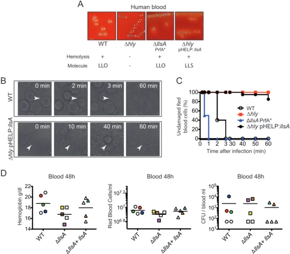
inducer yeast RNA core, LLS has been previously shown to display hemolytic activity (3). In the case of SLS, real-time imaging indicated that the secreted toxin induces hemo-lysis of RBCs in 30 s (9). We investigated whether LLS exerts hemohemo-lysis as efficiently as SLS. First, we determined in brain heart infusion (BHI) agar plates with human or mouse blood the hemolytic activity of the L. monocytogenes F2365 wild type (WT), F2365 Δhly, F2365 ΔllsA PrfA* (a strain that constitutively expresses listeriolysin O [LLO]), and F2365 Δhly(pHELP::llsA) (a strain that does not produce LLO but expresses LLS). F2365 Δhly was not hemolytic on human (Fig. 1A) or mouse (see Fig. S1 in the supplemental material) BHI blood agar plates. On the other hand, the F2365 WT and ΔllsA PrfA* strains caused hemolysis of both RBCs due to LLO activity, while the Δhly(pHELP::llsA) strain also caused hemolysis in human and mouse RBCs due to LLS activity (Fig. 1A; Fig. S1). Second, we performed live-cell microscopy to visualize human RBC hemolysis caused by the same F2365 strains. Real-time imaging indicated 100% RBC hemolysis in 3 min due to LLO activity when incubated with the F2365 WT (Fig. 1B, upper panel, and C) or in 1 min when incubated with F2365 PrfA* ΔllsA (Fig. 1C). Hemolysis was marked by the visual rupture of the RBC membranes, as previously described for SLS (9). F2365 Δhly caused no hemolysis, highlighting the potent activity of LLO (Fig. 1C). Surprisingly, only 15% of RBCs were lysed after 60 min of incubation with F2365 Δhly(pHELP::llsA) (Fig. 1B,
Blood 48h
A
B
Human bloodD
WT ∆hlyTime after infection (min) WT llsA PrfA* Undamaged R ed blo o d c ells (%) ∆hly ∆hly pHELP:llsA
0 min 2 min 3 min 60 min
0 min 10 min 40 min 60 min
WT ∆ hly pHELP: llsA Hemolysis Molecule + -LLO ∆hly pHELP: llsA + LLS ∆llsA PrfA* + LLO
-C
0 1 2 3 30 40 50 60 0 20 40 60 80 100 101 102 103 104 105 CFU / b lood m l 106.8 107 107.2 Red B lood C ells/ml Hemoglobin g/dl 14 16 18 20 22 WT llsA llsA + llsA WT llsA llsA + llsA WT llsA llsA + llsA Blood 48h Blood 48hFIG 1 Hemolytic and cytolytic properties of LLS. (A) Hemolysis of human blood BHI agar plates by L. monocytogenes F2365, L. monocytogenes F2365 Δhly, L. monocytogenes F2365 Δhly(pHELP::llsA), and L. monocytogenes ΔllsA PrfA*. The lower panel
shows the presence (⫹) or absence (⫺) of hemolysis and the L. monocytogenes molecule responsible for such hemolysis.
LLO, listeriolysin O; LLS, listeriolysin S. (B and C) Live microscopy of RBC hemolysis using phase-contrast microscopy at different times posttreatment with the same strains used in panel A. Panel C shows quantification of the hemolysis over time. Data from one representative experiment out of the three performed are shown. (D) Concentration of hemoglobin (grams per deciliter), number of RBCs per milliliter of blood, and number of L. monocytogenes CFU per milliliter of blood
of BALB/c mice injected intravenously with 104CFU of the indicated strains. Mice were killed at 48 h p.i., and a blood
sample was taken to assess the number of RBCs and the hemoglobin concentration. Mice whose blood contained more than 300 L. monocytogenes CFU/ml are colored to facilitate comparison of blood parameters.
mbio.asm.org
on April 13, 2017 - Published by
lower panel, and C), suggesting that the hemolytic activity of LLS is much less efficient than that of LLO, as shown in our experiments and as reported in the literature for SLS. In order to reveal the potential effects of the LLS hemolytic activity during L. mono-cytogenes infection in vivo, we infected intravenously conventional BALB/c mice with the F2365 WT, ΔllsA mutant strain, or its complemented strain and quantified the concentration of hemoglobin in blood, the number of RBCs, and the bacterial burden in blood at 48 h postinfection (p.i.). Consistent with real-time imaging performed in vitro, no significant changes in hemoglobin concentration (Fig. 1D, left panel) or RBC counts (Fig. 1D, center panel) were observed in mice infected with these three strains. Importantly, no correlation was observed between the number of L. monocytogenes cells in the blood (Fig. 1D, right panel) and the number of RBCs or hemoglobin (Fig. 1D; see Table S1 in the supplemental material). Taken together, these results show that LLS hemolytic activity is weak in vitro and that this activity does not affect RBC numbers during L. monocytogenes infection in vivo.
LLS expression does not confer resistance to phagocytic clearance. The weak
hemolytic activity of LLS prompted us to evaluate its contribution to L. monocytogenes survival in blood. In the case of GAS, WT strains are able to proliferate in human blood, while SLS-negative mutants are cleared, demonstrating a crucial role for SLS in the resistance to phagocytic killing in human blood (10). To explore whether LLS contrib-utes to phagocytic clearance, we incubated fresh human blood from three donors with L. monocytogenes F2365 WT, F2365 ΔllsA, and F2365(pHELP::llsA) strains. No significant difference could be observed between the three strains in phagocytic clearance (Fig. 2A). GAS can kill macrophages through the activation of an inflammatory pro-gramed cell death pathway mediated by SLS and streptolysin O (SLO) (11). Moreover, SLS induces alterations in keratinocyte inflammatory signaling cascades during GAS infection (15). These SLS properties prompted us to investigate a hypothetical role of LLS in cytokine secretion by macrophages infected by L. monocytogenes. We used an array kit detecting 111 different cytokines (Proteome Profiler mouse XL cytokine array) to compare the inflammatory responses of RAW264.7 macrophages infected with the F2365 WT strain (which does not express LLS in vitro, as shown above) to the responses
0h 2h 4h 24h 102 103 104 105 106 CFU/0.1ml b lood 0h 2h 4h 24h 102 103 104 105 106 0h 2h 4h 24h 102 103 104 105 106 WTllsA pHELP:llsA Donor3 Donor1 Donor2 RAW264.7 R e la ti v e c y to to x ic ity WT llsA pHELP: llsA 0 50 100 150 Caco2 R e la ti v e c y to to x ic ity WT llsA pHELP: llsA 0 50 100 150
A
B
FIG 2 LLS expression does not confer resistance to phagocytic clearance and is not cytotoxic for eukaryotic cells. (A) Survival in human whole blood of the L. monocytogenes F2365 WT, L. monocytogenes F2365 ΔllsA, and L. monocytogenes(pHELP::llsA) strains. CFU numbers were monitored at 2, 4, and 24 h p.i. Experiments with three independent blood donors with 6 replicates in each experiment were performed. Error bars represent standard deviation (SD). (B) Cytotoxicity (LDH release) relative to F2365 WT (100%) in RAW264.7 and Caco-2 cells infected
for 24 h (washed after 1 h of infection and with 40g/ml gentamicin added). Error bars represent SD.
mbio.asm.org
on April 13, 2017 - Published by
of cells infected with the ΔllsA deletion mutant or with the pHELP::llsA strain (which constitutively expresses LLS). No significant difference between the three groups (WT, ΔllsA, and pHELP::llsA strains) could be observed for secreted cytokine levels, including
interleukin-1 (IL-1), IL-6, and tumor necrosis factor alpha (TNF-␣), whose role is essential
in the response to L. monocytogenes infection (see Fig. S2 in the supplemental material) (16), suggesting that LLS does not alter cytokine production in macrophages. Overall, our results indicate that LLS does not contribute to L. monocytogenes survival during the blood stage.
LLS expressed by intracellular L. monocytogenes is cytotoxic for neither mac-rophages nor epithelial cells. The absence of a role for LLS in macmac-rophages contrasts
with the previously published role of this molecule as cytotoxic for diverse cell lines, including professional phagocytes (3). To revisit this concept, we evaluated LLS cyto-toxicity by measuring lactate dehydrogenase (LDH) release from RAW264.7 macro-phages and enterocyte-like Caco-2 cells. No differences in LDH release into culture supernatants were observed between the WT, ΔllsA, and pHELP::llsA strains in RAW264.7 and Caco-2 cells (Fig. 2B). Taken together, these results suggest that LLS secreted by intracellular L. monocytogenes is not cytotoxic for eukaryotic host cells.
llsA promoter activity is undetectable during infection of eukaryotic cells in vitro or infection of deep organs, such as spleen or liver, in vivo. Transcriptional fusion of the LLS promoter to a lux reporter plasmid previously demonstrated that LLS expression is specifically triggered in the intestine of orally infected mice and unde-tectable in other organs, such as liver and spleen (although these organs contained higher bacterial counts) (4). Bioluminescent signals in the intestine of germfree mice could also be detected, indicating that the intestine itself and not the gut microbiota activates the LLS promoter (4). Different compounds naturally present in the intestine,
like mucin, gastric fluid, pepsin, NaHCO3, short-chain fatty acids, ethanolamine, or even
6% O2or intestinal content of mice added ex vivo could not induce bioluminescence
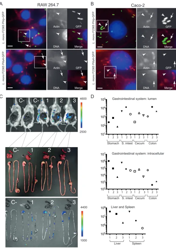
under in vitro conditions (4). In the present study, we investigated whether macro-phages or epithelial cells could trigger LLS expression. We generated a transcriptional fusion of the LLS promoter to the promoterless green fluorescent protein gene (gfp), which is more sensitive as a reporter than the luciferase systems in other Gram-positive bacteria (17). A similar green fluorescent protein (GFP) transcriptional reporter was used for hly, a gene essential for the intracellular lifestyle of L. monocytogenes. We next
infected Caco-2 epithelial cells and RAW264.7 macrophages with F2365InlB
(pAD-PllsA-GFP) or F2365InlB(pAD-Phly-GFP) for 2, 6, and 24 h. Figure 3 shows intracellular bacteria
that polymerize host cell actin (arrows) and bacteria not associated with actin (arrow-heads), presumably inside vacuoles after 5 h of gentamicin treatment. Intracellular
F2365InlB(pAD-Phly-GFP) produced a green fluorescence at 2, 6, and 24 h p.i. in both cell
lines tested, indicating that the GFP reporter is functional and that the hly promoter is active (Fig. 3A and B; see Fig. S3 and S4 in the supplemental material). Interestingly, the activity of the llsA promoter was undetectable at 2, 6, or 24 h p.i. in the cell lines tested (Fig. 3A and B; Fig. S3 and S4). No signal was observed for the llsA promoter at 2, 6, and 24 h p.i. in other cell lines (LoVo, HeLa, and Jeg-3) where the hly promoter was active (data not shown).
Furthermore, we investigated whether L. monocytogenes inoculated intravenously expresses LLS in deep organs, such as the spleen or the liver. We used the L. mono-cytogenes F2365llsA::luxstrain in which we fused the LLS promoter to the lux reporter
plasmid (4). Upon intravenous infection of BALB/c mice with 104cells of F2365llsA::lux, a
bioluminescent signal was detected in the abdomen of infected animals at 72 and 96 h p.i. To uncover the origin of the bioluminescent signals, abdominal skin and perito-neum dissection was performed. The ex vivo images of the gastrointestinal system and the livers and spleens removed from the body are shown in Fig. 3C. Interestingly, bioluminescent signals were only detected in the stomach and the intestine of infected mice, while being absent from the liver and spleen (which are the main organs targeted by L. monocytogenes). Importantly, although the bioluminescent signals were specifi-cally expressed in the gastrointestinal system, the liver and spleen contained at least
mbio.asm.org
on April 13, 2017 - Published by
L. mono F2365 P hly -GFP GFP Actin DNA Merge L. mono F2365 P llsA -GFP RAW 264.7 GFP Actin DNA Merge GFP Actin DNA Merge Caco-2 L. mono F2365 P hly -GFP L. mono F2365 P llsA -GFP GFP Actin DNA Merge
A
B
C-
1
2
3
C-C-
1
2
3
C-
1
2
3
4400 1000 4600 2500C
Gastrointestinal system: lumen101 102 103 104 105 102 103 104 105 106 107
Liver and Spleen
105
106
107
108
Gastrointestinal system: intracellular
D
Stomach S. intest Cecum Colon 1 2 3 1 2 3 1 2 3 1 2 3
Stomach S. intest Cecum Colon 1 2 3 1 2 3 1 2 3 1 2 3
Spleen Liver
1 2 3 1 2 3
FIG 3 Fluorescence microscopy and bioluminescence assays to evaluate the promoter activity of llsA. RAW264.7 cells (A) and Caco-2
cells (B) were cultured in 96-well plates and infected with L. monocytogenes F2365InlB(pAD-PllsA-GFP) or L. monocytogenes
F2365InlB(pAD-Phly-GFP). Host cells were infected for 6 h (washed after 1 h of infection and with 40g/ml gentamicin to kill
extracellular bacteria) and fixed. GFP is shown in green. Actin (red) and nuclei (blue) were labeled with phalloidin conjugated to Alexa
546 and Hoechst, respectively. Scale bars, 5 m. (C) Bioluminescence imaging showing induction of the LLS promoter in the
gastrointestinal system after intravenous inoculation of three mice with 104bacteria per BALB/c mouse. C⫺, noninfected control
mice. Images were acquired at 96 h p.i. with an IVIS Spectrum imaging system. The false color bar indicates the number of photons/second. (D) Bacterial counts in the stomach, small intestine (S. intest), cecum, and colon content (top), as well as in the stomach, small intestine, cecum, colon, liver, and spleen tissues (middle and bottom), of the same mice at 96 h p.i.
mbio.asm.org
on April 13, 2017 - Published by
more than 100-fold CFU than the stomach, small intestine, cecum, or colon (Fig. 3D). Altogether, these results demonstrate that the llsA promoter is not active or is ex-pressed at very low levels during infection of host cells, suggesting that the activation of LLS expression in the gastrointestinal system is not triggered by the infection of eukaryotic cells.
LLS does not contribute to cell infection and is not sufficient for L.
monocyto-genes vacuolar escape. Since we could not exclude that LLS produced during intes-tinal infection could impact the capacity of bacteria to infect eukaryotic cells, we investigated the potential contribution of LLS to infection. No differences in intracel-lular CFU counts were observed between the F2365 WT, F2365 ΔllsA, and F2365(pHELP:: llsA) strains at 2, 6, or 24 h p.i. in RAW264.7, HD11, and Caco-2 cells (Fig. 4A). These results show that LLS is not required during cellular infection by L. monocytogenes.
If LLS does not play a role in cell infection, this means that it should not affect vacuolar escape by L. monocytogenes. The capacity of LLS to mediate vacuolar rupture and to allow intracellular growth was evaluated in RAW264.7 macrophages. Extracel-lular and total L. monocytogenes numbers were distinguished by using inside-outside staining of fluorescently labeled bacteria, and cytosolic microorganisms were identified by actin staining. Approximately 80% of F2365 WT cells escaped from vacuoles and polymerized actin (Fig. 4B and E). As expected, LLO was required for vacuolar escape in macrophages, as no escape events were observed upon cell infection with F2365 Δhly (Fig. 4C and E). Importantly, no F2365 Δhly(pHELP::llsA) cells were found associated with actin (Fig. 4D and E), suggesting that these bacteria were trapped in phagosomes and that LLS expression is not sufficient to rupture the bacterial internalization vacuole in order to facilitate access to the host cytoplasm. Overall, our results indicate that LLS does not target eukaryotic membranes during infection.
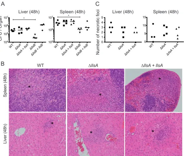
LlsB, but not LLS, has a role in spleen and liver colonization. SLS causes
extensive tissue disruption, inflammation, and necrosis of skin lesions and is required for S. pyogenes infection of skin and soft-tissues (9, 10, 12). Regarding LLS, as mentioned above we have shown that this molecule is specifically expressed in the intestine of intravenously and orally infected mice, where it alters the host intestinal microbiota and increases L. monocytogenes persistence (Fig. 3C and 4). LLS function in the intestine requires the activity of the LlsB enzyme, which is by homology with SLS putatively involved in the posttranslational modification of LLS. As reported, deletion of LlsB completely inactivates LLS hemolytic activity in vitro and decreases L. monocytogenes virulence in a mouse intraperitoneal infection model (3). To determine the impact of LLS on virulence once L. monocytogenes has crossed the intestine, we intravenously infected mice with the F2365 WT, F2365 ΔllsA, F2365 ΔllsB, F2365 ΔllsA, and F2365 ΔllsB complemented strains, and quantified the bacterial burdens in the liver and spleen. In agreement with the inactivity of LLS promoter in liver and spleen (4), and with the absence of a role for LLS in the blood and in the cell lines used in the present study, the ΔllsA mutant strain did not display significantly different bacterial loads in the spleen or liver compared with the WT strain (Fig. 5A). However, significant differences were observed between the WT and ΔllsB strains in the number of CFU isolated from
these organs (P⬍ 0.05) (Fig. 5A). Furthermore, histopathologic assessment of the liver
and spleen of mice infected with WT, ΔllsA mutant, and ΔllsA complemented strains showed no differences in the numbers, locations, and inflammatory cell types of necrotic foci (Fig. 5B and C). Together, these results demonstrate that LLS does not play an essential role in L. monocytogenes deep organ colonization once the intestine has been crossed and strongly suggest that LlsB performs additional important functions for virulence apart from putatively driving LLS posttranslational modification.
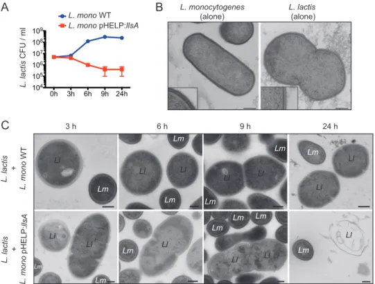
LLS promotes killing of target bacteria. Our results suggest that during in vivo
infections, LLS does not target eukaryotic cells. Our previous results (4) indicate that LLS is able to modulate the growth of Lactococcus lactis, Staphylococcus aureus, and L. monocytogenes lineage II strains as well as representatives of the Allobaculum and Alloprevotella genera during in vivo infection (4). To investigate a direct cytotoxic effect
mbio.asm.org
on April 13, 2017 - Published by
of LLS on target bacteria, diluted overnight cultures of L. monocytogenes F2365 strains (WT and the pHELP::llsA strain) and the target L. lactis were grown in coculture. After 3 h of coculture, there was a reduction in the growth of L. lactis only when cocultured with L. monocytogenes F2365(pHELP::llsA) (Fig. 6A). The effect of LLS on L. lactis growth was even higher at 6 and 9 h of coculture (Fig. 6A). Under transmission electron microscopy, L. lactis cultured alone or cocultured with the L. monocytogenes F2365 WT (which does not produce LLS under in vitro conditions [4]) for 3, 6, or 9 h showed the typical structure of Gram-positive cocci, with a thick, uniform, smooth cell wall and intact
∆hly pHELP:llsA
Actin Total Extra Actin Total Extra
Actin Total Extra
WT ∆hly WT hly hly pHELP: llsA 0 20 40 60 80 100 N ° of intracellular bacteria associated to a ctin
B
D
C
E
HD11 WTllsA pHELP: llsAWTllsA pHELP: llsAWTllsA pHELP: llsA 103 104 105 RAW 264.7 WTllsA pHELP: llsAWTllsA pHELP: llsAWTllsA pHELP: llsA 103 104 105 2h 2h 6h 6h 24h 24hA
Caco-2 103 104 105 106 107 CF U/W ell WTllsA pHELP: llsAWTllsA pHELP: llsAWTllsA pHELP: llsA 2h 6h 24hFIG 4 LLS expression does not confer an advantage during cell infection and is not sufficient to damage and rupture host cell vacuoles to access the host cytoplasm. (A) Numbers of viable intracellular L. monocytogenes F2365, L. monocytogenes F2365 ΔllsA, and L. monocytogenes(pHELP::llsA) cells in Caco-2, HD11, and RAW264.7 cells. The mean and standard error of the mean (SEM) are shown. CFU numbers were monitored at 2, 6, and 24 h p.i. (washed after 1 h of infection and with
40g/ml gentamicin added). Three independent experiments with 6 replicates in each experiment were performed. One
representative experiment is shown. (B, C, and D) RAW264.7 cells were seeded into 96-well plates and infected with the L. monocytogenes F2365 WT (B), L. monocytogenes F2365 Δhly (C), and L. monocytogenes F2365 Δhly(pHELP::llsA) (D) strains. Differential immunofluorescence staining for identification of extracellular versus total L. monocytogenes numbers was performed. Extracellular bacteria (Extra) were labeled with a secondary goat anti-rabbit Alexa 647 (blue), and total bacteria (total) were labeled with a secondary goat anti-rabbit Alexa 488 (green) after cell permeabilization. Actin was labeled by
using phalloidin conjugated to Alexa 546 (red). Nuclei were stained with Hoechst (gray). Bars, 5m. (E) Quantification of
the number of bacteria of the L. monocytogenes strains used in panels B, C, and D that access the host cytosol.
mbio.asm.org
on April 13, 2017 - Published by
cytoplasmic membrane attached to the wall (Fig. 6B and C). The cytoplasm was granular and evenly distributed in the cell. Some of the cells demonstrated a dividing septum, indicative of bacterial growth (Fig. 6B and C). After 3 h of coculture with L. monocytogenes F2365(pHELP::llsA), disruption of L. lactis cell wall integrity was observed (Fig. 6C). At 6, 9, and 24 h of coculture with L. monocytogenes F2365(pHELP:: llsA), increasing numbers of L. lactis cells showed more drastic changes, including cell wall wrinkles and even cellular lysis (Fig. 6C). These results confirm that LLS is the first SLS-like virulence factor of a bacterial pathogen able to promote death of target bacteria.
DISCUSSION
In 2008, SLS-like gene clusters were discovered in clinically relevant Gram-positive pathogens (including S. aureus and C. botulinum) and other nonpathogenic bacteria (7), leading to the identification of the LLS gene cluster in L. monocytogenes. SLS is a potent membrane-damaging agent and a major virulence factor contributing to GAS infection through rapid destruction of eukaryotic cells and tissue damage (6, 9–11, 15, 18–20). It has been proposed that SLS-like virulence factors from other Gram-positive pathogens also behave as potent cytotoxins (6, 14). Interestingly, functional experimental data of LLS activity on eukaryotic cells are scarce (3), despite the fact that LLS is almost exclusively detected within lineage I strains (the most frequent lineage among L.
mono-WT llsA llsA + llsA llsB llsB + llsB 104 105 106 107 105 106 107 WT llsA llsA + llsA llsB llsB + llsB * *
A
B
WT llsA llsA + llsA 0 1 2 3 4 5 WT llsA llsA + llsA 0 5 10 15C
*
*
*
*
*
*
Liver (48h) Spleen (48h)WT ∆llsA ∆llsA + llsA
Liver (48h) Spleen (48h) Liver (48h) Spleen (48h)
CFU / Organ
Number of necrotic foci
FIG 5 LLS role in spleen and liver colonization once the intestinal barrier has been crossed. (A) BALB/c mice were injected
intravenously with 104CFU of the indicated strains. Mice were killed at 48 h p.i., and spleens and livers were removed to assess the
bacterial load per organ. (Note that these numbers of CFU correspond to the half-organ used to assess bacterial load. For details, see the Materials and Methods section.) (B) Examples of spleen and liver tissues from the same infected mice. Asterisks show necrotic foci. (C) The number of necrotic foci in spleen and liver tissues from the same infected mice was evaluated. (Note that these numbers of necrotic foci correspond to the half-organ used for histopathology. For details, see the Materials and Methods section.)
mbio.asm.org
on April 13, 2017 - Published by
cytogenes clinical isolates) and that it has been related to the L. monocytogenes infectious potential in epidemiological and comparative genomic studies (21, 22). In the present work, we aimed to characterize the extent to which LLS potentially contributes to host infection by directly performing damaging activities on eukaryotic cells and tissues.
Using an array of molecular, cell biology, and histology techniques, we demonstrate that unlike SLS, (i) LLS causes very weak RBC hemolysis, (ii) it does not confer resistance to phagocytic clearance, (iii) it does not affect the levels of secreted cytokines by cells infected by L. monocytogenes, (iv) when expressed under the control of its native promoter or expressed through a constitutive promoter by intracellular L. monocyto-genes, it is not cytotoxic for epithelial cells and macrophages, (v) its constitutive expression by L. monocytogenes in the confined space of a phagocytic vacuole is not sufficient to rupture this membrane compartment, (vi) its expression is undetectable within host cells due to inactivity of its promoter under both in vitro and in vivo conditions, (vii) it does not contribute to eukaryotic cell infection, and (viii) it does not contribute to virulence in an intravenous infection murine model. Altogether, our results clearly demonstrate that the biological activity of LLS is distinct from that of SLS or CLS, showing that LLS does not target eukaryotic host cells and is not involved in inner organ infection during systemic stages of listeriosis.
It has been previously proposed that LLS is hemolytic on blood agar plates and cytotoxic for epithelial and phagocytic cell lines (3). Importantly, experimental condi-tions in vitro used a ratio of 100 bacteria per cell for 6 h (without explicit use of gentamicin) to demonstrate the cytotoxic effect of LLS on J774, C2-Bbe, and CT26 cells (3). The fact that we did not observe the same cytotoxic effect on Caco-2 or RAW264.7 cells after the same infection period (making use of gentamicin after 1 h p.i.), using an MOI of 5 for Caco-2 cells or an MOI of 2 for RAW264.7 cells, leads us to believe that
3 h 6 h 9 h 24 h L. lactis + L. mono WT 0h 3h 6h 9h 24h 104 105 106 107 108 109 L. lactis + L. mono pHELP: llsA
A
B
C
L. lactis (alone) L. monocytogenes (alone) Ll Lm Ll Ll Lm Ll Ll Ll Ll Lm Lm Ll Ll Ll Ll Ll Lm Lm Ll Lm Lm Lm Lm Lm Lm Lm L. lactis CFU / ml L. mono WT L. mono pHELP:llsAFIG 6 Effect of LLS on L. lactis. (A) Viable L. lactis at 3, 6, 9, and 24 h postinoculation in coculture with L. monocytogenes F2365 WT or L. monocytogenes F2365(pHELP::llsA). Error bars show SD. Data from one represen-tative experiment out of the three performed are shown. (B) L. lactis and L. monocytogenes F2365 WT seen by electron microscopy when cultured alone. Bacteria were grown in BHI for 24 h. Insets present an enlargement of
an area of the cell wall. Scale bars, 200m. (C) L. lactis cells cocultured with L. monocytogenes F2365 WT (upper
panels) or L. monocytogenes(pHELP::llsA) (lower panels) in BHI for 3, 6, 9, or 24 h. Scale bars, 200 nm.
mbio.asm.org
on April 13, 2017 - Published by
experimental conditions used in the previous study (3) may have influenced the in vitro
cell system conditions, finally leading to an increase of⬇20 to 30% of LDH release to
the medium due to LLS and also bacterial exposure. Moreover, the cytotoxic effect was only demonstrated by using a L. monocytogenes strain in which LLS was constitutively expressed using the pHELP promoter (3).
It has also been claimed that LLS contributes to virulence in a murine intraperitoneal model of infection (3). This conclusion was based on the reduced F2365 ΔllsB CFU numbers in the livers and spleens relative to those in the corresponding F2365 WT-infected mice. However, when we compared the virulence of the F2365 ΔllsA and F2365 ΔllsB mutants to that of the F2365 WT strain, we discovered an unexpected difference between these two deletion mutants: llsB but not llsA (the gene coding for the toxin LLS) contributed to virulence in our mouse intravenous model of infection, indicating that LlsB performs additional functions apart from the putative posttransla-tional modification of LLS. llsB is therefore the first gene from a dehydratase/dehydro-genase TOMM complex reported to perform additional functions apart from putative posttranslational modifications of the cluster-encoded toxin. An attractive hypothesis that remains to be validated is that LlsB participates in the posttranslational modifica-tion of another molecule outside the LLS operon.
Our present results show that although LLS constitutive expression [strain F2365(pHELP::llsA)] can damage RBCs in a blood agar plate or in a phosphate-buffered saline (PBS) suspension, the concentration of LLS produced by the epidemic L. mono-cytogenes F2365 WT strain during in vivo infection once the intestine has been crossed has minimal toxicity or even no effect on eukaryotic cells. On the contrary, LLS is highly toxic for prokaryotic targets such as L. lactis in vitro and modulates the host microbiota in vivo (4). These data demonstrate that LLS is the first SLS-like virulence factor of a bacterial pathogen that only targets prokaryotic cells in vivo. It is very important to highlight that in our present experiments, LLS expression is detected in the stomach and the intestine of mice after intravenous bacterial inoculation. Indeed, it has been previously shown that L. monocytogenes can be discharged back to the gastrointestinal system from the gallbladder (23, 24). Our results suggest that modulation of the host microbiota by LLS takes place not only during oral animal infection but also upon intravenous infection: this fact should be now taken into account during in vivo animal experiments with relevant epidemic L. monocytogenes strains expressing the LLS operon.
MATERIALS AND METHODS
Bacterial strains and cell lines. The epidemic lineage I L. monocytogenes strain F2365 of serotype 4b responsible for the 1985 California listeriosis outbreak (25) was used as parental strain (BUG3012; UIBC bacterial collection). The isogenic mutants and plasmids used in this study are listed in Table 1. Bacteria
TABLE 1 Bacterial strains used in this studya
BUG no. Mutation/relevant genotype Strain Reference
3012 Wild type L. monocytogenes 4b F2365 25
3651 PrfA* L. monocytogenes 4b F2365 This study
3671 Δhly L. monocytogenes 4b F2365 This study
3781 ΔllsA L. monocytogenes 4b F2365 4
3795 ΔllsA(pPl2::llsA) L. monocytogenes 4b F2365 4
3817 pHELP::llsA L. monocytogenes 4b F2365 4
3819 Δhly(pHELP::llsA) L. monocytogenes 4b F2365 This study
3672 ΔllsB L. monocytogenes 4b F2365 4
3975 ΔllsB(pPl2::llsB) L. monocytogenes 4b F2365 4
3783 ΔllsA PrfA* L. monocytogenes 4b F2365 This study
3824 inlB corrected L. monocytogenes 4b F2365 This study
4060 pAD-Phly-GFP L. monocytogenes 4b F2365InlB This study
4058 pAD-PllsA-GFP L. monocytogenes 4b F2365InlB This study
3763 LLS promoter fused to lux reporter system in pPL2 lux L. monocytogenes 4b F2365 4
4048 pAD-PllsA-GFP E. coli This study
4052 pAD-Phly-GFP E. coli This study
aThe strains shown are from the UIBC bacterial collection.
mbio.asm.org
on April 13, 2017 - Published by
were grown in brain heart infusion (BHI) medium with shaking at 200 rpm in tubes at 37°C. E. coli cells
were grown in LB broth. When required, antibiotics were added (chloramphenicol at 35g/ml for E. coli
or 7g/ml for L. monocytogenes). The tissue culture cells used in this study were from the RAW264.7
(BALB/c mouse macrophage cells; ATCC TIB-71), HD11 (avian macrophage cell line [26]), and Caco-2 (ATCC HTB-37) lines. Cells were maintained in Dulbecco’s modified Eagle’s medium (DMEM) (Gibco) with 2 mM GlutaMAX (4 mM for RAW264.7 cells) supplemented with 10% (20% for Caco-2) (vol/vol) fetal calf
serum (BioWest). Cells were grown at 37°C with 10% CO2.
Mutant construction. To construct L. monocytogenes F2365 Δhly, fragments containing 500 bp DNA flanking the open reading frames (ORFs) of hly were amplified by PCR and cloned into the suicide integrative vector pMAD as previously described (see Table S2 in the supplemental material) (27). The F2365 PrfA* mutant strain was designed inserting a point mutation (G145S) in PrfA, which rendered it constitutively active. The prfA*-A/prfA*-B and prfA*-C/prfA*-D (Table S2) oligonucleotides were designed to introduce a silent mutation in Cys144 (codon TGC¡TGT) and a missense mutation in Gly145 changing it to Ser145 (codon GGT¡TCT). The DNA fragment generated after splicing by overlap extension (SOEing) PCR was inserted into pMAD.
L. monocytogenes F2365 has a premature stop codon in inlB (codon no. 34 is TAA) (28). To facilitate in vitro cell infection and imaging, we generated an L. monocytogenes F2365 strain with a functional inlB
(L. monocytogenes F2365InlB
) by introducing a point mutation in codon 34 (TAA¡CAA). The oligonucle-otides InlB-new-A/InlB-new-B and InlB-new-C/InlB-new-D (Table S2) were used in SOEing PCR, and the PCR fragment was also cloned into pMAD.
To construct L. monocytogenes F2365 Δhly(pHELP::llsA), we electroporated L. monocytogenes F2365 Δhly with pMAD containing the promoter pHELP fused between two 500-nucleotide (nt) DNA fragments flanking the start codon of llsA (BUG3801 [4]). To construct the L. monocytogenes ΔllsA PrfA* strain, we electroporated L. monocytogenes F2365 PrfA*with pMAD-llsA from BUG3751 (4). Mutagenesis was performed by double recombination as described previously (27).
Bacterial cocultures and electron microscopy. Coculture assays of L. monocytogenes and L. lactis
(Institut Pasteur Collection CIP 70.56T) were performed for 24 h in 6% O2as previously described (4). At
3, 6, 9, and 24 h, part of the coculture was fixed for transmission electron microscopy, and another part of the culture was used to determine viable CFU on BHI agar plates. For biosafety reasons, bacterial strains or cocultures were inactivated by fixation with 2% glutaraldehyde (Sigma-Aldritch) in PHEM
(PIPES-HEPES-EGTA-MgSO4·7H2O) buffer at pH 7.1. After inactivation, cells were washed with PHEM buffer
and centrifuged. The pellet was resuspended in a small volume of PHEM buffer, and this suspension was taken up in capillary tubes (Wohlwendt Engineering) as described previously (29). The filled tube was
separated by clamping into segments of less than 2 mm and placed into the 200-m deep cavity of an
aluminum planchette, type A (Wohlwendt Engineering) filled with 1-hexadecene. With the flat side of the complementary type B planchette, the filled planchette was closed and frozen with the HPM 010 (Abra fluid).
Freeze substitution was performed in anhydrous acetone containing 1% osmium tetroxide (Merck).
1-Hexadecene is insoluble at⫺90°C in dry acetone. To allow access of the substitution mix to the sample,
small cracks were introduced under liquid nitrogen in the solid hexadecene by application of gentle
pressure using a precooled fine-point forceps (Dumont). Substitution was carried out at⫺90°C for 24 h,
at⫺30°C for 12 h, and at 0°C for 1 h each in a freeze substitution device FS8500 (RMC). Next, the samples
were washed with dry acetone and embedded stepwise in Epon (29). After heat polymerization, thin sections were cut with a UC6 microtome (Leica Microsystems, Inc.). Sections were collected on 200-mesh Formvar-coated cupper grids and poststained with 4% aqueous uranyl acetate and Reynold’s lead citrate. Images were taken at 120 kV with a Tecnai G2 transmission electron microscope (FEI) equipped with a US4000 camera (Gatan, Inc.).
Blood hemolysis assay on agar plates. Hemolysis was assessed by streaking 10 l of frozen bacterial cultures to isolate single colonies onto BHI agar plus 5% mouse (BALB/c) or human blood (French National Blood Service) and incubating the cultures for 24 h at 37°C.
Live imaging of blood hemolysis. Fresh whole human blood (French National Blood Service) was
centrifuged (2,000⫻ g, 10 min, 4°C). Three components were obtained at this stage: (i) the upper phase,
a clear solution of blood plasma; (ii) a middle thin layer of platelets and leukocytes; and (iii) at the bottom
RBCs. RBCs were collected and washed twice in cold 1⫻ Dulbecco’s PBS (DPBS; Gibco). An equivalent MOI
of 40 bacteria was incubated with 2 ml of RBC suspension (⬇10,000 RBCs) in 35-mm petri dishes (MatTek)
at 37°C. Bacterial overnight cultures were directly added to the petri dishes during microscopy acqui-sition. Live-cell imaging was performed during 60 min on a Zeiss Axio Observer spinning-disk confocal
microscope equipped with a 63⫻oil objective and driven by the MetaMorph software. Images were
acquired every 5 s for 60 min. One hundred RBCs were counted for hemolysis for each of the strains tested during the duration of the experiment. Three independent experiments were performed.
Whole-blood killing assays. Fresh human whole blood (French National Blood Service) was diluted
1/5 into RPMI, and 96-well tissue culture plates were seeded with 100 l of this suspension. The
L. monocytogenes strains were grown overnight in BHI, washed in PBS, and diluted in RPMI medium. A
total of 5⫻ 104bacteria were added per well. The mixture of bacteria and blood was incubated at 37°C
for 2, 4, and 24 h. The number of L. monocytogenes survivors was determined by serial dilution and colony counting on BHI agar plates. The experiments were repeated with blood from three independent human donors. Six technical replicates per bacterial strain and per blood donor were performed using independently derived clones of each of the strains. Statistical analyses were performed using the Student’s t test.
mbio.asm.org
on April 13, 2017 - Published by
Cell infection. Prior to infection, 96-well tissue culture plates were seeded with cells to attain 80% confluence on the day of infection. Overnight cultures of bacterial strains were washed three times in PBS and resuspended in infection medium (1% fetal bovine serum [FBS]) at an MOI of 2 (RAW264.7 and HD11) or 5 (Caco-2). Cells were centrifuged for 1 min at 1,000 rpm to synchronize infection. The cells were then incubated with the bacteria for 1 h at 37°C. Following this incubation, the cells were washed, and
extracellular bacteria were neutralized by adding complete medium containing 40g/ml of gentamicin.
At 2, 6, or 24 h postinfection, cells were washed with PBS and finally lysed in distilled water containing 0.1% Triton X-100. The number of viable intracellular L. monocytogenes cells was calculated by serial dilution and colony counting on BHI agar plates. These experiments employed 6 technical replicates per bacterial strain and were repeated three times with independent clones of each of the strains. Statistical analyses were conducted by using the Student’s t test.
Cytotoxicity LDH release assays and cytokine measurements. RAW264.7 and Caco-2 cells were
infected as indicated in the previous paragraph for 24 h (washed after 1 h of infection and with 40g/ml
gentamicin added to kill extracellular bacteria) where the supernatant was recovered and filtered. LDH levels were assayed with the kit LDH BR (Linear Chemicals) according to the kit instructions. Supernatants from RAW264.7 cells were also used to measure cytokine levels by using the cytokine array kit (Proteome Profiler mouse XL cytokine array; Becton, Dickinson) according to the manufacturer’s instructions.
Vacuolar escape. RAW264.7 cells were infected as indicated in the previous paragraphs for 6 h
(washed after 1 h of infection and with 40g/ml gentamicin added to kill extracellular bacteria) and fixed
with a paraformaldehyde (PFA) solution (4% in PBS) for 15 min at room temperature. Extracellular L. monocytogenes cells were labeled with a primary polyclonal goat anti-Listeria serum and with a secondary chicken anti-goat Alexa 647. Next, cells were permeabilized using 0.1% Triton X-100 for 4 min at room temperature, and total L. monocytogenes cells were labeled with the same primary antibody and a secondary chicken anti-goat Alexa 488 antibody. Actin was labeled by using phalloidin conjugated to Alexa 546. Nuclei were stained with Hoechst 33342 (dilution, 1/1,000). Samples were then rinsed four times in PBS and examined with a Zeiss Axiovert 135 epifluorescence microscope (Carl Zeiss, Inc.)
associated with a charge-coupled device (CCD) camera. Images were obtained with a⫻63 oil immersion
objective and processed with MetaMorph software (Universal Imaging). Cytosolic bacteria were consid-ered those stained with the Alexa 546 and Alexa 488 antibody but lacking Alexa 647 antibody labeling. Approximately 100 cells were counted in 3 representative fields to estimate the number of cytosolic L. monocytogenes cells.
Evaluation of llsA promoter expression with a GFP and a luciferase reporter system. A transcriptional fusion of the llsA promoter was created by cloning 500 bp upstream of the ATG of the respective gene with the gene encoding GFP-mut2 (30) (generating PllsA-GFP). This construct was cloned into SalI/SmaI-digested pAD vector (generating pAD-PllsA-GFP). Gene synthesis to construct PllsA-GFP was produced by Genecust (Luxembourg). pAD-PllsA-GFP was electroporated into L. monocytogenes
F2365InlB. hly was used as a control gene, the expression of which is upregulated during infection of
eukaryotic cells. The transcriptional fusion of the promoter of hly to GFP-mut 2 (pAD-Phly-GFP) was also
cloned into L. monocytogenes F2365InlB.
For epifluorescence analysis of promoter activity, RAW264.7 and Caco-2 cells were infected for 6 h
(washed after 1 h of infection and with 40g/ml gentamicin added) fixed with a PFA solution (4% in PBS)
for 15 min at room temperature, and permeabilized (0.1% Triton X-100 for 3 min in PBS). Samples were then rinsed four times in PBS, incubated with Hoechst and phalloidin conjugated to Alexa 546 for 30 min at room temperature, and rinsed four times in PBS. Samples were examined with a Zeiss Axiovert 135 epifluorescence microscope (Carl Zeiss, Inc.) associated with a charge-coupled device (CCD) camera.
Images were obtained with a⫻63 oil immersion objective and processed with MetaMorph software
(Universal Imaging).
For in vivo bioluminescence experiments, 8-week-old female BALB/c mice were infected by
intrave-nous inoculation with 104L. monocytogenes F2365llsA::lux(BUG3763) cells grown in BHI broth to an optical
density (OD) of 1.0 at 37°C. Bioluminescence imaging was accomplished using an IVIS Spectrum in vivo imaging system (Perkin Elmer) with a 5-min exposure time. Mice were anesthetized with isoflurane. For CFU determinations, whole luminal contents from stomach, small intestine, cecum, and colon, as well as tissues from stomach, small intestine, cecum and colon, liver, and spleen, were obtained, homogenized, and serially diluted and plated on Oxford agar plates (Oxoid). In order to determine the CFU numbers in the tissues of the gastrointestinal system, the tissues were washed three times in DMEM, incubated for
2 h in DMEM supplemented with 40g/ml gentamicin, and finally washed three times in DMEM.
Mouse infections. Six- to 8-week-old female BALB/c mice (Charles River, Inc., France) were injected
intravenously with 104CFU of the indicated strain. Mice were sacrificed at 48 h after infection (four mice
in each group), and livers and spleens were removed. Half of the organ was used to assess bacterial load, and the other half was used for histological analysis. To assess bacterial load, organs were homogenized and serially diluted. Dilutions were plated onto BHI plates and grown during 24 h at 37°C. Colonies were counted to assess bacterial load per organ. RBC and hemoglobin counts were determined using a Horiba scil Vet abc Plus veterinary hematology blood analyzer. Two independent experiments were carried out. Statistically significant differences were evaluated by the Mann-Whitney test. Pearson’s correlation coefficients were computed to measure correlations between blood parameters and L. monocytogenes CFU in blood.
Histological analysis. Liver and spleen tissue sections from mice intravenously infected with the F2365 WT, ΔllsA, and ΔllsA complemented strains sacrificed at 48 h were fixed in 10% neutral buffered formalin and routinely processed for the histopathological analysis. Four-micrometer sections per organ were stained with hematoxylin and eosin (H&E). The number of necrotic foci and the main cell type
mbio.asm.org
on April 13, 2017 - Published by
infiltrating necrotic areas were recorded. All of the slides were internally coded and analyzed blind. Statistically significant differences were evaluated by the Mann-Whitney test.
Ethics statement. This study was carried out in strict accordance with the French national and European laws and conformed to the Council Directive on the approximation of laws, regulations, and administrative provisions of the Member States regarding the protection of animals used for experi-mental and other scientific purposes (86/609/Eec). Experiments that relied on laboratory animals were performed in strict accordance with the Institut Pasteur’s regulations for animal care and use protocol, which was approved by the Animal Experiment Committee of the Institut Pasteur (approval no. 03-49). All human blood samples were anonymized and collected from the French National Blood Service under IRB approval no. HS2008-3470.
SUPPLEMENTAL MATERIAL
Supplemental material for this article may be found athttps://doi.org/10.1128/mBio
.00259-17.
FIG S1, EPS file, 0.5 MB. FIG S2, EPS file, 3 MB. FIG S3, EPS file, 5.2 MB. FIG S4, EPS file, 5.3 MB. TABLE S1, DOC file, 0.1 MB. TABLE S2, DOC file, 0.1 MB.
ACKNOWLEDGMENTS
We thank Esteban Chaves-Olarte for logistical support. P.C. is an International Senior Research Scholar of the Howard Hughes Medical Institute. The authors declare no conflict of interest.
This work was supported by the Institut Pasteur, the Institut National de la Santé et de la Recherche Médicale (INSERM Unité 604), the Institut National de la Recherche Agronomique (INRA Unité Sous Contrat 2020), Université Paris Diderot, grants from Région Île-de-France, the Institut Pasteur “Programmes Transversaux de Recherche” (PTR521 to J.P.C.), Agence Nationale de la Recherché (ANR-15-CE15-0017 StopBugEntry to J.P.C.), Fondation Le Roch Les Mousquetaires, European Research Council Advanced grant (670823 BacCellEpi to P.C.), and Région Île-de-France (DIM-MALINF to J.M.T.). The funders had no role in study design, data collection and interpretation, or the decision to submit the work for publication.
REFERENCES
1. Cossart P. 2011. Illuminating the landscape of host-pathogen interac-tions with the bacterium Listeria monocytogenes. Proc Natl Acad Sci
U S A 108:19484 –19491.https://doi.org/10.1073/pnas.1112371108.
2. Quereda JJ, García-Del Portillo F, Pucciarelli MG. 16 April 2016. Listeria monocytogenes remodels the cell surface in the blood-stage. Environ
Microbiol Rephttps://doi.org/10.1111/1758-2229.12416.
3. Cotter PD, Draper LA, Lawton EM, Daly KM, Groeger DS, Casey PG, Ross RP, Hill C. 2008. Listeriolysin S, a novel peptide haemolysin associated with a subset of lineage I Listeria monocytogenes. PLoS Pathog
4:e1000144.https://doi.org/10.1371/journal.ppat.1000144.
4. Quereda JJ, Dussurget O, Nahori MA, Ghozlane A, Volant S, Dillies MA, Regnault B, Kennedy S, Mondot S, Villoing B, Cossart P, Pizarro-Cerda J. 2016. Bacteriocin from epidemic Listeria strains alters the host intestinal microbiota to favor infection. Proc Natl Acad Sci U S A 113:5706 –5711. https://doi.org/10.1073/pnas.1523899113.
5. Quereda JJ, Cossart P, Pizarro-Cerdá J. 7 September 2016. Role of Listeria
monocytogenes exotoxins in virulence. Microb Toxinshttps://doi.org/10
.1007/978-94-007-6725-6_24-1.
6. Molloy EM, Cotter PD, Hill C, Mitchell DA, Ross RP. 2011. Streptolysin S-like virulence factors: the continuing sagA. Nat Rev Microbiol
9:670 – 681.https://doi.org/10.1038/nrmicro2624.
7. Lee SW, Mitchell DA, Markley AL, Hensler ME, Gonzalez D, Wohlrab A, Dorrestein PC, Nizet V, Dixon JE. 2008. Discovery of a widely distributed toxin biosynthetic gene cluster. Proc Natl Acad Sci U S A 105:5879 –5884. https://doi.org/10.1073/pnas.0801338105.
8. Alouf JE. 1980. Streptococcal toxins (streptolysin O, streptolysin S,
eryth-rogenic toxin). Pharmacol Ther 11:661–717. https://doi.org/10.1016/
0163-7258(80)90045-5.
9. Higashi DL, Biais N, Donahue DL, Mayfield JA, Tessier CR, Rodriguez K, Ashfeld BL, Luchetti J, Ploplis VA, Castellino FJ, Lee SW. 2016. Activation of band 3 mediates group A Streptococcus streptolysin S-based
beta-haemolysis. Nat Microbiol 1:15004.https://doi.org/10.1038/nmicrobiol
.2015.4.
10. Datta V, Myskowski SM, Kwinn LA, Chiem DN, Varki N, Kansal RG, Kotb M, Nizet V. 2005. Mutational analysis of the group A streptococcal operon encoding streptolysin S and its virulence role in invasive infection. Mol
Microbiol 56:681– 695.https://doi.org/10.1111/j.1365-2958.2005.04583.x.
11. Goldmann O, Sastalla I, Wos-Oxley M, Rohde M, Medina E. 2009. Strep-tococcus pyogenes induces oncosis in macrophages through the acti-vation of an inflammatory programmed cell death pathway. Cell
Micro-biol 11:138 –155.https://doi.org/10.1111/j.1462-5822.2008.01245.x.
12. Mitchell DA, Lee SW, Pence MA, Markley AL, Limm JD, Nizet V, Dixon JE. 2009. Structural and functional dissection of the heterocyclic peptide
cytotoxin streptolysin S. J Biol Chem 284:13004 –13012.https://doi.org/
10.1074/jbc.M900802200.
13. Gonzalez DJ, Lee SW, Hensler ME, Markley AL, Dahesh S, Mitchell DA, Bandeira N, Nizet V, Dixon JE, Dorrestein PC. 2010. Clostridiolysin S, a post-translationally modified biotoxin from Clostridium botulinum. J Biol
Chem 285:28220 –28228.https://doi.org/10.1074/jbc.M110.118554.
14. Molloy EM, Casjens SR, Cox CL, Maxson T, Ethridge NA, Margos G, Fingerle V, Mitchell DA. 2015. Identification of the minimal cytolytic unit for streptolysin S and an expansion of the toxin family. BMC Microbiol
15:141.https://doi.org/10.1186/s12866-015-0464-y.
15. Flaherty RA, Puricelli JM, Higashi DL, Park CJ, Lee SW. 2015. Strepto-lysin S promotes programmed cell death and enhances inflammatory signaling in epithelial keratinocytes during group A Streptococcus
mbio.asm.org
on April 13, 2017 - Published by
infection. Infect Immun 83:4118 – 4133. https://doi.org/10.1128/IAI .00611-15.
16. Stavru F, Archambaud C, Cossart P. 2011. Cell biology and immunology of Listeria monocytogenes infections: novel insights. Immunol Rev 240:
160 –184.https://doi.org/10.1111/j.1600-065X.2010.00993.x.
17. Guinane CM, Piper C, Draper LA, O’Connor PM, Hill C, Ross RP, Cotter PD. 2015. Impact of environmental factors on bacteriocin promoter activity in gut-derived Lactobacillus salivarius. Appl Environ Microbiol 81:
7851–7859.https://doi.org/10.1128/AEM.02339-15.
18. Betschel SD, Borgia SM, Barg NL, Low DE, De Azavedo JC. 1998. Reduced virulence of group A streptococcal Tn916 mutants that do not produce streptolysin S. Infect Immun 66:1671–1679.
19. Fontaine MC, Lee JJ, Kehoe MA. 2003. Combined contributions of strep-tolysin O and strepstrep-tolysin S to virulence of serotype M5 Streptococcus
pyogenes strain Manfredo. Infect Immun 71:3857–3865.https://doi.org/
10.1128/IAI.71.7.3857-3865.2003.
20. Engleberg NC, Heath A, Vardaman K, DiRita VJ. 2004. Contribution of CsrR-regulated virulence factors to the progress and outcome of murine skin infections by Streptococcus pyogenes. Infect Immun 72:623– 628. https://doi.org/10.1128/IAI.72.2.623-628.2004.
21. Maury MM, Tsai YH, Charlier C, Touchon M, Chenal-Francisque V, Leclercq A, Criscuolo A, Gaultier C, Roussel S, Brisabois A, Disson O, Rocha EP, Brisse S, Lecuit M. 2016. Uncovering Listeria monocytogenes hypervirulence by harnessing its biodiversity. Nat Genet 48:308 –313. https://doi.org/10.1038/ng.3501.
22. Moura A, Criscuolo A, Pouseele H, Maury MM, Leclercq A, Tarr C, Björk-man JT, DallBjörk-man T, Reimer A, Enouf V, Larsonneur E, Carleton H, Bracq-Dieye H, Katz LS, Jones L, Touchon M, Tourdjman M, Walker M, Stroika S, Cantinelli T, Chenal-Francisque V, Kucerova Z, Rocha EP, Nadon C, Grant K, Nielsen EM, Pot B, Gerner-Smidt P, Lecuit M, Brisse S. 2016. Whole genome-based population biology and epidemiological
surveil-lance of Listeria monocytogenes. Nat Microbiol 2:16185.https://doi.org/
10.1038/nmicrobiol.2016.185.
23. Briones V, Blanco MM, Marco A, Prats N, Fernández-Garayzábal JF, Suárez G, Domingo M, Domínguez L. 1992. Biliary excretion as possible origin of Listeria monocytogenes in fecal carriers. Am J Vet Res 53:191–193. 24. Hardy J, Margolis JJ, Contag CH. 2006. Induced biliary excretion of
Listeria monocytogenes. Infect Immun 74:1819 –1827.https://doi.org/10
.1128/IAI.74.3.1819-1827.2006.
25. Linnan MJ, Mascola L, Lou XD, Goulet V, May S, Salminen C, Hird DW, Yonekura ML, Hayes P, Weaver R, Audurier A, Plikaytis BD, Fannin SL, Kleks A, Broome CV. 1988. Epidemic listeriosis associated with
Mexican-style cheese. N Engl J Med 319:823– 828. https://doi.org/10.1056/
NEJM198809293191303.
26. Beug H, von Kirchbach A, Döderlein G, Conscience JF, Graf T. 1979. Chicken hematopoietic cells transformed by seven strains of defective avian leukemia viruses display three distinct phenotypes of
differentia-tion. Cell 18:375–390.https://doi.org/10.1016/0092-8674(79)90057-6.
27. Arnaud M, Chastanet A, Débarbouillé M. 2004. New vector for efficient allelic replacement in naturally nontransformable, low-GC-content,
Gram-positive bacteria. Appl Environ Microbiol 70:6887– 6891.https://
doi.org/10.1128/AEM.70.11.6887-6891.2004.
28. Nightingale KK, Milillo SR, Ivy RA, Ho AJ, Oliver HF, Wiedmann M. 2007. Listeria monocytogenes F2365 carries several authentic mutations po-tentially leading to truncated gene products, including inlB, and dem-onstrates atypical phenotypic characteristics. J Food Prot 70:482– 488. https://doi.org/10.4315/0362-028X-70.2.482.
29. Hohenberg H, Mannweiler K, Müller M. 1994. High-pressure freezing of cell suspensions in cellulose capillary tubes. J Microsc 175:34 – 43. https://doi.org/10.1111/j.1365-2818.1994.tb04785.x.
30. Balestrino D, Hamon MA, Dortet L, Nahori MA, Pizarro-Cerda J, Alignani D, Dussurget O, Cossart P, Toledo-Arana A. 2010. Single-cell techniques using chromosomally tagged fluorescent bacteria to study Listeria monocytogenes infection processes. Appl Environ Microbiol 76:
3625–3636.https://doi.org/10.1128/AEM.02612-09.
mbio.asm.org
on April 13, 2017 - Published by