Repair of stent graft-induced retrograde type A aortic dissection
using the E-vita open prosthesis
†
Michael Gorlitzer
a,*, Gabriel Weiss
a, Reinhard Moidl
a, Sandra Folkmann
a, Ferdinand Waldenberger
a,
Martin Czerny
band Martin Grabenwöger
aa
Department of Cardiovascular Surgery, Hospital Hietzing, Vienna, Austria
b Department of Cardiovascular Surgery, Bern University Hospital, Bern, Switzerland
* Corresponding author. Department of Cardiovascular Surgery, Hospital Hietzing, 1130 Vienna, Austria. Tel: +43-1-80110; fax: +43-1-80110-2729; e-mail: [email protected] (M. Gorlitzer).
Received 20 September 2011; received in revised form 10 December 2011; accepted 23 December 2011
Abstract
OBJECTIVES: Stent graft-induced retrograde type A dissection is a life-threatening complication after endovascular treatment of acute aortic type B dissections.
METHODS: From August 2005 to February 2011, retrograde aortic dissection occurred in 4 of 29 patients (13.8%) undergoing thoracic endovascular aortic repair (TEVAR) for acute complicated aortic type B dissection. Three patients underwent emergent surgical conver-sion immediately after TEVAR. The operative strategy was a combined surgical and endovascular approach (frozen elephant trunk tech-nique) using a specially designed hybrid prosthesis ( Jotec E-vita open). All operations were performed under moderate hypothermia (25–28°C) and selective bilateral antegrade cerebral perfusion. The mean duration of circulatory arrest was 56 ± 7 min. Operative data and the outcome of surgery were analysed retrospectively. Data were analysed retrospectively in the limited number of patients. RESULTS: All patients survived the surgical procedure. No stroke, paraplegia, renal failure or other major complications occurred. Postoperative CT scans revealed perigraft thrombus formation and stable aortic dimensions in all patients after 6 months. In one patient, the retrograde dissection remained primarily undetected and untreated. The patient died suddenly, with no clinical signs, within 7 days after stent graft implantation. Autopsy revealed cardiac tamponade due to retrograde type A aortic dissection.
CONCLUSIONS: Retrograde aortic dissection type A is a serious complication of thoracic endovascular repair of acute aortic type B dis-section. Despite the small number of patients investigated in this study, the frozen elephant trunk technique appears to be a feasible bail-out strategy for the treatment of these acute aortic events.
Keywords:Retrograde type A dissection• Endovascular aortic repair • Type B dissection
INTRODUCTION
Thoracic endovascular aortic repair (TEVAR) is used increasingly often as a less invasive treatment option than open surgery for patients with complicated Stanford type B thoracic aortic dissec-tions. A complicated type B aortic dissection is defined as pro-gression of the dissection and rapid aortic dilatation with the risk of aortic rupture, malperfusion due to compression of major ar-terial side branches which may affect lower extremities as well as visceral organs, refractory hypertension and pain [1].
Owing to its safety and efficacy, TEVAR has become the stand-ard procedure for complicated type B dissection [2,3].
The potential benefits of TEVAR, such as avoidance of thora-cotomy, extracorporeal circulation and cardiac ischaemia, have to be weighed against the considerable risk of acute or delayed retrograde type A dissection, stroke, paraplegia or access-related
complications at the femoral or iliac arteries. Another major concern associated with this minimally invasive procedure is whether the stiff stent graft or endovascular manipulation would injure the aorta. This may lead to the potentially lethal complica-tion of retrograde ascending aortic disseccomplica-tion [4–12].
The aim of this retrospective analysis was to report the inci-dence and clinical characteristics of retrograde aortic type A dis-sections after TEVAR, and describe a bail-out strategy using the frozen elephant trunk technique to counteract this potentially lethal complication.
MATERIALS AND METHODS
Patients
We performed a retrospective analysis of 29 consecutive patients who underwent TEVAR for acute complicated aortic dissection type B from August 2005 to February 2011.
†Presented at the 25th Annual Meeting of the European Association for
Cardio-Thoracic Surgery, Lisbon, Portugal, 1–5 October 2011.
© The Author 2012. Published by Oxford University Press on behalf of the European Association for Cardio-Thoracic Surgery. All rights reserved.
Indications for TEVAR were malperfusion in 5 patients, con-tained rupture in 5 patients, increasing diameter of the aorta in 4 patients and persistent pain or uncontrollable hypertension in 15 patients. The median interval between the onset of symptoms and implantation of the endovascular graft was 4 days (range, 0– 14 days). All patients underwent CT angiography of the entire aorta prior to the intervention in order to determine their ana-tomical feasibility for stent graft implantation. Furthermore, lumen diameters of the ascending aorta, the aortic arch and the descending aorta as well as the site of the primary entry tear were analysed.
Four patients [two men and two women; median age 62 years (age range, 51–70 years)] developed retrograde type A dissection.
In a 70-year old man, the retrograde dissection remained pri-marily undetected and untreated. The patient was in hospital with no clinical signs of rupture until Day 7 after stent graft im-plantation. A CT-angiography was planned on this specific day to evaluate the positioning of the stent graft and potential endo-leaks. The patient died suddenly, with no clinical signs, 7 days after stent graft implantation. Autopsy revealed cardiac tampon-ade due to retrogrtampon-ade type A aortic dissection.
Stent graft devices
Four types of stent grafts were used in this series. The Valiant Thoracic Stent Graft (Medtronic Endovascular, Santa Rosa, CA, USA) was used in 21 patients. The Gore Thoracic Excluder stent graft (WL Gore, Flagstaff, AZ, USA) was inserted in five patients. The E-vita thoracic stent graft ( Jotec Company, Hechingen, Germany) was used in three patients. The diameter of the stent graft was oversized by 0–10% in relation to the diameter of the native aorta in order to achieve tight sealing. Balloon dilatation was avoided in all patients. All procedures were performed in the angio suite in cooperation with, and in the presence of, an interventional radiologist together with a cardiovascular surgeon. All patients were operated under general anaesthesia, tracheal intubation and mechanical ventilation.
An angiography catheter was introduced by the Seldinger technique through the main access artery and from the contra-lateral groin percutaneously. A maximum dose of 5000 IU heparin was administered by the intravenous route. The angiog-raphy catheter was advanced to the ascending aorta under fluoroscopic guidance, the delivery system was positioned at the
desired level and the stent graft deployed. In 18 patients (62%) the origin of the left subclavian artery was covered with the stent graft in order to satisfactorily exclude the lesion.
A final aortography was performed to demonstrate stent graft
location and ensure that the false aneurysm had been properly excluded.
Operative strategy of retrograde type A dissection
after TEVAR with E-vita open stent graft
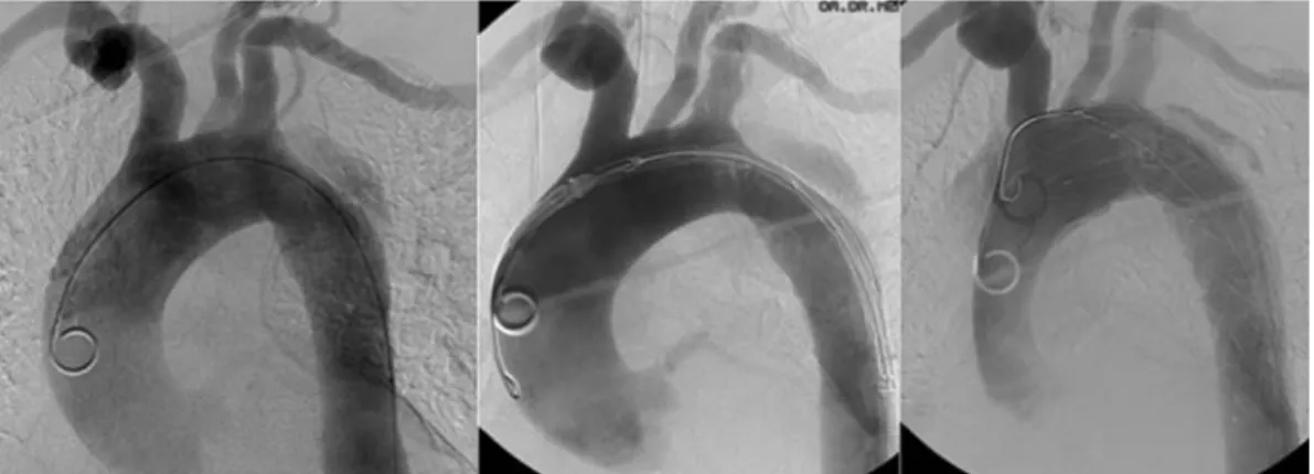
In cases of retrograde aortic type A dissection (Fig.1), we per-formed combined surgical and endovascular repair using a spe-cially designed hybrid prosthesis ( Jotec E-vita open).
Replacement of the ascending aorta, the aortic arch and the proximal descending aorta can be performed simultaneously by this combined single-session procedure.
The E-vita open hybrid device is composed of a vascular Dacron prosthesis in continuity with the self-expanding stent graft.
The arterial cannulation site was the right axillary artery via a short 8-mm Dacron prosthesis. Venous cannulation was intro-duced into the right atrium in the usual manner. All operations
were performed under moderate hypothermia (25–28°C).
Once a core temperature of 25°C had been achieved, the ascending aorta and the concave portion of the aortic arch were resected in a state of circulatory arrest. Selective antegrade cere-bral perfusion with 10 ml/kg/min cold blood was achieved via a catheter in the left carotid artery and the cannula in the right ax-illary artery after clamping the brachiocephalic artery. Oxygen saturation was measured by infrared spectroscopy during cere-bral perfusion. In one patient, the previously implanted stent graft could be extracted and removed easily from the descend-ing aorta. In the remaindescend-ing two patients the endovascular pros-thesis already in place at the descending aorta was shortened as far as possible.
As the next step, the E-vita open stent graft was introduced into the true lumen of the descending aorta or the remaining stent graft via the open aortic arch (Fig.2). The point of orienta-tion for placement of the stent was offspring of the subclavian artery. After antegrade implantation through the opened aortic arch into the descending aorta, the stent graft wasfixed to the aortic stump at the origin of the left subclavian artery using a continuous polypropylene suture supported with Teflon strips by a double-sandwich technique.
Figure 1:Retrograde aortic type A dissection during placement of a stent graft in a acute complicated type B dissection.
A O RTIC SURGER Y M. Gorlitzer
Reconstruction of the aortic arch with preservation of the supra-aortic branches and the convex portion of the aortic arch was performed. The ascending aorta and the concave portion of the aortic arch were replaced with an additional coated Dacron prosthesis and anastomosed to the reconstructed descending aorta [13,14]. The proximal anastomosis was performed at the sino-tubular junction preserving the aortic valve.
RESULTS
In three patients, the retrograde dissection was observed during the endovascular intervention in the angio suite. They were im-mediately transferred to the operating room for emergent surgi-cal conversion. All of these patients underwent combined surgical and endovascular repair of the affected aorta by the frozen elephant technique as described earlier [13,14]. Operative data are listed in Table1.
A valiant stent graft prosthesis was used in all three patients with retrograde type A aortic dissection. The site of the primary entry tear was at the concavity of the distal aortic arch in two patients, and at the convexity in one patient. In one patient who experienced retrograde-type dissection (median 44 vs. 37 mm), the diameter of the ascending aorta was remarkably larger and the distance from the primary entry tear to the left subclavian artery was shorter (median 9 vs. 15 mm) (Table2).
Operative data
The mean duration of circulatory arrest was 61 ± 3 min. The dur-ation of aortic cross-clamping and extracorporeal circuldur-ation was 92 ± 16 and 180 ± 27 min, respectively. Ventilation time was 2 ± 3 days, the stay at the intensive care unit 10 ± 8 days and the length of hospital stay 17 ± 15 days.
Clinical outcome
All patients survived the surgical procedure. No stroke, paraple-gia or other major neurological complications occurred. One patient developed renal failure requiring haemofiltration for 8 days. Postoperative CT scans revealed perigraft thrombus forma-tion and stable aortic dimensions in all patients after 6 months.
DISCUSSION
The technical feasibility and the low mortality and morbidity rates of TEVAR when compared with open surgical repair for the treatment complicated type B aortic dissection have been reported by several authors [2,3,15]. However, this endovascular intervention is not entirely devoid of risks. An increasing number of complications of endovascular therapy have been reported in the recent published literature [4–12].
Figure 2:The E-vita open stent graft is introduced into the remaining stent
graft located in the descending aorta via the open aortic arch.
Table 1: Descriptive characteristics of operated patients after rTAA
n, overall = 3 Demographics
Age, mean (SD), years 58 (±7)
Male,n (%) 1 (33%)
Operative data
Extracorporeal circulation time (min) 180 (±27)
Aortic cross-clamp time (min) 92 (±16)
Circulatory arrest time (min) 61(±3)
Total operation time (min) 330 (±125)
Postoperative ventilation time (days) 2 (±3)
Duration intensive care unit stay (days) 10 (±8)
Length of hospital stay (days) 17 (±15)
Unless otherwise indicated, data are number (percentage) (± standard deviation).
Table 2: Distances and diameters assessed by MSCT
Non-retrograde dissection (n = 26) Retrograde type A dissections (n = 3) Distance to subclavian artery (mm),
mean ± SD
15 ± 4.9 9 ± 4.1
Diameter descending aorta (mm), mean ± SD
37 ± 4.1 37 ± 5.5
True lumen diameter (mm), mean ± SD
17 ± 2.7 23 ± 1.5
False lumen diameter (mm), mean ± SD
20 ± 5.4 14 ± 7
Diameter abdominal aorta (mm), mean ± SD
30 ± 5.5 41 ± 5.2
Diameter ascending aorta (mm), mean ± SD
37 ± 3.5 44 ± 2.5
The underlying pathology of type B aortic dissection is a sys-temic illness. The natural progression of this aortic pathology, in-cluding expansion of the false lumen and the diameter of the aorta followed by further extension of the dissection or even rupture, has been observed in one-third of patients who under-went initial treatment 5 years after the condition was diagnosed [16,17].
The life-threatening situation of retrograde type A aortic dis-section after TEVAR is caused by stent-related manipulation and the fragility of the entire aortic wall [15].
One of the sources of potential injury to the aortic wall are the proximal bare springs which are designed to provide strong
radial force and ensure proximal fixation of the stent graft.
Retrograde type A dissections are most commonly caused by endovascular prostheses with proximal bare springs. [6,10, 15, 18]. Balloon dilatation bears the additional risk of a patient devel-oping further dissection in the fragile aortic wall [12,15,19].
In our series, a prosthesis with a proximal bare spring was used in 25 patients. The tip of the proximal bare springs induced a retrograde type A dissection in four patients. In the present series, we performed no balloon inflation on the dissected wall during placement of the stent graft.
The location of the primary entry tear influences retrograde type A dissection in acute type B dissections. In an experimental circulatory model, Dziodzio et al. [20] described retrograde propagation of the dissecting membranes when the primary entry tear was located at the concavity. The same has been reported for intramural haematoma. In contrast to the site of convexity, the site of concavity is devoid of anatomical barriers such as supra-aortic vessels [21].
In the recent cohort, the majority of patients experiencing retrograde dissection after TEVAR had a primary entry tear at the concavity of the distal aortic arch.
The shape and diameter of the ascending aorta must be taken into account in the treatment of complicated type B dissections. The dissection may not be limited to the descending aorta, but may involve the ascending aorta as well due to generalized propagation of this disease. Therefore, assessment of the ascend-ing aorta is of substantial importance in plannascend-ing effective and safe treatment of complicated type B aortic dissections. In our patient group, the dilated ascending aorta may influence the oc-currence of retrograde type A dissection after TEVAR as described in Table2.
The principal aims of surgery for an acute type A dissection by the conventional technique of replacement of the ascending aorta and the aortic arch are to ensure the patient’s survival and a low morbidity rate. The management of retrograde type A aortic dissection after TEVAR has not been standardized yet. Published reports focus on the treatment of retrograde type A aortic dissection in the ascending aorta, aortic arch replacement, or both, but do not address the lesion that develops into the antegrade or retrograde component of the dissection. In cases of type B aortic dissection, the site of the primary entry tear is located in the descending aorta and cannot be effectively elimi-nated by single replacement of the ascending aorta and the aortic arch.
The single-stage hybrid approach implies conventional open surgical treatment of the ascending aorta and the aortic arch combined with open antegrade stent grafting of the proximal descending aorta via a median sternotomy. A running suture line at the offspring of the left subclavian artery ensures closure of the primary lesion. The frozen elephant trunk technique thus
permits treatment of the underlying pathology in the proximal descending aorta [22].
In summary, this single-stage hybrid approach permits safe, ef-fective and simultaneous treatment of the ascending aorta, the aortic arch and the descending aorta in patients with acute com-plicated type B aortic dissection who undergo TEVAR and develop retrograde type A dissection. The procedure is asso-ciated with a good clinical outcome in patients with this poten-tially lethal complication. Based on these findings, the frozen elephant trunk procedure should be considered as a valuable option in patients with acute complicated type B aortic dissec-tion if associated with dilatadissec-tion of the ascending aorta or pos-sible potential components for retrograde type A dissection like a short distance to the subclavian artery or the concavity of the aortic arch as possible primary entry tear.
LIMITATIONS
Given the need for acute intervention in patients with compli-cated type B aortic dissection following retrograde type A dissec-tions after TEVAR, the present investigation was conducted as a retrospective intention-to-treat analysis.
Although the patient population is small (n = 3), the results support the thesis that retrograde type A aortic dissection can be effectively treated by means of a combined surgical and endovascular repair of the thoracic aorta. The small sample size was due to the low incidence of this disease. Taking into account the retrospective fashion of this study and the small number of patients, the enlargement of the ascending aorta and the short distance to the subclavian artery are the most notice-able parameters in the case of retrograde dissection and should be considered before stent graft implantation in type B
dissec-tions. A multi-centre study should be performed to confirm
thesefindings. Besides, a prolonged follow-up period will help to document the advantages of the method.
ACKNOWLEDGEMENTS
The authors would like to thank Meinhart from the Karl Landsteiner Institute for Cardiovascular Surgery Research for his support in developing this article.
Conflict of interest: none declared.
REFERENCES
[1] Pretre R, Von Segesser LK. Aortic dissection. Lancet 1997;349:1461–4.
[2] Dake MD, Kato N, Mitchell RS, Semba CP, Razavi MK, Shimono Tet al.
Endovascular stent-graft placement for the treatment of acute aortic dis-section. N Engl J Med 1999;340:1546–52.
[3] Nienaber CA, Fattori R, Lund G, Dieckmann C, Wolf W, von Kodolitsch Y et al. Nonsurgical reconstruction of thoracic aortic dissection by stent-graft placement. N Engl J Med 1999;340:1539–45.
[4] Misfeld M, Notzold A, Geist V, Richardt G, Sievers HH. Retrograde type A dissection after endovascular stent grafting of type B dissection. Z Kardiol 2002;91:274–77.
[5] Oertel F, Sirch J, Bockler D, Klein M, Raithel D, Beyer M. Delayed type A dissection after primary successful interventional treatment with endo-luminal stent graft of the descending thoracic aorta. Thorac Cardiovasc Surg 2002;50:180–1. A O RTIC SURGER Y M. Gorlitzer
[6] Pasic M, Bergs P, Knollmann F, Zipfel B, Muller P, Hofmann Met al. Delayed retrograde aortic dissection after endovascular stenting of the descending thoracic aorta. J Vasc Surg 2002;36:184–6.
[7] Fanelli F, Salvatori FM, Marcelli G, Bezzi M, Totaro M, Vagnarelli Set al.
Type A aortic dissection developing during endovascular repair of an
acute type B dissection. J Endovasc Ther 2003;10:254–9.
[8] Bethuyne N, Bove T, Van den BP, Goldstein JP. Acute retrograde aortic dissection during endovascular repair of a thoracic aortic aneurysm. Ann Thorac Surg 2003;75:1967–9.
[9] Neuhauser B, Czermak BV, Fish J, Perkmann R, Jaschke W, Chemelli A et al. Type A dissection following endovascular thoracic aortic stent graft
repair. J Endovasc Ther 2005;12:74–81.
[10] Fattori R, Lovato L, Buttazzi K, Di BR, Gavelli G. Extension of dissection in stent graft treatment of type B aortic dissection: lessons learned from endovascular experience. J Endovasc Ther 2005;12:306–11.
[11] Kpodonu J, Preventza O, Ramaiah VG, Shennib H, Wheatley GH III,
Rodriquez-Lopez Jet al. Retrograde type A dissectionafter endovascular
stenting of the descending thoracic aorta: is therisk real? Eur J Cardiothorac Surg 2008;33:1014–8.
[12] Totaro M, Miraldi F, Fanelli F, Mazzesi G. Emergency surgery for retro-grade extension of type B dissection after endovascular stent graft repair.
Eur J Cardiothorac Surg 2001;20:1057–8.
[13] Gorlitzer M, Weiss G, Thalmann M, Mertikian G, Wislocki W, Meinhart J et al. Combined surgical and endovascular repair of complex aortic path-ologies with a new hybrid prosthesis. Ann Thorac Surg 2007;84:1971–6. [14] Tsagakis K, Pacini D, Di Bartolomeo R, Gorlitzer M, Weiss G,
Grabenwoger Met al. Multicenter early experience with extended aortic
repair in acute aortic dissection: is simultaneous descending stent graft-ing justified? J Thorac Cardiovasc Surg 2010;14:S116–20.
[15] Dong ZH, Fu WG, Wang YQ, Guo da Q, Xu X, Ji Yet al. Retrograde type
A aortic dissection after endovascular stent graft placement for treatment
of type B dissection. Circulation 2009;119:735–41.
[16] Winnerkvist A, Lockowandt U, Rasmussen E, Rådegran K. A prospective study of medically treated acute type B aortic dissection. Eur J Vasc Endovasc Surg 2006;32:356–7.
[17] Hata M, Sezai A, Niino T, Yoda M, Wakui S, Unosawa Set al. Prognosis
for patients with type B acute aortic dissection: risk analysis of early
death and requirement for elective surgery. Circ J 2007;71:1279–82.
[18] Rubin S, Bayle A, Poncet A, Baehrel B. Retrograde aortic dissection after a stent graft repair of a type B dissection: how to improve the endovas-cular technique. Interact Cardiovasc Thorac Surg 2006;5:746–8. [19] Neuhauser B, Greiner A, Jaschke W, Chemelli A, Fraedrich G. Serious
complications following endovascular thoracic aortic stent-graft repair
for type B dissection. Eur J Cardiothorac Surg 2008;33:58–63.
[20] Dziodzio T, Juraszek A, Reineke D, Jenni H, Zermatten E, Zimpfer Det al.
Experimental acute type B aortic dissection: different sites of primary entry tears cause different ways of propagation. Ann Thorac Surg 2011;
91:724–7.
[21] Grimm M, Loewe C, Gottardi R, Funovics M, Zimpfer D, Rodler Set al.
Novel insights into the mechanisms and treatment of intramural hema-toma affecting the entire thoracic aorta. Ann Thorac Surg 2008;86:453–6. [22] Urbanski PP. Retrograde extension of type B dissection after
endovascu-lar stent graft repair. Eur J Cardiothorac Surg 2002;21:767–8.
APPENDIX. CONFERENCE DISCUSSION
Dr T. Schachner (Innsbruck, Austria): These results reported by you and the group around Martin Grabenwöger are important, on the one hand to show us the life-threatening character of complicated type B dissection and, on the other hand, that this complication with retrograde dissection is perhaps
under-reported in the literature. Myfirst question is about bare springs. I
re-member a case I operated on six months ago where I clearly saw a bare spring as a source of retrograde type A dissection following TEVAR. My ques-tion is, did you or do you recommend the use of completely covered stent grafts in this setting?
Dr Gortlitzer: This is a very interesting point regarding the causes of retro-grade type A dissections. There are several publications describing that bare springs induce retrograde type A dissections, and in our series, all cases with retrograde type A dissections had stent grafts with uncovered bare springs. So I
think the industry at least should be advised to produce specially designed stent grafts for the treatment of acute complicated type B dissections.
Dr Schachner: And did you undertake long-term follow-up of these
patients tofind out if there were perhaps cases of retrograde dissection that
contributed to late mortality or to a reoperation at another centre?
Dr Gorlitzer: Out of our 29 patients, we had a follow-up period of a median 15 months, and we did not observe any late retrograde type A dis-sections. That could be because the series is a little bit too small.
Dr Schachner: And my third question is about the interesting finding that the dilated ascending aorta was a risk factor and, if I understood you
correct-ly, yourflow chart indicates that if you have an ascending aorta of 4 cm or
more, you go for a frozen elephant repair, including ascending, arch and des-cending aortic replacement or repair. Is this true for all patients, or is this true for patients with connective tissue disease?
Dr Gorlitzer: Based on our experiences, we have developed this flow chart which is generally applied wherever possible. If the ascending aorta is enlarged, we assume that the whole aorta is sick and not only the descending aorta. In cases with an enlarged ascending aorta, we perform the frozen ele-phant technique.
Dr B. Zipfel (Berlin, Germany): I want to add a comment concerning the bare stents. There is multicentre Registry data published in Circulation by Dr. Eggebrecht from Essen, showing no clear correlation to the proximal end of the stent grafts; this is also our experience. We have also seen per-forations by the stent grafts as dissections remote from the stent grafts. So the reason is not really clear, and it is hard to say that whether this com-plication is related to a particular type of stent graft. As you pointed out, this complication seems to be related mainly to type B dissection as a
primary indication. This finding is similar in the Registry data and also in
our own experience.
What is your strategy if you have an entry very close to the subclavian artery or even involving the subclavian artery? In my experience, this seems to have a higher risk for stent graft-associated Type A dissection. Would this also be a reason for you to use the open stent graft technique?
Dr Gorlitzer: It is true that the distance to the subclavian artery influences the outcome of the treatment with endovascular devices. Overall, the series are too small to give guidelines or to develop a strategy depending on the distances to the subclavian artery, so usually we over-cover the subclavian artery.
Dr C. Etz (Leipzig, Germany): I would like you to speculate on the aetiology again. What do you think is the role of oversizing? Do you have any data regarding over-sized stent grafts or would that be something that you could just give us your opinion on?
Dr Gorlitzer: Usually we do not oversize in type B dissections. If indicated, we oversize to a maximum of 10% in acute type B dissections.
Dr Etz: So that could not be the reason for the retrograde type As? Dr Gorlitzer: No.
Dr C. Mestres (Barcelona, Spain): I think you raised a very important point with this contribution because, of course, there are so many things that we do not know yet; this may require further elaboration. It is my feeling that these anatomical factors that you mentioned (also mentioned in one of the previous papers), such as the distance to the left subclavian artery and the convexity and concavity, are going to play a very important role at the time of decision-making. I think that regardless of the type of disease, the problem is who treats those patients with complicated acute type B dissection, which is, by definition, a surgical disease, but which is managed by many other people who have no idea of surgery. This is one of the problems. I think that the value of your contribution is that if more information is collected in the future, it could be a major advance to go back to surgery. It is likely that we have to prove it in the future because I am absolutely sure that acute complicated type B dissection continues to be a surgical disease that has to be managed by cardiothoracic and vascu-lar surgeons rather than anybody else, and this is something that I encour-age you to follow.
Dr Gorlitzer: I completely agree with you. This is a disease which has to be managed by cardiac surgeons in a multidisciplinary setting.
Dr H. Jakob (Essen, Germany): I just want to ask you about the so-called
ag-gressive hemiarch, or as Martin Grabenwöger says,‘arch light’. You leave in
diseased tissue. Have you ever seen problems later on after this kind of ap-proach? I personally would prefer to be radical.
Dr Gorlitzer: Yes, I know. But we did not see any problems after leaving the eyelet. We published this as a light arch replacement, and we did not have any problems after this repair.