British Journal of Anaesthesia 92 (3): 419±21 (2004)
DOI: 10.1093/bja/aeh062 Advanced Access publication January 22, 2004
Concentration of rocuronium in cerebrospinal ¯uid of patients
undergoing cerebral aneurysm clipping
²
T. Fuchs-Buder
1*, M. Strowitzki
2, K. Rentsch
3, J. U. Schreiber
1, S. Philipp-Osterman
1and
S. Kleinschmidt
11
Department of Anaesthesia and Critical Care and
2Department of Neurosurgery, University of Saarland,
Homburg/Saar, Germany.
3Institute for Clinical Chemistry University Hospital of Zurich, Switzerland
*Corresponding author: DeÂpartement d'AnestheÂsie et de ReÂanimation, Centre Hospitalier Universitaire Nancy/ Brabois, 4 Rue du Morvan, F-54500 Vandoeuvres-Les-Nancy, France. E-mail: [email protected]
Background. This study assessed the concentration of rocuronium in the cerebrospinal ¯uid (CSF) of patients undergoing cerebral aneurysm clipping, and investigated whether the mode of administration (single bolus vs continuous infusion) in¯uenced the CSF concentration.
Methods. Twenty patients with subarachnoid haemorrhage were randomly allocated to receive a bolus dose (bolus group), or a bolus followed by a continuous infusion of rocuronium (infusion group) (n=10 for each group). Arterial blood and ventricular CSF were sampled 2 h after the rocuronium bolus. Samples were analysed by liquid chromatography electrospray ionization-tandem mass spectrometry.
Results. Rocuronium could be detected in all the CSF samples. The mean (range) CSF concen-tration was 2.2 (0.9±4.6) ng ml±1in the bolus group and 12.4 (2.4±34.6) ng ml±1in the infusion group; P<0.01.
Conclusions. This study demonstrated that rocuronium, normally not considered to cross the blood±brain barrier, is regularly found in the CSF of patients undergoing cerebral clipping; continuous infusion of the drug led to higher plasma and CSF concentrations than after a single bolus dose.
Br J Anaesth 2004; 92: 419±21
Keywords: blood±brain barrier, mass spectrometry high pressure liquid chromatography; neuromuscular blocking agents, rocuronium; surgery, cerebrovascular
Accepted for publication: July 8, 2003 Neuronal nicotinic acetylcholine receptors (nAChR) are ubiquitously present in CNS and involved in chemical signalling in the brain.1When given in suf®cient amounts, neuromuscular blocking agents (NMBA) interact with these nAChR, leading dose dependently to activation or inhibition of different nAChR subtypes.2 To estimate the clinical relevance of this interaction, quantitative data on the cerebrospinal ¯uid (CSF) concentration of NMBAs are required. Whilst NMBAs are considered not to cross the intact blood±brain barrier, these molecules may pass into the CSF when its function is impaired.3 4 Only a little information about re-entry of rocuronium, a NMBA used in neurosurgical patients, into the CSF is available. This, study aimed ®rst to investigate the CSF concentration of rocuronium in patients undergoing cerebral aneurysm clipping, and secondly to assess whether the mode of
administration (single bolus or continuous infusion) has any in¯uence on its CSF concentration.
Methods and results
The study was approved by the hospital Ethics Committee; the patients or their next of kin gave informed consent or assent, respectively. Twenty patients undergoing cerebral aneurysm clipping with CSF drainage were randomly allocated to the bolus group or infusion group. Anaesthesia was induced with thiopentonal (3±5 mg kg±1), fentanyl (1±2 mg kg±1), and rocuronium (0.6 mg kg±1); patients in the infusion group also received a continuous ²Declaration of interest: This study was supported in part by a grant from Organon, The Netherlands.
infusion of rocuronium to maintain constant neuromuscular block, that is 90±95% twitch depression. Anaesthesia was maintained with continuous infusions of midazolam (0.05± 0.2 mg kg±1 h±1) and fentanyl (2±5 mg kg±1 h±1). The patients' lungs were ventilated to normocarbia with iso¯urane 0.5% and nitrous oxide 60% in oxygen. Before the beginning of the craniotomy, an external CSF drainage was placed into the lateral ventricle by the neurosurgeon via a single bore hole approach.
Baseline arterial blood samples (5 ml), needed to control to linearity of the analytical method, were taken before induction of anaesthesia. Baseline CSF samples were obtained from patients undergoing elective surgery under spinal anaesthesia. In the ®rst ®ve of 10 patients in each group, blood and CSF were sampled at 1, 2, and 4 h after the initial dose of rocuronium. In the second ®ve patients, blood and CSF were sampled at 2 h. After collection, the samples were centrifuged at 4°C, and 1 ml of plasma or CSF was added to tubes containing 0.2 ml of 1 M sodium hydrogen phosphate solution also as a stabilizer. These samples were kept frozen at ±70°C until analysis. The concentration of rocuronium was determined by liquid chromatography electrospray ionization-tandem mass spectrometry.5 The detection limit of the method was 0.5 ng ml±1 for rocuronium in CSF and 5 ng ml±1for rocuronium in plasma, and the inter-assay coef®cient of variation was less than 8%. Data are expressed as individual values or as median or mean and range. Patient characteristics and rocuronium concentrations at 2 h in the two groups were compared using the Mann±Whitney test. The time course of the plasma and CSF concentration of rocuronium was compared with a one-wayANOVAfor repeated measures followed by a Bonferroni
correction. Differences were considered signi®cant when P<0.05.
One patient in the infusion group had to be excluded because of violation of the study protocol. The physical characteristics and surgical data were comparable between the two groups (Table 1). Preliminary analysis after 10 patients (®ve in each group) revealed that in the infusion group the concentration of rocuronium was highest after 2 h; the corresponding values at 1, 2, and 4 h were 3.8 (2.4±5.3), 5.7 (2.4±13.2) (P<0.05 compared with 1 h), and 3.8 (1.1± 7.5) ng ml±1, respectively, n=5. The respective values in the bolus group were 2.2 (0.6±7.5), 2.0 (0.9±4.5), and 1.8 (0.7± 4.7) ng ml±1; n.s, n=5. The ®nal analysis demonstrated that the CSF concentration of rocuronium at 2 h was 2.2 (0.9± 4.6) ng ml±1in the bolus group (n=10) and 12.4 (2.4±34.6) ng ml±1in the infusion group (n=9); P<0.01 (Table 1).
Discussion
In the present study rocuronium could be detected in all CSF samples, and a continuous infusion led to higher CSF concentrations than a single bolus dose. In the ®rst ®ve of 10 patients in each group, the time course of CSF penetration of rocuronium was assessed; we focused on a 4-h period
because it has recently been demonstrated that in the patient population studied the CSF concentration of NMBA may be highest at this time point.4Based on the results of the ®rst part of the study blood and CSF were only sampled at 2 h in the second ®ve of 10 patients in each group.
Our results are surprising, as in previous studies NMBAs were not regularly detected in the CSF. Tassonyi and co-workers found atracurium in the CSF of only ®ve out of 10 patients undergoing cerebral aneurysm clipping.4However, their lower limit of detection was 10 ng ml±1. In the present study the lower detection limit for rocuronium in the CSF was 0.5 ng ml±1, allowing quanti®cation of very small amounts of the drug. Moreover, most studies investigating the CSF concentration of NMBA have sampled the CSF in the lumbar subarachnoid space.3 4Circulation of CSF from the brain to the lumbar subarachnoid space takes about 90 min, and approximately 0.25% of total CSF volume is replaced by freshly formed CSF each minute. Thus, sampling of CSF from the lumbar subarachnoid space re¯ects neither the time course of the passage of NMBA into the CSF or its maximum concentration in the CNS. In the present study, the low detection limit of the analytical method and sampling directly from the ventricular system may have contributed to the consistent section of rocuronium in the CSF.
There is controversy in the literature about the origin of NMBA detected in the CSF. Eddlestone and co-workers3 thought that the presence of atracurium in the CSF in two out of 10 patients (80 and 1460 ng ml±1) could be accounted for by blood contamination. This factor cannot be excluded in our study. However, CSF containing haemolysed blood was only observed in one patient. Moreover, the CSF concentrations of rocuronium were dose-dependent, a continuous infusion leading to higher concentrations than a single bolus, and in both groups the CSF concentration was within a narrow range (Table 1).
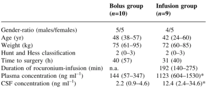
Table 1 Physical characteristic, surgical data, and rocuronium plasma and CSF concentrations. Data are absolute values (sex ratio), mean or median (range, orSD); n.a., not applicable; *P<0.01. Plasma and CSF concentrations were determined 2 h after the initial dose of rocuronium. Hunt and Hess classi®cation: 0, unruptured aneurysm; 1, asymptomatic or minimal headache and slight nuchal rigidity; 2, moderate to severe headache, nuchal rigidity, but no neurological de®cit other than cranial nerve palsy; 3, drowsiness, confusion, or mild focal de®cit; 4, stupor, mild to severe hemiparesis, possible early decerebrate rigidity, vegetative disturbance; 5, deep coma, decerebrate rigidity, moribund appearance.10Time to surgery: time between
diagnosis of subarachnoid haemorrhage and aneurysm clipping
Bolus group Infusion group (n=10) (n=9) Gender-ratio (males/females) 5/5 4/5
Age (yr) 48 (38±57) 42 (24±60)
Weight (kg) 75 (61±95) 72 (60±85)
Hunt and Hess classi®cation 2 (0±3) 2 (0±3) Time to surgery (h) 40 (57) 31 (40) Duration of rocuronium-infusion (min) n.a. 192 (140±275) Plasma concentration (ng ml±1) 144 (57±347) 1123 (604±1530)*
CSF concentration (ng ml±1) 2.2 (0.9±4.6) 12.4 (2.4±34.6)*
Fuchs-Buder et al.
There is a dose-dependent relationship between an NMBA causing inhibition or activation of nAChR. Thus, the possible clinical consequences of the CSF concentration of rocuronium measured in this study have to be considered. Cholinergic neurotransmission plays a role in regulation of respiratory pattern, and inhibition of nAChR may lead to central respiratory depression.6As recently shown, NMBAs may inhibit the major brain a4b2 human nAChR,2and thus lead to central respiratory inhibition. However, the concen-trations required to produce inhibition of nACh receptors were in the mM range, being about 1000-fold higher than the concentrations detected in this study.2 NMBAs may also produce excitatory CNS effects. Pancuronium and vecuronium may cause a sustained increase in cytosolic calcium by initiating prolonged activation of brain nAChR, thus leading to uncontrolled neuronal excitability and seizure activity.7 Case reports of accidental intrathecal injection of NMBAs con®rmed this property.8 9However, about 10 000-fold higher concentrations than those measured in this study were necessary to elicit seizures.6 Thus, according to the current literature, no convincing evidence exists of major CNS effects from the concentra-tions of rocuronium found in the CSF in this study.
References
1 Tassonyi E, Charpantier E, Muller D, Dumont L, Bertand D. The role of nicotinic acetylcholine receptors in the mechanisms of anesthesia. Brain Res Bull 2002; 57: 133±50
2 Chiodini F, Charpentier E, Muller D, Tassonyi E, Fuchs-Buder T, Bertrand D. Blockade and activation of the human neuronal nicotinic acetylcholine receptors by atracurium and laudanosine. Anesthesiology 2001; 94: 643±51
3 Eddlestone JM, Harper NJN, Pollard BJ, Edwards D, Gwinnutt CL. Concentrations of atracurium and laudanosine in cerebrospinal ¯uid and plasma during intracranial surgery. Br J Anaesth 1989; 63: 525±39
4 Tassonyi E, Fathi M, Hughes GJ, et al. Cerebrospinal ¯uid concentrations of atracurium in neurosurgical patients. Acta Anaesthesiol Scand 2002; 46: 1236±41
5 Gutteck-Amsler U, Rentsch KM. Quanti®cation of the aminosteroidal non-depolarizing neuromuscular blocking agent rocuronium and vecuroniumin plasma with liquid-chromatography-tandem mass spectrometry. Clin Chem 2000; 46: 1413±5
6 Shao XM, Feldman JL. Pharmacology of nicotinic receptors in the preBotzinger complex that mediate modulation of respiratory pattern. J Neurophysiol 2002; 88: 1851±8
7 Cardone C, Szenohradszky J, Yost S, Bickler PE. Activation of brain acetylcholine receptors by neuromuscular drugs. Anesthesiology 1994; 80: 1155±61
8 Goonewardene TW, Sentheshanmuganathan S, Kamalanathan S, Kanagasunderam R. Accidental subarachnoid injection of gallamine: a case report. Br J Anaesth 1975; 47: 889±93 9 Foster PA. Potassium depletion and the central action of curare.
Br J Anaesth 1956; 28: 488±94
10 Hunt WE, Hess RM. Surgical risk as related to time of intervention in the repair of intracranial aneurysms. J Neurosurg 1968; 28: 14±20
MAC of xenon in swine