Published online 20 July 2005
Original article
Scope and significance of non-uniform classification practices in
breast cancer with non-inflammatory skin involvement: a
clinicopathologic study and an international survey
U. Gu¨th
1* , G. Singer
2, A. Scho¨tzau
3, I. Langer
4, H. Dieterich
5, C. Rochlitz
6, L. Herberich
1,
W. Holzgreve
1& E. Wight
1Departments of1Gynecology and Obstetrics,4Surgery and6Medical Oncology, University Hospital Basel, Switzerland;2Institute of Pathology, University of Basel, Switzerland;3JPS Institute for Biomathematics, Basel, Switzerland;5Women’s Hospital and Breast Center Rheinfelden, Rheinfelden, Germany Received 2 May 2005; revised 13 June 2005; accepted 14 June 2005
Background:The study evaluates the scope of non-uniform classification practices concerning breast carcinomas with non-inflammatory skin involvement.
Patients and methods: We compared the clinical course of patients with histologically proven non-inflammatory skin involvement: 119 (65.4%) with clinically obvious ‘classical’ skin changes (Group A) and 63 (34.6%) with no or only discreet changes (Group B). A questionnaire was circulated to path-ology departments in 24 countries to assess the practice concerning the placement of skin- involved breast carcinomas in the TNM classification.
Results: Patients in Group B showed a significantly better disease specific survival (P = 0.0002). Eighty-six respondents (70.5%) of the survey preferred the ‘histological view’ and classified tumors with only histological proven skin involvement as T4b/stage IIIB. The opposing classification principle (‘clinical view’), which dictates that T4b breast cancer is a clinical diagnosis and the classical signs must be present, was supported by 31 respondents (25.4%).
Conclusions:A large number of breast cancer patients with non-inflammatory skin involvement are only histologically proven and show, compared with cases exhibiting the classical clinical signs, sig-nificant differences in clinical course and prognosis. In general, both subsets were aggregated in one T category/stage (T4b/IIIB). This results in a considerable distortion of the reported statistical data. Key words:breast carcinoma, skin involvement, TNM classification, prognostic factors, survey
Introduction
According to the current edition of the American Joint Commit-tee on Cancer (AJCC)/International Union Against Cancer (UICC) TNM (tumor–node–metastasis) staging system [1, 2], non-inflammatory breast carcinomas with direct extension to the skin are classified as T4b lesions. These tumors, eliminating cases with distant metastasis, are included in stage III (stage IIIB: T4 N0–2 M0, stage IIIC: any T N3 M0), which is considered to be synonymous with locally advanced breast cancer (LABC).
Only tumors accompanied by macroscopic and typically read-ily discernible ‘classical’ skin changes, such as ulceration, edema, peau d‘orange and satellite skin nodules, should be placed in the T4b category. It is stated explicitly that discreet skin changes, such as dimpling, retraction of the nipple and other changes, often caused by shortening of Cooper’s ligaments
due to infiltration by malignant disease, may occur in T1–3 disease and, therefore, do not allow classification in the T4 category. Tumors that showed histologically proven skin in-volvement but not the accompanying ‘classical’ clinical changes have been in a grey area of the TNM nomenclature and allowed a certain leeway in the interpretation of these cases. It was only in 2001 that the 2nd edition of the TNM Supplement [3] estab-lished how to classify this subgroup of cases within the T cat-egory. It is required that for a lesion to be classified as T4b, the previously mentioned clinical (macroscopic) features must be present. Microscopic invasion of the dermis alone, without the accompanying classical clinical signs is not sufficient for plac-ing a lesion in the T4b category and the T classification is based solely on tumor size (T1–3).
The goal of the current review was, on one hand, to assess via an international survey the classification practice of breast can-cer cases with non-inflammatory skin involvement in the period before precise recommendations were drafted (in the 1990s). On the other hand, we demonstrate distinct clinical entities with significant differences in terms of long-term clinical outcome
*Correspondence to: Dr U. Gu¨th, University Hospital Basel, Department of Gynecology and Obstetrics, Spitalstrasse 21, 4031 Basel, Switzerland. Tel:+41-61-2659028; Fax: +41-61-2659037; E-mail: [email protected] U. Gu¨th and G. Singer contributed equally to the work.
of patients who exhibit histologically proven non-inflammatory skin involvement with and without classic clinical signs. Our report shows that a lack of uniformity in the classification of these cases results in considerable distortions in the epidemio-logical picture of T4b breast cancer.
Patients and methods
The clinicopathologic study
Between January 1988 and August 1999, 184 women with newly diagnosed breast carcinoma and histologically proven non-inflammatory skin involve-ment who had no local recurrence or a history of contralateral breast cancer were evaluated and treated at the Department of Gynecology and Obstetrics of the University Hospital Basel (Basel, Switzerland), the Department of Surgery of the University Hospital Basel, and the Gynecological Hospital and Breast Center Rheinfelden (Rheinfelden, Germany). Patients with Paget’s disease and inflammatory carcinoma (criteria: tumors affecting at least one-third of the breast, showing clinically the simultaneous presence of diffuse edema, erythema, warmth, tenderness and skin biopsy revealing lymphangiosis carcinomatosa) were excluded from the study.
Bilaterality, multicentricity and male gender were also exclusion criteria. Two patients who presented with additional histologically proven chest wall involvement (pT4c) were not considered in the analysis.
The data of 182 breast cancer patients (accounting for 7.8% of all newly diagnosed breast carcinomas within the study period) were the basis of the current analysis. Staging was performed for all patients in accordance with the current (6th) edition of the UICC/AJCC TNM classification. As a second step, reclassification was undertaken to assess the disease stage (tumor size and lymph node involvement) of tumors independent of the morphologic parameter ‘skin involvement’. This clinical/histopathological feature was no longer taken into consideration and was eliminated from our classification. All tumors were reclassified based on tumor size and, therefore, the category T4b was replaced with the categories T1–3. In this manner, patients who had stage IIIB disease underwent restaging.
Based on the clinical degree of skin involvement reported in the patient’s records, all patients were placed into one of two groups. One hundred and nineteen patients (65.4%) presented with clinically obvious classical skin changes (ulceration, edema, peau d‘orange and satellite skin nodules), and therefore fulfilled the current criteria for the T4b classification (Group A). Sixty-three patients (34.6%) had histologically proven skin involvement but no or only discreet (e.g. retraction or dimpling) clinical changes to the over-lying skin or the nipple (Group B).
Histopathological analyses were performed at the Institute of Pathology, University of Basel. A differentiation between pathological skin involve-ment of the epidermis and the dermis was not conducted. Histopathological analyses also included grading according to the Bloom–Richardson–Elston scheme and immunohistochemical staining for estrogen and progesterone receptors. Each patient underwent a staging work-up, which included a re-cording of clinical history, physical examination, routine blood studies, chest X-ray, sonography of the liver and additional diagnostic studies that were needed to rule out metastatic disease. To discuss the clinical and pathological features, all cases were evaluated in a multidisciplinary tumor board. There was no standard therapeutic approach during the study period (Table 3). All patients were followed until their death or for a minimum of 5 years if they remained alive. These patients were seen a maximum of 3 months before conclusion of the study.
Statistical methods
Using the Kaplan–Meier method, disease-specific survival (DSS) was cal-culated from the date of diagnosis to the date of death, or for patients who
remained alive, to the date of last follow-up. Non-malignancy-related deaths were censored in the statistical analyses according to the same method used for patients who were alive and disease-free. Statistical differences between groups in terms of survival curves were analyzed using the log-rank test. Comparisons between nominal parameters were made with the Fisher exact test. Statistical analyses were performed with SPSS 12.0 software (SPSS Inc., Chicago, IL).
The survey
A questionnaire was designed to assess the practice concerning the place-ment of breast cancer cases with non-inflammatory skin involveplace-ment within the TNM classification (Table 1). Four distinctive clinicopathological con-stellations of skin involvement, including different degrees of clinical skin changes, had to be assessed as to whether the criteria of the T4b category were fulfilled (answer choice: T4b), or if the criteria were not fulfilled and the case had to be classified according to tumor size (answer choice: T1–3). An invitation to contribute to this international survey was targeted at specialists in breast pathology of pathology departments of universities, university hospitals or associated hospitals. The questionnaire, together with an accompanying letter, was circulated by e-mail or fax. Most of the col-leagues were also personally contacted by telephone. Replies could be sent in by e-mail, fax or post. Data collection started in September 2004 and ended in December 2004. Replies were received from the USA, Canada, Australia, New Zealand, Asia (Hong Kong, Japan) and Europe (Austria, Belgium, Denmark, France, Finland, Germany, Greece, Hungary, Italy, Ire-land, Luxembourg, the Netherlands, Norway, Portugal, Spain, Sweden, Switzerland and the UK). Of the 150 institutions replying to the question-naire, 28 did not use the TNM classification in their reports. In these insti-tutions, the pathologists only gave morphologic descriptions of the gross and histological extent of skin involvement, leaving the placement within the TNM classification to the surgeon or oncologist.
Table 1. Survey: questionnaire for the use of TNM classification in breast cancer with non-inflammatory skin involvement
Question:
How did you classify breast carcinomas with non-inflammatory skin involvement using the T category at your institution in the years 1990–2000?
There are the following four constellations:
pT4b pT1-3* A. histological: skin involvement
clinical: unambiguous skin changes (e.g. exulceration, edema) B. histological: skin involvement
clinical: subtle or minor skin changes C. histological: skin involvement
clinical: no skin changes
D. histological: no evidence of skin involvement clinical: suggestive skin changes *pT1–3: T category according to tumor size
Please answer the four constellations as follows: pT4b pT1–3 yes no or no yes
Results
The clinicopathologic study
Patient, tumor and treatment characteristics of the 182 breast cancer patients with histologically proven non-inflammatory skin involvement are summarized in Tables 2 and 3 and in Figure 1. The median age of patients was 73 years in both groups. The mean tumor diameter in group A was significantly higher (6.1 cm versus 3.1 cm; P <0.0001). The distribution of
disease stages according to current UICC/AJCC criteria, after disregarding skin involvement (T4) in favor of tumor size (T1–3), is shown in Figure 1. In comparison to study group B, the patients with classical clinical skin involvement (group A) presented significantly more often in advanced stages (stage IIIC/IV; P <0.0001).
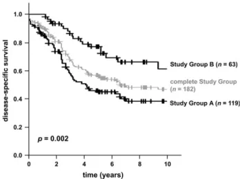
The clinical outcome of patients with only histologically proven skin involvement (group B) was significantly superior (P = 0.0002) to that of group A patients (Figure 2). The 5-year adjusted survival rates were 46.1% in group A and 77.1% in group B; the 10-year rates were 38.4% and 61.7%, respectively.
The survey
One hundred and twenty-two institutes replied to the question-naire and assessed the four clinicopathologic constellations in terms of TNM classification as shown in Table 4. Six answer combinations of the four constellations were offered. The
Table 2. Patient/tumor characteristics
Characteristic Group A Group B Total no. of patients (%) 119 (65.4) 63 (34.6) Age (years)
Median 73 73
Range 40–93 38–90 Premenopausal status (%) 18 (15.1) 9 (14.3) Follow-up time (months)
Median 37 64 Range 1–199 16–198 Tumor size (cm) Mean 6.1 3.1 Range 1.1–21.0 0.7–12.0 Histological grade (%) Grade 1 2 (1.7) 3 (4.7) Grade 2 46 (38.7) 26 (41.3) Grade 3 66 (55.5) 34 (54.0) Unknown 5 (4.2) 0 Hormone receptor status (%)
ER-positive 96 (80.7) 54 (85.7) ER: estrogen receptor
Table 3. Treatment data
Treatment type Group A Group B Surgery (%)
Lumpectomy+ axillary dissection 4 (3.4) 12 (19.0) Mastectomy+ axillary dissection 85 (71.4) 38 (60.3) Simple mastectomy 19 (16.0) 8 (12.8) Tumor excision 3 (2.5) 5 (7.9) No surgery 8 (6.7) 0 Systemic therapy (%) Postoperative chemotherapy 20 (16.8) 18 (28.6) Preoperative chemotherapy 15 (12.6) 2 (3.2) Postoperative hormonal therapy 87 (73.1) 47 (74.6) Preoperative hormonal therapy 5 (4.2) 1 (1.6) Radiation therapy (%) 28 (23.5) 20 (31.7) No pre-/postoperative therapy (%) 18 (15.1) 8 (12.8)
Figure 1. Distribution of UICC/AJCC TNM stage groupings among 182 breast cancer patients with non-inflammatory skin involvement. The parameter ‘skin involvement’ (T4 category) was disregarded, and all tumors were placed in the T1, T2, or T3 category.
Figure 2. Disease-specific survival (DSS) among 182 patients with breast cancer and histologically proven skin involvement (P = 0.0002). +: censored.
distribution by percentage is listed in Figure 3. Two centers (1.6%) reported not using the T4 category in cases of non-inflammatory skin involvement. All tumors were classified according to their size.
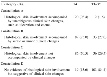
The key question to assess the classification principles con-cerning breast cancer with non-inflammatory skin involvement was Constellation C (histological skin involvement not accom-panied by clinical changes). Eighty-six respondents (70.5%) reported that they also use the T4b category based on histological features alone (‘histological view’). The opposing classification principle dictates that breast cancer with non-inflammatory skin involvement is a clinical diagnosis and the classical signs (ulceration, edema, peau d‘orange) must be present (Constel-lation A: T4; Constel(Constel-lation B and C: T1–3). This ‘clinical view’ was supported by 31 respondents (25.4%).
Discussion
A fundamental weakness of the majority of studies investigating incidence and outcome for non-inflammatory types of T4 breast cancer [4–18] is the lack of clearly defined inclusion criteria
concerning interpretation of the feature ‘skin involvement’ within the T4 category. In particular, the study protocols often do not specify whether cases with histologically proven skin involvement but no corresponding clinical picture were also regarded as T4b and included in the analyses. This, however, would have been important. The results of the clinical part of our study demonstrate that breast cancer cases with only histological skin involvement and no corresponding clinical features, compared with cases exhibiting the classical advanced local extent, are distinct entities with significant differences in clinical course and prognosis. These findings confirm our results of a one-center study evaluating 76 patients with non-inflammatory skin involvement [5].
Within the entire group of patients with non-inflammatory skin involvement, the subset of patients without the classical clinical signs is not negligible, but accounts for 35% of cases. The aggregation of both entities within one category violates the rules of the stage model upon which the TNM system is based. The TNM concept means that only clearly defined homogenous entities with similar prognostic impact may be placed together in one category/stage and placement in a higher category/stage generally corresponds to a poorer prognosis. When both entities are aggregated in one category, considerable heterogeneity of the T4b category with a broad distribution of cases among the subsets of disease stages is inevitable. Approximately 70% of the cases without the clinical features (group B) have malignant locoregional extent of TNM stage I/II. These patients run the risk of being falsely regarded as having more advanced disease. The results of our survey show that historically the majority of both distinct entities were not differentiated in terms of tumor classification. A total of 25.4% of the respondents reported that breast cancer cases with skin involvement were recorded in the ‘adjusted’ form (only cases with histologically proven skin in-volvement accompanied by classical clinical changes were classified as T4b). The majority of the respondents (70.5%), however, also classified tumors with only histological proven skin involvement but without the clinical correlate as pT4b/stage IIIB. Due to the non-uniformity in the practice of classification, heterogeneous data from this T category/stage, which are retro-spectively difficult to control and reproduce, were compiled in the tumor registries and database collections. Our survey en-compasses the years from 1990 to 2000, a period from which a large part of the currently accepted epidemiological data is derived. Therefore, while interpreting the data of the T4 cate-gory and stage III (especially T4b catecate-gory and stage IIIB), distortions could be present resulting from the fact that tumors with more favorable prognoses were falsely classified, from the current view, as T4.
The survey reveals two incompatible points of view concern-ing the classification in the TNM system of breast cancer cases with non-inflammatory skin involvement: the histological and the clinical point of view. The clinical view defined T4 carci-noma to be a clinical diagnosis where the typically readily discernible skin changes, such as exulceration, edema, peau d‘orange and satellite skin nodules, must be present. Advocates of the histological view were of the opinion that clinical staging may be performed in some cases, but pathologic staging is more
Table 4. The survey: the assessment of four clinicopathologic constellations of the participating centers
T category (%) T4 T1–3* Constellation A
Histological skin involvement accompanied by unambiguous clinical skin changes, such as ulceration and edema
120 (98.4) 2 (1.6) Constellation B
Histological skin involvement accompanied by subtle or minor clinical changes
89 (73.0) 33 (27.0) Constellation C
Histological skin involvement not accompanied by clinical changes
86 (70.5) 36 (29.5) Constellation D
No evidence of histological skin involvement but suggestive of clinical skin changes
19 (15.6) 103 (84.4) *T1–3: T category according to tumor size.
Figure 3. Distribution of the six answer combinations of the four (A–D) clinicopathologic constellations of the survey.
accurate [19], and also classified tumors with only histological skin involvement and no corresponding clinical features in the T4 category. Through a lack of clear guidelines in the TNM classification, there were ambiguities and a certain amount of leeway concerning the definition of ‘skin involvement’ in the past. These inconsistencies are reflected in six different answers of the four clinicopathological constellations in our survey. When the TNM Supplement in 2001 established recommenda-tions about how to use the TNM classification uniformly in these cases, the dispute seemed to be resolved in favor of the clinical view. It was once again substantiated that only the recognized classical clinical signs (edema, peau d ‘orange or ulceration of the skin, satellite skin nodules) are to be classified as T4. Any other skin changes, as well as microscopic invasion of the skin (dermis) without the above-mentioned clinical features, do not affect the classification [3]. Our survey demonstrates that the clinical view that is currently accepted as valid was put into practice by only a minority of the respondents (25.4%) as a basis for classification. Nevertheless, this recommendation also allows for leeway of interpretation. The following example shows that the confusion concerning classification principles could not be entirely dispelled. An enquiry was posed to the AJCC in summer 2004 on how to classify an invasive ductal carcinoma with histological infiltration of the epidermis but no associated classical clinical signs. The curator stated that direct skin invasion is defined as full thickness involvement including the epidermis; if the epidermis is intact with only focal dermal involvement, then it is not considered to be T4 but classified by the size of the primary tumor (G. MacGrogan, personal com-munication). This statement emphasizes again a histological feature, i.e. skin involvement of the epidermis to the border of deeper skin layers, as being crucial for classification. This view is only an individual interpretation of the TNM Supplement and cannot be by all means supported by the TNM nomenclat-ure or in the literatnomenclat-ure.
The following critical points of the survey must be discussed. (1) The definition of ‘histological skin involvement’ was not specified exactly. Three colleagues replied that the depth of skin involvement (dermis versus epidermis) is the crucial point for confirming histological skin involvement. In addi-tion, for some pathologists, the invasion of the lymphatic vessels by tumor was of pathognomonic significance. How-ever, without the clinical picture of inflammatory carcinoma this feature is generally not considered in the T category; these cases are classified by the size of the tumor [20]. (2) In Constellation B of our survey, subtle or minor skin
changes as a clinical feature without further specifications had to be assessed. Our goal in posing this question was to investigate (and obviously most colleagues also had this understanding) how the classification principles were im-plemented with respect to the clinicopathological changes described in the TNM nomenclature as being dimpling or retraction of the skin. We have deliberately foregone the use of the literal quotation of the TNM text, so as not to formu-late the question in an all-too suggestive manner. Further-more, Constellation B is of secondary importance. The
crucial question that shows whether the clinical or the histological view is expressed, is the question of how cases are classified when there is histological skin involvement without any clinical changes (Constellation C).
(3) The survey was targeted only to pathologists and not to clinicians. Potentially, surgeons or oncologists, especially in facilities where the pathologists do not report the TNM classification, could have proposed more clinical views in the interpretation of the four clinicopathologic constella-tions. On the other hand, it is undoubtedly incorrect that the histological view is a typical perception of only pathol-ogists. In cases of doubt, many clinicians do not put trust in the clinical aspect, but prefer the histological view and con-cede that a microscopically verified finding, not identified by the naked eye in preoperative physical examination, may have been overlooked.
(4) A survey, generally, cannot provide an exact picture of clas-sification practices. Our survey demonstrates, however, a clear trend that worldwide non-uniform classification principles were used in the interpretation and classification of breast cancer with non-inflammatory skin involvement. Although our survey depicted the classification practices of the 1990s, we believe that our results also mirror the current standard. Most participants were contacted personally by phone to introduce the study questions. A frequent answer given was that classification of breast cancer with non-inflammatory skin involvement, lacking the classical clinical signs, is a well known and often discussed, but unresolved, problem. We had the impression that the contents of the TNM Supplement concern-ing this subject was often not recognized or put into practice. Only three respondents supplemented the questionnaire with a commentary that classification principles had changed in their departments since the year 2000.
The following factors could also lead, regardless of clear guidelines, to persistent non-uniform use of the TNM classifi-cation for breast cancer with non-inflammatory skin involve-ment in the future.
(1) In the TNM Supplement the clinical view has become the standard for the classification of these tumors. This means that the finding of the clinician has a higher diagnostic value than results of the microscopic examination. This attitude broke with the generally accepted fundamental principle that pathologic staging is more accurate. The clinical view in the TNM nomenclature is a unique exception. Unique excep-tions of classification rules run the risk, notably in the spe-cialized subset as breast carcinoma with non-inflammatory skin involvement, not to be put into practice.
(2) Some clinical pictures are unclear and cannot be easily clas-sified based on the rigid definitions of the TNM classifica-tion. Characteristics of different phenomena may overlap and depend on the subjective perception of the observer. The following examples may illustrate this fact. (a) The differences and transitions between a skin retraction, a roughness and an incipient small ulceration are blurred and hard to delineate. (b) Primary breast carcinomas may
acquire pigmentation that can mimic an incipient classic skin infiltration. This phenomenon of epidermotrophic tu-mors, a result of phagocytosis of melanin by tumor cells, can, in rare cases, become pigmented to a degree that the observer may be induced to an erroneous diagnosis of ma-lignant melanoma [21, 22]. (c) How should the clinician distinguish an edema/peau d‘orange (inclusion criterion for T4) from a localized redness (no inclusion criterion)? These clinically visible features are also frequently not even verified by additional histological evaluation. The presence of these changes usually correspond to involvement of dermal lymphatic channels or obstruction of these channels by the tumor [23, 24]; in contrast, there is no pattern of histological findings specifically associated with the clin-ical diagnosis [25] and histopathologic evidence of skin involvement may be elusive or unconfirmed at the time of pathologic examination [24–26]. For cases in which clinical signs are observed but histological evidence is lacking, the current edition of the TNM Supplement recommends that the surgeon should inform the pathologist to guarantee its consideration and to prevent pathological understaging [20]. The classification of these cases should be based on a consensus reached by the surgeon and pathologist and depend on the degree of clinical involvement [5]. This rec-ommendation is uncharacteristically vague compared with other TNM guidelines and recommendations.
Our analysis indicates a need for further debates concerning a revision of the T4 category. A conceivable proposal could be that cases with non-inflammatory skin involvement should no longer be classified in a separate T category (T4b) but rather simply through their size (T1–3). Further studies should be conducted to support our proposal to assess the prognostic impact of the morphologic parameter ‘skin involvement’ inde-pendent of tumor size and disease stage.
References
1. Greene F, Page D, Fleming I et al. (eds). AJCC Cancer Staging Manual, 6th edition. New York: Springer 2002.
2. Sobin L, Wittekind C (eds). TNM Classification of Malignant Tumors, 6th edition. New York: John Wiley & Sons 2002.
3. Wittekind C, Henson DE, Hutter RVP et al. (eds). TNM Supplement. A Commentary on Uniform Use, 2nd edition. New York: Wiley-Liss 2001.
4. El-Tamer M, Hussain S, Weedon J et al. Prognoses of T4 breast cancer subsets. Ann Surg Oncol 2002; 9: 340–345.
5. Guth U, Moch H, Herberich L, Holzgreve W. Noninflammatory breast carcinoma with skin involvement. Cancer 2004; 100: 470–478. 6. Poole GV, Thigpen JT, Vance RB, Barber WH. Management of women
who present with T4 breast cancer. Am Surg 2004; 70: 662–667. 7. Price A, Kerr GR, Rodger A. Primary radiotherapy for T4 breast
cancer. Clin Oncol (R Coll Radiol) 1992; 4: 217–221.
8. Rao DV, Bedwinek J, Perez C et al. Prognostic indicators in stage III and localized stage IV breast cancer. Cancer 1982; 50: 2037–2043. 9. Rubens RD, Armitage P, Winter PJ et al. Prognosis in inoperable stage
III carcinoma of the breast. Eur J Cancer 1977; 13: 805–811. 10. Sutherland CM, Mather FJ. Long-term survival and prognostic factors
in patients with regional breast cancer (skin, muscle, and/or chest wall attachment). Cancer 1985; 55: 1389–1397.
11. Touboul E, Lefranc JP, Blondon J et al. Multidisciplinary treatment approach to locally advanced non-inflammatory breast cancer using chemotherapy and radiotherapy with or without surgery. Radiother Oncol 1992; 25: 167–175.
12. Touboul E, Lefranc JP, Blondon J et al. Primary chemotherapy and preoperative irradiation for patients with stage II larger than 3 cm or locally advanced non-inflammatory breast cancer. Radiother Oncol 1997; 42: 219–229.
13. Valagussa P, Zambetti M, Bignami P et al. T3b-T4 breast cancer: factors affecting results in combined modality treatments. Clin Exp Metastasis 1983; 1: 191–202.
14. Valagussa P, Zambetti M, Bonadonna G et al. Prognostic factors in locally advanced noninflammatory breast cancer. Long-term results following primary chemotherapy. Breast Cancer Res Treat 1990; 15: 137–147.
15. Wieland AW, Louwman MW, Voogd AC et al. Determinants of prog-nosis in breast cancer patients with tumor involvement of the skin (pT4b). Breast J 2004; 10: 123–128.
16. Yildirim E, Semerci E, Berberoglu U. The analysis of prognostic fac-tors in stage III-B non-inflammatory breast cancer. Eur J Surg Oncol 2000; 26: 34–38.
17. Zucali R, Kenda R. Small size T4 breast cancer. Natural history and prognosis. Tumori 1981; 67: 225–230.
18. Hortobagyi GN, Ames FC, Buzdar AU et al. Management of stage III primary breast cancer with primary chemotherapy, surgery, and radia-tion therapy. Cancer 1988; 62: 2507–2516.
19. Rosen P. Invasive duct carcinoma. Assessment of prognosis, morpho-logic prognostic markers, and tumor growth rate. In Rosen P (ed.): Rosen’s Breast Pathology, 2nd edition. Philadelphia: Lippincott Williams & Wilkins 2001; 325–364.
20. Wittekind C, Greene FL, Henson DE et al. TNM Supplement. A Com-mentary on Uniform Use, 3rd edition. New York: Wiley-Liss 2004. 21. Sau P, Solis J, Lupton GP, James WD. Pigmented breast carcinoma. A
clinical and histopathologic simulator of malignant melanoma. Arch Dermatol 1989; 125: 536–539.
22. Azzopardi JG, Eusebi V. Melanocyte colonization and pigmentation of breast carcinoma. Histopathology 1977; 1: 21–30.
23. Morrow M. Physical examination of the breast. In Harris J, Lippman ME, Morrow M, Osborne CK (eds): Diseases of the Breast, 3rd edition. Philadelphia: Lippincott Williams & Wilkins 2004; 29–32.
24. Speyer J, Muggia F. Management of locally advanced breast cancer. In Roses D (ed.): Breast Cancer, 1st edition. Philadelphia: Churchill Livingstone 1999; 493–502.
25. Rosen P. Unusual clinical presentations of carcinoma. In Rosen P (ed.): Rosen’s Breast Pathology, 2nd edition. Philadelphia: Lippincott Williams & Wilkins 2001; 653–687.
26. Sloane J. Infiltrating carcinoma-pathological types. In Sloane J (ed.): Biopsy Pathology of the Breast, 2nd edition. London: Arnold 2001; 170–214.