Production of a Functional Catalytic Antibody ScFv-NusA Fusion
Protein in Bacterial Cytoplasm
Lei Zheng, Ulrich Baumann and Jean-Louis Reymond*
*To whom correspondence should be addressed. Tel: +41-31-631-4325,
Fax: +41-31-631-8057, E-mail: jean-louis.reymond@ioc.unibe.ch
Department of Chemistry and Biochemistry, University of Bern, Freiestrasse 3, CH-3012 Bern, Switzerland
Received November 4, 2002; accepted February 10, 2003
Functional expression of catalytic antibodies in the cytoplasm of E. coli is potentially of great interest in searching for new catalysts by genetic selection. Herein, a cata-lytic antibody single chain Fv (ScFv) 14D9, which catalyzes a highly enantioselective protonation, was expressed as a NusA fusion protein under the T7 promoter. A func-tional disulfide-containing ScFv fusion protein was obtained in the oxidizing envi-ronment of bacterial cytoplasm. The 14D9 ScFv could not be overexpressed alone without NusA fusion. The highly soluble NusA protein most likely retards aggregate formation of ScFv and indirectly supports correct folding and disulfide bridge forma-tion in the fusion construct ScFv-NusA. The ScFv-NusA fusion product shows highly enantioselective, specific, hapten inhibited catalytic activity comparable to its parent monoclonal antibody, 14D9. The NusA fusion method might be generally helpful for functional antibody expression in vivo and for the new development of biocatalysts by genetic selection.
Key words: catalytic antibody; cytoplasmic expression; enantioselectivity; single chain Fv; NusA.
Abbreviations: ScFv, Single chain variable fragment; NusA, N utilization substance protein A.
Recombinant antibody fragments are the workhorses of antibody research since they offer a convenient format for mutational manipulation of their sequences and func-tional specificities (1). Although all antibodies have a common framework and thus possess very similar struc-tures, their expression in recombinant format is not uni-form and must be optimized on a case by case basis, some of which yield very respectable expression levels (2). Available expression formats include the periplasmic expression of Fab-fragments (3), which may be human-ized (2), the single-chain Fv format (ScFv) (4), and the helix-stabilized format (5). In relation to our interest in the development of catalytic antibodies (6–7), we have been particularly interested in finding a general format for the cytoplasmic expression of functional antibody fragments (8). Indeed such a format would make it possi-ble to establish genetic selection schemes for the optimi-zation of catalytic function by working on the catalysis of reactions essential to cell survival, such as the liberation of an essential growth factor from an inactive precursor, or the catalysis of a metabolically essential reaction within an auxotrophic host (9, 10).
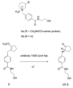
Herein we report a novel method for the production of a functional ScFv fragment in the oxidizing bacterial cytoplasm. The system is demonstrated by the catalytic antibody 14D9, a catalytic antibody against piperidinium hapten 1a (11), which catalyzes the highly enantioselec-tive protonation of enol ethers such as 2 to give optically pure carbonyl products such as (S)-ketone 3 (12), with absolute control of enantioselectivity (14). The kinetic
constants for the reaction (kcat/kuncat = 104, k
cat = 0.4 s–1 at
pH6.0) make it one of the fastest and most practical cata-lytic antibodies reported to date (15). The ScFv-fragment is fused with the NusA protein (N utilization substance protein A), which shows the highest solubility in E. coli (16). A stain bearing a defect in the reductase for thiore-doxin and glutathione, which has been shown to accumu-late active oxidizing enzymes in the cytoplasm (17), pro-vides a potentially favorable environment for disulfide bond formation of single chain Fv. We show that the com-bination of NusA fusion and an oxidizing cytoplasmic environment facilitates the expression of a functional catalytic antibody fragment.
MATERIALS AND METHODS
Construction of Single Chain Fv 14D9—The Fd and
kappa chain gene fragments of 14D9 were amplified by PCR as described (18). Amplified PCR products were digested with XhoI-SpeI for the Fd gene fragment and SacI-XbaI for the kappa chain gene fragment, and ligated into phage display vector pcomb3H (19) to give pcomb3H-14D9. All PCRs were carried out with Vent proof-reading polymerase (New England Biolabs) to ensure the highest fidelity. The VL and VH domains were PCR amplified with primer pairs, VL_Bn 5 ¢-GCGGCCAGCGAGCTCGT-GACACAGTC-3¢ and Linker_rev 5¢-GAACCTTCAGAGC- TTTTGCCGCTACCGGAAGTGCTGCCTTTGATCTCAAG-CTTTGTGCC-3¢ for the VL fragment, Linker_for 5¢- GCGGCAAAAGCTCTGAAGGTTCTGGCAGCACCAAAG-GTCAGGTTCAGCTGCTGCTCGAGCAG-3¢ and VH_end2 CGTAGTCGACTCAGG-AGAGACGGTGACCGT (the SalI site is underlined) for the VH fragment. Single chain Fv was assembled following a standard protocol (4).
with VL53 5 ¢-GCAGTGACATATGGAGCTCGTGATGAC-ACAGTCTCC-3¢ (the NdeI site is underlined) instead of VL_Bn. The plasmid pET43.1a was digested with NdeI–
XhoI, the large fragment purified from an agrose gel was
ligated with an amplified ScFv fragment treated with
NdeI–SalI to give plasmid pET-14D9. Both clones were
confirmed by DNA sequencing.
Cytoplasmic Expression and Purification—BL21(DE3)
TrxB–,Gor–, under the name of Origami™, and strain
BL21(DE3), are available from Novagen. The procedures for growth of the bacteria and induction of single chain Fv were conducted according to a standard protocol. Briefly, an overnight preculture grown in LB medium supplemented with responding antibiotics [50 mg/ml Carbenicillin, 35 mg/ml Kanamycin, and 15 mg/ml Tetra-cycline for BL21(DE3) TrxB–,Gor– ; 50 mg/ml
Carbenicil-lin for BL21(DE3)] was incubated in 1 liter of the same medium at 1:500 dilution and grown until mid exponen-tial stage (A600 = 0.5) at 22°C. Induction was started by adding isopropyl thio-b-D-galactoside (IPTG) to the cul-ture at a final concentration of 1mM, and growth was continued for 15 h at 22°C. The cells were pelleted, resus-pended in lysis buffer (Tris 20 mM pH 8.0, 300 mM NaCl, 10 mM imidazole), and the cytoplasmic proteins were extracted from the cells by sonication and subsequent centrifugation. The supernatant was loaded onto a Ni2+
-NTA (IMAC) column (Qiagen), and the column was washed extensively with washing buffer (20 mM Tris/ HCl, 300 mM NaCl, 40 mM imidazole pH 8.0). The fusion ScFv was eluted with elution buffer (20 mM Tris/HCl, 300 mM NaCl, 250 mM imidazole pH 8.0).
ScFv Preparation from the Fusion Protein—The fusion
protein in PBS buffer (pH7.4) was digested with Thrombin protease (Roche) for 12 h at 22°C. The ratio of fusion protein to protease was optimized and set to 0.5 unit/mg protein, and the protease-treated solution was loaded directly onto the Ni2+-NTA column. The
pass-through was fed onto a hapten affinity column, in which the hapten was coupled covalently to DEAE-Sepharose (Pharmacia) (20). The column was washed with the same buffer and the ScFv was eluted with 0.2 mM Glycine/ HCl, pH3.0, and immediately neutralized with 1M Tris/ HCl, pH 9.0. The eluate was dialyzed against PBS buffer (pH7.4) overnight at 4°C and concentrated with Cen-triprepTM YM-10 and YM-30 (Amicon, Millipore).
The integrity of the recombinant fusion ScFv and its purification were checked by SDS/PAGE on a homogene-ous 15% gel with Coomassie Brilliant Blue staining. The final protein concentrations were measured by UV spec-trophotometry with extinction coefficient A280 = 1.35 cm2·mg–1.
Catalytic Activity Assay—Catalytic reactions were
car-ried out by mixing 0.6 ml of a 10 mM stock solution of sub-strate 2 in acetonitrile/water 1:1 (Fig. 1) with 19.2 ml pro-tein solutions at a concentration of 1 mg/ml in PBS
(pH7.4) at 22°C (final concentration of 2: 300 mM). An identically prepared solution containing 0.2 ml of 5 mM inhibitor 1b (final concentration: 50 mM) was used to check specific inhibition. All reactions were monitored by HPLC. The substrate and product were separated on an analytical RP-C18 column (218TP54, 22 cm ´ 0.45 cm, 300 Å poresize, Vydac, USA). Retention times were as fol-lows (eluent: 1.5 mL.min–1 H
2O/acetonitrile = 75:25,
detection by UV at 230nm): tR(2) = 3.3 min, tR(3) = 15.7
min. The percentage conversion as indicated in Fig. 3 was calculated from peak integration.
Enantioselectivity Assay—Substrate 2 was incubated
with the antibody samples as above on a 200 ml scale. The entire reaction mixture was injected onto RP-C18-HPLC and the product ketone 3 peak was collected and lyophi-lized. The residue was taken up in hexane/isopropanol = 1:1, and analyzed on a chiral-phase column (Chiralpak OD, Daicel, 22 cm ´ 0.45 cm), which allows the separation of the two enantiomeric products (eluent: hexane/isopro-panol = 7:3; 1ml/min) as tR[(R)-3 = 8.6 min, tR(S)-3] = 12.4
min. The absolute configuration of the products was established earlier by chemical correlation (21).
RESULTS
The antibody 14D9 Fab domain was cloned from the 14D9 hybridoma cell line (Details of the hapten-affinity column preparation and use will be described elsewhere). To assemble the two variable domains, we used a known modified 18-amino acid linker derived from the classical Gly-Ser linker, including three charged residues (Lys, Lys, Glu) to enhance the solubility of the ScFv construct (Details of the hapten-affinity column preparation and use will be described elsewhere). The assembled ScFv was cloned into the vector pET43.1a downstream of NusA, separated by a histidine tag (6´) and a 2–amino acid spacer containing a thrombin cleavage site.
Fig. 1. Enantioselective protonation catalyzed by antibody 14D9.
Plasmid pET-14D9nus was transformed into
Escheri-chia Coli cells BL21(DE3), TrxB–,Gor–, which have an
oxidizing cytoplasmic environment favorable for the expression of disulfide-bridged proteins (Details of the hapten-affinity column preparation and use will be described elsewhere). These cells grow quite slowly com-pared to the BL21(DE3) wild type. After induction over-night, the fusion protein was purified by Ni2+-NTA
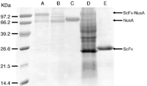
chro-matography (IMAC). Two main bands were observed in the reducing SDS-PAGE (Fig. 2, lane A), one correspond-ing to a molecular mass of ca. 82 kDa, the other with a relatively low molecular mass appearing close to NusA (Fig. 2, lane C, theoretical mass of 54 kDa). Between these two main bands, some smeared bands suggested the degradation of unfolded fusion proteins; the second main band (NusA) might be the remainder of such degra-dation. The expression yield was calculated from total elution from the IMAC and reached a satisfactory level of 3 mg/litre LB medium.
Purification of the ScFv-NusA construct by hapten-affinity chromatography gave 14D9 ScFv and NusA in equal amounts, this due to the cleavage by proteolytic impurities during sample dialysis and concentration after elution from the column. The 14D9 ScFv fragment was, therefore, cleaved on purpose by treatment with thrombin, for which a cleavage site is present in the 20– amino acid linker between NusA and the ScFv, and puri-fied by filtration through a Ni2+-NTA column followed by
hapten-affinity chromatography. The product isolated showed only one band at 28 kDa, indicative of a high purity and integrity (Fig. 2, lane E). The overall yield of the expressed ScFv domain thus isolated from the NusA-fusion protein amounted to 0.5 mg/liter LB medium. For comparison, the 14D9 Fab fragment was prepared from the hybridoma-produced monoclonal antibody by papain digestion (25), and purified by hapten-affinity chroma-tography.
The functionality of the 14D9 ScFv-NusA fusion pro-tein and the cleaved 14D9-ScFv was compared to the 14D9-Fab fragment by testing the chemical catalysis of
the enantioselective protonation of enol ether 2 to pro-duce ketone (S)-3, which can be recorded by HPLC (Fig. 1). The protein samples were tested at an equal concen-tration of 1 mg/ml (as measured by optical density) at pH 7.4 in the presence or absence of the activesite-specific hapten inhibitor 1b. All three samples catalyzed the for-mation of ketone 3 to a comparable level. In all three cases, the activity was quantitatively inhibited to the background level in the presence of the 14D9-specific hapten 1b. Chiral-phase column analysis of the ketone formed showed that all three samples catalyzed the for-mation of the S-enantiomer of ketone 3 exclusively, in agreement with the absolute control of enantioselectivity achieved by antibody 14D9 for this reaction (15), with the amount of racemate present in the sample corresponding to the level of the background reaction as calculated from the hapten-inhibited reaction. To examine disulfide bridge formation, the purified 14D9 ScFv-NusA and ScFv were treated with 10 mM b-mercaptoethanol before activity measurement. There was a complete loss of catalysis in the sample, indicating the importance of disulfide bridges to functionality, and confirming that correct disulfide bond formation occurred in the cyto-plasm.
To investigate the solubilizing ability of the NusA pro-tein, pET-14D9nus was transformed into BL21(DE3) wild type, which should not support disulfide bridge for-mation in the cytoplasm. The purified cytoplasmic extract showed a pattern similar to the expression in oxi-dizing cytoplasm, but with a very low relative depth ratio of intact fusion protein to NusA (Fig. 2, lane B). In addi-tion, the fusion protein band located at 82 kDa disap-peared after storage overnight at 4°C, indicating degra-dation and structural instability due to the lack of disulfide bonds in the reducing cytoplasm.
Fig. 2. Expression and purification of 14D9-ScFv NusA fusion constructs. Lane A: 14D9 ScFv-NusA fusion protein expressed in the cytoplasm of BL21(DE3) TrxB–, Gor– cells after purification
with IMAC. Lane B: 14D9 ScFv-NusA fusion protein expressed in the cytoplasm of BL21(DE3) cells after purification with IMAC. Lane C: NusA protein expressed in the cytoplasm of BL21(DE3) cells after purification with IMAC. Lane D: Total cell extract for the expression of 14D9 ScFv without NusA in BL21(DE3) TrxB–, Gor–
cells. Lane E: ScFv purified from a hapten affinity column after fil-tration through a Ni2+-NTA column.
Fig. 3. Catalytic activity assay for 14D9 antibodies. Percent conversion of enol ether 2 to ketone 3 is shown as a function of time. Measured by peak integration of RP-HPLC. Conditions: 300 mM substrate 2 in aq. PBS with 1.5% v/v acetonitrile, 1 mg/ml protein, 25°C. (solid square): catalytic reaction of 14D9 ScFv-nusA fusion protein; (open square): inhibition of 14D9 ScFv-nusA fusion protein with 50 mM hapten 1b; (solid triangle): catalytic reaction of 14D9 Fab purified from hybridoma; (open triangle): inhibition of 14D9 Fab with 50 mM hapten 1b; (solid circle): catalytic reaction of 14D9 ScFv cleaved from the 14D9 ScFv-nusA fusion protein; (open circle): inhibition of 14D9 ScFv with 50 mM hapten 1b.
Recombinant antibody expression is strongly depend-ent on primary sequence (26), and the success obtained with the NusA fusion construct might be entirely due to the 14D9 itself. To test this point, the direct expression of 14D9-ScFv without NusA was also investigated. Thus, the NusA 6x histidine tag and spacer were deleted, and the 14D9-ScFv fragment was cloned downstream of the T7 promoter to give plasmid pET-14D9. Expression of this plasmid yielded most of the 14D9-ScFv as an inclu-sion body, which was retained in the cytoplasmic pellet even at low temperature (22°C) (Fig. 2, lane D). Hapten affinity chromatography gave only a tiny peak of ScFv that was too small to be quantitated and isolated. These experiments show that 14D9-ScFv alone is not expressed in a functional soluble form, thereby highlighting the key role of NusA-tag as a solubilizing agent for the antibody.
DISCUSSION
Antibody expression in recombinant format must be appreciated from the point of view of functionality, con-venience, and overall yield. Here we present the overex-pression of a catalytic antibody fragment 14D9, which accelerates an abiological, enantioselective protonation reaction on synthetic substrate 2 with a rate enhance-ment of kcat/kuncat = 104. This rate enhancement is very
good for an antibody, but by no means comparable to that of an enzyme, and requires fair amounts of functional protein to provide a signal in the HPLC assay used for activity detection. Considering that the activity appears in direct proportion to the amount of functional protein and that there is no possibility for signal amplification as is possible with ELISA-type activity tests, the evidence for functionality presented here can be considered as par-ticularly strong. The evidence is reinforced by the
obser-As for convenience, the cloning procedure used to con-struct the 14D9-ScFv NusA fusion vector from the 14D9 hybridoma cell line involved only a minimal set of stand-ard operations. Purification of the expressed fusion pro-tein by simple IMAC using the His-tag provided an anti-body sample of sufficient purity for activity detection. Remarkably, the 14D9-ScFv fragment is not expressed in a soluble functional form when expressed alone within the oxidizing cytoplasm of E. coli BL21(DE3), TrxB–,
Gor–, but appears almost exclusively as insoluble
inclu-sion bodies. While the ScFv-NusA fuinclu-sion product is formed in a stable form under these conditions, expres-sion in the wild-type reducing cytoplasm provided only an unstable product that was quickly degraded. These results suggest that NusA proteins act as solubilizing agents, which allows the ScFv fragment to properly fold without forming insoluble aggregates. However, proper folding is tied to the formation of the structurally essen-tial disulfide bridges. Indeed the rapid degradation of the ScFv portion of the fusion construct under the reducing wild-type conditions suggests that ScFv is not properly folded under these conditions, but yet remains in a solu-ble form and is, therefore, particularly vulnerasolu-ble to the action of proteases. To compare in vivo folding with the in
vitro refolding procedure, the 14D9 ScFv inclusion body
was refolded by two popular ScFv refolding protocols (31,
32). All attempts to refold failed, implying that some
fac-tors in the cytoplasm, probably chaperones, might facili-tate the folding of ScFv in vivo. Our experiment with antibody 14D9, whose ScFv fragment is not suited for cytoplasmic expression, suggests that the NusA fusion format should be easily transferable to other antibodies. This NusA fusion method might be generally useful as an alternative for functional antibody expression.
The calculated yield of 3 mg/liter of functional fusion protein obtained lies well within the range accessible without optimization for antibody expression systems. The lower specific activity of the 14D9 ScFv-NusA fusion protein compared with the purified 14D9Fab and 14D9 ScFv fragments is partly explained by its higher molecu-lar mass, but is certainly also due to the fact that the pro-tein purified by IMAC is not pure and contains NusA from which the ScFv has been cleaved (Fig. 2), as evi-denced by the fact that only 1.4 mg/liter of 14D9 ScFv-NusA fusion protein was isolated after hapten-affinity chromatography. The key point is that no refolding oper-ation was undertaken to isolate an active sample of 14D9 ScFv-NusA fusion protein, and that the activity of this sample establishes that the ScFv-NusA fusion protein is indeed functional as expressed in the cytoplasm. While this is not necessarily important for expression, it becomes critical from the perspective of setting up a genetic selection system to improve the catalytic effi-ciency of antibodies. Indeed, selection schemes based on metabolic selection are envisioned as the best approach Fig. 4. Enantioselectivity assay for 14D9 antibodies. The peak
of product ketone 3 from the analytical RP-C18 column was col-lected and lyophilized, and the residue taken was up in hexane/iso-propanol = 1:1, and analyzed on a chiral-phase column (Chiralpak OD, Daicel, 22 cm ´ 0.45 cm), which allows the separation of the two enantiomeric products (eluent: hexane/isopropanol=7:3; 1 ml/min) as tR(R)-3 = 8.6 min, tR(S)-3 = 12.4 min. (A) racemic ketone 3 pro-duced by treatment of enol ether 2 with 0.1% TFA, R/S = 49%/51%. (B): (S)-ketone 3 from the reaction with 14D9-Fab, R/S = 6%/94%; (C): (S)-ketone 3 from the reaction with 14D9 ScFv-nusA fusion pro-tein, R/S = 8%/92%; (D): (S)-ketone 3 from the reaction with 14D9 ScFv protein, R/S = 3%/97%.
to improve the activity of catalytic antibodies over rounds of mutation and selection. Cytoplasmic expression of functional antibodies is particularly important in that respect as most genetic selection schemes should involve the catalysis of chemical reactions taking place within the cytoplasm, such as the catalysis of key metabolic reactions (11, 12) or the liberation of essential growth factors from synthetic precursors. Catalytic antibody 14D9 has been shown to catalyze a variety of processes, such as epoxide and acetal hydrolysis (12, 27–30). Some of these might be amenable to the release of an essential growth factor, for example biotin, from a specifically syn-thesized substrate, and future experiments will address this point.
This work has been supported by the Swiss National Science Foun-dation. The pComb3H-myc vector was a kind gift of Dr. Carlos Bar-bas, The Scripps Research Insitute, La Jolla.
REFERENCES
1. Pluckthun, A. (1991) Antibody engineering: advances from the use of Escherichia coli expression systems. Bio/Technology 9, 545–551
2. Carter, P., Kelley, R.F., Rodrigues, M.L., Snedecor, B., Covarru-bias, M., Velligan, M.D., Wong, W.L., Rowland, A.M., Kotts, C.E., Carver M.E., Yang, M., Bourell, J.H., Shepard, H.M., and Herner, D. (1992) High level Escherichia coli expression and production of a bivalent humanized antibody fragment. Bio/ Technology 10, 163–167
3. Barbas, C.F. and Wagner, J. (1995) Synthetic human antibod-ies: selecting and solving functional proteins. Methods: a Com-panion to Methods Enzymol. 8, 94–103
4. Skerra, A. and Plückthun, A. (1988) Assembly of a functional immunoglobulin Fv fragment in Escherichia coli. Science 240, 1038–1041
5. Arndt, K.M., Muller, K.M., and Pluckthun, A. (2001) Helix-sta-bilized Fv (hsFv) antibody fragments: substituting the con-stant domains of a Fab fragment for a heterodimeric coiled-coil domain. J. Mol. Biol. 31, 221–228
6. Tramontano, A., Janda, K.D., and Lerner, R.A. (1986) Catalytic antibodies. Science 234, 1566–1570
7. Pollack, S.J., Jacobs, J.W., and Schultz, P.G. (1986) Selective chemical catalysis by an antibody. Science 234, 1570–1573 8. (a) Thomas, N.R. (1996) Catalytic antibodies: reaching
Adoles-cence? Nat. Prod. Rep. 13, 479–511 (b) Stevenson, J.D. and Thomas, N.R. (2000) Catalytic antibodies and other biominetic catalysts. Nat. Prod. Rep. 17, 535–537
9. Proba, K., Ge, L., and Plückthun, A. (1995) Functional anti-body single-chain fragments from the cytoplasm of Escherichia coli: influence of thioredoxin reductase(TrxB). Gene 159, 203– 207
10. Smiley, J.A. and Benkovic, S.J. (1994) Selection of catalytic antibodies for a biosynthetic reaction from a combinatorial cDNA library by complementation of an auxotrophic Escheri-chia coli: antibodies for orotate decarboxylation. Proc. Natl Acad. Sci. USA 91, 8319–8323
11. Tang, Y., Hicks, J.B., and Hilvert, D. (1991) In vivo catalysis of a metabolically essential reaction by an antibody. Proc. Natl Acad. Sci. USA 88, 8784–8786
12. Reymond, J.L., Janda, K.D., and Lerner, R.A. (1991) Antibody catalysis of glycosidic bond hydrolysis. Angew. Chem. Int. Ed. Engl. 30, 1711–1713
13. Jahanghiri, G.K. and Reymond, J.L. (1994) Antibody-catalyzed hydrolysis of enol ethers. 2. Structure of the antibody – transi-tion state complex and origin of the enantioselectivity. J. Amer. Chem. Soc. 116, 11264–11274
14. Shabat, D., Itzhaky, H., Reymond, J.L., and Keinan, E. (1995) Antibody catalysis of a reaction otherwise strongly disfavored in water. Nature 374, 143–146
15. Reymond, J.L., Reber, R.A., and Lerner, R.A. (1994) Enantiose-lective, multigram-scale synthesis with a catalytic antibody. Angew. Chem. Int. Ed. Engl. 33, 475–477
16. Wilkinson, D.L. and Harrison, R.G. (1991) Predicting the solu-bility of recombinant proteins in Escherichia coli. Bio/Technol-ogy 9, 443–448
17. Prinz, W.A., Aslund, F., Holmgren, A., and Beckwith, J. (1997) The role of the thioredoxin and glutaredoxin pathways in reducing protein disulfide bonds in the Escherichia coli cyto-plasm. J. Biol. Chem. 272, 15661–15667
18. Takahashi, N., Kakinuma, H., Liu, L., Nishi, Y., and Fujii, I. (2001) In vitro abzyme evolution to optimize antibody recogni-tion for catalysis. Nat. Biotechnol. 19, 563–567
19. Barbas, C.F., Kang, A.S., Lerner, R.A., and Benkovic, S.J. (1991) Assembly of combinatorial antibody libraries on phage surfaces: the gene III site. Proc. Natl Acad. Sci. USA 88, 7978– 7982
20. Details of the hapten-affinity column preparation and use will be described elsewhere
21. Sinha, S.C. and keinan, E. (1995) Catalytic antibodies in organic synthesis. Asymmetric synthesis of (–)-a- multistritin. J. Amer. Chem. Soc. 117, 3653–3654
22. Huse, W.D., Sastry, L., Iverson, S.A., Kang, A.S., Alting-Mees, M., Burton, D.R., Benkovic, S.J., and Lerner, R.A. (1989) Gen-eration of a large combinatorial library of the immunoglobulin repertoire in phage lambda. Science 246, 1275–1281
23. Filpula, F., Mcguire, J., and Whitlow, M. (1993) Production of single-chain Fv monomers and multimers in Antibody Engi-neering (McCafferty, J., Hoogerboom, H.R., and Chiswell, D.J., eds.) pp. 253–268, IRI Press, New York
24. Derman, A.I., Prinz, W.A., Berlin, D., and Beckwith, J. (1993) Mutations that allow disulfide bond formation in the cyto-plasm of Escherichia coli. Science 262, 1744–1747
25. Porter, R.R. (1959) The hydrolysis of rabbit g-globulin and anti-bodies with crystalline papain. J. Biochem. 73, 119–127 26. Plückthun, A., Krebber, A., Krebber, C., Horn, U., Knüpfer, U.,
Wenderoth, R., Nieba, L., Proba., K., and Riesenberg, D. (1996) Producing antibodies in Escherichia coli:from PCR to fermen-tation in Antibody Engineering (McCafferty, J., Hoogerboom, H.R., and Chiswell, D.J., eds.) pp. 203–252, IRI Press, New York
27. Sinha, S.C., Keinan, E., and Reymond, J.L. (1993) Antibody-catalyzed reversal of chemoselectivity. Proc. Natl Acad. Sci. USA 90, 11910–11913
28. Sinha, S.C., Keinan, E., and Reymond, J.L. (1993) Antibody-catalyzed enantioselective epoxide hydrolysis. J. Amer. Chem. Soc. 115, 4893–4894
29. Shabat, D., Sinha, S.C., Reymond, J.L., and Keinan, E. (1996) Catalytic antibodies as probes of evolution: modelling of a pri-mordial glycosidase. Angew. Chem. Intl. Ed. Engl. 35, 2628– 2630
30. Shabat, D., Shulman, H., Itzhaky, H., Reymond, J.L., and Keinan, E. (1998) Enantioselectivity vs. kinetic resolution in antibody catalysis: formation of the (S) product despite prefer-ential binding of the (R) intermediate. Chem. Commun. 1759– 1760
31. Buchner, J., Pastan, I., and Brickmann, U. (1992) A method of increasing the yield of properly folded recombinant fusion pro-teins: single-chain immunotoxins from renaturation of bacte-rial inclusion bodies. Anal. Biochem. 205, 263–270
32. Tsumoto, K., Shinoki, K., Kondo, H., Uchikawa, M., Juji, T., and Kumagai, I. (1998) Highly efficient recovery of functional single-chain Fv fragments from inclusion bodies overexpressed in Escherichia coli by controlled introduction of oxidizing rea-gent—application to a human single-chain Fv fragment. J. Immunol Methods. 219, 119–129