Decisional role of the dorsolateral prefrontal cortex
in ocular motor behaviour
C. Pierrot-Deseilligny,
1R. M. MuÈri,
2C. J. Ploner,
3B. Gaymard,
1S. Demeret
1and S. Rivaud-Pechoux
11INSERM 289 and Service de Neurologie 1, HoÃpital de la
SalpeÃtrieÁre, Assistance Publique-HoÃpitaux de Paris, Paris, France,2Eye Movement Research Laboratory and
Department of Neurology, Inselspital, Bern, Switzerland and 3Klinik fuÈr Neurologie, ChariteÂ, Berlin, Germany
Correspondence to: Professor Ch. Pierrot-Deseilligny, Service de Neurologie 1, HoÃpital de la SalpeÃtrieÁre, 47 Bd de l'HoÃpital, 75651 Paris cedex 13, France
E-mail: [email protected]
Summary
Three patients with a unilateral cortical lesion affecting the dorsolateral prefrontal cortex (DLPFC), i.e. Brodmann area 46, were tested using different para-digms of re¯exive saccades (gap and overlap tasks), intentional saccades (antisaccades, memory-guided and predictive saccades) and smooth pursuit movements. Visually guided saccades with gap and overlap, latency of correct antisaccades and memory-guided saccades and the gain of smooth pursuit were normal, compared with controls. These results con®rm our anatomical data showing that the adjacent frontal eye ®eld (FEF) was unimpaired in these patients. The speci®c pattern of abnormalities after a unilateral DLPFC lesion, com-pared with that of the FEF lesions previously reported,
consists mainly of: (i) a bilateral increase in the per-centage of errors in the antisaccade task (misdirected re¯exive saccades); (ii) a bilateral increase in the vari-able error in amplitude, without signi®cant decrease in the gain, in the memory-guided saccade task; and (iii) a bilateral decrease in the percentage of anticipatory sac-cades in the predictive task. Taken together, these results suggest that the DLPFC plays a crucial role in the decisional processes, preparing saccades by inhibit-ing unwanted re¯exive saccades (inhibition), maintain-ing memorized information for ongomaintain-ing intentional saccades (short-term spatial memory) or facilitating anticipatory saccades (prediction), depending upon cur-rent external environmental and internal circumstances. Keywords: antisaccades; prediction; prefrontal cortex; saccades; spatial memory
Abbreviations: ACC = anterior cingulate cortex; DLPFC = dorsolateral prefrontal cortex; FEF = frontal eye ®eld; fMRI = functional MRI; MGS = memory-guided saccade; PEF parietal eye ®eld; PPC = posterior parietal cortex; SEF = supplementary eye ®eld
Introduction
The prefrontal cortex is essential in effective and skilfully organized behaviour. One fundamental principle of the prefrontal cortex may be adaptive neural coding, since many neurons in this cortical region adapt their properties speci®cally to carry information, producing a dense, distrib-uted representation of related inputs, actions, rewards and other information (for a review see Duncan, 2001). Another principle of prefrontal function is to guide or inhibit future responses that require temporal integration of events for purposeful actions. Constantinidis et al. (2002) recently showed, using simultaneous recordings in the monkey dorsolateral prefrontal cortex (DLPFC), inhibitory inter-actions between prefrontal neurons active at different time intervals. They proposed that the inhibitory function of the prefrontal cortex plays an important role in controlling the
timing of neuronal activity and shaping the temporal ¯ow of information processing.
In humans, the role of the DLPFC in eye movement control, i.e. Brodmann area 46 (Rajkowska and Goldman-Rakic, 1995), is not yet fully understood. One reason may be the problem of ®nding patients with isolated lesions of this region. As shown by functional imaging, the DLPFC and the frontal eye ®eld (FEF) in humans lie close together. Consequently, lesions often involve both regions, rendering any inferences as to their respective functions equivocal. Indeed, although eye movement studies with lesions includ-ing prefrontal structures have existed since the 1980s (Guitton et al., 1985; Pierrot-Deseilligny et al., 1991b; Braun et al., 1992), a study testing different aspects of eye movement control in patients with lesions restricted to the DLPFC has Brain 126 ã Guarantors of Brain 2003; all rights reserved
Patients
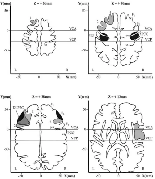
Three right-handed patients, two females and one male, were examined. Their mean age was 53 years (range: 34±73 years). The lesions were vascular ischaemic and remained mainly cortical, not involving the subcortical regions such as the basal ganglia and the internal capsule. Two patients had a left-sided lesion and one patient a right-sided lesion, docu-mented by CT or MRI scan (Fig. 1). For reconstruction of the patient's lesions, we used four brain sections: +60 mm, +50 mm, +28 mm and +12 mm parallel above the anterior commissure±posterior commissure line taken from the atlas of Talairach and Tournoux (1988). The +50 mm section was chosen to show the FEF in the precentral gyrus as de®ned by recent functional imaging studies (Paus, 1996; Heide et al., 2001). The +28 mm section was chosen to show the invariable portion of Brodmann's area 46 in the DLPFC in the middle portion of the middle frontal gyrus as de®ned by cytoarchitectonic criteria (Rajkowska and Goldman-Rakic, 1995). This region also shows activation during functional imaging of normal subjects performing memory-guided saccades (O'Sullivan et al., 1995; Sweeney et al., 1996). The other two sections (+60 mm and +12 mm) were chosen to show the extension of the lesions above and below both critical eye movement areas. The DLPFC was damaged, but the FEF was spared by the lesions in all three cases. The parietal eye ®eld (PEF) and supplementary eye ®eld (SEF), located at a distance from the DLPFC region, were also spared (see Discussion).
All patients were examined within the ®rst month after the vascular accident. None of them was taking medications acting upon on the CNS. Patient 1 (age: 57 years) had a cephalalgia as the initial and unique symptom. Clinical examination was normal. On MRI, an ischaemic lesion affecting a small part of the left prefrontal region was found (Fig. 1). The remainder of the CNS was normal. After investigations, it was stated that this lesion was due to a venous thrombosis. Patient 2 (age: 34 years) had slight aphasia (dif®culty in ®nding some words) as the initial and unique sign. The clinical examination was otherwise normal. On MRI, an ischaemic and isolated lesion of the left prefrontal region was found (Fig. 1), probably secondary to
Eye movements were recorded by means of direct current electro-oculography in complete darkness, using four Ag± AgCl electrodes (two horizontal temporal and two vertical on one eye to control blinks). The subject's head was immobilized. The electrical signal was ampli®ed and ®ltered (bandwidth: 0±100 Hz), and the spatial resolution was 0.5°. Visual cues were presented at a distance of 95 cm with red LEDs embedded in a curved ramp. LEDs were 0.15° and 5 cd/m2in luminance. Each session was preceded by 10 min
of dark adaptation. The velocity threshold criterion for de®nition of saccades was 30°/s. Data were sampled with a frequency of 200 Hz. System calibration was performed before each paradigm. For further details, see Pierrot-Deseilligny et al. (1991b). The whole examination lasted ~45 min. The following paradigms were tested.
Gap task
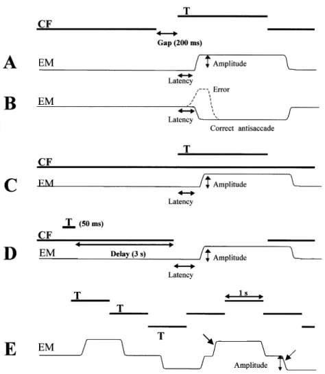
In the gap task, re¯exive visually guided saccades were tested (Fig. 2A). The central ®xation point was switched off 200 ms (i.e. gap) before the onset of a lateral target, located 25° to the right or the left of the central ®xation point. The subjects were instructed to ®xate the central ®xation point, and to look at the lateral target as soon as it appeared. The target was presented randomly to the right or left, with unpredictable timing. Left and right saccade latencies were calculated for each subject by averaging 20 measurements in each direction. The percentage of express saccades, with latency comprised between 80 and 120 ms (Fischer and Ramsperger, 1984), was also determined. Lastly, the saccade gain (amplitude of the ®rst saccade over eccentricity of the target) was measured.
Antisaccade task
In the antisaccade task, the visual presentation was the same as in the gap task, except that the subject was instructed to look in the opposite direction to that of the suddenly appearing lateral target, without ®rst looking at the target (Fig. 2B). Twenty trials were made in each lateral direction. The percentage of errors (misdirected saccades, i.e. reaching or simply initially directed towards the target), the latency of these misdirected saccades and the latency of correct
antisaccades (made in the direction opposite to the target) were determined for each direction. Furthermore, the per-centage of express saccades (with latency comprised between 80 and 120 ms) among the misdirected saccades was also calculated in patients. In controls, this percentage could not be determined since the number of misdirected saccades was too small.
Overlap task
In the overlap task, the central ®xation point remained switched on during the presentation of the lateral visual target
(Fig. 2C). All other conditions and measurements were the same as for the gap task.
Memory-guided saccade task
In the memory-guided saccade (MGS) task, the subject ®xated a central ®xation point while the lateral target was ¯ashed for 50 ms, with unpredictable direction and eccentri-city (between 10 and 30°; Fig. 2D). The central ®xation point was switched off 3 s after the ¯ashed target, which was the go signal for the subject to make a saccade to the remembered position of the ¯ash. Then, the lateral target was switched on and the subject made a corrective saccade if necessary. Fig. 1 Lesions of the three patients. Four transverse brain sections parallel above the anterior
commissure±posterior commissure (AC±PC) line with the Talairach coordinate frame (Talairach and Tournoux, 1988) show the location of the frontal eye ®eld (top, right), in the precentral gyrus and sulcus, and the invariable portion of Brodmann's area 46 in the dorsolateral prefrontal cortex (DLPFC; bottom, left), at the level of the middle frontal gyrus (F2), as black areas. Lesions of the patients are in grey (1, 2 and 3). Note that in all three patients, the lesions damaged the DLPFC but spared the FEF. cs = central sulcus; DLPFC = dorsolateral prefrontal cortex; F1, F2and F3= superior, middle and inferior frontal gyrus, respectively; FEF = frontal eye ®eld; L = left; PCG = precentral gyrus; pcs = precentral sulcus; R = right; VCA = vertical anterior commissure line; VCP = vertical posterior commissure line; x, y and z, distance from the saggital plane, the coronal plane (through the anterior commissure) and the AC±PC line, respectively.
Saccade latency and gain were averaged from 20 saccades in each lateral direction. Furthermore, interquartile ranges were used to describe a subject's gain variability (variable error in amplitude). For the amplitude analysis, we studied the ®rst saccade made after the central ®xation point was switched off (Pierrot-Deseilligny et al., 1991b). Additional saccades just after the initial saccades were rare and did not in¯uence MGS errors. The data on ®nal eye positions were therefore similar to those of initial saccades and are not presented, so as to avoid redundancy.
Predictive saccade task
The subject was instructed to follow a luminous target which appeared 25° to the right, then was displaced to the centre,
25° to the left, back to the centre and ®nally to the original position (Fig. 2E). The target remained in each position for 1 s. Therefore, target direction, amplitude and timing were entirely predictable in this paradigm. Six such consecutive cycles were repeated three times. A saccade was considered as anticipatory, i.e. not visually guided, when latency was <70 ms (Smit and Van Gisbergen, 1989) or if it occurred even before target onset. The percentage of centrifugal anticipatory saccades was calculated from 18 saccades in each direction. The ®rst saccade was excluded from analysis. The gain of centrifugal anticipatory saccades was also determined. The centripetal saccades, which are very different in nature from centrifugal saccades in terms of both triggering and amplitude (Findlay, 1981), were not studied.
Fig. 2 Saccade paradigms. (A) Visually guided saccade with gap: latency and amplitude are measured. (B) Antisaccade, with the same stimulation as in A but with the instruction to look in the opposite direction to the target: latency of correct antisaccades and the percentage of errors (misdirected re¯exive saccades) are measured. (C) Visually guided saccade with overlap, with the same instruction as in A but with the central ®xation (CF) remaining switched on: latency and amplitude are measured. (D) Memory-guided saccade, with the go signal given by the extinction of the CF (after a delay of 3 s): latency and amplitude are measured. (E) Predictive saccades, with the instruction to follow the targets: the amplitude and the percentage of anticipatory saccades (oblique arrows) are measured. EM = eye movement; T = target.
Smooth pursuit task
In the smooth pursuit task, the subject was instructed to follow a horizontal target moving sinusoidally with peak velocities of 23°/s (0.25 Hz). The mean rightward and leftward gains (peak eye velocity over peak target velocity) were calculated from 10 consecutive cycles.
Statistical analysis
Since lesions were both left- and right-sided, we grouped the results of the patients into ipsilateral and contralateral results. In the control group, the results for rightward and leftward saccades were analysed statistically. There was no signi®cant side difference (Mann±Whitney test) in any performance. Therefore, the left and right data were pooled for each subject.
Statistical analysis was performed using a non-parametric test (Kruskal±Wallis test) for percentages, comparing control, ipsilateral and contralateral performance. When results were statistically signi®cant, we compared control performance with ipsilateral or contralateral performance, respectively, using Mann±Whitney test. For latency and the amplitude gain, a parametric test was used (Student's t test).
Results
The results of the gain in the gap, overlap and smooth pursuit tasks for controls and patients are presented in Table 1. Results were similar in both groups, with no statistically signi®cant difference for the gain values in any of the three paradigms. Furthermore, smaller standard errors in the patient group than in controls indicate that the results in the latter were particularly homogeneous.
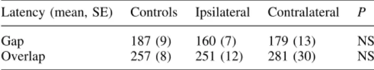
Table 2 shows latency values in the gap and overlap tasks. There was no statistically signi®cant difference between the two groups, with, however, a greater variability in the patient group, as indicated by their standard errors.
The median percentage of express saccades (comprised between 80 and 120 ms) in the gap task was 8% in controls (range: 0±27%), and 42% (range: 35±48%; P < 0.01, Mann± Whitney test) for ipsilateral saccades and 16% (range: 6±27%; NS) for contralateral saccades in patients. This increase in the percentage of ipsilateral express saccades obviously is related to the slight decrease observed in the latency of ipsilateral saccades made in the gap task (160 ms versus 187 ms in controls, P = 0.12). These results were similar in the three patients, including the patient with a right-sided lesion. There is no obvious explanation for this increase in the percentage of ipsilateral express saccades or for the tendency for a decrease in ipsilateral saccade latency in the gap task.
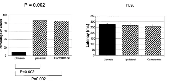
The percentage of errors in the antisaccade task (Fig. 3, left side) was signi®cantly increased in the patient group: the median was 86% (range: 28±100%) for ipsilateral saccades (P = 0.002, Mann±Whitney test) and 85% (range: 28±92%)
for contralateral saccades (P = 0.002, Mann±Whitney test), respectively. In the control group, the median percentage of errors was 8% (range: 0±17%). The latency of the few correct antisaccades (made in the opposite direction to the target) was normal (Fig. 3, right side): 270 ms (SE = 22) for ipsilateral saccades and 260 ms (SE = 22) for contralateral saccades in patients versus 280 ms (SE = 9) in controls (NS, Student's t test). The number of errors (misdirected saccades made towards the target) in controls was too small to be used for statistical purposes in the study of latency. However, the latency of these re¯exive misdirected saccades in patients was signi®cantly shorter than that of correct antisaccades: 223 ms (SE = 15) for ipsilateral saccades (P < 0.01, Student's t test) and 203 ms (SE = 11) for contralateral saccades (P < 0.01), but not as short as that observed in the gap task. Furthermore, the percentage of express saccades among these re¯exive misdirected saccades was less than in the gap task ipsilaterally (median: 10%; range: 5±42%) and slightly higher contralaterally (median: 18%; range: 13±33%), therefore, without the same asymmetry as in the gap task.
For MGS, there was a bilateral, slight, not signi®cant increase in latency [Fig. 4, left; mean: 381 ms (SE = 10) for ipsilateral saccades with P = 0.001, and 347 ms (SE = 5) for contralateral saccades with P = 0.001] in the patient group, compared with controls (mean: 321 ms, SE = 13). In the MGS task, the subjects may make a saccade to the target just after the ¯ash, i.e. a visually triggered saccade. Such trials were excluded from the analysis of MGS. However, it could be of interest to determine the percentage of such saccadic errors since they are a re¯ection of the control of re¯exive saccade suppression. In controls, there were 4.5% (median; range: 0±12%) of such saccades, but in patients the percentage was 42% (range: 23±53%) for ipsilateral saccades (P < 0.01, Mann±Whitney test) and 39% (range: 8±57%) for contral-ateral saccades (P < 0.01). Therefore, saccade suppression during the MGS task was much less ef®cient in these patients with a prefrontal dysfunction than in controls.
Table 1. Gain of the gap, overlap and smooth pursuit tasks
Gain (mean, SE) Controls Ipsilateral Contralateral P
Gap 0.92 (0.04) 0.92 (0.02) 0.91 (0.01) NS
Overlap 0.94 (0.04) 0.92 (0.02) 0.96 (0.02) NS
Smooth pursuit 0.92 (0.05) 0.95 (0.03) 0.90 (0.01) NS NS = not signi®cant.
Table 2. Latency of the gap and overlap tasks
Latency (mean, SE) Controls Ipsilateral Contralateral P
Gap 187 (9) 160 (7) 179 (13) NS
Overlap 257 (8) 251 (12) 281 (30) NS
In patients, the gain of MGS was 0.79 (SE = 0.05) ipsilaterally and 0.74 (SE = 0.13) contralaterally on average, compared with controls with 0.92 on average (SE = 0.02) (Fig. 4, middle), and was not signi®cantly decreased. However, we cannot rule out the possibility that this lack of statistical signi®cance was simply due here to the relative weakness of the sample of three patients. In contrast, the variable error of the gain (Fig. 4, right) was signi®cantly increased, bilaterally: mean of 0.43 (interquartile range = 0.36±0.48) for ipsilateral saccades (P = 0.012), and of 0.32 (interquartile range = 0.23±0.34) for contralateral saccades (P = 0.037) compared with controls, with a mean of 0.21 (interquartile range = 0.06±0.23).
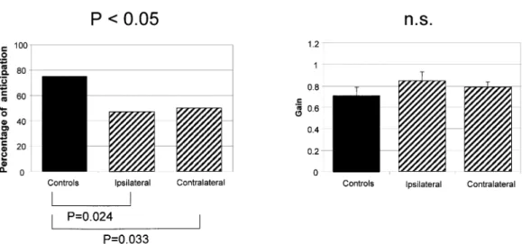
Lastly, in the predictive task (Fig. 5, left side), the percentage of anticipatory saccades was signi®cantly de-creased in the patient group: 47% (median; range = 39±52%) for contralateral saccades (P = 0.033) and 42% (median;
range = 27±52%) for ipsilateral saccades (P = 0.023). The controls made 75% (median; range = 30±100%) of anticipa-tory saccades. The gain of these anticipaanticipa-tory saccades was not different in patients and controls (Fig. 5, right side): mean of 0.72 (SE = 0.08) in controls, and mean of 0.84 (SE = 0.08) for ipsilateral saccades and 0.79 (SE = 0.05) for contralateral saccades in patients. The variability of the gain of anticipa-tory saccades was high in both controls and patients, with therefore no signi®cant difference between the two groups.
In none of the paradigms were there any obvious differ-ences in individual results between the patients with a left lesion and the patient with a right lesion.
Discussion
In these patients with a unilateral DLPFC lesion, we observed a marked impairment of antisaccades, MGS and predictive Fig. 4 Memory-guided saccades (MGS). Left, latency of MGS (bars indicate +1 SE); middle, gain of MGS; right, variable error of the gain. Note that latency of patients was only slightly increased, bilaterally, compared with that of controls, and the gain was only slightly decreased, whereas the variable error of the gain was signi®cantly increased, bilaterally. Top, Kruskal±Wallis test; bottom, Mann±Whitney test.
Fig. 3 Antisaccade task. On the left side, median percentage of errors (misdirected re¯exive saccades made towards the target), and, on the right side, latency of correct antisaccades (made in the opposite direction to the target) (bars indicate +1 SE). Note that the percentage of errors of patients was increased bilaterally, compared with that of controls, whereas the latency of correct antisaccades was similar to that of controls. Top, Kruskal±Wallis test; bottom, Mann±Whitney test.
saccades. Before discussing each of these abnormalities and the speci®c role of the DLPFC in saccade control, the normality of the other results also deserves some comments since they allow us to assert that the cortical areas triggering saccades were spared by the DLPFC lesions, in particular the nearby FEF.
Integrity of cortical areas triggering saccades
Three areas are involved in saccade triggering (Pierrot-Deseilligny et al., 1995, 2002b; Leigh and Zee, 1999) (Fig. 6): the PEF, located in the intraparietal sulcus (Pierrot-Deseilligny et al., 1991a; MuÈri et al., 1996a; Perry and Zeki, 2000; Heide et al., 2001; Simon et al., 2002); the SEF, located in the upper part of the paracentral sulcus (Grosbras et al., 1999), i.e. medially in the superior frontal gyrus; and the FEF, mainly located at the intersection between the superior frontal sulcus and the precentral sulcus, with some lateral extension in the precentral gyrus (Petit et al., 1993; Paus, 1996; Heide et al., 2001; Lobel et al., 2001; Milea et al., 2002) and anteriorly to the precentral sulcus (Blanke et al., 2000; Rosano et al., 2002). The PEF is involved mainly in the triggering of re¯exive saccades in the gap task, since lesions affecting this area result in increased latency of such saccades (Pierrot-Deseilligny et al., 1991a; Braun et al., 1992; Heide and Kompf, 1998). In contrast, after lesions affecting the DLPFC, the SEF or the FEF, the latency of these saccades is not increased (Pierrot-Deseilligny et al., 1991a). Lesion studies suggest that the SEF is involved mainly in the control of saccade sequences (Gaymard et al., 1993) or saccades combined with body movements (IsraeÈl et al., 1995), but such paradigms were not tested here. However, the SEF, like the PEF, is located at a distance from the DLPFC, and our anatomical data clearly show that these two areas were not damaged. In contrast, the FEF is located much closer to the DLPFC and, even though our anatomical study also suggeststhat the FEF was not damaged in our patients, it nevertheless was important to con®rm that this area was also functionally spared. The FEF is involved in the control of all intentional saccades, including MGS and predictive saccades (Rivaud et al., 1994; IsraeÈl et al., 1995; Gaymard et al., 1999). Since MGS and predictive saccades are controlled upstream of the FEF by the DLPFC, the relatively subtle differences in the abnormalities resulting from lesions of the FEF and the DLPFC will be discussed below. The antisaccade paradigm also involves both areas, but the normality of the latency of correct antisaccades in our patients suggests that the FEF was spared (see next section). In contrast, some eye movements, such as visually guided saccades in the overlap task (Rivaud et al., 1994; Gaymard et al., 1999) and smooth pursuit (MacAvoy et al., 1991; Rivaud et al., 1994; Morrow and Sharpe, 1995; Heide et al., 1996; Gaymard et al., 1999), appear to be controlled more speci®cally by the FEF. The normality of overlap saccade latency and smooth pursuit gain in our patients are strong arguments supporting the preser-vation of the FEF, as well as a con®rmation that these eye movements do not crucially involve the DLPFC. The involvement of the FEF in overlap task latency, but not in gap task latency, may be explained by an active disengage-ment of visual ®xation in the former but not in the latter (Rivaud et al., 1994; Gaymard et al., 1999), in which ®xation is already suppressed when the target appears. Lastly, it should be noted that the gain of contralateral visually guided saccades in the gap and overlap tasks was impaired in FEF lesions (Rivaud et al., 1994; Gaymard et al., 1999) but was normal here, which is another argument suggesting that the FEF was spared in our patients. Taken together, our anatomical and functional results suggest that not only the SEF and the PEF, but also the FEF were spared in these patients, allowing us to de®ne, therefore, the speci®c spectrum of eye movement abnormalities due to isolated DLPFC lesions.
Fig. 5 Predictive saccades. Left, median percentage of anticipatory saccades; right, gain of anticipatory saccades (bars indicate +1 SE). Note that the percentage of anticipatory saccades of patients was decreased bilaterally compared with that of controls, whereas the gain of these saccades was similar to that of controls. Top, Kruskal±Wallis test; bottom, Mann±Whitney test.
Antisaccades
In our patients, the percentage of errors was markedly increased, bilaterally, in the antisaccade paradigm, but the latency of correct antisaccades was normal. In this paradigm, two different physiological processes are involved: (i) inhibition of unwanted re¯exive visually guided saccades towards the target (i.e. the `errors'), these errors being triggered mainly by the PEF, as already stated, when inhibition is lacking; and (ii) triggering of correct anti-saccades in the opposite direction to the target, when inhibition has been effective. Since the latency of these correct antisaccades is increased after isolated FEF lesions (Rivaud et al., 1994; Gaymard et al., 1999), it may be deduced that the FEF is crucially involved in this second process, which is expected for the triggering of such an intentional saccade. In contrast, the FEF does not appear to be crucially involved in the ®rst process, i.e. the inhibition of unwanted re¯exive saccades, since the percentage of these saccades (the `errors') remains normal after FEF lesions (Pierrot-Deseilligny et al., 1991a; Rivaud et al., 1994; Gaymard et al., 1999). However, it is true that the monkey
has been shown to contain suppressive sites, i.e. cells whose stimulation does not elicit saccades but prevents saccade triggering (Burman and Bruce, 1997). Since these cells project to the rostral pole of the superior colliculus, this area could play a side role in re¯exive saccade inhibition by increasing the activity of the ®xation system. Nevertheless, the inhibition of re¯exive saccades, which is under the control of the frontal lobe (Guitton et al., 1985), appears to depend more speci®cally upon the DLPFC, as suggested by a previous lesion study (Pierrot-Deseilligny et al., 1991a) and the results obtained using PET scan (Doricchi et al., 1997) or functional MRI (fMRI) (Sweeney et al., 1996; MuÈri et al., 1998). The results of the present study fully con®rm earlier ®ndings in humans by showing, once more, with a bilateral increase in the percentage of errors, that re¯exive saccade inhibition, i.e. the ®rst physiological process involved in the antisaccade paradigm, is mainly under the control of the DLPFC. Furthermore, our results also con®rm, by the normality of correct antisaccade latency, that the second physiological process involved in the antisaccade paradigm, i.e. the triggering of correct antisaccades, is not under the Fig. 6 Cortical areas involved in saccades. After receiving visual information in the occipital lobe and
after visuospatial integration in the PPC, a saccade may be either triggered re¯exively, mainly by the PEF, or triggered intentionally by the FEF, an area which also appears to be involved in active visual ®xation. If a re¯exive saccade must be inhibited, the DLPFC appears to play a crucial role (1). This area is also involved in short-term spatial memory (2) and prediction (3) when anticipatory saccades must be performed. With these three different actions, the DLPFC could play an important role in the decisional processes controlling ocular motor behaviour. The SEF could be involved in motor programmes including several successive saccades, or saccades combined with other body movements, whereas the CEF appears to activate all the areas controlling intentional saccades via a motivation process. ACC = anterior cingulate cortex; CEF = cingulate eye ®eld; cs = central sulcus; DLPFC = dorsolateral prefrontal cortex; FEF = frontal eye ®eld; ips = intraparietal sulcus; ls = lateral sulcus; pcs = precentral sulcus;
PEF = parietal eye ®eld; PPC = posterior parietal cortex; RF = brainstem reticular formation; SC = superior colliculus; SEF = supplementary eye ®eld; 1, 2, 3 = the main actions of the DLPFC; + = saccade triggering; ± = saccade inhibition.
control of the DLPFC. Therefore, in the antisaccade paradigm (at least with a gap), there is now cumulative evidence based on purely FEF lesions (Pierrot-Deseilligny et al., 1991a; Rivaud et al., 1994; Gaymard et al., 1999) and purely DLPFC lesions (Pierrot-Deseilligny et al., 1991a; and our present results) that the inhibition of re¯exive saccades is under the control of the DLPFC and the triggering of correct antisaccades is under the control of the FEF (see also Connolly et al., 2002). Yet, a degree of confusion persists in the literature on this point, mainly because, besides the activation of the DLPFC, an activation of the FEF has also been observed during the antisaccade paradigm using func-tional imaging (Sweeney et al., 1996; Doricchi et al., 1997; MuÈri et al., 1998; Connolly et al., 2002), which has sometimes led to the conclusion that this area may be involved in saccade inhibition (Cornelissen et al. 2002): on the basis of lesion study results, this would seem to be an erroneous interpretation, since the FEF is indeed involved in visual ®xation and the triggering of correct antisaccades in the antisaccade paradigm, but this area does not appear to be crucial for saccade inhibition, at least when a gap is used. Thus, the visual ®xation and/or the preparation of the triggering of correct antisaccades (Connolly et al., 2002) probably generates the increase in the metabolism of the FEF observed in neuroimaging studies of the antisaccade para-digm. However, it should also be noted that an active inhibition of the FEF during this paradigm (probably originating in the DLPFC) may also result in a further increase in the metabolism of this area in such studies (Kimmig et al., 2001), which does not imply, therefore, that this inhibition is generated in and starts from the FEF.
The inhibition function of the DLPFC could be exerted directly downstream on the brainstem or the superior colliculus, as suggested by experimental results (Everling et al., 1999; Trappenberg et al., 2001) and a human lesion study in which the superior colliculus was damaged (Pierrot-Deseilligny et al., 1991c), via the direct prefronto-collicular tracts (Leichnetz et al., 1981). Indeed, lesion studies suggest that the other cortical areas controlling saccades, including the FEF, are not involved in this function (see above). An exception to this is the anterior cingulate cortex (ACC), the posterior part of which could also be involved in anti-saccades, as suggested by an fMRI study (Paus et al., 1993) and lesion studies, (Gaymard et al., 1998; Milea et al., 2003a). However, the ACC could act early in saccade control, preparing all areas involved in intentional saccades, such as the FEF, the SEF and the DLPFC, but not the area mainly triggering re¯exive saccades, i.e. the PEF (Gaymard et al., 1998). This preparation could be due to a physiological process called motivation. With a relative lack of pre-activation of the DLPFC after an ACC lesion, the re¯exive saccades under the control of the PEF could be less restrained, with a consequent increase in errors in the antisaccade paradigm. Be that as it may, the percentage of errors in the antisaccade paradigm appears to be a good marker of DLPFC
control, and is probably also the easiest to test among the saccade paradigms.
Memory-guided saccades
In our patients with a DLPFC lesion, MGS latency was slightly increased, but not signi®cantly different from that of controls. The percentage of unwanted re¯exive visually triggered saccades made just after the ¯ash was increased, compared with controls, suggesting a relative disinhibition of re¯exive saccades related to the prefrontal dysfunction (as for antisaccades, see above). The gain of MGS was moderately decreased, but not signi®cantly different from that of controls, perhaps because of the small numbers in the patient group. In contrast, the variable error of the gain was markedly increased. The MGS paradigm is used in saccade physiology to study short-term spatial memory, which is the working memory controlling current, ongoing behaviour (Goldman-Rakic, 1996). In monkeys, electrophysiological and inactiva-tion studies have shown that the DLPFC is involved in the control of short-term spatial memory used in MGS paradigms (Funahashi et al., 1993; Roberts et al., 1994; Goldman-Rakic, 1996; Sawaguchi and Iba, 2001). In humans, lesion studies have suggested that several cortical areas, including the DLFPC, are involved in MGS (Pierrot-Deseilligny et al., 1991b, 1993, 1995, 2002b; IsraeÈl et al., 1995). These results have been corroborated by functional imaging, showing that a large network of frontoparietal areas is active during such a paradigm (O'Sullivan et al., 1995; Sweeney et al., 1996; Heide et al., 2001). However, recent functional imaging studies have questioned a predominant role of the DLPFC in maintenance of spatial information within this network, and suggest rather that the DLPFC may be responsible mainly for the selection of memory-guided behavioural responses, with maintenance being dependent on premotor and parietal cortices (Rowe et al., 2000). It should be noted that the MGS paradigm comprises three successive phases, involving different types of physiological mechanisms (Fig. 2) (for a review see Pierrot-Deseilligny et al., 2002a): (i) a ®rst phase of perception, during which the visual stimulus (a peripheral ¯ashed target) is presented, involving both the visual (occipital) and attentional (parietal) areas; (ii) a second phase, related to memorization (during the delay), starting after the visual stimulus presentation and under the control of the cortical area involved in spatial memory; in fact, the beginning of this second phase corresponds to visuospatial integration, a posterior parietal process occurring just after the visual stimulus presentation and allowing the subject to know the memorized position of the stimulus in relation not only to the eyes but probably also to the body; and (iii) after the go signal, the ®nal phase of movement, during which the MGS is triggered by the frontal and parietal motor areas, and accuracy of spatial memory, re¯ected by that of the saccade, is measured. The results of lesion studies have shown that the accuracy of MGS is impaired after lesions affecting either the DLPFC, the posterior parietal cortex (PPC) or the FEF
in line both with ®ndings from lesion studies in monkeys (Funahashi et al., 1993; Sawaguchi and Iba, 2001) and with cortical network models of spatial working memory (Compte et al., 2000). In the latter, the spatial tuning of a network simulating DLPFC neurons, i.e. its variable error, critically depends on network size and increases with smaller networks. We propose that a similar phenomenon could explain the increase in variable error observed in our patients, where the size of the DLPFC network coding a distinct position in space is necessarily smaller than in healthy controls. It therefore appears reasonable to assume that space is coded differen-tially in the DLPFC and the FEF. This is corroborated further by the fact that the increase in MGS variable error is bilateral after a unilateral DLPFC lesion, whereas the decrease in the gain is clearly contralateral to the lesion after FEF damage (Rivaud et al., 1994; Gaymard et al., 1999). This means that, in humans, spatial information held in the DLPFC is less lateralized than the motor execution of MGS by the FEF. Accordingly, the patterns of abnormalities of saccade amp-litude concerning the gain and the variable error are clearly different after pure FEF or DLPFC lesions.
However, although lesion studies in humans have shown that the DLPFC, the PPC and the FEF are indeed essential for the correct performance of MGS, such studies are no more able than functional imaging studies to tell us how the control of these different cortical areas is organized chronologically and which of them is more particularly involved in short-term spatial memory. We recently used transcranial magnetic stimulation (TMS)Ða method with a good temporal reso-lutionÐto determine at which speci®c time the MGS paradigm is controlled by these different cortical areas. The results of these studies have shown that (i) the right PPC is involved before 300 ms after the appearance of the target, probably for visuospatial integration; (ii) the DLPFC is involved, bilaterally, during the memorization phase, corres-ponding to short-term spatial memory; and (iii) the FEF is involved in saccade triggering (MuÈri et al., 1996b, 2000; Wip¯i et al., 2001). Therefore, the DLPFC controls short-term spatial memory in humans, as in the monkey, probably up to delays of 15±20 s, after which the medial temporal region could take over the control of medium- and long-term spatial memory (Ploner et al., 1998, 2000; for a review see
Predictive saccades
Predictive saccades were also impaired in our patients, with a decrease in the percentage of anticipatory saccades but preservation of their gain. In this paradigm, in which both the location of the target and the timing of its occurrence are predictable, healthy subjects normally start to perform anticipatory saccades after a few cycles of appearance of the targets. Such anticipatory saccades are, therefore, non-visually guided saccades and also represent another type of intentional saccades. Based on studies of patients with focal FEF lesions, it has been shown that the FEF controls both the gain and the frequency of these anticipatory saccades, mainly contralaterally (Rivaud et al., 1994; Gaymard et al., 1999). These saccades are also disturbed in some degenerative diseases, such as Parkinson's disease (Crawford et al., 1996), and after putamen lesions (Vermersch et al., 1996), suggest-ing that subcortical structures are also important in the control of this paradigm. However, although the FEF probably triggers such intentional anticipatory saccades, it has yet to be determined which cortical area actually prepares them. Our current results suggest, for the ®rst time in human studies, that the DLPFC is involved speci®cally in the control of timing of predictive saccades, by showing a decreased percentage of anticipatory saccades. This abnormality was clearly bilateral in our patients with DLPFC lesions, whereas it was mainly contralateral in patients with FEF lesions (Rivaud et al., 1994). A greater delay of processing in the absence of a lateral target, possibly due to weaker spatial information, could explain the reduction in the frequency of anticipatory saccades in the predictive saccade task after a DLPFC lesion. This result is also consistent with recent functional studies showing that the DLPFC is the key structure for tasks requiring time production or memory of temporal intervals (Basso et al., 2003). Thus, our results suggest that the DLPFC could be the neural basis for working memory in both spatial and temporal domains. The gain of anticipatory saccades was not affected in our patients, compared with controls, whereas it was impaired contralaterally to the lesion in FEF damage, as in all other saccade tasks (Rivaud et al., 1994; Gaymard et al., 1999). Therefore, in the predictive saccade task, the bilateral decrease in anticipatory saccades with preservation of the gain of these saccades appears to be speci®c to
unilateral DLPFC damage and could be due mainly to the impairment of short-term spatial memory.
General considerations
It may be helpful to begin by summarizing the differences between the patterns of abnormalities observed in humans after lesions localized in the DLPFC in the present study and those previously reported after lesions localized in the FEF (Rivaud et al., 1994; Gaymard et al., 1999; Ploner et al., 1999) (Table 3). After a unilateral DLPFC lesion, visually guided saccades and smooth pursuit are normal, whereas, after a unilateral FEF lesion, saccade latency is increased bilaterally in the overlap task, the gain is decreased contralaterally in the gap and overlap tasks, and the gain is decreased mainly ipsilaterally in smooth pursuit. In the antisaccade paradigm, exactly the opposite patterns of abnormalities exist, with a percentage of errors that is increased after a DLPFC lesion but normal after an FEF lesion, and an increased latency of correct antisaccades after an FEF lesion, but not after a DLPFC lesion. In the MGS paradigm, latency is markedly increased bilaterally after an FEF lesion, but only slightly increased after a DLPFC lesion. Furthermore, MGS accuracy is impaired contralaterally after an FEF lesion and bilaterally after a DLPFC lesion, with a marked systematic error in amplitude (decreased gain) in the former and mainly an increased variable error in amplitude in the latter. In predictive saccades, the percentage of anticipa-tory saccades is decreased in both cases, but more bilaterally after a DLPFC lesion, with preservation of the gain in the latter case whereas the gain is impaired contralaterally in the
former case. Thus, for the control of almost all eye movement characteristics, the FEF and DLPFC appear to act differently. Consequently, damage to these areas may be depicted on the basis of the speci®c ocular motor abnormalities described in our current study on the DLPFC or in our previous reports on the FEF.
How can we synthesize the multiple functions of the DLPFC in ocular motor control? A common aspect of all these functions is the involvement in the control of the short-term behaviour of ocular motor performance at different time intervals. In a ®rst, decisional phase, the brain has to decide whether or not to inhibit a saccade, then, if appropriate, to facilitate saccade triggering such as in the predictive task, and ®nally to hold on to spatial information (short-term working memory). Furthermore, in a recent fMRI study, it was shown that the DLPFC is strongly involved when the subject has to make a self-selection of the direction (left or right) of a forthcoming intentional visually guided saccade (Milea et al., 2003b), which represents another type of decisional process. In simple terms, the DLPFC has to interfere at the right moment during ongoing motor behaviour, which con®rms, on the functional level, the proposed shaping of the temporal ¯ow of information processing of the prefrontal cortex (Constantinidis et al., 2002). In contrast, the FEF appears to have a more executive role in saccade physiology, controlling the triggering of all intentional saccades, the amplitude of contralateral saccades, and is also active during visual ®xation and smooth pursuit.
In conclusion, the results of the current study in patients with lesions restricted to the DLPFC reveal a distinct pattern of ocular motor disturbances with mainly an impaired Table 3. Comparison of eye movement abnormalities after DLPFC and FEF lesions
Unilateral FEF lesions Unilateral DLPFC lesions Ipsilateral Contralateral Ipsilateral Contralateral
Visually guided saccades: gap task Latency N N N N
Gain N ¯ N N
Visually guided saccades: overlap task Latency N N
Gain N ¯ N N
Antisaccades Latency* N N
% errors N N
Memory-guided saccades Latency N N
Gain N ¯ N N
Variable error N N
Predictive (anticipatory) saccades % N ¯ ¯ ¯
Gain N ¯ N N
Smooth pursuit Gain ¯¯ ¯ N N
The results in the third and fourth columns are from Rivaud et al. (1994), Gaymard et al. (1999) or Ploner et al. (1999) and those in the ®fth and sixth columns are from the current study. DLPFC = dorsolateral prefrontal cortex; FEF = frontal eye ®eld; N = normal (or not signi®cant); = signi®cantly increased; ¯ = signi®cantly decreased; ¯¯ = more marked abnormality than ¯; * Latency of correct antisaccades.
reaction times in patients with frontal and parietal lesions. Brain 1992; 115: 1359±86.
Burman DD, Bruce CJ. Suppression of task-related saccades by electrical stimulation in the primate's frontal eye ®eld. J Neurophysiol 1997; 77: 2252±67.
Compte A, Brunel N, Goldman-Rakic PS, Wang XJ. Synaptic mechanisms and network dynamics underlying spatial working memory in a cortical network model. Cereb Cortex 2000; 10: 910± 23.
Connolly JD, Goodale MA, Menon RS, Munoz DP. Human fMRI evidence for the neural correlates of preparatory set. Nat Neurosci 2002; 5: 1345±52.
Constantinidis C, Williams GV, Goldman-Rakic PS. A role for inhibition in shaping the temporal ¯ow of information in prefrontal cortex. Nat Neurosci 2002; 5: 175±80.
Cornelissen FW, Kimmig H, Schira M, et al. Event-related fMRI responses in the human frontal eye ®elds in a randomized pro- and antisaccade task. Exp Brain Res 2002; 145: 270±4.
Crawford T, Goodrich S, Henderson L, Kennard C. Predictive responses in Parkinson's disease: manual key presses and saccadic eye movements to regular stimulus events. J Neurol Neurosurg Psychiatry 1989; 52: 1033±42.
Doricchi F, Perani D, Incoccia C, et al. Neural control of fast-regular saccades and antisaccades: an investigation using positron emission tomography. Exp Brain Res 1997; 116: 50±62.
Duncan J. An adaptive coding model of neural function in prefrontal cortex. Nat Rev Neurosci 2001; 2: 820±9.
Everling S, Dorris NC, Klein RM, Munoz DP. Role of primate superior colliculus in preparation and execution of anti-saccades and pro-saccades. J Neurosci 1999; 19: 2740±54.
Findlay JM. Spatial and temporal factors in the predictive generation of saccadic eye movements. Vision Res 1981; 21: 347±54.
Fischer B, Ramsperger E. Human express saccades: extremely short reaction times of goal directed eye movements. Exp Brain Res 1984; 57: 191±5.
Funahashi S, Bruce CJ, Goldman-Rakic PS. Dorsolateral prefrontal lesions and oculomotor delayed-response performance evidence for `mnemonic scotomas'. J Neurosci 1993; 13: 1479±97.
human revealed with functional magnetic resonance imaging. Cereb Cortex 1999; 9: 705±11.
Guitton D, Buchtel HA, Douglas RM. Frontal lobe lesions in man cause dif®culties in suppressing re¯exive glances and in generating goal-directed saccades. Exp Brain Res 1985; 58: 455±72.
Heide W, KoÈmpf D. Combined de®cits of saccades and visuo-spatial orientation after cortical lesions. Exp Brain Res 1998; 123: 164±71.
Heide W, Kurzidim K, KoÈmpf D. De®cits of smooth pursuit eye movements after frontal and parietal lesions. Brain 1996; 119: 1951±69.
Heide W, Binkofski F, Seitz RJ, et al. Activation of frontoparietal cortices during memorized triple-step sequences of saccadic eye movements: an fMRI study. Eur J Neurosci 2001; 13: 1177±89. IsraeÈl I, Rivaud S, Gaymard B, Berthoz A, Pierrot-Deseilligny C. Cortical control of vestibular-guided saccades in man. Brain 1995; 118: 1169±83.
Kimmig H, Greenlee MW, Gondan M, Schira M, Kassubek J, Mergner T. Relationship between saccadic eye movements and cortical activity as measured by fMRI: quantitative and qualitative aspects. Exp Brain Res 2001; 141: 184±94.
Leichnetz GR, Spencer RF, Hardy SGP, Astruc J. The prefrontal corticotectal projection in the monkey: an anterograde and retrograde horseradish peroxidase study. Neuroscience 1981; 6: 1023±41.
Leigh RJ, Zee DS. The neurology of eye movements. 3rd edn. New York: Oxford University Press; 1999.
Leung HC, Gore JC, Goldman-Rakic PS. Sustained mnemonic response in the human middle frontal gyrus during on-line storage of spatial memoranda. J Cogn Neurosci 2002; 14: 659±71. Lobel E, Kahane P, Leonards U, et al. Localization of the human frontal eye ®elds: anatomical and functional ®ndings from functional magnetic resonance imaging and intracerebral electrical stimulation. J Neurosurg 2001; 95: 804±15.
MacAvoy MG, Gottlieb JP, Bruce CJ. Smooth-pursuit eye movement representation in the primate frontal eye ®eld. Cereb Cortex 1991; 1: 95±102.
stimulation elicits ocular deviation and saccade suppression. Neuroreport 2002; 13: 1359±64.
Milea D, LeheÂricy S, Rivaud-PeÂchoux S, et al. Antisaccade de®cit after anterior cingulate cortex resection. Neuroreport. 2003a; 14: 283±7.
Milea D, Lobel E, LeheÂricy S, et al. Frontal correlates of decision where to look. J Neurosci. In press 2003b.
Morrow MJ, Sharpe JA. De®cits of smooth-pursuit eye movement after unilateral frontal lobe lesions. Ann Neurol 1995; 37: 443±51. MuÈri RM, Iba-Zizen MT, Derosier C, Cabanis E, Pierrot-Deseilligny C. Location of the human posterior eye ®eld with functional magnetic resonance imaging. J Neurol Neurosurg Psychiatry 1996a; 60: 445±8.
MuÈri RM, Vermersch AI, Rivaud S, Gaymard B, Pierrot-Deseilligny C. Effects of single-pulse transcranial magnetic stimulation over the prefrontal and posterior parietal cortices during memory-guided saccades in humans. J Neurophysiol 1996b; 76: 2102±6.
MuÈri RM, Heid O, Nirkko AC, et al. Functional organisation of saccades and antisaccades in the frontal lobe in humans: a study with echo planar functional magnetic resonance imaging. J Neurol Neurosurg Psychiatry 1998; 65: 374±7.
MuÈri RM, Gaymard B, Rivaud S,Vermesch A, Hess CW, Pierrot-Deseilligny C. Hemispheric asymmetry in cortical control of memory-guided saccades. A transcranial magnetic stimulation study. Neuropsychologia 2000; 38: 1105±11.
Nyffeler T, Pierrot-Deseilligny C, Felblinger J, Mosimann UP, Hess CW, Muri RM. Time-dependant hierarchical organization of spatial working memory: a transcranial magnetic stimulation study. Eur J Neurosci 2002; 16: 1823±7.
Nyffeler T, Pierrot-Deseilligny C, P¯ugshaupt T, von Wartburg R, Hess CW, MuÈri RM. Parallel and serial processing components in memory-guided saccade control. A TMS study. Neuroreport. In press 2003.
O'Sullivan EP, Jenkins IH, Henderson L, Kennard C, Brooks DJ. The functional anatomy of remembered saccades: a PET study. Neuroreport 1995; 6: 2141±4.
Paus T. Location and function of the human frontal eye ®eld: a selective review. Neuropsychologia 1996; 734: 475±83.
Paus T, Petrides M, Evans AC, Meyer E. Role of the human anterior cingulate cortex in the control of oculomotor, manual, and speech responses: a positron emission tomography study. J Neurophysiol 1993; 70: 453±69.
Perry RJ, Zeki S. The neurology of saccades and covert shifts in spatial attention. An event-related fMRI study. Brain 2000; 123: 2273±88.
Petit L, Orssaud C, Tzourio N, Salamon G, Mazoyer B, Berthoz A. PET study of voluntary saccadic eye movements in humans: basal ganglia±thalamocortical system and cingulate cortex involvement. J Neurophysiol 1993; 69: 1009±17.
Pierrot-Deseilligny C, Rivaud S, Gaymard B, Agid Y. Cortical control of re¯exive visually guided saccades. Brain 1991a; 114: 1473±85.
Pierrot-Deseilligny C, Rivaud S, Gaymard B, Agid Y. Cortical control of memory-guided saccades in man. Exp Brain Res 1991b; 83: 607±17.
Pierrot-Deseilligny C, Rosa A, Masmoudi K, Rivaud S, Gaymard B. Saccade de®cits after a unilateral lesion affecting the superior colliculus. J Neurol Neurosurg Psychiatry 1991c; 54: 1106±9. Pierrot-Deseilligny C, IsraeÈl I, Berthoz A, Rivaud S, Gaymard B. Role of the different frontal lobe areas in the control of the horizontal component of memory-guided saccades in man. Exp Brain Res 1993; 95: 166±71.
Pierrot-Deseilligny C, Rivaud S, Gaymard B, MuÈri RM, Vermersch AI. Cortical control of saccades. Ann Neurol 1995; 37: 557±67. Pierrot-Deseilligny C, MuÈri RM, Rivaud-PeÂchoux S, Gaymard B, Ploner CJ. Cortical control of spatial memory in humans: the visuo-oculomotor model. Ann Neurol 2002a; 52: 10±9.
Pierrot-Deseilligny C, Ploner CJ, MuÈri RM, Gaymard B, Rivaud-PeÂchoux S. Effects of cortical lesions on saccadic eye movements in humans. Ann NY Acad Sci 2002b; 956: 216±29.
Ploner CJ, Gaymard B, Rivaud S, Agid Y, Pierrot-Deseilligny C. Temporal limits of spatial working memory in humans. Eur J Neurosci 1998; 10: 794±7.
Ploner CJ, Rivaud-PeÂchoux S, Gaymard BM, Agid Y, Pierrot-Deseilligny C. Errors of memory-guided saccades in humans with lesions of the frontal eye ®eld and the dorsolateral prefrontal cortex. J Neurophysiol 1999; 82: 1086±90.
Ploner CJ, Gaymard B, Rivaud-PeÂchoux S, et al. Lesions affecting the parahippocampal cortex yield spatial memory de®cits in humans. Cereb Cortex 2000; 10: 1211±6.
Rajkowska G, Goldman-Rakic PS. Cytoarchitectonic de®nition of prefrontal areas in the normal human cortex. II. Variability in locations of areas 9 and 46 and relationship to the Talairach Coordinate System. Cereb Cortex 1995; 5: 323±37.
Rivaud S, MuÈri RM, Gaymard B, Vermersch AI, Pierrot-Deseilligny C. Eye movement disorders after frontal eye ®eld lesions in humans. Exp Brain Res 1994; 102: 110±20.
Roberts AC, De Salvia MA, Wilkinson LS, et al. 6-Hydroxydopamine lesions of the prefrontal cortex in monkeys enhance performance on an analog of the Wisconsin Card Sort Test: possible interactions with subcortical dopamine. J Neurosci 1994; 14: 2531±44.
Rosano C, Krisky CM, Welling JS, et al. Pursuit and saccadic eye movement subregions in human frontal eye ®eld: a high-resolution fMRI investigation. Cereb Cortex 2002; 12: 107±15.
Rowe JB, Toni I, Josephs O, Frackowiak RS, Passingham RE. The prefrontal cortex. Response selection or maintenance within working memory? Science 2000; 288: 1656±60.
Sawaguchi T, Iba M. Prefrontal cortical representation of visuospatial working memory in monkeys examined by local inactivation of muscimol. J Neurophysiol 2001; 86: 2041±53. Simon O, Mangin JF, Cohen L, Le Bihan D, Dehaene S. Topographical layout of hand, eye, calculation, and language-related areas in the human parietal lobe. Neuron 2002; 33: 475±87. Smit AC, Van Gisbergen JAM. A short-latency transition in saccade