Lymph node homing cells biologically
enriched for yS T cells express multiple
genes from the T19 repertoire
Meredith A. O'Keeffe, Sylvia A. Metcalfe, Michelle D. Glew, Tim Bowden,
Sean Mclnnes, Wayne G. Klmpton, Ross N. P. Cahill,
1Wayne R. Hein and
Ian D. Walker
The Department of Veterinary Science, The University of Melbourne, Parkville, Victoria, Australia 3052 1The Basel Institute for Immunology, Grenzacherstrasse 487, Postfach CH-4005 Basel, Switzerland Key words: yd T cells, DNA sequence, gene expression, gene family, SRCR repeat, WC1
Abstract
Sheep yd T cells have been shown serologlcally to express T19, a membrane protein of
180-200 kDa which Is a member of the scavenger receptor superfamily. Previous work from this laboratory resulted in the detection of a murtigene family of T19-llke genes In the sheep genome. In this study nucleotlde sequences from several T19 genes were determined and are reported along with the corresponding segments of a number of expressed mRNA molecules. A segment of a single sheep T19-llke gene was sequenced and these data, along with the corresponding sequences from cloned T19-llke cDNA molecules from sheep and cow, were used to design an ollgonucleotlde primer system suitable for amplification of corresponding segments of many T19 genes and their cDNAs. Between 30 and 40% of cloned T19 genes were amenable to amplification using the selected primers, and sequence analysis of cloned PCR products confirmed that different T19 genes encode unique amino acid sequences. The expression of multiple T19 genes was established using cDNA molecules obtained from a single sample of sheep lymphocyte mRNA. The possible role of the T19 family of genes is discussed.
Introduction
The T19 protein of sheep lymphocytes occurs exclusively on the surface of sheep yd T cells (1-3). Although it has no known counterpart in mouse or man, it occurs in cow and other artiodactyls, and a cow counterpart of T19 known as WC1 has recently been cloned as a cDNA molecule and sequenced (4). It is a single polypeptide of 1413 amino acids which consists of 11 contiguous repeating units, each of -110 amino acids, a short hinge region, a 36 amino acid (presumptive) membrane anchor and a cytoplasmic segment of 131 amino acids. The extracellular repeats all bear substan-tial homology to one another and one large block of repeats (repeats 2-6) of WC1 is -80% identical to the subsequent block containing repeats 7-11; each repeat is referred to as a scavenger receptor cysteine rich (SRCR) domain (5). The SRCR domain has been conserved throughout evolution and the presence of this domain defines an increasingly large family of membrane proteins, the scavenger receptor super-family. Other members of this family include CD5 (6), CD6 (7), complement factor I (8), sea urchin speract receptor (9),
CyCAP (a protein which binds cyclophilin C) (10) and M130. M130 is a recently identified macrophage differentiation anti-gen of unknown function which bears greater structural similarity to WC1 than any other of the scavenger receptor superfamily members reported to date (11). The T19-WC1 structure has not yet been accorded a CD number and, in this work, these genes and their corresponding cDNAs are referred to operationally as T19 as they all originated from sheep tissues.
We recently demonstrated the existence of a surprisingly large number of T19-like genes (50 or more) within the sheep genome using a fragment of the WC1 clone as a molecular probe (25). Four cloned genomic isolates were shown by direct sequence analysis to derive from unique but closely related genes and it was concluded that most if not all of 27 clonal isolates which bound the WC1 probe were most likely derived from non-identical genes. Several questions arose as a consequence of the demonstrated multiplicity of T19-like genes in sheep. Firstly, were all genes transcriptionally and Correspondence to- I. D Walker
translationally functional? Which of them were expressed in 76 T cells of sheep? Were T19-like genes expressed in cells and tissues other that y5 T cells? All available serological evidence suggests that the T19 protein is confined to y6 T cells but the epitopes responsible for its detection may be encoded by only one or two of many T19 genes, some of which may encode polypeptides expressed in CD4+ or CD8+ T cells and lacking such epitopes. To answer these and other questions it was necessary to develop a means of detecting the expression of specific T19 genes which would enable a distinction to be made between T19 genes and other genes from the scavenger receptor superfamily. It was decided to concentrate on the T19 cytoplasmic domain for two reasons. Firstly this segment is non-repetitious, i.e. it occurs only once within the WC1 sequence, and secondly it bears no significant homology to the cytoplasmic domains of other members of the scavenger receptor superfamily. The work herein describes the development of a technique which accom-plishes this along with its application to the analysis of T19 gene expression in circulating sheep lymphocytes. We find within a population of lymph cells that several T19 genes are expressed and this finding establishes that many, perhaps all, T19 genes are transcriptionally functional.
Methods Cells
Efferent lymphocytes were collected from cannulated sheep prescapular lymph nodes (12), aliquoted into batches of 5 x 10⁸ cells, centrifuged and either frozen immediately at -70°C or used freshly for mRNA preparation. The average proportion of 76 T cells within the efferent lymph samples was -20% (determined by FACS analysis).
Recombinant T19 genomic clones
The isolation and partial characterization of 21 recombinant isolates in the EMBL 3 cloning vector, which contained molecular inserts homologous in sequence to a particular T19-specific probe, has previously been described (25). T19 genomic inserts from selected clones were excised from phage DNA using EcoRI and ligated into the pUC18 cloning vector which was linearized by digestion with EcoRI. Competent DH5a Escherichia coli cells were electroporated with the ligation products and recombinant T19 genomic clones were isolated on selective media.
Preparation of mRNA
Batches of 5 x 10⁸ sheep lymphocytes were suspended in 4 ml of GTC (4 M guanidine thiocyanate, 25 mM trisodium citrate) containing 0.1 M |J-mercaptoethanol and were dis-rupted using a Polytron device. The cells were then diluted threefold in a preheated (70°C) dilution buffer containing 6 x SSC, 10 mM Tris, pH 7.4 (HCI), 1 mM EDTA, 0.25% SDS and 0.05 M fi-mercaptoethanol. A 2 ^l quantity of biotinylated oligo(dT) (Promega, Madison, Wl) was added to the disrupted cell solution and the mixture was incubated at 70°C for 15 min. The poly(A)+ RNA species was then isolated with streptavidin conjugated to paramagnetic particles using the Poly ATract mRNA Isolation System IV (Promega), according
to the manufacturer's instructions. Approximate RNA concen-trations were determined using DNA Quick Strips [Interna-tional Biotechnologies, Inc. (IBI), New Haven, CT].
Reverse transcription of mRNA
Aliquots of ~1 \ig of mRNA were incubated with 1 pJ of oligo{dT)i2_ie primer (0.5 mM; Amersham Australia, Sydney) for 10 min at 70cC and then placed on ice. Four microlitres of fivefold concentrated reaction buffer [250 mM Tris, pH 8.3 (HCI), 375 M KCI, 15 mM MgCI, 50 mM dithiothreitol (DTT)] was added together with an additional 2 (xJ of 0.1 M DTT and four dNTPs (final concentration 500 \iM each). Sample volumes were increased to 19 |il with dH2O and then incub-ated at 37°C for 2 min. The mRNA samples were reverse transcribed by the addition of 200 U of Moloney murine leukaemia virus reverse transcriptase (RNase H minus) (Gibco BRL, Gaithersburg, MD) and incubated at 37°C for 1 h. The enzyme was heat inactivated at 70°C for 10 min and the resultant RNA-DNA hybrids were diluted to a final volume of
indH2O.
Polymerase chain reaction (PCR)
Vent (exo~) DNA polymerase, which is a highly thermostable DNA polymerase with a half-life of 1.8 h at 100°C, was used to conduct the PCR reaction of this study. This DNA polymerase is purified from the thermophilic archaebacterium Thermococcus litoralis and was from New England Biolabs (Beverley, MA).
Generally the PCR reactions were carried out in 50^1 volumes with the following reagents: Vent buffer [10 mM KCI, 10 mM (NH4)2SO41 20 mM Tris, 2 mM MgSO4, 0.1% Triton X-100, pH 8.8. (HCI)], 0.4 mM of each dNTP, 0.2 ^M primers, 2 U Vent (exo~) DNA polymerase (New England Biolabs) and template (1-500 pg). The concentrations of MgSO4 and primers were altered to optimize the amplification from specific genomic templates as indicated in the text.
The PCR reactions were performed using a Hybaid Omni-gene thermal cycler. All of the genomic DNA templates were subjected to a programme consisting of the following 40 cycles:
Cycle 1 denaturation at 95°C for 5 min.
Cycles 2-39 denaturation at 95°C for 30 s, annealing at 45-50°Cfor 15 s (annealing temperatures were optimized for specific genomic templates) and extension at 72°C for 90 s.
Cycle 40 extension for 3 min at 72°C.
PCR reactions carried out on reverse-transcribed mRNA consisted of 40 cycles identical to cycles 2-39 used for genomic DNA templates.
The sequences of the pair of oligonucleotides used (shown in the 5'3' orientation) was: M F C T Q / C T T G C T G A A G C -TGTGTAT and 15R-CCTGTCTGAGAGGACCTCA.
DNA sequencing
DNA sequence was determined by the method of Sanger etal. (13) using T7 polymerase (Pharmacia Biotech, North Ryde, NSW, Australia) and the T7 Deaza sequencing kit supplied by Promega. Automated DNA sequencing was also performed on genomic PCR isolates by fluorescence based
...I
KCB» SKCB10 Hinge ^ (T) 1.06 KbpB
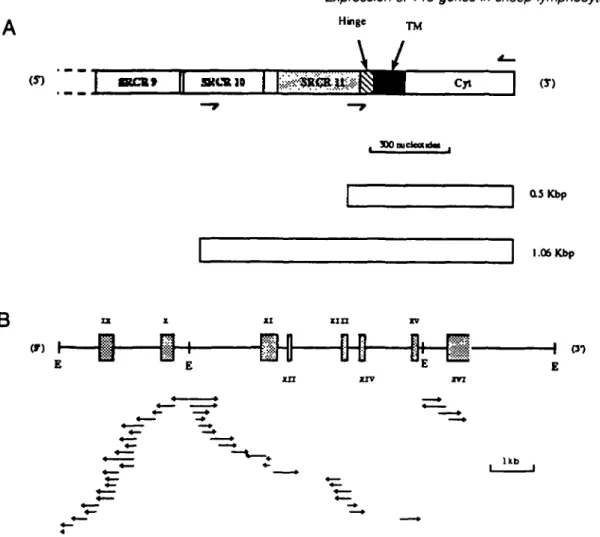
(T) H (J-) U bFig. 1. Cloning and sequence analysis of clone 18 DMA. The C-temninal (3') segment of bovine WC1 is depicted in (A) and the locations of the oligonucleotides used to construct 1.06 and 0.5 kbp probes are indicated by arrowheads above or below the locations of their complementary sequences and the PCR products generated (by their use as primers) and used as molecular probes are depicted under the WC1 schematic. The structure of clone 18 DNA is shown in (B) and sequences corresponding to putative exons are boxed. Exons are labelled (Roman numerals) from the ninth SRCR repeat of WC1. fcofll sites are indicated (E) and the direction and extentlon of DNA sequencing runs are depicted by arrowheads. The sequence of exon XI is incomplete.
sequencing reactions using the PRISM Ready Reaction Dye Deoxy Terminator Cycle Sequencing Kit (Applied Biosystems, Foster City, CA) according to the manufacturer's instructions. Genomic PCR isolates were sequenced directly using the same forward and reverse primers that were used to amplify these products. PCR products resulting from LcDNA samples (see text) were first cloned into the T-tailed pGEM-T vector (Promega) and then sequenced using forward and reverse universal pUC/M13 primers (IBI).
Results
The overall strategy was to design oligonucleotide primers based on two or more sequences which were identical to or highly conserved between the cytoplasmic domains of several T19 gene or cDNA sequences. Oligonucleotides would then be constructed to facilitate amplification by PCR of intervening sequences using either cDNA or genomic recombinant DNA as templates. Sequence data for this purpose were obtained
from three sources: firstly, the published WC1 sequence of Wijngaard etal. (4); secondly, from a particular T19 genomic recombinant (clone 18); and thirdly, from a cloned T19 cDNA from mRNA derived from enriched •$ T cells.
The partial stmcture of T19 genomic clone 18
The construction of a sheep genomic library and the success-ful cloning of 27 independent T19 recombinants from it has already been described (25). One recombinant, clone 18, was selected for sequence analysis because of its ability to bind multiple oligonucleotides based on the published WC1 sequence. We chose to examine preferentially the non-repeti-tious segment of this genomic clone which encoded the membrane anchor and the cytoplasmic segments rather than a segment containing exclusively SRCR repeats. Accordingly, three EcoRI fragments of clone 18 DNA which bound a 1.06 kbp probe derived by PCR from the WC1 template (25) were subcloned. As depicted in Fig. 1, the probe facilitated detection of homologous DNA corresponding to the non-repetitious membrane and cytoplasmic segments of WC1 as
well as the last (3' proximal) two SRCR repeats and part of a third. The three EcoRI restriction fragments which bound radioactive 1.06 kbp probe (of approximate size 3, 3 and 6 kbp) were cloned. One of the 3 kbp cloned fragments and the 6 kbp fragment both bound a truncated (0.5 kbp) probe (Fig. 1) which lacked the SRCR DNA segments: the remaining 3 kbp fragment did not, and its probable 5' location was therefore suspected. The strategy used to sequence the three restriction fragments is depicted in Fig. 1 along with the locations of the 1.06 and the 0.5 kbp probes used. Selected segments of the nucleotide sequence data obtained from
clone 18 DNA along with amino acid sequences are presented in Fig. 2. Several major features are apparent.
(i) Putative exon sequences are defined by both the uninter-rupted runs of amino acids which they encode and by the presence of consensus 3' and 5' donor and acceptor sequences immediately flanking the exon sequences (Fig. 2).
(ii) At the level of both nucleotide sequence and amino acid sequence a high level of homology between clone 18 and the corresponding segment of WC1 exists (Fig. 2). (iii) Two essentially complete SRCR repeats of the T19 gene
A *
K.
C t o n * 1 l ACACCACACA^KrrCCGCCTCCTCMCCCCaiCTCTCCCTCTGCCCCCACACTGCACATCCTTCACCACNGCTCCTGCGCCHCCATCTCTC^TCACCCCTG C«CCT«ACCAT(XCC»CCTarrCTCCACCCACCTCGCCTGTCCACAAGCCCTCAATCCCACMGCTCTCCTCACTTCGCCOCAGCATCACGa CCACCTCWCCATCCCCCTCTCCTGTCCACKCACCTCGCCTCTGCACAACCCCTCAATCCCACGCCCTCTCCTCACTTCCCaittCCATCAKCCCCATC 3000DtlX
3100 3 2 0 0 CTCCCACACCCCCATGGATGATCTCACCCTCTCCATCATCTGCAGTCACCTTGCCTSTGGGGACAGTGGACGTCTCAACACCT^ CTGCCCCAGCCCCATGGMCATATCACTCTCTCCGTGATCTGCACACAGCTTCGATCrrGGCGAC^GTGGAACTCTCAA^^ 3 3 0 0 GCTTCTACACCCCCCTG<OTACATCTAATTCAGTCTCGGAAAACTGATACCTCTCTCTGGCAGTGTCCTirrc CCIICTA(aCCCCCCTCOrrACATTTAATrCAGTGTCCCAAAATCGATACCTCTCTCTGGCAGTGTCCT IC TGGCCCATGGAAATACAGTTCATCCTCTC • • • . 1 1 X 1 CCAACGACCAACCCTACATCTCCTGCCCAcT 3 4 0 0 3500 TACTCATGGANCCTCCCANANCCCGANGTCCTCTGT GTGTGT 3 6 0 0 CAGCACCTCCCCTCTGCCCAG<XCXTGGAAGCCGTGCGCTCTCCAGCATTTGGCCCTGGAAATGGGAGCATCTGCCTGGACGAGGTGCAGTGCGCGCGCC 3 7 0 0• • * * xjixn
C<3U^GTCCTCCCTCT<WCACTCTCCTGCG«CtCCTGGGGGCAGANCANCTGCAAGCACMGGA^• " * 1 * * * * * *• •
ATTCCCCACGACTACAGCAGGTACT* ACCTCAAGTCCTCTCCCTKGTATCTTCTCCCTGCCTGCGCTCCTCTGCCTCATCCTCCCAGeCCTTCTC ATCTTCTCCCxmixiv
3600 3900 XJVJ.XV. • • ** » • • X V I X V I • • AGTMCCTGGAACTCCTCCTGCCTCTCAJWa^GCNAGGAGCAAGTNCNCCCAGAGAAGGACGATGGNKCGAGGTCCTCTCACACAGGCTCTTGTCTGAAC rCGAACTCCTTCTC * * • • * * * * TTCTCCykakGACCCTGCTCATCCTCGAGAAGCACAAGAGAGCCTCTCCCTGCTCCAGGGAAAGAAAGCCGACGCTGCGTACGATGATCrrGAACTCACTG 4000 4100 4200 4300 CCCTCCGAACATCCCCAGTGACTTTCTCGTGA CCCTGGGAACATCCCCACTGACTTTCTCGTCA 4332B
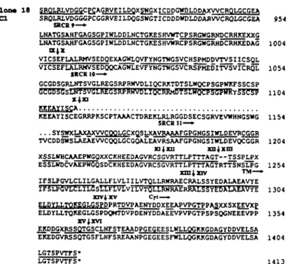
C l o n a 1 8 SROLRI.VDGGCPCAGRVEII.DOXSWGXICDDGWt)I,DDAXWCROLGCGEA M d SRQLRLVDGGGPCGGRVEILDQCSKGTICDDDWDLODARWCROLGCGEA SBCR»—• 954 LNATGSAHFGAGSGPIWLDDLNCTGKESHVHTCPSRGWGR1TOCRHKEXXG LNATGSAHFGAGSGPIHLDDLNCTGKESHVWRCPSRGWGRHDCRHKEDAG 1004 «•* VICSEFLALRMVSEDQEXAGWLQVrYNGTWGSVCHSPMDDVTVSIICSQL VICSEFLALRMVSEDQQCAGWLEVTYNG'nJGSVCRSPMEDITVSVICRSL 1 0 5 4 SRCR 10—» GCGDSGR^SVGLREGSRPRWypLIQCRKTDTSLWQCPSGPKKrSSCSP GCGDSGSLNTSVGLREGSRPRWVDLIQCRKMDTSLHQCPSGPHKKS3CSP 1 1 0 4 4 KEEAYISCA KEEAYISCEGRRPKSCPTAAACTDREKLRLRGGDSECSGRVEVWHNGSWG 1 1 5 4 SRCR11—» . . . SYSWaAXAXWCOOLGCXOSLXAVRAAArGPGNGSIWLPEVRCGGR TVCDDSWSI-AZAEWCQQLGCGQALEAVRSAAFGPGNGSIWLPEVQCGGR 1204 n j x n xnjxm XSSLWBCAAEPWGQXXCKHEEDAGVRCSGVRTTLPNTAGT—TSSPLPX ESSLWDCVAEPWGQSDCKHEEDAGVRCSGVRTTLPTTTAG"TRTTSNSLPG 1254 T M — • irSLPGVLCLILGALLFLVLIILVTQLLRWRAECRALSSYEDAXAEAVYE IFSLPGVLCLILGSLLFLVLVILVTQLLRHRAERRALSSYEDALAEAVYE 1304 nv|xv Cyt—• ELDYLLTOKEGLGSPDPRTDVPAEWYDDXEEAPVPGTPPA3XXSXEEVXP ELDYU.TQKEGLGSPDQMTDVPDENYDDAEEVPVPGTPSPSQGNEEEVPP 1354 XV J XVI EKDDGXRSSQTGSCLNTSTEAADPGEGEESLWLLQGKKGDAGyDDVELSA EKEDGVRSSQTGSFLNFSREAANPGEGEESFHLLQGKKGDAG*DDVELSA 14 04 1413Fig. 2. The nucleotide and predicted amino acid sequences of the putative exons of T19 clone 18. (A) The nucleotide sequence of clone 18 (top) is written after deletion of all intron DNA sequences and is compared to the corresponding segments of WC1 (bottom). Nucleotldes which differ between the two sequences are asterisked (above the clone 18 sequence). The dotted lines in the clone 18 sequence represent incomplete data and the exon junctions are indicated using arrowheads above the clone 18 sequence. A parallel comparison of the predicted amino acid sequences is presented in panel B and amino acid residues are numbered using the WC1 numbering system, vertical arrowheads in (B) indicate the position of amino acids coded at exon-intron boundaries and the commencements of SRCR motifs are indicated under the WC1 sequence. Uncertain nucleotides and amino acids in both montages are represented as N and X respectively and identities between upper and lower sequences in (B) are represented by the solid line between them.
from which clone 18 was derived were uninterrupted by introns (Fig. 1, exons IX and X); the same is likely to be true for exon XI but its sequence was not completed in this work. The putative hinge domain was encoded by a single discrete exon: the proposed membrane anchor region of this gene occurred on an adjacent exon along with 10 nucleotides of the cytoplasmic tail. The remainder of the cytoplasmic segment was subdivided into three small exons.
Sequence analysis of T19 cDNA from enriched y6 T cells To accumulate more comparative sequence data on the T19 gene family in sheep a cDNA clone obtained from enriched 76 T cells was cloned and then sequenced. This cDNA clone, which was derived by PCR amplification of cDNA followed by cloning, was previously used as a probe in Southern blot analysis (25) and shown to bind an essentially identical spectrum of sheep genomic restriction fragments as the corresponding WC1 fragments (Fig. 1, 1.06 kbp probe). The sequence of this sheep cDNA along with the corresponding sequence of WC1 and sheep genomic clone 18 is presented in Fig. 3. Comparison of the three sequences suggested two conserved nucleotide segments on which to base the construction of oligonucleotides to conduct subsequent PCR reactions. The locations of these oligonucleotides, 14F and 15R, are listed under the sequences to which they correspond:
the 15R primer was the reverse complement of the sequence under which it is listed in Fig. 3. The utility of the 14F, 15R primer pair is in distinguishing genomic DNA and cDNA templates on the basis of PCR product length is apparent from Fig. 3. Specifically, the PCR product predicted by priming T19 genomic DNA with this set of oligonucleotides traverses most of exons 14 and 15 (-250 bp) and the intervening intron (~1 kbp in clone 18) thereby distinguishing it unambiguously from material amplified from cDNA which would be at least 1 kbp shorter.
Analysis of cloned T19 genomic recombinants
The two oligonucleotide primers 14F and 15R should prime efficient PCR reactions on all three of the DNA molecules on which their sequences are based. It was anticipated that they may also have the potential to prime on other T19 DNA molecules, possibly even those with imperfectly complement-ary priming sites. To assess this possibility samples of 27 T19 genomic recombinant DNA samples, described previously, were used as templates for PCR reactions. Figure 4 depicts samples of the nine PCR products which were successfully obtained, each derived from different genomic templates by PCR optimization (details in Fig. 5 legend) after subsequent purification of the major species from each reaction. The nucleotide sequences of each of the nine products were determined and they are listed in Fig. 5(A). Five of the nine
CAACACCTCT CTTCGTCTCA CCCAACCTTC TACACCCCCC TCCCTACATT TAATTCACTC TCCCAAAATC CATAC am . . . _ _ _ _ . . . . . . . . • • • • . . . A C - - - - - . - - - e x . . . . .
i C CT
• a crcTCTCice CACTCTCCTT CTCCCCCATC CAAATACACT TCATCCTCTC CAAACCACCA ACCCTACATC TCATC M S
•|6t aall asm ~c c— T— A —c—
• a TCAACCAACA ACACCCAACA CCTCTCCAAC TCCTCCCCCC TCCACACACA CACACAACCT CCCCCTCACC CCACC l a s T * t o a l l a s m — e C - TC-C CA T—TTCAB— A—AC f f l II C - C —
WOX ACACACCCAC TCCTCACCCC CCCTCCACCT CTCCCACAAC CCCTCCTCCC CCACCCTCTC CCATCACTCC TCGAC m
l i t aall asm ~c._e-T A c c c I I T A N
CCTCCCACAC CCTCACCTCC TCTCTCACCA CCTCCCCTCT CCCCAMCCC TCCAACCCCT CCCCTCTCCA CCATT M a a l l asm — trt —CAH -c~ -c—c A
II »-» —C — H H T-C N
C-C---• C l TCCCCCTCCA AATCCCACCA TCTCCCTCCA CCACCTCCAC TCCCCCCCCC CCCACTCCTC CCTCTCCCAC TCTCT tm
j t t a a U a s m c TC C
Clam* I I c _ .G _ . H- c C
• a TCCCCACCCC TCCCCCCACA CCCACTCCAA CCACCACCAC CATCCTCCTC TCACCTCCTC TCCTCTAACC ACAAC • * • >
Hi "ail csm -» A — A—A —
II N - A N " " C
*
ATTCCCCACC ACCACACCAC CCACCACAAC AACCTCAAHT TCTCTCCCTC CCATCTTCTC CCTCCCTCCC CTTCT M l A T T A
-- .T - T — T C - C K - T
I
• C l CTCCCTTATC CTCCCCTCCC TTCTCTTCCT CCTCCTCCTC ATCCTCCTCA CTCACCTACT CACATCCACA CCACA M lit aall K a .--C-— - . AC-CA — . — . - - - C -„.£.. -c— —
CCCCACACCC TTATCCACCT ATCAACATCC TCITCCTCAA CCTCTCTATC ACCACCTCCA TTACCTTCTC ACACA « • > . . K A O - A — — T - — - - . . . - . . . . . — • - — TC- . . . . .
• 0 1 CAAC«AACCT CTCCCCACCC CACATCAKAT CACTCATCTC CCTCATCAAA ATTATCATCA TCCTCAACAA CTACC • • Tit a*U an*\ -,; c '
Claaa I I , . .r, c N C
• 0 1 ACICCCTCCA ACTCCTTCTC CCT'.TI.ACJJ. CAATCACCAC CAACTCCCCC CALACAACCA SCACCCCCTC ACCTC < i n l i t oall a m C--T c c - ; —T
II » — - — - . . - - - c - - c KN- --ecu -H-H--
C--T--IWC-CTCTCACACA CCCICTTTCC TCAAtTTCTC CASACAACCA CCTAATCCTC CKCAACCACA ACACACCTTC TCCCT CT --C T — c ccc _ _ _ . . - - . . . . — ^T. . . . — . — . .c— c - - T — c — - - - - - A - - - - C - - — - •
• 0 1 CCTCCACCOO AACAAACCCC ATCCTr.CCTA TCATCATCTT CAACTCACTC CCCTCCCAAC ATCCCCACTC ACTTT «U> l i t aall asm ,, A
-daman R r f
•01 CTCCTSA e n
l i t aall AB Clau l i
Fig. 3. Sequences of the corresponding segments of three T19/WC1 genes. The nucleotide sequence of a cloned fragment of a sheep cDNA clone obtained from enriched 76 T cells is shown aligned to the corresponding regions of WC1 (upper sequence) and clone 18 (lower sequence). The oligonucleotides constructed for subsequent PCR amplifications are shown as directional arrows below the sequences selected. The junctions between consecutive exons (of clone 18 DNA) are indicated by vertical arrows. The translational end-points of the coding regtons are indicated by a heavy dot. Asterisks within the nucleotide sequences have been introduced to maximize sequence homology between the three sequences compared. The symbol N indicates an uncertain nucleotide assignment. The 14F primer sequence is identical to the sequence above it but the 15R sequence is the complement of the sequence above it.
kk M A1 A2 12 M 13 IS 18 19 20 21
Flg. 4. Analysis of PCR products from nine T19 genomic clones. Small DNA samples from nine genomic clones described by I. D. Walker et al. (25) were each used as templates for separate PCR reactions. In all cases the 14F and 15R primers described in Methods were used to obtain the amplified products. The PCR conditions used were as described in the text with the following alterations in the primer and MgSO4 concentrations of the PCR reaction in order to
maximize the yield of each PCR product. Products from clones 18, 19, 20 and 21 were amplified using the primers at a concentration of 0.2 \iM and MgSO« at 2 mM. The remaining clones also required the primers at this concentration but with 4 mM MgSO«. Lane M contained 0.5 jig of SSP1 DNA cut with EccRI, and lanes A1 and A2 contained known amounts (250 and 350 ng respectively) of the previously purified PCR products of clone 18 in order to aid with quantitation of the products for subsequent nucleotide sequencing.
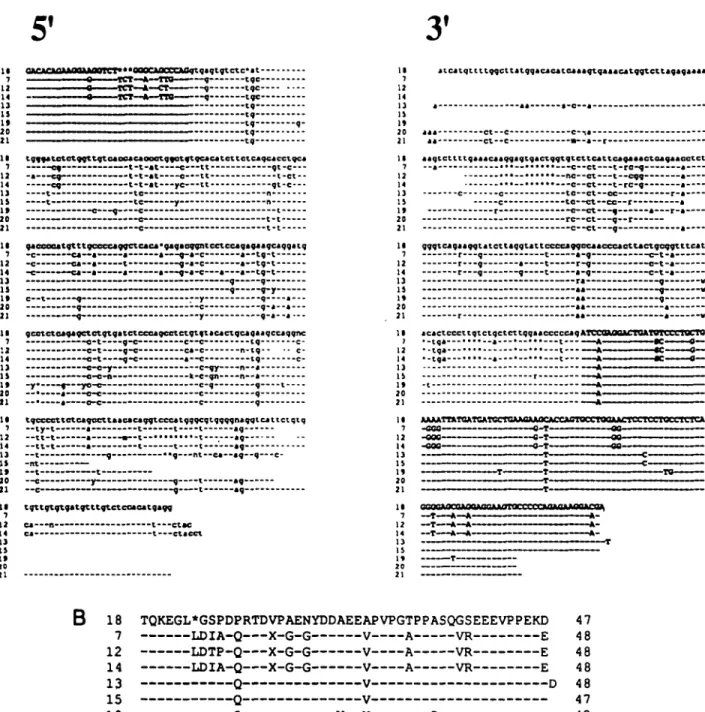
sequences listed are unique and differ by multiple nucleotides from their closest correspondent. Two additional unique sequences have exact correspondents which differ at no position over the lengths compared. Acceptable consensus donor and acceptor sites occur in the intron sequences immediately flanking exons. Specifically, the invariant sequence .. . GGT occurs at the 3' end of every exon 14 sequence in Fig. 5(A) and the sequence . . . AG immediately precedes the first nucleotide of exons 15 (Fig. 5A). These conform precisely to the minimum consensus sequences located at exon-intron boundaries (14). Differences between the nucleotide sequences occur in exons as well as introns and the predicted amino acid sequences of exon segments are shown in Fig. 5(B). No in-frame stop codon occurs within the sequence of any exon. It was essential to establish that the differences in nucleotide sequences between cloned T19 isolates were not due to sequence errors introduced by the PCR reactions used to obtain amplification. Accordingly three PCR reactions were conducted using 5-50 pg of WC1 cDNA as template and primers as described above, and from each separate reaction several cloned isolates were sequenced. The sequence data (not shown) from all clones (<2000 molecules in all) concorded perfectly with that published for the corresponding segment of WC1. It was concluded that the error rate of the PCR reaction was much lower than the observed variation between specific T19 sequences of Fig. 5(A).
Analysis of T19 mRNA
Preliminary experiments in which poly(A)-containing RNA samples obtained from a number of sheep lymphoid tissues
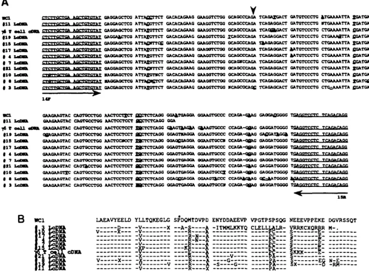
were examined by Northern blot analysis revealed weak and heterodisperse patterns when probes for T19 were used (data not shown). In contrast mRNA for the glycolytic enzyme glyceraldehyde-3-phosphate dehydrogenase was detected on the same Northern blots as a sharp intense species with little evidence of degradation. These difficulties in detecting T19 transcripts were bypassed by copying mRNA molecules into cDNA using reverse transcriptase and then amplifying specific segments of T19 reverse transcripts by PCR using the 14F-15R primer system as described in the previous section for analysis of genomic fragments. The sequence data in Fig. 6(A) are of the inserts from individual clonal isolates obtained by inserting PCR products into the pGEM-T vector. pGEM-The sequences obtained are aligned to one another, to the segment of cDNA obtained from enriched y> T cells and to the corresponding segment of WC1; the corresponding predicted amino acid sequences are aligned in Fig. 6(B). In all, 10 sequences were obtained. Eight of them differ at multiple positions from their closest correspondents obtained from lymph cell mRNA, from the sequence obtained from enriched y5 T cells, from WC1 and from any of the exon sequences of Fig. 5. Two of the sequences obtained, those from lymph cDNA (LcDNA) clone 17 and LcDNA clone 4, are identical over the region examined. LcDNA clone 3 encodes an in-frame stop codon which would result in a truncated cytoplasmic region finishing at recombinant 1359 of WC1. The functional significance of this is not known. The possibility that one or two nucleotide differences are due to errors introduced by the PCR reaction cannot be absolutely ruled out but the low error frequency estimated previously by analysing the PCR products from the WC1 template (<1 per 2000 nucleotides) conclusively negates the possibility that such errors are an important contributor to the pattern of variation apparent in Fig. 6(A).
Discussion
The results of this paper confirm and extend a previous report from this laboratory that the sheep T19 protein of y5 T cells is encoded by one or more genes of a large gene family (25). The first suggestion that this may be the case was based upon restriction digest analysis of T19 (WC1) DNA in cow (4). We established that a similar situation prevailed in sheep by demonstrating sequence differences between four genomic T19 recombinants selected from a total of 27 clonal isolates (25). This paper utilizes the PCR reaction to extend the earlier analysis and to discriminate against other members of the scavenger receptor superfamily by concentrating on only the cytoplasmic regions of T19 genes. The results in Fig. 5 establish beyond doubt that most of the genomic recombin-ants isolated in our earlier work are from unique T19 genes rather than simply overlapping fragments of one or a few T19 genes. It is notable where sequences from exon-intron boundaries were encountered that the nucleotide sequences of introns immediately flanking exons conformed to consensus sequences required for functional splicing of pre-mRNA mole-cules (14). The absence of internal stop codons within putative exons of T19 genes (Fig. 5) also implied that the sequences of these genes were constrained to preserve translational potential. The sequence analysis of the T19 insert of
recombin-A
5'
3'
II 7 12 14 1) IS It 20 21 II 7 12 14 1) 1] It 20 21 II 7 '.2 14 1) IS 20 11 II 7 12 14 11 15 1» 20 21 tgffatctetggttgteaocacaooetgfotftgcacatcttctcaqcacctqca 0 ^ . — . . . t - t - a t c tt g t - c — -t-t-at---tc ye— t t -— c g c— c— c— c -gaoooaatgtttgceccaggctcaca'gagaogqatcctccagaaaaqcaqgatq -c - - c a ~ a ~ a —a—-fl-a-c- a—tg-t -c —«a—a——-t-» —g-c a--c -• • a--ca—a--—a—-—-a-—g-aa--c™a a—tg-t-gcetctea^agctetgtgatctcccagcetctgtftacactqcagaaqccaqgnc . . c - t g-e—•- —c—• ig . . . . — c - t -g-« ca-e n-ig-- • - c- —e-w--fc-c-ga— -ty-t-I -ty-t-I tgccccttctcagccttaacacaqqtcccatgqgcqtqqqqpaqqtcattctqtq 7 12 14 1] II If 20 21 nt — t — -—c— tgttgtftaatgtttgtctcoaoatgavg c a - -ctac •ctacct II 7 12 14 11 IS It JO 21 II 7 12 14 13 IS It 20 21 II 7 12 14 1} 19 It 20 21 12 14 1) IS It 20 21 II 7 12 14 1] IS It 20 21 II 7 12 14 1] IS It 20 21 itcatqttttqqcttatqqacacatcaaagtqaaautggtcttagagaaaa aagtcttttqaaacaaqgagtqactqqtgtcttcattcagjaactaagaacotct ct t c — c t — c c ct - -ro-g- —cqq- -rc-g-c to—ct—cc—r r c—ct g a r-r e —c t g — r c—ct g ggqtcagaaqqtatcttaqgtatt r—q t gqocaacceaottaetgcqytttcat , - , c-t-a r-•-9— --t r-9 • • - - - — c-t-a "' " 9 • . . . --« -tqa- •tqa-AAAATTMOATCATI .cooB 18 TQKEGL*GSPDPRTDVPAENYDDAEEAPVPGTPPASQGSEEEVPPEKD 47
7 LDIA-Q X-G-G V A VR E 4 8
!2 LDTP-Q X-G-G V A VR E 4 8
14 LDIA-Q X-G-G V A VR E 48
13
QV D 48
15
-QV 47
19
Q _ v— V C 42
20 Q V 42
21
QV 42
Fig. 5. The nucteotlde alignment of nine products of cloned T19 genomic templates obtained after PCR amplification using the 14F and 15R pair of primers. Panel A (51) corresponds to the sequence of these products obtained using the 14F primer for sequencing and panel A (3')
corresponds to the reversed complement of the sequences obtained using the 15R primer. Nucleotides in bold represent sequences of exons 14 (5') and 13 (3'). Dashes indicate identity to the clone 18 sequence and asterisks indicate deletions relative to clone 18. The intron sequence Is in lower case. Alignment of the predicted amino acid translations of the exons of panel A is depicted in panel B. The asterisk in the amino acid sequence of clone 18 represents a deletion relative to clones 7, 12 and 14. Dashes indicate identity to the clone 18 sequence. The symbol X signifies a sequence uncetainty.
ant clone 18 revealed that at least three predicted SRCR domains were encoded on separate, unbroken exons as might have been anticipated: the predicted hinge, transmem-brane and cytoplasmic segments of this gene were distributed over five separate exons. Several sequence elements origin-ally identified in WC1 were found in T19 clone 18 and occurred
in the same order as in WC1. This observation does not support a possibility that differential splicing events of T19 pre-mRNA molecules are common although much more work is necessary to comprehensively rule out such a possibility. The 3' end of the clone 18 sequence, distal to the translational stop codon, contains two contiguous poly(A) addition sites
GAOGAGCTCa A T T A f i C r r C T GACACACAAO GAGCAGCTCO ATTAfiGTTCT GACACACAAO HffTOTnTAT GAGOAGCTCO ATTAXOTTCT GACACAGAAG tGGA&CTCG ATTAXOTTCT GACACASAAC m i T O T U T A T 0AOOA0CTC0 A T T A 0 I T T C C OACACAGAAG OPTOTdTAT OACOAOCTCO A T T A X U U C T OACACAOAAO OAOOAOCTCO ATTAXOTTCT OACACAOAAO OAGGAGCTCG ATTAXOTTCT CACACAGAA6 GACOAUCTCU A T T A f i l l l C T OACACAGAAC •AOOAQCTCO ATTAXOTSCT OACACAOAAO •AQGAOCTCQ ATTAfiKCTCT OACACAflAAQ OACOAOCTCO ATTAXOTTCT OACACAGAAC
QAAGGTCTQG GAAGGTCTOO QAAGOTCTOO GAACOTCTGC OAASOTCTGO QAAUU11IUU GAAGOTCTOG GAAGGTCTGG OAAOOTCTOO GAAQOTCTGQ GAAG8TCT0G GAAGGTCTGQ KAOCOCAQC XQATOATGCT XQATOATGCT fiOATOATGCT SGATGATGC4 XCATQATCCT fiOATOATCCT fiOATCATCCT fiCATOATCCT XQATOATCXT fiOATOATCCT XCATCflrGCT CCATOATCCT
GOJ^^TGAQGA QQJVAOTOCCC CCAGJ^^'G^U^G GAOQJ^QCOGC 7
OOA
^ f t ' 7 T t ^ry i / a ^ ^ i w r i r f f CCAGA-Q0M! CACGATOGC0 TCJmQTf.'CTC
O^UkOTvU^GOA OflAftOTCCCC CCJ^^A^^j^UkO GJ^QGA^^LGG^ T^Al^Q^CCT^^^ OOAaCOAGOA OGAAOTGCai CCAOAHJBAO aAOGATCGGO T C A n d T T C T r GGAOTOAGGA OOAAOTOCCC CCACA-OBAG OAOOATOOGO T n A m r r r T T C TTABAg GGAOTOAOOA OOAAOTOCCC CCAOA-QBHO aAOGATGGGO 1
OttUTTOAOQA OOAAOTOCCC CCAOA-OQAO OAOOATOGOO 1 GOAOTOAGOA OOAAOTOCCC CCA0A-G4AG GAGCATGGGO TOAOnTCCTC GGAOTGAOGA OOAAOTCCCX CCAGA-OSAO OAOCATOGGC T f l i . GGAGCGACOA GGAAOT^CCC CCAOAa£^A0 Q C ^ ^ T O O O O J OGAGTGAOGA OOAAOTOCai CCAOA-OflAO OAOQATOOOO 1
LAEAVYEELD YLLTQKEGLG SPDQMTDVPD ENYDDAEEVP VPGTPSPSQG NEEEVPPEKE DGVRSSQT D V X — A - S A -ITMMLKKYQ CLELLLALR" VRRJCCXORRR M-.
¥
-KJ»
SKKK-A
Fig. 6. Comparative nucieotide and amlno acid sequences of T19 cDNA molecules obtained from sheep lymph. Panel A depicts an alignment of nucieotide sequences of the ten cloned PCR products (LcDNA) derived from sheep lymphocyte cDNA. PCR reactions were performed using 14F and 15R primers- the corresponding regions of the yB T cell enriched cDNA sample and that of WC1 are also shown. The cloned PCR products were sequenced using M13 Universal primer. The primers used for PCR amplification are Indicated in underlined, plain text. Nucleotides which differ between the 12 sequences are shown in bold, underlined text. The asterisk indicates the In-frame stop codon present in LcDNA clone 3 and the arrowhead indicates the splicing site of exons 14 and 15. Panel B depicts an alignment of amino acid translations of the PCR amplified region of the ten LcDNA clones and the corresponding regions of WC1 and the •& T cell enriched sheep cDNA sequences Dashes indicte amino acids conserved in all sequences and only amino acids which differ are indicated. The full stop at the end of LcDNA clone 3 Indicates the premature termination of this sequence due to an in-frame stop codon.
•lap 1 CSC IMVDGGCACA(»VEiai:BGEM<»Va>OTl<DLEOAjnrVCroUXGIIAV0AIJ<H£^^
hp«Kl Rap I CXIS Kap « NCI
Fig. 7. Alignments of CD6 SRCR repeat 2 with selected homologous repeats from T19 (clone 18) and WC1. In both panels (A and B) residues present in the SRCR domains selected which are identical to corresponding residues In SRCR repeat 2 of human CD6 (top lines) are depicted in black and only differences are listed. Identity levels between CD6 repeat 2 and other SRCR sequences are listed as percentages. Gaps introduced to maximize the homoiogy between the various sequences are indicated by an asterisk.
and in this respect, too, the gene corresponding to clone 18 has the potential to encode a functional and typical mature mRNA. In summary, the analysis of T19 genomic sequences
found no evidence for transcriptionally or translationally non-functional pseudogenes among the cloned templates amen-able to PCR amplification and sequence analysis.
In the course of this work it was noted that the amino acid sequence predicted by clone 18 exon IX exhibited an unusually high level of amino acid sequence homology to a segment of the human CD6 sequence (7). This latter structure is a cell surface protein of T cells, some B cells and thymo-cytes. It is reportedly present on yd T cells (15). No function is yet known for CD6 although the relationship between the SRCR domains of CD6 and corresponding segments of the murine macrophage scavenger receptor, CD5, and T19 has previously been noted (4). There are three SRCR domains in CD6 and Fig. 7 depicts an alignment of the second of these domains with its flanking domains from CD6, with domain 9 of clone 18 and with selected domains from WC1. The mutual homology between WC1 domains 4,6 and 9, clone 18 domain 9 and the second SRCR domain of human CD6 is striking and far exceeds corresonding internal homology levels obtained when the domains of CD6 are compared with one another or when WC1 domains 4, 6 and 9 are compared with other T19 domains. These internal homology levels are in the range of 25-35%. Repeat 7 of the macrophage differentiation antigen M130, which is very similar to SRCR domains 4 and 8 of WC1 (11), also shares this high homology with domain 2 of CD6 (-60%). It is most likely that this observation denotes an unusual evolutionary conservatism whereby these (CD6-like) domains are subject to structural constraints which do not apply to other SRCR domains. To account for this observation it is tempting to invoke a specialist recognition function shared by these homologous domains. CD6 has been reported to occur on y6 as well as a0 T cells at least in human (15), but the evidence presently available in sheep suggests that the T19 and CD6 molecules may be expressed in a mutually exclusive manner (16) whereas M130 is expressed on all circulating monocytes and most tissue macrophages (11).
The issue of which T19 genes were expressed in a particular lymphoid tissue was addressed using a sample of mRNA derived from unfractionated lymph cells obtained by cannulat-ing a sheep efferent lymph node. This cell population con-tained 20% y& T cells. In addition, a sample of 76 T cells enriched by panning was used as an alternative source of mRNA. The results of experiments in which mRNA molecules were reverse transcribed, then amplified by the PCR reaction and finally cloned and sequenced are presented in Fig. 6(A). The notable feature of these experiments is that they demon-strate the expression of a number of T19 genes in sheep lymphocytes. In fact, of the ten clonal isolates examined in this study and obtained using the same primer system only two isolates had exactly the same sequence. The expression of multiple T19 genes in sheep lymphocytes biologically enriched for yd T cells was not totally surprising given earlier serological evidence that certain epitopes of WC1 (T19) are found selectively on discrete subpopulations of bovine y6 lymphocytes (3,4). This serological heterogeneity implied the existence of multiple protein variants within the T19 population of molecules, and the demonstration in this work of multiple unique T19 mRNA molecules within a single lymphocyte population strongly suggests that the serological variation within the T19 population can be determined genetically (rather than post-synthetically). Nevertheless, no evidence was presented in this work that the expression of T19 genes is confined to yS T cells. Whereas there is no serological
evidence to support the expression of any T19 molecule on other T cells or on B cells, it is formally possible that such molecules are indeed expressed but lack epitopes which enable their detection using existing T19-specific mAbs. Work is now in progress to explore the possibility that CD8+ and/ or CD4+ T cells also contain T19 mRNA molecules in spite of their lack of reactivity with T19-specific mAb. Even if the expression of the T19 repertoire were indeed confined to yd T cells, it would be of considerable interest to know whether, within an individual yb T cell, a single T19 gene were expressed. The demonstration that only one T19 gene was expressed in individual y& T cells would imply a cell- or subset-specific function for T19 molecules.
We previously speculated that T19 may constitute a family of lymphocyte homing molecules (25). The basis for this speculation was a series of earlier observations by others that f5 T cells resident in a number of diverse epithelial tissues each constitute a discrete subset as evidenced by their respective yd TCRs (17-20). The specificity of 76 TCRs per se cannot account for the resident status of such T cells (21) and it was attractive to invoke a different multigene family, particular members of which might encode cell surface ligands which contribute specific tissue trophism to the cells which express them. The T19 family, or at least some members of it, seemed well suited to such a role, given their apparent restriction to y6 T cells and the fact that different T19 polypep-tides could be expressed in different subsets of yd T cells. If recirculating lymphocytes are considered to be a discrete subset restricted to the blood and lymph (as opposed to epithelial tissues) by a T19 structure then the expression of at least nine unique T19 genes within this population would not necessarily have been anticipated. This number should be considered an underestimate for two reasons. Firstly, there is every reason to expect that additional cloned mRNA copies could have been rescued had more intensive work been undertaken. Secondly, the same primer system used to enable the amplification of T19 sequences from cDNA was found to be limited to 30-40% of cloned T19 gene fragments, most of which contained at least part of the segment which the primer system amplified. In short, one or two expressed genes in this population would have been more in keeping with a role for T19 in the specific homing behaviour of lymphocytes. In the context of the proposed role of T19 as a migration ligand of lymphocytes it is important to note that the mRNA segments examined in this work do not correspond to the extracellular segments of the T19 structures which would be expected to engage a notional cognate receptor on vascular endothelium. It is formally possible that the SRCR segments of T19 genes expressed in the cell population selected may exhibit less sequence variation than the cytoplasmic segments do. Even if this were so, the relevance of the diversity of the cytoplasmic segments would still be puzzling.
A recurring theme in sheep immunobiology which distingu-ishes this species from rodents and humans is that genes which code for proteins involved in immune function are often more numerous and more diverse in sheep than in the two latter species. For example, there are at least nine different MHC class II p chains in sheep and probably many more (22). In contrast, there are two in mouse and five in man (23). There are at least five Cy TCR loci in sheep which differ