The Effects of Dietary Mannanoligosaccharides on Cecal Parameters
and the Concentrations of Enteric Bacteria in the Ceca
of Salmonella-Challenged Broiler Chicks
1P. Spring,* C. Wenk,† K. A. Dawson,† and K. E. Newman‡,2
*Institute for Animal Science, ETH Zurich, Universitaetsstr, 2, CH-8092 Zurich, Switzerland; †Department for Animal Science, University of Kentucky, Lexington, Kentucky 40546; ‡North American Bioscience Center,
3031 Catnip Hill Pike, Nicholasville, Kentucky 40356 ABSTRACT The ability of different enteric pathogens
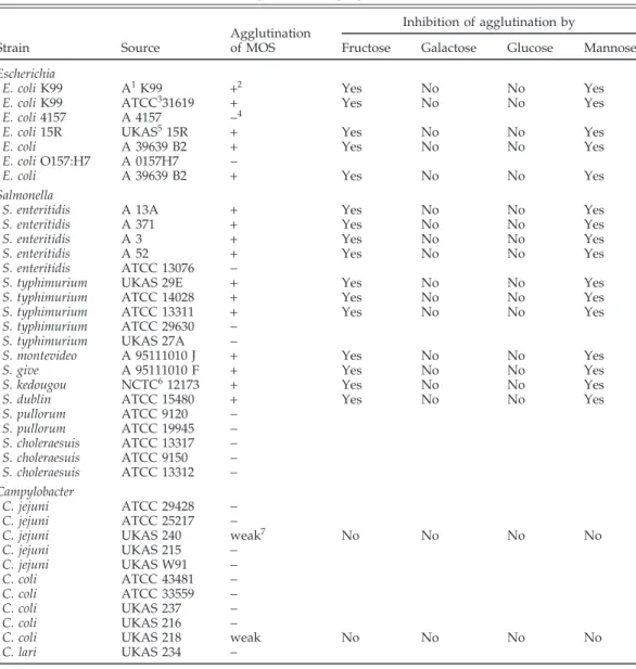
and coliforms to trigger agglutination of yeast cells (Sac-charomyces cerevisiae, NCYC 1026) and a yeast cell wall preparation (MOS) was examined. Five of seven strains of Escherichia coli and 7 of 10 strains of Salmonella typhimu-rium and Salmonella enteritidis agglutinated MOS and Sac. cerevisiae cells. Strains of Salmonella choleraesuis, Salmonella pullorum, and Campylobacter did not lead to agglutination. Two strains that agglutinated MOS (S. typhimurium 29E and Salmonella dublin) and one nonagglutinating strain (S. typhimurium 27A) were selected as challenge organ-isms for in vivo studies in chicks under controlled con-ditions.
In a series of three trials in which 3-d-old chicks were orally challenged with 104cfu of S. typhimurium 29E, birds
(Key words: Salmonella, chicken, mannans, yeast, ceca)
2000 Poultry Science 79:205–211
INTRODUCTION
Newly hatched birds lack complex gastrointestinal mi-croflora and are especially prone to colonization of enteric pathogens (Milner and Shaffer, 1952). However, the appli-cation of cultures of cecal microflora obtained from healthy adult birds (competitive exclusion cultures) to the newly hatched chick has been shown to increase the complexity of their gastrointestinal microflora and give protection against enteric pathogens (Nurmi and Rantala, 1973; Stavric et al., 1985; Corrier et al., 1995). Many enteric pathogens must attach to the mucosal surface of the gut wall to establish themselves in the gastrointestinal tract (Costerton et al., 1978, Fuller et al., 1981). One mode of action of competitive exclusion (CE) cultures to exclude pathogens is related to their ability to increase the
compe-Received for publication April 5, 1999. Accepted for publication September 30, 1999.
1Research was supported by a grant from Alltech, Inc., Nicholasville, KY 40356.
2To whom correspondence should be addressed: knewman@ alltech-bio.com.
205
receiving 4,000 ppm of dietary MOS had reduced cecal S. typhimurium 29E concentrations (5.40 vs 4.01 log cfu/ g; P< 0.05) at Day 10. In a second series of three trials with S. dublin as challenge organism, the number of birds that tested salmonella positive in the ceca at Day 10 was less when MOS was part of the diet (90 vs 56%; P<0.05). To test the effect of MOS on concentrations of bacteria that do not express Type 1 fimbriae, a challenge trial was conducted with S. typhimurium 27A. However, strain 27A did not colonize the birds sufficiently to evaluate whether MOS affected its cecal concentration. Mannanoligosac-charide did not significantly reduce the concentrations of cecal coliforms (P<0.10) although they were numerically lower. It had no effect on cecal concentrations of lactoba-cilli, enterococci, anaerobic bacteria, lactate, volatile fatty acid, or cecal pH.
tition for attachment sites (Schneitz et al., 1993). Because attachment is often mediated through binding of bacterial lectins to receptors containing D-mannose (Eshdat et al., 1978), it may be possible to block the lectins with mannose or similar sugars and to inhibit bacterial attachment.
Oyofo et al. (1989a) tested the effect of different sugars on the in vitro adherence of Salmonella typhimurium to epithelial cells from 1-d-old chicks. They reported inhibi-tion of adherence by methyl-a-D-mannoside and man-nose by more than 90%. The inhibition of the adherence of S. typhimurium by mannose (Oyofo et al., 1989a) is reflected in decreased colonization of S. typhimurium when mannose is added to the diet of young chicks (Oy-ofo et al., 1989b,c). Because relatively high concentrations of mannose are required to control colonization of patho-genic bacteria, the cost of using pure mannose in commer-cial production is prohibitive even for short periods. However, mannose-based carbohydrates occur naturally
Abbreviation Key: BGA=brilliant green agar; CE=competitive exclusion; MOS=mannanoligosaccharide; OD=optical density; VFA
in many products, such as yeast cell walls or different gums, which are available at reasonable prices. The hy-pothesis tested in this study was that mannanoligosac-charides (MOS) from yeast cell walls are able to decrease the concentrations of enteric pathogens that express Type 1 fimbriae in poultry.
The objectives of these studies were to screen different bacterial strains for their ability to agglutinate MOS in yeast cell preparations and to determine the effect of MOS on cecal fermentation parameters, cecal microflora, and enteric pathogen and coliform colonization in chicks un-der controlled conditions.
MATERIALS AND METHODS
Mannanoligosaccharides
The MOS preparation used in these studies was the commercial product Bio-Mos.3The product contains yeast cell wall fragments derived from Saccharomyces cerevisiae. The cell wall fragments are obtained by centrifugation from a lysed yeast culture. The pellet containing the yeast cell wall fragments is then washed and spray dried.
Agglutination of Yeast Products
The ability of the bacteria to adhere to the mannan associated with the yeast cell wall was measured with an agglutination test. Agglutination of MOS (yeast cells) by different enteric pathogens and coliforms in the presence and absence of specific sugars was qualitatively examined (Table 1). These tests were conducted using a modification of the method described by Mirelmann et al. (1980). To standardize the concentration of bacteria, bacterial cells were grown for 24 h on the following media: 10 g/L Bacto-Peptone4; 5 g/L yeast extract; and 5 g/L NaCl (Firon
et al., 1983). Cells were harvested by centrifugation and suspended in PBS (0.05 M, pH 7.2) to reach an optical density at 660 nm (OD660) of 1.0. Mannanoligosaccharide
was suspended at 1 g/L in PBS (OD660 = 1.60). A 24-h
culture of Sac. cerevisiae NCYC 1026, grown in Tryptic Soy Broth4, was harvested by centrifugation, and cells were suspended in PBS to reach an OD similar to the one of the MOS suspension (OD660=1.60). The yeast and MOS
suspensions were blended for 1 min in a kitchen blender to separate clumping cells and MOS particles. Agglutina-tion tests were then performed with 1.0 mL of the bacterial suspension and 1.0 mL of the MOS or yeast suspension. Agglutination was determined microscopically. To deter-mine the inhibitory effect of carbohydrates on agglutina-tion, each agglutinating strain was first incubated for 5 min in PBS containing 50 mM glucose, mannose, galac-tose, or fructose.
3Alltech Inc., Nicholasville, KY 40356. 4Difco Laboratories, Detroit, MI 48232.
5Standard Safety Equipment Co., Palatine, IL 60067. 6Avian Farms, Sommerset, KY 42633.
Animal Trials
Experimental Design. All animal trials were con-ducted following the animal care protocol IACUC 94-0004A approved by the University of Kentucky Institu-tional Animal Care and Use Committee. The ability of salmonella to colonize the intestinal tracts of chickens was evaluated in six separate trials. In each of the first two trials, the birds were divided into two control and two treatment groups containing 10 birds each. Trial 3 was conducted with 26 birds per group to study salmo-nella colonization over time. The fourth and fifth trials also used four groups of 10 birds, but because of low hatchability only 36 birds were assigned to treatment groups (9 birds per group). Birds were hatched and housed in bacterial isolation chambers. All diets met NRC requirements for young birds (NRC, 1994). Gastrointesti-nal microflora of birds were standardized at the beginning of each experiment by inoculating each bird with 1×10⁴ cfu of one of the challenge organisms on Day 3. Cecal parameters were determined on Day 10. Dietary treat-ments in all trials included an unsupplemented diet (con-trol) and a diet supplemented with 4,000 ppm of dietary Bio-Mos.
Bacterial Isolation Chambers.Five bacterial isolation chambers5were used for these studies. One chamber
con-taining the incubator was outfitted with a bacterial trap filled with disinfecting solution; the other four contained plastic housing boxes (3,840 cm2×320 cm2per bird) and
were equipped with regular inlet ports. The chambers were sanitized as described by Makin and Tzipori (1980). Feed, water, and 500 g wood shavings were placed in autoclave bags and were placed in the chambers prior to fumigation.
Eggs. Eggs (broiler type, line 246) were sanitized by spraying with a disinfectant solution (2.0% H2O2, 0.8%
quaternary ammonium, and 0.25% acetic acid) and were misted daily during incubation. On Day 19, eggs were dipped for 30 s in disinfectant solution (37 C) and were transferred into the sterile incubator, which was placed in one bacterial isolator.
Standard Inoculum.Newly hatched chicks were inoc-ulated with a standard inoculum to provide similar basic microflora for each bird at the beginning of each experi-ment. The standard inoculum was derived from the hatching debris from a commercial hatchery. The debris were mixed and stored in 100-g aliquots at−80 C. The inoculum was tested to be salmonella-free according to the approved standard method for isolation of salmonel-lae (Wallace et al., 1992). Microbial counts on the debris revealed the following bacterial concentrations: total bac-teria, 2.5×10⁷cfu/g (Viand Levure Agar at pH 6.7; Barnes et al., 1979); coliforms, 8.03×10⁶cfu/g (MacConkey Agar); enterococci, 7.50×10⁶cfu/g (KF Streptococcal Agar); and lactobacilli, <1,000 cfu/g (Rogosa Agar). The defrosted debris was mixed 1:5 with 0.1% peptone water, blended in a sterile kitchen blender, and each bird was given 0.25 mL from the tip of a pipette (1.25×10⁶ cfu/bird). Six h
as-TABLE 1. Inhibitory effects of fructose, galactose, glucose and mannose on bacterial agglutination of mannanoligosaccharide preparations (MOS)
Inhibition of agglutination by Agglutination
Strain Source of MOS Fructose Galactose Glucose Mannose
Escherichia
E. coli K99 A1K99 +2 Yes No No Yes
E. coli K99 ATCC331619 + Yes No No Yes
E. coli 4157 A 4157 −4
E. coli 15R UKAS515R + Yes No No Yes
E. coli A 39639 B2 + Yes No No Yes
E. coli O157:H7 A 0157H7 −
E. coli A 39639 B2 + Yes No No Yes
Salmonella
S. enteritidis A 13A + Yes No No Yes
S. enteritidis A 371 + Yes No No Yes
S. enteritidis A 3 + Yes No No Yes
S. enteritidis A 52 + Yes No No Yes
S. enteritidis ATCC 13076 −
S. typhimurium UKAS 29E + Yes No No Yes
S. typhimurium ATCC 14028 + Yes No No Yes
S. typhimurium ATCC 13311 + Yes No No Yes
S. typhimurium ATCC 29630 −
S. typhimurium UKAS 27A −
S. montevideo A 95111010 J + Yes No No Yes
S. give A 95111010 F + Yes No No Yes
S. kedougou NCTC612173 + Yes No No Yes
S. dublin ATCC 15480 + Yes No No Yes
S. pullorum ATCC 9120 − S. pullorum ATCC 19945 − S. choleraesuis ATCC 13317 − S. choleraesuis ATCC 9150 − S. choleraesuis ATCC 13312 − Campylobacter C. jejuni ATCC 29428 − C. jejuni ATCC 25217 −
C. jejuni UKAS 240 weak7 No No No No
C. jejuni UKAS 215 − C. jejuni UKAS W91 − C. coli ATCC 43481 − C. coli ATCC 33559 − C. coli UKAS 237 − C. coli UKAS 216 −
C. coli UKAS 218 weak No No No No
C. lari UKAS 234 −
1Culture Collection of Alltech Inc., Nicholasville, KY. 2The MOS particles were clumping together. 3American Type Culture Collection, Rockville, MD. 4No MOS particles were clumping together.
5Culture Collection of the University of Kentucky, Department of Animal Science, Lexington, KY. 6National Collection of Type Cultures, Central Public Health Laboratory, London, U.K.
7A few particles were clumped, but the majority were free.
signed to four groups of 10 chicks each and were trans-ferred into the bacterial isolation chambers.
Husbandry.Room temperature was adjusted to 34 C for the first day and was then gradually lowered to 28 C at Day 10. Light was on continuously for the first 24 h after the chicks were transferred into the chamber and for 20 h each of the following days. Feed and water were consumed ad libitum. The feed was a corn-soybean meal diet that was formulated to meet NRC recommendations and contained no antimicrobial supplement (NRC, 1994). Challenge Cultures and Their Application.Challenge cultures of S. typhimurium (strains 29E and 27A) and S. dublin for in vivo studies were grown in the basal peptone-yeast extract medium described earlier. The cultures were then diluted in sterile 0.1% peptone solution to a final
concentration of 4×10⁴cfu/mL. Concentrations of
salmo-nellae were determined on brilliant green agar (BGA) using spread-plate technique. One quarter of a milliliter of the diluted culture (1×10⁴cfu/bird) was given to each bird from the tip of a pipette.
Sampling and Sample Analysis.After 10 d, the chicks from each group were sacrificed by asphyxiation with CO2, and both ceca were aseptically removed. The content
of one cecum was placed in a sterile test tube, weighed, and diluted 1:10 with sterile 0.1% peptone solution. The empty cecum was cut longitudinally and placed in lactose broth. Decimal dilutions of each sample were prepared in 0.1% peptone solution. Salmonellae were enumerated on BGA containing 30 mg naladixic acid/L after 24 h incubation at 37 C. Naladixic acid was added to the media
to facilitate the selection of the antimicrobial-resistant challenge organisms. The tube containing the empty ce-cum in lactose broth was incubated at 37 C for 24 h for enrichment of salmonellae (Wallace et al., 1992). Cecal samples that were salmonella-negative on BGA plates, but positive in the enrichment test, were assigned as 1.50 log cfu/g (Nisbet et al., 1993). Samples that were con-firmed negative in the enrichment test were assigned a concentration of 1 cfu/g (0 log cfu/g) (Nisbet et al., 1993). Four birds were randomly selected from each group for analysis of the concentrations of different groups of intes-tinal bacteria. Lactobacilli, enterococci, coliforms, and an-aerobic bacteria were enumerated in duplicate using the pour-plate technique on Rogosa SL agar, KF streptococcal agar, McConkey agar, and reinforced clostridial agar,7
re-spectively.
Determination of pH, Volatile Fatty Acid, and Lactate Concentrations.The content of the second cecum was diluted 1:10 in distilled water, and pH was determined with a combined electrode.8Samples were stored at−20 C until analyses of volatile fatty acids and lactate. Volatile fatty acid analysis was conducted on a Hewett-Packard model 5890 gas chromatograph,9fitted with a 180-cm × 4-mm glass column, containing 10% SP 1000 and 1% H3PO4on 100/120 Chromosorb WAW10with a
modifica-tion of the method described by Erwin et al. (1961). D-and L-lactate were determined spectrophotometrically with D- and L-lactate dehydrogenase, respectively, in the presence of nicotinamide adenine dinucleotide (Brandt et al., 1980).
Statistical Analysis.Data were analyzed by ANOVA appropriate for the randomized complete block design; chambers (10 birds) were considered to be the experimen-tal unit. Differences between treatment means were as-sessed by least significant difference test (Kuehl, 1994). Bacterial concentrations were subject to log 10 transfor-mation prior to analysis. To evaluate the overall effects of MOS supplementation, the data was initially blocked by trial. No significant trial by treatment interactions were observed, and the data were pooled across the trials. Sta-tistical analyses were conducted using the general linear models procedure of SAS威 (SAS Institute, 1985).
RESULTS
Agglutination
Five of seven strains of Escherichia coli and 7 of 10 strains of S. typhimurium and Salmonella enteritidis agglutinated MOS (Table 1). Salmonella montevideo, Salmonella give, Sal-monella kedougou, and S. dublin also triggered agglutina-tion. Addition of Salmonella choleraesuis and Salmonella pullorum strains did not lead to agglutination of the MOS
7All media were purchased from Difco Laboratories. 8Orion Research Inc., Boston, MA 02129.
9Hewlett-Packard, Palo Alto, CA 94304. 10Supelco Inc., Bellofonte, PA 16823.
particles. Agglutination associated with these Gram-neg-ative bacterial strains could be inhibited with mannose or fructose but not with glucose or galactose. However, it generally took 9 to 16 times more fructose to bring about the inhibitory responses observed with mannose (data not shown). This result suggests that fructose was less effective as a blocking agent. Agglutination patterns of MOS were similar to patterns of S. cerevisiae (data not shown). Different strains of Campylobacter were also tested for their abilities to agglutinate MOS (Table 1). Only two strains showed some agglutination; however, these ag-glutinations were very weak and were not affected by addition of carbohydrates.
Challenge Trials
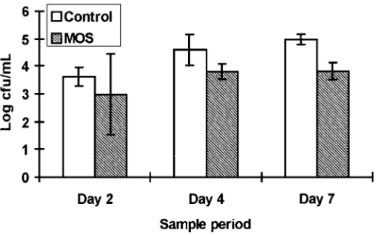
The concentrations of salmonellae and coliforms in chicks challenged with S. typhimurium 29E are shown in Table 2. The concentrations of salmonella were consis-tently lower in birds fed MOS than in birds fed the unsup-plemented diet. Salmonellae concentrations were be-tween 4.97 and 5.73 cfu/g for the control and ranged from 3.79 to 4.29 log cfu/g for the MOS treatment. Over the three trials, MOS-supplemented chicks had lower S. typhimurium 29E concentrations by about 25-fold (4.01 vs. 5.40 log cfu/g; P<0.05). Concentrations of coliforms also tended to be lower (P< 0.1) when MOS was added to the feed (8.47 vs. 8.71 log cfu/g). Concentrations of lacto-bacilli, enterococci, and anaerobic bacteria were not af-fected by treatment. Volatile fatty acid concentrations, lactic acid concentrations, and pH were not affected by dietary treatment. Average values were as follows: pH 5.61, acetate 61.7 mM, propionate 5.9 mM, butyrate 12.2 mM, and lactate 9.0 mM. In Trial 3, the cecal S. typhimu-rium 29E concentrations in the unsupplemented group rose from 3.61 log cfu/g at 2 d to 4.97 log cfu/g at 7 d after challenge (Figure 1). Salmonella typhimurium 29E concentrations in the MOS treatment group were lower (P<0.05) than those of the unsupplemented control group 7 d after challenge. Birds receiving MOS had average cecal salmonella concentrations of 2.98 log cfu/g at 2 d and 3.79 log cfu/g at 7 d after challenge.
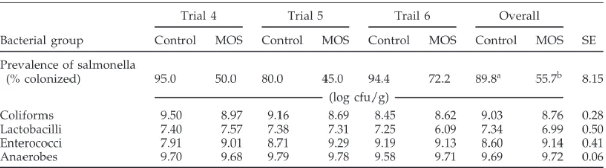
The percentage of birds from which S. dublin could be recovered was lower in the MOS-treated groups than in the control group in all three trials (Table 3). In Trial 6, 90% of the birds in one of the groups receiving MOS were colonized with S. dublin. As a result, the mean reduction in the percentage of colonized birds with MOS addition appears lower in Trial 6 (24%) than in Trial 4 (47%) or Trial 5 (44%). Over the three trials, MOS decreased the number of salmonella-positive birds from 89.8 to 55.7% (P<0.05). Concentrations of coliforms, lactobacilli, entero-cocci, and anaerobic bacteria were not different in the two treatment groups. Cecal pH, VFA, and lactate concen-trations were in a similar range as in the trials with S. typhimurium and were not affected by treatment (data not shown).
Salmonella typhimurium 27A, a nonaggultinating strain of salmonella, colonized the chicks at a very low rate.
TABLE 2. Effect of dietary mannanoligosaccharide preparations (MOS) on concentrations of different bacterial populations in the ceca of chicks maintained in microbiological isolators and
challenged with Salmonella typhimurium 29E1
Trial 1 Trial 2 Trail 3 Overall
Bacterial
group Control MOS Control MOS Control MOS Control MOS SE
(log cfu/g) Salmonella 5.73 4.29 5.50 3.95 4.97 3.79 5.40a 4.01b 0.08 Coliforms 8.78 8.40 8.87 8.73 8.48 8.29 8.71 8.47 0.05 Lactobacilli <6.00 <6.00 <6.00 <6.00 7.41 7.61 Enterococci 8.12 8.35 8.26 8.25 8.03 8.24 8.13 8.28 0.05 Anaerobes 9.37 9.14 9.20 9.11 9.21 8.91 9.26 9.05 0.04
a,bValues in same row within trial with no common superscript differ significantly (P<0.05). 1n=6.
This organism could only be recovered from the ceca of 3 of 40 chicks that were challenged with doses of up to 1
×104cfu/bird. Two control chicks and one bird receiving MOS were salmonella positive at 10 d, which made it impossible, using the bacterial isolation chambers, to eval-uate the effects of MOS on the colonization of birds by this strain.
DISCUSSION
The colonization of bacteria on mucosal tissues is recog-nized as an important step in the infectious process. To colonize the mucosal surfaces, bacteria must first bind to the epithelial cells of these tissues. One way of binding to epithelial cells is through attachment of Type-1 fimbriae (Ofek et al., 1977). A variety of strains of E. coli, Salmonella, and Campylobacter were tested for the presence of Type-1 fimbriae by means of yeast and MOS agglutination. No differences were observed between agglutination pattern of the two yeast products. The majority of the strains of E. coli and S. typhimurium and S. entritidis tested aggluti-nated both the yeast culture and MOS. All agglutination observed in these enteric bacteria was mannose sensitive. This result is in agreement with findings by Duguid (1964) and Duguid et al. (1966) who reported high frequency of
FIGURE 1.Effect of dietary mannanoligoscharrides on the concentra-tion of salmonellae in the chicks maintained in microbial isolators for 2, 4, and 7 d after challenge with Salmonella typhimurium 29E. Bars represent standard deviations of the mean values.
Type-1 fimbriae expression in S. typhimurium, Salmonella typhi, and S. enteritidis. However, Mirelmann et al. (1980) only found 4 of 13 strains of S. typhimurium to express Type-1 fimbriae. The poultry-specific salmonella types, Salmonella pullorum and Salmonella gallinarum, did not trig-ger agglutination in the work of Duguid (1964) or in the study presented here. Four of seven tested strains of E. coli agglutinated MOS. Mirelmann et al. (1980) reported that about half of the tested strains of E. coli expressed Type-1 fimbriae. None of the tested Campylobacter strains caused a mannose-sensitive agglutination of MOS. How-ever, mannose was reported to inhibit adherence of C. jejuni to epithelial cells in vitro (McSweegan and Walker, 1980), and protective effects with dietary mannose were also reported against Campylobacter jejuni colonization in chickens (Schoeni and Wong, 1994). These authors did not determine whether the Campylobacter strains used in their study expressed mannose-sensitive fimbriae. There-fore, it is not possible to say whether inhibition of attach-ment or other mechanisms were involved in their studies. Fucose is a constituent of the mucus the primary site of Campylobacter colonization, and inhibition of Campylo-bacter colonization has also been reported with addition of fucose to the diet (Cinco et al., 1984).
Standardization of experimental conditions is im-portant to achieve reproducible results. Initial results ob-tained from challenge studies with birds from a commer-cial hatchery were not reproducible. It was hypothesized that the birds might have been exposed to different bacte-rial flora at the hatchery, which could explain differences between trials. Birds that would have been exposed to a more complex microflora at the hatchery would exert a stronger CE effect in the gastrointestinal tract and would, consequently, be colonized with the challenge organisms at lower rates. Therefore, the experimental conditions were modified to standardize microflora of each bird at the beginning of the experiment by removing the native microflora of the egg through sanitation and then exposing the newly hatched bird to a constant level of microorganisms derived from hatching debris in bacterial isolators. This process allowed for the standardization of the microflora at the beginning of the experiment and allowed the composition of the cecal gastrointestinal mi-croflora, cecal pH, and cecal acid concentrations of the
TABLE 3. Effect of dietary mannanoligosaccharide preparations (MOS) on concentrations of different bacterial populations in the ceca of chicks maintained in microbiological isolators and
challenged with Salmonella dublin1
Trial 4 Trial 5 Trail 6 Overall
Bacterial group Control MOS Control MOS Control MOS Control MOS SE Prevalence of salmonella (% colonized) 95.0 50.0 80.0 45.0 94.4 72.2 89.8a 55.7b 8.15 (log cfu/g) Coliforms 9.50 8.97 9.16 8.69 8.45 8.62 9.03 8.76 0.28 Lactobacilli 7.40 7.57 7.38 7.31 7.25 6.09 7.34 6.99 0.50 Enterococci 7.91 9.01 8.71 9.29 9.19 9.13 8.60 9.14 0.41 Anaerobes 9.70 9.68 9.79 9.78 9.58 9.71 9.69 9.72 0.06
a,bValues in same row within trial with no common superscript differ significantly (P<0.05). 1n=6.
birds to be similar in each experiment and each treat-ment group.
The main goal of these trials was to evaluate the effects of MOS on salmonella colonization in chicks. The focus was on Salmonella strains expressing Type-1 fimbriae. We hypothesized that MOS might affect colonization of such strains through blocking bacterial attachment to the gut mucosa. In a first series of three trials, dietary addition of MOS decreased the cecal concentration of S. typhimurium 29E. To further investigate whether MOS could decrease the concentration of a strain of Salmonella that colonizes birds with a cecal concentration less than 104cfu/g, an-other series of trials was conducted using S. dublin as a challenge organism. Mannanoligosaccharide treatment also reduced the number of birds from which S. dublin could be isolated in these trials. The challenge trials with S. dublin and S. typhimurium 29E suggest that MOS can lower cecal salmonella colonization of strains that colo-nize at high or low concentrations. The study conducted over time showed that MOS reduced cecal S. typhimurium 29E concentrations 7 d after challenge. To observe cecal S. typhimurium 29E concentration over a longer period of time, with and without withdrawing MOS, might help to further investigate what effect MOS exerts on Type-1 fimbriated enteric pathogens. An inhibitory effect on salmonella colonization in chicks has also been shown with live yeast culture. Line et al. (1995) reported de-creased colonization of S. typhimurium with dietary Sac-charomyces boulardii. However, different modes of action might be involved with live yeast cells. Live yeast cells excrete metabolites, which are known to affect the compo-sition of the gastrointestinal microflora (Girard, 1996) and could, therefore, alter the CE effect of the indigenous microflora. In addition, the strain of S. typhimurium used by Line et al. (1995) has not been screened for presence of Type-1 fimbriae. Therefore, it can not be determined whether the observed reduction was due to yeast cell wall mannan or other mechanisms.
The decrease in concentrations of Salmonella expressing Type-1 fimbriae was not accompanied by changes in cecal parameters such as decreases in pH or increase in propi-onic acid concentrations, which are known to reduce sal-monella concentrations, which suggests that other modes
of action are responsible for the decrease salmonella con-centrations with dietary MOS. In vitro agglutination re-sults suggest that adsorption of salmonellae by MOS, which would keep them from adhering to the intestinal wall, could be a possible mode of action. Oyofo et al. (1989a) tested the effect of different sugars on the adher-ence of S. typhimurium to epithelial cells of 1-d-old chicks in vitro. They reported inhibition of adherence by methyl-a-D-mannoside and mannose by more than 90%. In differ-ent in vivo trials, mannose has also been shown to decrease cecal colonization of S. typhimurium in young chicks (Oy-ofo et al., 1989b,c). The fact that MOS had no effect on the concentrations of lactobacilli and enterococci, two bac-terial species that are known to not express Type-1 fim-briae, further suggests that MOS affects bacterial concen-trations in the gastrointestinal tract by adsorbing bacteria and keeping them from adhering to the gut wall.
Dietary MOS has been shown to decrease the preva-lence of strains of Salmonella expressing Type-1 fimbriae in young chicks under laboratory-scale conditions. No changes in cecal parameters such as a major shift in bacte-rial populations or changes in pH or VFA concentrations, which are known to affect salmonella, were observed with MOS addition. Field trials will be required to show how the reduction noticed under small-scale conditions will transfer into commercial production systems. Be-cause MOS can be considered animal, environmental, and consumer friendly, it might offer an additional and inter-esting tool that can be included in a hazard analysis criti-cal control point program of poultry production.
REFERENCES
Barnes, E. M., C. S. Impey, and B.J.H. Stevens, 1979. Factors affecting the incidence and anti-salmonella activity of the anaerobic caecal flora of the young chick. J. Hyg. Camb. 82:263–283.
Brandt, R. B., S. A. Seige, M. G. Walters, and M. H. Bloch, 1980. Spectrophotometric assay for D-lactate in plasma. Anal. Biochem. 102:39–46.
Cinco, M., E. Banfi, E. Ruaro, D. Crevatin, and D. Crotti, 1984. Evidence for L-fucose-(6-deoxy-L-galactopyranose) medi-ated adherence of Campylobacter ssp. to epithelial cells. FEMS Microbiol. Lett. 21:347–351.
Corrier, D. E., D. J. Nisbet, C. M. Scanlan, A. G. Hollister, D. J. Caldwell, L. A. Thomas, B. M. Hargis, T. Tomkins, and J. R. DeLoach, 1995. Treatment of commercial broiler chickens with a characterized culture of cecal bacteria to reduce salmo-nella colonization. Poultry Sci. 74:1093–1101.
Costerton, J. W., G. G. Geesey, and K. J. Cheng, 1978. How bacteria stick. Sci. Am. 238:86–95.
Duguid, J. P., 1964. Functional anatomy of Escherichia coli with special reference to enteropathogenic E. coli. Rev. Latinoam. Microbiol. 7(Suppl. 13-4):1–16.
Duguid, J. P., E. S. Anderson, and I. Campbell, 1966. Fimbriae and adhesive properties in salmonellae. J. Pathol. Bacteriol. 92:107–138.
Erwin, E. S., G. J. Marco, and E. M. Emery, 1961. Volatile fatty acid analysis of blood and rumen fluid by gas chromatogra-phy. J. Dairy Sci. 44:1768–1771.
Eshdat, Y., I. Ofek, Y. Yehuidit-Gan, Y. Yashouv-Gan, N. Sharon, and D. Mirelmann, 1978. Isolation of a mannose-specific lec-tin from E. coli and its role in the adherence of the bacteria to epithelial cells. Biochem. Biophy. Res. Com. 85:1551–1559. Firon, N., I. Ofek, and N. Sharon, 1983. Carbohydrate specificity of the surface lectins of Escherichia coli, Klebsiella pneumoniae and Salmonella typhimurium. Carbohydr. Res. 120:235–249. Fuller, R., S. B. Houghton, and B. E. Brooker, 1981. Attachment
of Streptococcus faecium to the duodenal epithelium of the chicken and its importance in colonization of the small intes-tine. Appl. Environ. Microbiol. 41:1433–1441.
Girard, I. D., 1996. Characterization of stimulatory activities from Saccharomyces cerevisiae on the growth and activities of ruminal bacteria. Ph.D. Diss., University of Kentucky, Lex-ington, KY.
Kuehl, R. O., 1994. Statistical Principles of Research Design and Analysis. Duxbury Press, Belmont, CA.
Line, E. J., N. J. Stern, N. A. Cox, and J. S. Bailey, 1995. Saccharo-myces treatment to diminish Campylobacter and Salmonella in chicks. Poultry Sci. 74(Suppl. 1):201. (Abstr.).
Makin, T. J., and S. Tzipori, 1980. Inexpensive techniques for the production and maintenance of gnotobiotic piglets, calves and lambs. Aust. Vet. J. 56:353–358.
McSweegan, E., and R. I. Walker, 1980. Identification and charac-terization of two Campylobacter jejuni adhesins for cellular and mucus substrates. Infect. Immun. 53:141–148.
Milner, K. C., and M. F. Schaffer, 1952. Bacteriologic studies of experimental salmonella infection in chicks. J. Infect. Dis. 90:81–96.
Mirelmann, D., G. Altman, and Y. Eshdat, 1980. Screening of bacterial isolates for mannose-specific lectin activity by ag-glutination of yeast. J. Clin. Microbiol. 11:328–331.
NRC, 1994. Nutrient requirement of poultry. National Academy Press, Washington, DC.
Nisbet, D. J., D. E. Corrier, and J. DeLoach, 1993. Effect of mixed cecal microflora maintained in continuous culture and of dietary lactose on Salmonella typhimurium colonization in broiler chicks. Avian Dis. 37:528–535.
Nurmi, E., and M. W. Rantala, 1973. New aspect of salmonella infection in broiler production. Nature 241:210–211. Ofek, I., D. Mirelmann, and N. Sharon, 1977. Adherence of E.
coli to human mucosal cells mediated by mannose receptors. Nature 265:623–625.
Oyofo, B. A., J. R. DeLoach, D. E. Corrier, J. O. Norman, R. L. Ziprin, and H. H. Mollenhauer, 1989a. Effect of carbohy-drates on Salmonella typhimurium colonization in broiler chickens. Avian Dis. 33:531–534.
Oyofo, B. A., J. R. DeLoach, D. E. Corrier, J. O. Norman, R. L. Ziprin, and H. H. Mollenhauer, 1989b. Prevention of Salmo-nella typhimurium colonization of broilers with D-mannose. Poultry Sci. 68:1357–1360.
Oyofo, B. A., R. E. Droleskey, J. O. Norman, H. M. Mollenhauer, R. L. Ziprin, D. E. Corrier, and J. R. DeLoach, 1989c. Inhibition by mannose of in vitro colonization of chicken small intestine by Salmonella typhimurium. Poultry Sci. 68:1351–1356. SAS Institute, 1985. SAS威 User’s Guide. SAS Institute Inc.,
Cary, NC.
Schneitz, C., L. Nuotio, and K. Lounatma, 1993. Adhesion of Lactobacillus acidophilus to avian intestinal epithelial cells me-diated by the crystalline bacterial cell surface layer (S-layer). J. Appl. Bacteriol. 74:290–294.
Schoeni, J.C.L., and A.C.L. Wong, 1994. Inhibition of Campylo-bacter jejuni colonization in chicks by defined competitive exclusion cultures. Appl. Environ. Microbiol. 60:1191–1197. Stavric, S., T. M. Gleeson, B. Blanchfield, and H. Pivnick, 1985. Competitive exclusion of salmonella from newly hatched chicks by mixtures of pure bacterial cultures isolated from fecal and cecal contents of adult birds. J. Food. Prot. 48:778–782.
Wallace, H. A., V. R. Bruce, G. June, F. Satchell, and P. Sherrod, 1992. Salmonella. Pages 57–58 in: Bacterial Analytical Man-ual. 7th ed. AOAC International, Arlington, VA.