Gene expression in cultured endometrium from women
with different outcomes following IVF
Nick A. Bersinger
1
, Dorothea M. Wunder, Martin H. Birkha¨user and Michael D. Mueller
Department of Obstetrics and Gynaecology, Inselspital, Berne University Hospital, University of Berne, Murtenstrasse 35,
Berne CH-3010, Switzerland
1
Correspondence address. Fax: þ41-31-63-20953; E-mail: nick.bersinger@dkf.unibe.ch
Estradiol and progesterone are crucial for the acquisition of receptivity and the change in transcriptional activity of target genes in
the implantation window. The aim of this study was to differentiate the regulation of genes in the endometrium of patients with
recurrent implantation failure (IF) versus those who became pregnant after in vitro fertilization (IVF) treatment. Moreover, the
effect of embryo-derived factors on endometrial transcriptional activity was studied. Nine women with known IVF outcome (IF,
M, miscarriage, OP, ongoing pregnancy) and undergoing hysteroscopy with endometrial biopsy were enrolled. Biopsies were
taken during the midluteal phase. After culture in the presence of embryo-conditioned IVF media, total RNA was extracted
and submitted to reverse transcription, target cDNA synthesis, biotin labelling, fragmentation and hybridization using the
Affy-metrix Human Genome U133A 2.0 Chip. Differential expression of selected genes was re-analysed by quantitative PCR, in which
the results were calculated as threshold cycle differences between the groups and normalized to Glyceraldehyde phosphate
dehy-drogenase and b-actin. Differences were seen for several genes from endometrial tissue between the IF and the pregnancy groups,
and when comparing OP with M, 1875 up- and 1807 down-regulated genes were returned. Real-time PCR analysis confirmed
up-regulation for somatostatin, PLAP-2, mucin 4 and CD163, and down-regulation of glycodelin, IL-24, CD69, leukaemia
inhibi-tory factor and prolactin receptor between Op and M. When the different embryo-conditioned media were compared, no
signifi-cant differential regulation could be demonstrated. Although microarray profiling may currently not be sensitive enough for
studying the effects of embryo-derived factors on the endometrium, the observed differences in gene expression between M
and OP suggest that it will become an interesting tool for the identification of fertility-relevant markers produced by the
endometrium.
Keywords: endometrium; gene profiling; IVF; miscarriage; implantation failure
Introduction
Two conditions have to be met in order to obtain a pregnancy: good
embryo quality and high endometrial receptivity. Successful
implan-tation is only possible during a very short phase within the luteal
phase, the so-called ‘implantation window’ which appears to be of
about 5 days in duration (Days 20 – 24 in a physiological menstrual
cycle) (Bergh et al., 1992). The two hormones estradiol (E
2) and
pro-gesterone are crucial for the acquisition of receptivity and the change
in transcription activity of target genes (Wilcox et al., 1999).
Pre-viously these genes were investigated one by one for their role in
implantation. The first reports studying the implantation window
glob-ally with a large-scale genomic array (Affymetrix
w) compared the
expression profiles in endometrium between the late proliferative
with the midluteal phase (Kao et al., 2002) or between days LHþ2
and LHþ7 in spontaneous ovulatory cycles (Riesewijk et al., 2003).
In this latter investigation performed with the HG-U95A array, 211
genes yielded a fold change of 3 or higher between the two time
points. For many of these a role in the context of endometrial
recep-tivity or even fertility had not been previously described, but the
stron-gest transcriptional up-regulation (107-fold) was observed for
glycodelin A (GdA, synonyms PP14, PEP, PAEP). It is a secretory
epithelial endometrial product (Mueller et al., 2000) whose serum
levels are high in the late luteal phase (after the progesterone peak)
of the menstrual cycle and steeply decline thereafter (Joshi et al.,
1982). Endpoint polymerase chain reaction (PCR) and subsequent
densitometric analysis yielded a 3.1-fold glycodelin gene
up-regulation in the endometrium during the implantation window
when compared with the late follicular phase tissue (Kao et al.,
2002). Another microarray study, involving an almost identical
proto-col (day LHþ8 versus LHþ3 in spontaneous cycle) and the same chip
(U95A), confirmed the validity of the method and identified 107
dif-ferentially expressed genes (Mirkin et al., 2005). At the same time
the Spanish/Dutch team from the first U95A study (Riesewijk et al.,
2003) obtained gene expression profiles from endometrium after
con-trolled ovarian stimulation (COS) with an even larger, more recently
developed array (Affymetrix HG-U133A), and differential expression
was again found for more than 200 genes when compared with
endo-metrial tissue obtained in natural cycles at similar timing (Horcajadas
et al., 2005). This was considered a surprising observation, and it was
concluded that current ovarian stimulation for treatment by in vitro
fertilization (IVF) was far from optimal, and this issue was further
strengthened by a reduction (9.8-fold) of GdA mRNA in COS (IVF)
compared with spontaneous cycles.
Gene transcription rates and expression profiles in patients with
implantation failure (IF) and recurrent miscarriage in comparison
with ongoing, uneventful pregnancy are not well investigated to
# The Author 2008. Published by Oxford University Press on behalf of the European Society of Human Reproduction and Embryology. All rights reserved.
date. It has been shown that monoamino oxidase A expression was
deficient in the endometrium of women with IF (Henriquez et al.,
2006). Moreover, the genes for C4BPA, CRABP2 and OLFM1 were
found to be differentially expressed in the endometrium of patients
with unexplained recurrent miscarriage when compared with normal
fertile women (Lee et al., 2007).
The endometrium is receiving signals from the embryo floating in
the uterine cavity at the time of implantation, but these have not
been systematically investigated due to problems of sensitivity. A
recent study with gene profiling on purified endometrial stromal
cells cultured in the presence and absence of trophoblast-conditioned
media found 4817 genes to be differentially regulated as a function of
the presence of these media (Hess et al., 2007).
The first aim of our study was to investigate and differentiate the
transcriptional up- and down-regulation of genes in patients with
recurrent IF versus those who became pregnant after IVF treatment,
and particularly between miscarried and ongoing clinical pregnancies.
The second aim was to concurrently study the effect of early
embryo-derived factors on the transcriptional activity in the cultured
endometrium at the time of implantation, by the addition of
embryo-conditioned medium from successful and unsuccessful IVF cycles as
a culture supplement. To our knowledge, results from studies using
such an approach have not been published to date.
Materials and Methods
Patient selection
Using the clinical records from our Department, women fulfilling the following inclusion criteria for this study were selected. First, an endometrial tissue sample, biopsied using the Pipelle-de-Cornier suction curette and stored frozen (liquid nitrogen) in medium containing 10% (v/v) serum and 20% (v/v) dimethyl sulphoxide, must have been available in sufficient quantity from a previous hysteroscopic investigation for infertility and, second, the patient has been undergoing treatment by IVF and embryo transfer in our clinic, where the pre-embryo transfer embryo-conditioned media are routinely kept in store. The study protocol (collection of endometrium) was approved by the Ethical Committee of the University of Berne, and informed consent was obtained from the patients for both endometrial investigation as well as for the IVF treatment. Three IVF-embryo transfer outcome groups were defined: (A) IF as defined by a negative serum hCG (,2 mIU/ml) result between 14 and 17 days after embryo transfer; (B) implantation followed by early pregnancy (,12 weeks) loss; and (C) clinical, ongoing pregnancy. In order to obtain enough RNA after the explant culture in the presence of the differ-ent embryo-conditioned media supplemdiffer-ents (see below) three or four patidiffer-ents were selected for each of these groups as it has been suggested elsewhere (Catalano et al., 2003), and their tissue was pooled at the time of setting up the suspension cultures (see below). The mean age of the women providing these three tissue pools was 33.7 + 4.5 (SD) years and did not differ between the groups. No endometriosis was documented in these patients by laparoscopy. All tissues were taken during the implantation window in the luteal phase: the mean cycle day at biopsy was 23.6 + 1.8 (SD) and the cycle length, again without difference between the three groups, was 30.0 + 1.6 (SD) days.
IVF procedure
IVF was performed 3 – 4.5 months after hysteroscopy and the collection of the endometrial biopsy in all groups. COS using the ‘long’ protocol after GnRH agonist down-regulation, oocyte retrieval, intracytoplasmic sperm injection (Tesarik and Sousa, 1995) and embryo transfer were performed as described elsewhere (Wunder et al., 2005). Embryos were cultured in microdroplets (20 ml) of IVF G1 medium under mineral oil (both from Vitrolife, Kungsbacka, Sweden) until they were transferred on Day 3 after oocyte retrieval. Due to the legal constraints in Switzerland, embryos from the same patient were trans-ferred. On the day of embryo transfer, 10 ml of this embryo-conditioned medium was collected, diluted 1:10 by the addition of 90 ml of the same medium to prevent evaporation and stored at 2208C until used as a cell culture supplement (see below). Embryo-conditioned media from the same
patients providing endometrial tissue (see above) were pooled into the same groups A (IF), B (pregnancy loss) and C (ongoing pregnancy). Blank IVF G1 medium provided a fourth media pool.
Endometrial explant suspension culture
The endometrial tissue was thawed by immersion of the cryotube in a water bath (378C), washed twice with 15 ml Dulbecco’s MEM containing 25 mM Hepes, glutamine, 10% fetal bovine serum, penicillin, streptomycin and fungi-zone (all from Gibco-Invitrogen, Paisley, Scotland). We have decided to perform explant suspension cultures; the advantages are an intact three-dimensional environment during the culture experiment and the assurance to obtain sufficient amounts of RNA for hybridization, but with this system we would not be able to know the source (epithelial or stromal) of the mRNA. The tissue was coarsely dissected with scalpels on a sterile glass petri dish, and a suspension prepared by several gentle passages through a syringe fitted with an 18-gauge needle. The resulting explant size was such that central necro-sis would not occur during the short duration of the culture experiment. The suspensions of each biopsied endometrium group (A, B or C) were divided into four cultures in a 24-well plate and 2 ml of complete medium without phenol red, with 25 mM Hepes, glutamine, 2% fetal bovine serum, penicillin, streptomycin and fungizone (all from Gibco-Invitrogen, Paisley, Scotland). After 24 h of culture (378C, 5% CO2in air), the medium was replaced with the same but the serum was omitted, and after 24 h of such conditions of quies-cence fresh medium containing embryo-conditioned supernatant was added: serum-free MEM (total volume 500 ml, containing 50 ml embryo medium in G1), sterile filtered through a 0.22 mm low protein absorption polyvinylidene fluoride membrane (Millipore, Volketswil, Switzerland). Each outcome-defined pool of embryo-conditioned medium (A, B, C and blank IVF G1 medium) was added to each endometrial tissue pool (A, B and C), yielding a total of 12 culture conditions. Due to the small amounts of tissue and particu-larly embryo media available, no replicate cultures were set up. After 24 h of culture, the suspension was centrifuged, the resulting supernatants frozen for further analyses and the endometrial cell pellets immediately subjected to total RNA extraction (see below).
Total RNA extraction
RNA was extracted from cellular pellets collected after suspension culture using the SV Total RNA on-column Isolation kit manufactured by Promega (Madison, WI, USA). The manufacturer’s protocol was followed, and this included an on-column DNAse treatment step. The quantity and quality of the obtained RNA was analysed spectrophotometrically with a Nanodropw photometer (Wilmington, DE, USA) and electrophoretically on an (Eukaryote Total) RNA Nano 6000 Chip (2100 Bioanalyser, Agilentw Technologies, Palo Alto, CA, USA). The total RNA yield per culture condition ranged between 6.17 and 14.19 mg, and the absorbance ratio (260 – 280 nm) for all extractions was 2.03 + 0.09 (mean + SD). Second elution fractions, as suggested in the extraction protocol to be added to increase yield, were discarded since their quality was consistently poorer (A260/280 ratio between 1.64 and 1.88). Both 18S and 28S fractions were seen as sharp peaks in the bioanalyser output. The RNA samples were stored at 2808C until the target synthesis and hybridization experiments were performed.
Gene array hybridization
Hybridizations were performed with the Human Genome U133A 2.0 Chip, obtained from Affymetrix Inc. (Santa Clara, CA, USA), covering 18 400 tran-scripts and variants including 14 500 characterized human genes. Three micro-grams of total RNA per culture condition were used for target synthesis which includes reverse-transcription and synthesis of double-stranded cDNA accord-ing to the Affymetrix GeneChip Expression Analysis Technical Manual. Fol-lowing phenol/chloroform extraction, the purified cDNA was used for an in vitro transcription reaction by using the IVT labelling kit (Affymetrix) to syn-thesize cRNA in the presence of a biotin-conjugated ribonucleotide analogue. An average of 196 mg of labelled cRNA from each reaction was purified on RNeasy Mini columns (Qiagen, Hilden, Germany) and average size of the cRNA molecules was assessed on RNA Nano 6000 Chips as above. The cRNA targets were incubated at 948C for 35 min in Fragmentation buffer and the resulting fragments of 50 – 150 nucleotides were again monitored
using the Bioanalyser. All synthesis reactions were carried out using a PCR machine (T1 Thermocycler; Biometra, Go¨ttingen, Germany) to ensure the highest possible degree of temperature control. The hybridization cocktail (130 ml) containing fragmented biotin-labelled target cRNA at a final concen-tration of 0.05 mg/ml was transferred into Affymetrix Human Genome U133A 2.0 Chips and incubated at 458C on a rotator in a hybridization oven 640 (Affy-metrix) for 16 h at 60 rpm. The arrays were washed and stained on a Fluidics Station 400 (Affymetrix) according to standard Affymetrix recommendations for performing the EukGE-WS2v4 protocol which includes an antibody ampli-fication to increase the signal strength. The arrays were scanned according to the default settings using the GeneChip System confocal scanner (Hewlett-Packard, Santa Barbara, CA). Raw DAT image files were generated using MicroArray Suite (MAS 5, Affymetrix). Further data analysis was conducted using Gene-Springw GX (Agilent Technologies) software.
RT and quantitative PCR of selected transcripts
From the results obtained by the automatic ranking with the Gene-Springw software (Tables I and II), the transcripts shown in Tables III and IV were selected for quantitative investigation of the amplification either for their extent of up- or down-regulation, or for their relevance in the context of repro-ductive function. The total RNA samples extracted from the pelleted endo-metrial explant tissue after culture (0.5 mg, see above) were reverse transcribed using the Moloney murine leukaemia virus enzyme (M-MLV, Gibco-Invitrogen, Paisley, Scotland, final concentration 10 U/ml, total sample volume 20 ml). Quantitative PCR was performed with cDNA from 10 ng RNA per well, in duplicate, using the Taqmanw system and Assays-on-Demandw with the primers/probe samples (single-tube system) listed in Tables III and IV on a Model ABI-7900 sequence detector (all from Applied Biosystems, Foster City, CA, USA). Threshold cycle (Ct) plot data were subsequently normalized for each marker and each cluster comparing the different endometrial tissues or the different embryo-conditioned media used in culture. Glyceraldehyde phosphate dehydrogenase (GAPDH, Hs0099999905_m1) and b-actin (Hs00242273_m1) were used as a housekeep-ing gene control.
Results
Automatic screening of the raw data identified the genes which were
found to be up- or down-regulated by a factor of 2 or more depending
on the arrays that were compared (endometria or medium supplement;
ongoing pregnancy, miscarriage or IF). These genes, with their factor
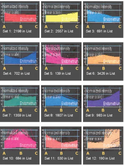
of up- or down-regulation, are shown in Table I. The data were then
analysed by the Genespring SX software; this returned the clustering
for the comparison of the different endometrial tissues such as IF
(group A), miscarriage (group B) and healthy ongoing pregnancy
(group C) on one hand, and of the different embryo media added to
the culture (blank G1 medium, groups A, B and C as above) on the
other. The clusters for the endometria are shown in Fig. 1. The
system identified 12 different sets of patterns with moderate or
strong up- or down-regulation. These differences were seen either
for all pregnancies when compared with IFs (groups BþC versus A,
e.g. sets 3, 5 7 and 11) or, more importantly for the aim of this
project which is the identification of women with recurrent
mis-carriage, between groups C versus B (sets 2, 4, 8, 9, 10 and 12).
Sets with only small variations (sets 1, 2 and 6) were discarded
from the analysis. Similarly, those with an up- or down-regulation
for both groups A and C versus group B (sets 10 and 11) were
removed. The most interesting clusters were thus identified in sets 4
(702 genes up-regulated but not further analysed due to a marginally
lower expression level in group B compared with A), 8 (1807 genes
down-regulated), 9 (983 genes up-regulated) and particularly 12
with fewer genes (190) than in the others but with the strongest
up-regulation.
Real-time PCR analysis of genes selected from the array results
(Tables I and II) and listed in the corresponding Tables III and IV
confirmed significant up-regulation in endometrial tissue from
implan-tation cycles in comparison with IFs for CD163 (P ¼ 0.002) and, to a
lesser extent, PLAP-2 (P ¼ 0.049, Fig. 2). More interestingly, when
comparing the difference in transcriptional activity between ongoing
pregnancies and miscarriages (groups C versus B, Fig. 3), strong
differences were observed for somatostatin, PLAP-2, mucin 4 and
CD163 were found (all increased in group C, P , 0.001). Many of
the other studied molecules were down-regulated between pregnancy
and IF endometrium (Fig. 2). For collagen IV, CEACAM-1, LIF and
fibronectin no difference was observed between groups with
success-ful and failed implantation (Fig. 2). IL-24, CD69, LIF, prolactin
receptor (PRL-R) and glycodelin were strongly down-regulated
between endometrial tissue from women with ongoing and miscarried
pregnancies (all P , 0.001, Fig. 3). Laminin and fetuin B, on the other
hand, distinguished between implantation and failure groups (Fig. 2)
but did not depend on the outcome of an initially established
preg-nancy (Fig. 3). For Carcinoembryonic antigen-related cell adhesion
molecule-1 (CEACAM-1) the findings were inconsistent; a limited
up-regulation of this gene was observed in the ongoing pregnancy in
comparison with the miscarried group, but this was not the case in
the combined implantation (BþC) versus the non-implantation
group (A). When the different embryo-conditioned media were
com-pared between these groups, no significant up- or down-regulation
could be confirmed by real-time PCR (data not shown).
Discussion
The up- or down-regulation of the genes selected from our microarray
experiment results (Tables III and IV) could be largely confirmed by
our real-time RT-PCR experiments, though the extent of the variation
did not necessarily reflect the factor that was returned by the automatic
(Gene-Springw) analysis of the array hybridization. In particular,
highly significant (P ¼ 0.0001 or lower) up- or down-regulations
between endometrial tissues from women with ongoing compared
with miscarried pregnancies were confirmed in RT-PCR for
somato-statin (up), PLAP-2 (up), Mucin-4 (up), glycodelin (down), IL-24
(down), CD69 (down) and LIF (down).
Leukaemia inhibitory factor (LIF) and glycodelin (PP14, PAEP) are
of particular interest in the context of implantation. LIF had been
suggested to play a role in the embryo implantation process in mice
(Stewart et al., 1992), but such a function had not been directly
firmed in the human, and the information in the literature is not
con-sistent. LIF production by cultured endometrial epithelial cells has
been demonstrated (Laird et al., 1997), and uterine flushings at the
time of implantation (LHþ7 to LHþ10) contained more LIF
protein in fertile women than in those with unexplained infertility
(Laird et al., 1997). In this study we could not confirm, using
quanti-tative PCR, the differential gene expression for LIF observed in the
array hybridization experiment between implantation versus
non-implantation cycles (Fig. 2). Similarly, in cultured endometrial
explants (thus also taking the full tissue into account) no differences
in LIF protein production between fertile and infertile women were
found in one study (Delage et al., 1995), while in another this was
the case in the midluteal (but not in the proliferative) phase
(Hambart-soumian E, 1998). On the other hand, a more recent study on uterine
flushings towards the end of the cycle (Day 26) reported lower LIF
concentrations in subsequently pregnant women (Le´de´e et al.,
2002), which would be in better agreement with our observations
made in this study and the reduced gene expression observed
pre-viously in the endometrium (Horcajadas et al., 2005). The
contradic-tory observations with immuno-analytical LIF protein determinations
in uterine flushings could be explained by cycle-dependent secretion
variations between fertile and infertile women, or simply by the
Table I. List of genes, up- or down-regulated by a factor of at least 2.0 between different endometrial tissues (in alphabetical order).
ORIGINAL SOURCE FILE DATA Outcome Group
Gene Gene Name C versus B C versus A B versus A
Up-regulation, .2
210431_at alkaline phosphatase, placental-like 2 14.53
208588_at apoptosis inhibitor 4.73 2.70
210828_s_at aryl hydrocarbon receptor nuclear translocator 2.36
206576_s_at carcinoembryonic antigen-related cell adhesion molecule 1 5.07
211883_x_at carcinoembryonic antigen-related cell adhesion molecule 1 5.47 8.91
201884_at carcinoembryonic antigen-related cell adhesion molecule 5 9.59
215049_x_at CD163 antigen 3.01
208653_s_at CD164 antigen, sialomucin 2.94
207315_at CD226 antigen 4.25 2.43
203507_at CD68 antigen 2.07
214049_x_at CD7 antigen (p41) 2.19
211189_x_at CD84 antigen (leukocyte antigen) 2.48
203757_s_at CEA-related cell adhesion molecule 6 3.67
202310_s_at collagen, type I, alpha 1 2.80
202311_s_at collagen, type I, alpha 1 2.94
202403_s_at collagen, type I, alpha 2 3.32 2.07
202404_s_at collagen, type I, alpha 2 4.16
213992_at collagen, type IV, alpha 6 2.06
213622_at collagen, type IX, alpha 2 3.53 2.50
212488_at collagen, type V, alpha 1 2.18
212489_at collagen, type V, alpha 1 2.79
221729_at collagen, type V, alpha 2 4.20
221730_at collagen, type V, alpha 2 2.93 2.23
212091_s_at collagen, type VI, alpha 1 2.43
212940_at collagen, type VI, alpha 1 2.45
213428_s_at collagen, type VI, alpha 1 2.20
201438_at collagen, type VI, alpha 3 4.06 2.99
211343_s_at collagen, type XIII, alpha 1 3.25 2.11
211809_x_at collagen, type XIII, alpha 1 2.36
207802_at cysteine-rich secretory protein 3 7.60
201325_s_at epithelial membrane protein 1 5.32
209365_s_at extracellular matrix protein 1 4.18
210521_s_at fetuin B 5.34
216377_x_at human placental alkaline phosphatase-like gene 50region 4.61
210141_s_at inhibin, alpha 2.25 4.52 2.01
209540_at insulin-like growth factor 1 (somatomedin C) 2.06
203851_at insulin-like growth factor binding protein 6 3.97 2.24
204989_s_at integrin, beta 4 2.13
209827_s_at interleukin 16 (lymphocyte chemoattractant factor) 2.58
209792_s_at kallikrein 10 4.03
210089_s_at laminin, alpha 4 2.43
210990_s_at laminin, alpha 4 13.50 7.47
208949_s_at lectin, galactoside-binding, soluble, 3 (galectin 3) 2.76
202291_s_at matrix Gla protein 7.76
203876_s_at matrix metalloproteinase 11 (stromelysin 3) 4.39 2.26
203878_s_at matrix metalloproteinase 11 (stromelysin 3) 13.06
204580_at matrix metalloproteinase 12 (macrophage elastase) 9.42 6.30
220541_at matrix metalloproteinase 26 9.13 7.01
210015_s_at microtubule-associated protein 2 8.68 6.92
204895_x_at mucin 4, tracheobronchial 17.42
220158_at placental protein 13-like protein 3.39
208131_s_at prostaglandin I2 (prostacyclin) synthase 7.40
213921_at somatostatin 43.93 13.04
203145_at Sperm-associated antigen 5 4.02 2.58
203167_at tissue inhibitor of metalloproteinase 2 3.45
201147_s_at tissue inhibitor of metalloproteinase 3 2.25
201148_s_at tissue inhibitor of metalloproteinase 3 4.76 3.42
203085_s_at transforming growth factor, beta 1 (Camurati-Engelmann disease) 4.66 2.71
201506_at transforming growth factor, beta-induced, 68kDa 7.53
Down-regulation, ,0.5
205357_s_at angiotensin II receptor, type 1 0.421
209795_at CD69 antigen (p60, early T-cell activation antigen) 0.388 0.349
204533_at chemokine (C-X-C motif) ligand 10 0.382 0.215
207442_at colony stimulating factor 3 (granulocyte) 0.473
203591_s_at colony stimulating factor 3 receptor (granulocyte) 0.274 0.434
206504_at cytochrome P450, family 24, subfamily A, polypeptide 1 0.109
217245_at early lymphoid activation protein 0.194
different antibodies that had been used in the two studies (Laird et al.,
1997; Le´de´e et al., 2002). LIF is highly glycosylated and comes in
three independently regulated transcripts; these may have been
recog-nized to varying degrees by the different antibodies due to epitope
masking. Gene expression studies, like the one presented here,
over-come this problem but do not give us information on the biological
function of the protein in fertility and implantation regulation. What
is new and has not been reported to date is the significant difference
in LIF gene expression between ongoing and miscarried pregnancies
(Fig. 3), with levels more than eightfold (¼2
3.03) lower in the
women whose gestations went successfully to term. It is therefore
necessary not only to determine LIF gene expression quantitatively
with PCR in a larger group of women, but also to measure the different
LIF protein transcripts with a high sensitivity immunoassay as a
func-tion of the menstrual cycle.
For glycodelin, not only an immunosuppressive (Vigne et al., 2001)
but also a contraceptive role has been proposed (Bolton et al., 1987;
Oehninger et al., 1995). While the former role favours embryo
implan-tation, the latter one is acting against. These two functions were
suggested to depend on the glycosylation pattern (Dell et al., 1995),
and it is thus possible that these two are produced in a sequential
manner, i.e. the immunosuppresive component acting at the moment
of embryo implantation and the contraceptive action functioning
outside the implantation window (Seppala et al., 1997). Our
glycode-lin findings in this study (reduced transcription levels in successful
pregnancies), the proposed contraceptive action of this molecule
under defined conditions, and the up-regulation of glycodelin in the
presence of 2,3,7,8-TCDD (dioxin) which we have previously
demon-strated (Mueller et al., 2005), are thus in agreement. Reports on serum
glycodelin levels in the literature are not easy to interpret, and no
normal day-to-day cycle levels obtained with recent
immuno-analytical methodology are available. In one study (Wood et al.,
1990), serum glycodelin concentrations did not differ during the
implantation phase between successful and unsuccessful outcome in
assisted conception cycles. In another one (Suzuki et al., 2000)
which used a new enzyme immunoassay method, increased levels in
implantation IVF cycles were observed 8 days after embryo transfer
when compared with IF cycles. On the other hand, serum glycodelin
Table I. Continued
ORIGINAL SOURCE FILE DATA Outcome Group
Gene Gene Name C versus B C versus A B versus A
206101_at extracellular matrix protein 2, female organ and adipocyte specific 0.420
205782_at fibroblast growth factor 7 (keratinocyte growth factor) 0.335 0.297
219250_s_at fibronectin leucine rich transmembrane protein 3 0.329
207345_at follistatin 0.423
206859_s_at glycodeln / PP14 0.146 0.148
206010_at hyaluronan binding protein 2 0.464
202410_x_at insulin-like growth factor 2 (somatomedin A) 0.208 0.143
205302_at insulin-like growth factor binding protein 1 0.172 0.408
212143_s_at insulin-like growth factor binding protein 3 0.430
208084_at integrin, beta 6 0.447
208261_x_at interferon, alpha 10 0.335 0.405
204415_at interferon, alpha-inducible protein (clone IFI-6 – 16) 0.425 0.335
209417_s_at interferon-induced protein 35 0.325
214453_s_at interferon-induced protein 44 0.372 0.46
205067_at interleukin 1, beta 0.447
206926_s_at interleukin 11 0.336 0.282
219115_s_at interleukin 20 receptor, alpha 0.158 0.480
206569_at interleukin 24 0.387 0.320
205207_at interleukin 6 (interferon, beta 2) 0.425 208193_at interleukin 9 0.145 0.231
205266_at leukaemia inhibitory factor (cholinergic differentiation factor) 0.291 0.260
203101_s_at mannosyl (alpha-1,6-)-glycoprotein beta-1,2-GlcNAc-transferase 0.361
203365_s_at matrix metalloproteinase 15 (membrane-inserted) 0.325 0.254
204574_s_at Matrix metalloproteinase 19 0.500
204259_at matrix metalloproteinase 7 (matrilysin, uterine) 0.366
214952_at neural cell adhesion molecule 1 0.210
209652_s_at placental growth factor, VEGF-related protein 0.437
201981_at pregnancy-associated plasma protein A 0.413
205220_at putative chemokine receptor 0.320
219140_s_at Retinol binding protein 4, plasma 0.118 0.179
209719_x_at serine (or cysteine) proteinase inhibitor B (ovalbumin), member 3 0.076
210413_x_at serine (or cysteine) proteinase inhibitor B (ovalbumin), member 4 0.118
211906_s_at serine (or cysteine) proteinase inhibitor B (ovalbumin), member 4 0.054
218681_s_at stromal cell-derived factor 2-like 1 0.478
206990_at tenascin R (restrictin, janusin) 0.483
220462_at TGF-beta induced apotosis protein 2 0.145
205599_at TNF receptor-associated factor 1 0.269
207332_s_at transferrin receptor (p90, CD71) 0.415
206943_at TGF, beta receptor I (activin A receptor type II-like kinase, 53kDa) 0.378
202688_at tumour necrosis factor (ligand) superfamily, member 10 0.492
209499_x_at tumour necrosis factor (ligand) superfamily, member 13 0.438 0.316
210314_x_at tumour necrosis factor (ligand) superfamily, member 13 0.405
211495_x_at tumour necrosis factor (ligand) superfamily, member 13 0.449
concentrations were found to be lower in IVF cycles leading to
preg-nancy before the initiation of the ovarian stimulation (Day 2 of the
treatment cycle) (Andersen et al., 1992). In the study presented
here, we are observing reduced glycodelin transcription levels even
in a different cycle than the one in which the women achieved a
suc-cessful IVF pregnancy; this is in agreement with the study by
Ander-sen et al. (1992) and will increase the value of an endometrial biopsy
for fertility investigations. It is not possible, however, to distinguish
between glycosylation isoforms in transcription profiling for obvious
reasons.
The structural, matrix constituent proteins yielded less consistent
results. For laminin (a4), discordant observations between the array
hybridization (up 13.5-fold) and real-time PCR (significantly down
by 2
1.04fold, P ¼ 0.003) were made for ongoing pregnancy
endometrium versus IF. We have also observed, in the microarray,
up-regulations between 2.1- and 4.2-fold for various forms of collagen
between implantation and non-implantation endometria. Mirkin et al.
(2005) also reported a 2.4-fold up-regulation for collagen IV between
the midluteal and the early luteal phase in a spontaneous cycle. In
quantitative PCR, however, we were unable to demonstrate an
expression difference between implantation and non-implantation
endometrial tissue and, when comparing endometrium from ongoing
with miscarried pregnancy patients, we found a significantly
decreased expression level.
For somatostatin no biological function in relation to infertility and the
endometrium is known to date. Somatostatin is present in pre-ovulatory
follicular fluid, but no association with follicular size, fertility
par-ameters or embryo morphology was noted (Holst et al., 1994), and
Table II. List of genes, up- or down-regulated by a factor of at least 2.0 between different supplemented embryo-conditioned media or medium blank (G1).
ORIGINAL SOURCE FILE DATA Outcome Group
Gene Gene Name C versus B C versus G1 B versus G1 A versus G1
Up-regulation,.2
210081_at advanced glycosylation endproduct-specific receptor 4.96
222257_s_at angiotensin I CE (peptidyl-dipeptidase A) 2 3.76
203645_s_at CD163 antigen 4.06
205758_at CD8 antigen, alpha polypeptide (p32) 2.27
205114_s_at chemokine (C-C motif) ligand 3 2.02 2.00
220351_at chemokine (C-C motif) receptor-like 1 3.29
218975_at collagen, type V, alpha 3 2.13
202533_s_at dihydrofolate reductase 3.27
220630_s_at eosinophil chemotactic cytokine 4.83
222112_at EGF receptor substrate EPS15R 7.78
214701_s_at fibronectin 1 2.30
211414_at Glutaminase 3.79
211372_s_at Human soluble type II IL-1 receptor mRNA 2.216 2.96 2.21
209540_at insulin-like growth factor 1 (somatomedin C) 3.03 2.67
204949_at intercellular adhesion molecule 3 2.14 2.58
214569_at interferon, alpha 5 3.323 3.17 3.07
207901_at interleukin 12B (natural killer cell stimulatory factor 2) 2.18
211516_at interleukin 5 receptor, alpha 2.07 12.25
204584_at LC1 adhesion molecule 5.17 2.32
203876_s_at matrix metalloproteinase 11 (stromelysin 3) 4.62
203930_s_at microtubule-associated protein tau 8.41 8.21
210289_at N-acetyltransferase 8 (camello like) 18.27
210355_at parathyroid hormone-like hormone 2.26
216638_s_at prolactin receptor 3.34
208205_at protocadherin alpha 3 2.90
206664_at sucrase-isomaltase 8.675 6.11 8.51
214034_at type 1 TNF receptor shedding aminopeptidase regulator 4.44 2.41
Down-regulation,,0.5
208218_s_at activin A receptor, type IB 0.353 0.138
206112_at ankyrin repeat domain 7 0.125
205467_at caspase 10, apoptosis-related cysteine protease 0.440
203645_s_at CD163 antigen 0.475
211189_x_at CD84 antigen (leukocyte antigen) 0.331
210945_at collagen, type IV, alpha 6 0.464
214587_at collagen, type VIII, alpha 1 0.245 0.236 0.373
208399_s_at endothelin 3 0.222 0.160 0.362
203193_at estrogen-related receptor alpha 0.396
205829_at hydroxysteroid (17-beta) dehydrogenase 1 0.226
203820_s_at IGF-II mRNA-binding protein 3 0.465
208402_at IL-17 (cytotoxic T-lympho-assoc’d serine esterase 8) 0.094 0.124
216190_x_at integrin beta 1 (fibronectin receptor, antigen CD29) 0.272
205718_at integrin, beta 7 0.353
221165_s_at interleukin 22 0.494
207538_at interleukin 4 0.417
207952_at interleukin 5 (colony-stimulating factor, eosinophil) 0.405 0.346
214270_s_at MT-associated protein, RP/EB family, member 3 0.184
Outcome group A, implantation failure; B, implantation occurred but pregnancy miscarried before 12 weeks of gestation; C, successful ongoing pregnancy; G1, blank medium control (Vitrolife G1 medium).
there are no recent publications in this context in the literature. It is
dif-ficult to speculate on a pregnancy-promoting function of somatostatin at
this stage. The hormone has recently been found to inhibit the
phospha-tidyl 3-kinase (PI3K) signalling pathway via its receptor-2 (sst2,
Bous-quet et al., 2006), and this receptor has previously been identified in
the endometrium throughout the menstrual cycle (Green et al., 2002).
The ability of endometrial cells to migrate on collagen IV substrate is
PI3K mediated (Gentilini et al., 2007) and growth factor dependent
(Cao et al., 2007). However, it is not possible to assess the relevance
of endometrial somatostatin on circulating, growth hormone-dependent
IGF-I which is produced in the liver.
Fetuin B is a variant of fetuin A, similarly produced in the liver and
the human placenta and playing a role in fetal life and calcium
metab-olism. In contrast to fetuin A, it is present in the serum at
concen-trations that are higher in women than in men (Denecke et al.,
2003). No information on fetuins in the endometrium could be
found in the context of infertility. Carcinoembryonic antigen-related
cell adhesion molecule-1 (CEACAM-1) is expressed in epithelial
tissues and was suggested to play a role in trophoblast – endometrium
interaction (Bamberger et al., 2006). This would be in agreement with
the up-regulated expression that we have observed here between
ongoing and miscarrying pregnancy groups in both gene array and
quantitative PCR. CD163 is only expressed in monocytes and
macro-phages, which are present in the endometrium and play a role as
mediators in inflammation processes. CD163 protein production was
shown in vitro to be inducible by anti-inflammatory agents such as
interleukin-10 and glucocorticoids (Buechler et al., 2000). In our
context it is interesting to note the correlation between up-regulated
CD163 gene expression in the successful pregnancy group and the
anti-inflammatory environment to which this antigen is associated.
While we have confirmed, in real-time PCR, clear up- or
down-regulations for several markers between pregnancy and
non-pregnancy endometrium, and even more interestingly between
ongoing and miscarried pregnancies, this was not the case between
the different embryo-conditioned media. Array hybridization returned
an up-regulation, between embryo media from ongoing and aborted
pregnancies, for prolactin receptor (PRL-R), fibronectin 1 and CD8/
p32 antigen, and a down-regulation for CD163 antigen and collagen
IVa6 (Tables III and IV). None of these findings could be confirmed
by real-time PCR between these different embryo-conditioned
cul-tures. However, between the different endometrial tissues significant
differences were found for most of these: pRL-R, CD8, fibronectin
and collagen IV were down, whereas CD163 was up (Figs. 2 and 3).
These discrepancies could be explained by the presence of implanting
and non-implanting embryos in the same culture medium which had
subsequently been used as a supplement in our endometrial cultures,
or by an insufficient sensitivity of our ‘bio-assay’ system (the
culture). Embryo-conditioned media came in 10 ml samples and thus
ended up in a 1:100 dilution due to the distribution into different
patient groups and the suspension culture volume of 0.5 ml. One
large investigation with gene profiling, using the Affymetrix
HG-U133A microarray on purified endometrial stromal cells cultured
Table III.Selected markers characterized for their up- or down-regulation between different endometrial tissues, as a function of subsequent pregnancy, in explant culture.
Marker name Outcome Group Factor AoD
Up-regulated Fold up Somatostatin C versus B 43.92 Hs00356144_m1 Mucin-4 C versus B 17.42 Hs00366414_m1 PLAP-2a C versus B 14.53 Hs00741068_g1 Fetuin B C versus B 5.35 Hs00608480_m1 CEACAM-1b C versus B 5.07 Hs00236077_m1 Laminin, _4 C versus A 13.50 Hs00158588_m1 CD163 antigen C versus A 3.01 Hs00174705_m1
Down-regulated Fold down
Glycodelin / PP14 C versus B 26.74 Hs00171462_m1
LIFc C versus B 23.44 Hs00171455_m1
Interleukin-24 C versus B 22.59 Hs00169533_m1
CD69 antigen C versus B 22.58 Hs00156399_m1
Outcome group A, implantation failure; B, implantation occurred but pregnancy miscarried before 12 weeks of gestation; C, successful ongoing pregnancy. a
Alkaline phosphatase, placental-like 2.
bCarcinoembryonic antigen-related cell adhesion molecule-1. c
Leukaemia inhibitory factor.
Table IV. Selected markers characterized for their up- or down-regulation between different embryo-conditioned media, depending on outcome, added in explant culture.
Marker name Outcome Group Factor AoD
Up-regulated Fold up
Prolactin receptor C versus B 3.34 Hs00168739_m1
Fibronectin 1 C versus B 2.30 Hs00277509_m1
CD8 antigen (p32) C versus B 2.27 Hs00233520_m1
Down-regulated Fold down
CD163 antigen C versus B 22.11 Hs00174705_m1
Collagen IV _6 C versus B 22.16 Hs00361494_m1
Figure 1: Genespringw expression analysis comparing the three groups of endometrial tissue; the 12 sets were generated automatically.
Each coloured line corresponds to a gene; and the number of lines making up each of the 12 sets is given under each graph. A, endometrium from patients never getting pregnant (implantation failure); B, endometrium from women with miscarriage (pregnancies lost before 12 weeks of gestation); C, endometrium from women who later succeeded in a healthy pregnancy proceeding to term.
Figure 3: Quantitative PCR analysis of selected genes (in alphabetical order) in successful implantations: comparison between cultured endometrial tissue from women with healthy, ongoing pregnancies and women with pregnancies which terminated in miscarriage.
Values are expressed in DDCt, i.e. in logarithms on base 2, normalized first against both b-actin and GAPDH reference gene expression, and then against successful, ongoing pregnancy (DCt ¼ 20¼ 1, faint horizontal line). Thick lined boxes and whiskers represent group B (miscarriage) and thin lined boxes/whiskers group C (ongoing pregnancy). Significantly (P , 0.05) up- or down-regulated markers are labelled with an asterisk.
Figure 2: Quantitative PCR analysis of selected genes (in alphabetical order) in cultured endometrial tissue: comparison between implantation failures and successful implantations (comprising both ongoing pregnancies and miscarriages).
Values are expressed in DDCt, i.e. in logarithms on base 2, normalized first against both b-actin and GAPDH reference gene expression, and then against implantation failure (DCt ¼ 20¼ 1, faint horizontal line). Thin lined boxes and whiskers represent group A (implantation failure) and thick line boxes/whiskers groups B and C (pregnancy). Significantly (P , 0.05) up- or down-regulated markers are labelled with an asterisk.
in the presence and the absence of trophoblast-conditioned media, was
recently published (Hess et al., 2007). A total of 4817 genes were
found to be differentially regulated as a function of the presence of
signals from the trophoblast, so we believe that this line of
investi-gation, i.e. embryo-derived factors and signals, should nevertheless
be continued. It also has to be noted that preimplantation genetic
testing is illegal in Switzerland. For this reason we were unable to
ascertain the genetic integrity of the transferred embryos by
fluor-escent in situ hybridization techniques and to conclude that IF (in
our group A) was solely due to inadequate endometrial function.
In conclusion, while this microarray approach (at least with the
high-density arrays) may not be currently sensitive enough for the study of
the effects of embryo-derived factors on the endometrium in vitro,
gene expression profiling will become an interesting tool for the
identi-fication of new fertility-related and obstetrically relevant markers
pro-duced by the endometrium in natural and stimulated cycles. The
findings from the few previous profiling studies have stimulated
quan-titative investigations on LIF and glycodelin, two proteins whose roles
in implantation modulation are far from being understood. Our findings
here indicate that microarray technology will, by the identification of
new markers, provide useful information towards the understanding
of unexplained IF and pregnancy loss in the future.
Acknowledgements
Our thanks go to Dr Philippe Demougin, Life Science Training Facility of the Biocentrum (University of Basel, Switzerland) for tuition during the gene expression array experiments, to Susanne von Wyl and her team in the IVF unit, to Anne Vaucher in the research laboratory for technical assistance and to the Department of Clinical Research (DKF, University of Berne) for making their equipment (real-time PCR) available.
Funding
University of Berne: Department of Obstetrics and Gynaecology and
Department of Clinical Research.
References
Andersen CY, Westergaard LG, Teisner B, Byskov AG, Ziebe S, Helledie L, Petersen K, Westergaard JG. Changes induced in serum protein profiles by ovarian stimulation during in-vitro fertilisation - embryo transfer treatment: a comparison between conception and non-conception cycles. Hum Reprod 1992;7:585– 591.
Bamberger AM, Minas V, Kalantaridou SN, Radde J, Sadeghian H, Loning T, Charalampopoulos I, Brummer J, Wagener C, Bamberger CM et al. Corticotropin-releasing hormone modulates human trophoblast invasion through carcinoembryonic antigen-related cell adhesion molecule-1 regulation. Am J Pathol 2006;168:141 – 150.
Bergh PA, Navot D. The impact of embryonic development and endometrial maturity on the timing of implantation. Fertil Steril 1992;58:537– 542. Bolton AE, Clough KJ, Stoker RJ, Pockley AG, Mowles EA, Westwood OM,
Chapman MG. Identification of placental protein 14 as an immunosuppressive factor in human reproduction. Lancet 1987;14:593– 595.
Bousquet C, Guillermet J, Saint-Laurent N, Archer E, Lopez F, Fanjul M, Ferrand A, Fourmy D, Pichereaux C, Monsarrat B et al. Direct binding of p85 to sst2 somatostatin receptor reveals a novel mechanism for inhibiting PI3K pathway. EMBO J 2006;25:3943 – 3954.
Buechler C, Ritter M, Orso E, Langmann T, Klucken J, Schmitz G. Regulation of scavenger receptor CD163 expression in human monocytes and macrophages by pro- and antiinflammatory stimuli. J Leukoc Biol 2000;67:97 – 103.
Cao Z, Liu LZ, Dixon DA, Zheng JZ, Chandran B, Jiang BH. Insulin-like growth factor-I induces cyclooxygenase-2 expression via PI3K, MAPK and PKC signaling pathways in human ovarian cancer cells. Cell Signal 2007;19:1542 – 1553.
Catalano RD, Yanaihara A, Evans AL, Rocha D, Prentice A, Saldi S, Print CG, Charnock DS, Sharkey AM, Smith SK. The effect of RU-486 on the gene expression profile in an endometrial explant model. Mol Hum Reprod 2003;9:465– 473.
Delage G, Moreau JF, Taupin JL, Freitas S, Hambartsoumian E, Olivennes F, Fanchin R, Letur H, Frydman R, Chaouat G. In-vitro endometrial secretion of human interleukin for DA cells/leukaemia inhibitory factor by explant cultures from fertile and infertile women. Hum Reprod 1995;10:2483 – 2488. Dell A, Morris HR, Easton RL, Panico M, Patankar M, Oehninger S, Koistinen R, Koistinen H, Seppala M, Clark GF. Structural analysis of the oligosaccharides derived from glycodelin, a human glycoprotein with potent immunosuppressive and contraceptive activities. J Biol Chem 1995;270:24116– 24126.
Denecke B, Graber S, Schafer C, Heiss A, Woltje M, Jahnen W. Tissue distribution and activity testing suggest a similar but not identical function of fetuin-B and fetuin-A. Biochem J 2003;376:135– 145.
Gentilini D, Busacca M, Di Francesco S, Vignali M, Vigano` P, Di Blasio AM. PI3K/Akt and ERK1/2 signalling pathways are involved in endometrial cell migration induced by 17beta-estradiol and growth factors. Mol Hum Reprod 2007;13:317– 322.
Green VL, Richmond I, Maguiness S, Robinson J, Helboe L, Adams IP, Drummond NS, Atkin SL. Somatostatin receptor 2 expression in the human endometrium through the menstrual cycle. Clin Endocrinol (Oxf) 2002;56:609– 614.
Hambartsoumian E. Endometrial leukemia inhibitory factor (LIF) as a possible cause of unexplained infertility and multiple failures of implantation. Am J Reprod Immunol 1998;39:137– 143.
Henriquez S, Tapia A, Quezada M, Vargas M, Cardenas H, Rios M, Salvatierra AM, Croxatto H, Orihuela P, Zegers F et al. Deficient expression of monoamine oxidase A in the endometrium is associated with implantation failure in women participating as recipients in oocate donation. Mol Hum Reprod 2006;12:749– 754.
Hess AP, Hamilton AE, Talbi S, Dosiou C, Nyegaard M, Nayak N, Genbecev O, Mavrogianis P, Ferrer K, Kruessel J et al. Decidual stromal cell response to paracrine signals from the trophoblast: amplification of immune and angiogenic modulators. Biol Reprod 2007;76:102 – 117.
Holst N, Haug E, Tanbo T, Abyholm T, Jacobsen MB. Somatostatin in human follicular fluid. Hum Reprod 1994;9:1448 – 1451.
Horcajadas JA, Riesewijk A, Polman J, Van Os R, Pellicer A, Mosselman S, Simon C. Effect of controlled ovarian hyperstimulation in IVF on endometrial gene expression profiles. Mol Hum Reprod 2005;11:195– 205. Joshi SG, Bank JF, Henriques ES, Makarachi A, Matties G. Serum levels of a
progestagen-associated endometrial protein during the menstrual cycle and pregnancy. J Clin Endocrinol Metab 1982;55:642 – 648.
Kao LC, Tulac S, Lobo S, Imani B, Yang YP, Germeyer A, Osteen K, Taylor RN, Lessey BA, Giudice LC. Global gene profiling in human endometrium during the window of implantation. Endocrinology 2002;143:2119 – 2138. Laird SM, Tuckerman EM, Dalton CF, Dunphy BC, Li TC, Zhang X. The
production of leukaemia inhibitory factor by human endometrium: presence in uterine flushings and production by cells in culture. Hum Reprod 1997;12:569– 574.
Le´de´e N, Lapre´e G, Taupin JL, Dubanchet S, Frydman R, Chaouat G. Concentration of leukaemia inhibitory factor (LIF) in uterine flushing fluid is highly predictive of embryo implantation. Hum Reprod 2002;17:213– 218. Lee J, Oh J, Choi E, Park I, Han C, Kim DH, Choi BC, Kim JW, Cho C. Differentially expressed genes implicated in unexplained recurrent spontaneous abortion. Int J Biochem Cell Biol 2007;39:2265 – 2277. Mirkin S, Arslan M, Churikov D, Corica A, Diaz JI, Williams S, Bocca S,
Oehninger S. In search of candidate genes critically expressed in the human endometrium during the window of implantation. Hum Reprod 2005;20:2104 – 2117.
Mueller MD, Vigne JL, Vaisse C, Taylor RN. Glycodelin: a pane in the implantation window. Semin Reprod Med 2000;18:289– 298.
Mueller MD, Vigne JL, Streit M, Tee MK, Raio L, Dreher E, Bersinger NA, Taylor RN. 2,3,7,8-tetrachlorodibenzo-p-dioxin increases glycodelin gene and protein expression in human endometrium. J Clin Endocrinol Metab 2005;90:4809 – 4815.
Oehninger S, Coddington CC, Hodgen GD, Seppala M. Factors affecting fertilisation: endometrial placental protein 14 reduces the capacity of human spermatozoa to bind to the human zonal pellucida. Fertil Steril 1995;63:377– 383.
Riesewijk A, Martin J, Van Os R, Horcajadas JA, Polman J, Pellicer A, Mosselman S, Simon C. Gene expression profiling of human endometrial receptivity on days LHþ2 versus LHþ7 by microarray technology. Mol Hum Reprod 2003;9:253 – 264.
Seppala M, Koistinen H, Koistinen R, Dell A, Morris HR, Oehninger S, Clark GF. Glycodelins as regulators of early events of reproduction. Clin Endocrinol 1997;46:381 – 386.
Stewart CL, Kaspar P, Brunet LJ, Bhatt H, Gadi I, Ko¨ntgen F, Abbondanzo SJ. Blastocyst implantation depends on maternal expression of leukaemia inhibitory factor. Nature 1992;359:76 – 79.
Suzuki Y, Sugiyama R, Fukumine N, Usuda S, Itoh H, Isaka K, Takayama M, Teisner B. Clinical applications of serum placental protein 14 (PP14) measurement in the IVF-ET cycle. J Obstet Gynecol Res 2000;26:295 – 302. Tesarik J, Sousa M. Key elements of highly efficient intracytoplasmatic sperm injection technique: Caþ fluxes and oocyte cytoplasmic dislocation. Fertil Steril 1995;64:770 – 776.
Vigne JL, Hornung D, Mueller MD, Taylor RN. Purification and characterization of an immunomodulatory endometrial protein, glycodelin. J Biol Chem 2001;276:17101 – 17105.
Wilcox AJ, Baird DD, Weinberg CR. Time of implantation of the conceptus and loss of pregnancy. New Engl J Med 1999;340:1769 – 1799.
Wood PL, Iffland CA, Allen E, Bentick B, Burton P, Shaw RW, Bell SC. Serum levels of pregnancy-associated endometrial a2-globulin (a2-PEG), a glycosylated a-lactoglobulin homologue, in successful and unsuccessful assisted conception. Hum Reprod 1990;5:421– 426.
Wunder DM, Kretschmer R, Bersinger NA. Concentrations of leptin and C-reactive protein in serum and follicular fluid during assisted reproductive cycles. Hum Reprod 2005;20:1266 – 1271.
Submitted on February 23, 2008; resubmitted on May 30, 2008; accepted on June 2, 2008