Isolated reduction of haematocrit does not compromise in vitro blood coagulation
4
0
0
Texte intégral
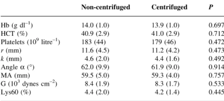
(2) Haematocrit and in vitro blood coagulation. saline so that the ®nal haematocrit, after mixing with the entire plasma and platelet fraction, yielded a ®nal haematocrit of 40, 30, 20, or 10%, respectively. Platelets, haemoglobin, and haematocrit were subsequently measured in the reconstituted blood (Coulterâ Ac T8, Coulter Corporation, Miami, FL, USA). Blood coagulation was assessed using a computerized Thrombelastographâ Coagulation Analyser (CTEGâ #3000, Haemoscope, Morton Grove, IL, USA) with celite activation.14 The following variables of the TEGâ trace were analysed: reaction time (r, normal value 12.0 (2.3) mm)Ðthe time from the start of the recording until the amplitude reaches 2 mm; coagulation time (k, normal value 4.2 (1.6) mm)Ðthe time from the end of reaction time (r) until the amplitude achieves 20 mm; maximum amplitude (MA, normal value 63.5 (4.5) mm) of the TEGâ tracing represents the absolute strength of the clot; angle a (normal value 60.2 (6.7)°)Ðthe angle formed by the slope of the TEGâ tracing from the r to the k value. Clot lysis was represented by the percentage decrease in the area under the curve at 60 min (Lys60, normal value <15%) after MA.9 One millilitre of citrated blood was pipetted into a tube containing 1% celite, and 340 ml of celite-activated citrated blood was added to a TEGâ cup containing 20 ml 2 M CaCl2 for recalci®cation. Clot strength (G) was calculated from the MA as: G=(50003MA)/(96±MA).15 16. Statistical analysis The effect of blood centrifugation, pipetting and reconstitution was assessed by comparing TEGâ variables before and after processing using paired t-tests. The effect of progressive in vitro haemodilution was assessed with repeated measures ANOVA and post-hoc paired t-test with Bonferroni correction. Data are presented as mean (SD). P-values less than 0.05 were considered signi®cant.. Results Blood handling including centrifugation did not affect blood coagulation (Table 1). Progressive reduction of haematocrit from 40 to 10% did not result in compromised blood coagulation as assessed by TEGâ (Fig. 1). The platelet count was unaffected, reaction time (r) and coagulation time (k) decreased, and angle a and MA increased with decreasing haematocrit levels. Clot strength (G) increased with decreasing haematocrit levels. Clot lysis at 60 min decreased minimally with the reduction in haematocrit.. Discussion The current study indicates that an isolated reduction of haematocrit does not compromise in vitro blood coagulation. In contrast, with decreasing haematocrit, all TEGâ variables unanimously changed towards an accelerated. Table 1 Effect of blood centrifugation, pipetting and reconstitution (centrifuged) on blood coagulation. Note, there are no signi®cant differences. Hb=haemoglobin, HCT=haematocrit r=reaction time, k=coagulation time, MA=maximum amplitude, G=clot strength and Lys60=clot lysis at 60 min. Hb (g dl±1) HCT (%) Platelets (109 litre±1) r (mm) k (mm) Angle a (°) MA (mm) G (103 dynes cm±2) Lys60 (%). Non-centrifuged. Centrifuged. P. 14.0 (1.0) 40.9 (2.9) 183 (44) 11.6 (4.5) 4.6 (2.0) 62.0 (9.9) 59.5 (5.0) 8.4 (1.9) 4.4 (2.0). 13.9 (1.0) 41.0 (2.9) 179 (46) 11.2 (4.2) 4.4 (1.6) 61.9 (9.0) 59.3 (4.0) 8.3 (1.7) 4.2 (1.4). 0.697 0.712 0.472 0.473 0.492 0.914 0.757 0.533 0.445. blood coagulation pro®le resulting in increased clot strength (G). It was recently shown that there is virtually no thrombin generation in the absence of platelets.17 With the addition of red blood cells, however, thrombin generation gradually recovered and it was speculated that red blood cells would participate in the haemostatic process through exposure of procoagulant phospholipids at their outer cell membrane.17 18 In the presence of a normal number of platelets, as in the current study, in vitro blood coagulation appears not to be compromised signi®cantly by a reduced number of red blood cells. Indeed, all TEGâ variables indicated accelerated blood coagulation with a decreasing haematocrit resulting in an increased clot strength (G) (Fig. 1). How relevant is this in vitro study for the clinical care of surgical patients? Of course, the in vitro situation is different from the clinical care of surgical patients, but only the in vitro situation allows the assessment of the isolated effect of a relevant change in haematocrit on blood coagulation. In surgical patients, low haematocrits are usually encountered after advanced surgical blood loss and asanguineous infusion therapy to maintain normovolaemia. Therefore, the patients have lost also coagulation factors and platelets. In this situation, coagulopathy will result from decreased levels of coagulation factors and platelets as well as the effects of dilution with crystalloids and colloids.9 Interestingly, Mezzano and colleagues found that platelet dysfunction and severity of renal dysfunction but not the haematocrit were independent predictors for haemostatic disorders in uraemic patients.19 Therefore, it may appear somewhat simplistic to advocate the use of allogeneic red blood cell transfusions for the speci®c purpose of optimizing blood coagulation.6 The use of TEGâ analysis may also be criticized. We chose TEGâ analysis because, in a variety of clinical situations, TEGâ was the most sensitive method to predict surgical blood loss or the occurrence of bleeding complications.20±25 TEGâ is increasingly used in studies on blood coagulation14 15 26 and in vivo and in vitro haemodilution results in similar TEGâ changes in. 247.
(3) Iselin et al.. Fig 1 Effect of the isolated reduction in haematocrit from 40 to 10% on in vitro blood coagulation. P values refer to the overall ANOVA, * indicates P<0.017 vs haematocrit of 40%. Hb=haemoglobin, r=reaction time, k=coagulation time (k), MA=maximum amplitude, G=clot strength (G), and Lys60=clot lysis at 60 min. humans.27 Interestingly, the results of the present study are in accordance with an in vivo study in rabbits demonstrating that a reduction of the haematocrit from 36 to 22% was not associated with an increased blood loss because of a subsequent standard splenic incision.8 The current study suggests that a reduction of haematocrit even beyond this range is not associated with signi®cantly compromised blood coagulation. The results are also in agreement with previous ®ndings that clot strength (G) tended to be higher in platelet rich plasma when compared with whole blood.16 -The current study assessed the effect of an isolated decrease in haematocrit on in vitro blood coagulation with constant platelet count and unchanged coagulation factors. This is different from a previous study by Egli and colleagues.9 where the effect of a progressive in vitro haemodilution with a concomitant dilution of platelets and coagulation factors was investigated. Accordingly, different results were found. At 60% haemodilution, corresponding to a haematocrit of approximately 17%, blood coagulation was largely unchanged and MA decreased.9 In contrast, blood coagulation was accelerated in the current study at 20%. haematocrit with an increase in MA (Fig. 1). This may be explained by preserved platelet count and coagulation factors. In conclusion, the isolated reduction of the haematocrit appears not to compromise in vitro blood coagulation and recommendations to raise the haematocrit to at least 30% with the use of allogeneic blood transfusions to optimize blood coagulation6 should be viewed with reservation considering the potential adverse effects of allogeneic blood transfusions.1. Acknowledgement Financially supported by the Institute of Anaesthesiology, University Hospital Zurich, Switzerland.. References. 248. 1 Spahn DR, Casutt M. Eliminating blood transfusions: New aspects and perspectives. Anesthesiology 2000; 93: 242±55 2 Hebert PC, Wells G, Blajchman MA, et al. A multicenter,.
(4) Haematocrit and in vitro blood coagulation. 3 4. 5. 6 7 8 9. 10 11 12 13 14 15. randomized, controlled clinical trial of transfusion requirements in critical care. N Engl J Med 1999; 340: 409±17 Younes RN, Rogatko A, Vydelingum NA, Brennan MF. Effects of hypovolemia and transfusion on tumor growth in MCA-tumorbearing rats. Surgery 1991; 109: 307±12 Sinclair S, James S, Singer M. Intraoperative intravascular volume optimisation and length of hospital stay after repair of proximal femoral fracture: randomised controlled trial. BMJ 1997; 315: 909±12 Spahn DR, Leone BJ, Reves JG, Pasch T. Cardiovascular and coronary physiology of acute isovolemic hemodilution: a review of nonoxygen-carrying and oxygen-carrying solutions. Anesth Analg 1994; 78: 1000±21 Valeri CR, Crowley JP, Loscalzo J. The red cell transfusion trigger: has a sin of commission now become a sin of omission? Transfusion 1998; 38: 602±10 Crowley JP, Metzger JB, Valeri CR. The volume of blood shed during the bleeding time correlates with the peripheral venous hematocrit. Am J Clin Pathol 1997; 108: 579±84 Quaknine-Orlando B, Samama CM, Riou B, et al. Role of the hematocrit in a rabbit model of arterial thrombosis and bleeding. Anesthesiology 1999; 90: 1454±61 Egli GA, Zollinger A, Seifert B, Popovic D, Pasch T, Spahn DR. Effect of progressive haemodilution with hydroxyethyl starch, gelatin and albumin on blood coagulation. An in vitro thrombelastography study. Br J Anaesth 1997; 78: 684±9 Strauss RG, Stans®eld C, Henriksen RA, Villhauer PJ. Pentastarch may cause fewer effects on coagulation than hetastarch. Transfusion 1988; 28: 257±60 Treib J, Haass A, Pindur G. Coagulation disorders caused by hydroxyethyl starch. Thromb Haemost 1997; 78: 974±83 Ruttmann TG, James MFM, Aronson I. In vivo investigation into the effects of haemodilution with hydroxyethyl starch (200/0.5) and normal saline on coagulation. Br J Anaesth 1998; 80: 612±6 Ruttmann TG, James MF, Viljoen JF. Haemodilution induces a hypercoagulable state. Br J Anaesth 1996; 76: 412±4 Camenzind V, Bombeli T, Seifert B, et al. Citrate storage affects Thrombelastographâ analysis. Anesthesiology 2000; 92: 1242±9 Nielsen VG, Geary BT, Baird MS. Evaluation of the contribution of platelets to clot strength by thrombelastography in rabbits: the role of tissue factor and cytochalasin D. Anesth Analg 2000; 91: 35±9. 249. 16 Khurana S, Mattson JC, Westley S, O'Neill WW, Timmis GC, Sa®an RD. Monitoring platelet glycoprotein IIb/IIIa-®brin interaction with tissue factor-activated thromboelastography. J Lab Clin Med 1997; 130: 401±11 17 Peyrou V, Lormeau JC, Herault JP, Gaich C, P¯iegger AM, Herbert JM. Contribution of erythrocytes to thrombin generation in whole blood. Thromb Haemost 1999; 81: 400±6 18 Zwaal RF, Comfurius P, Bevers EM. Mechanism and function of changes in membrane-phospholipid asymmetry in platelets and erythrocytes. Biochem Soc Trans 1993; 21: 248±53 19 Mezzano D, Tagle R, Panes O, et al. Hemostatic disorder of uremia: the platelet defect, main determinant of the prolonged bleeding time, is correlated with indices of activation of coagulation and ®brinolysis. Thromb Haemost 1996; 76: 312±21 20 Chau TN, Chan YW, Patch D, Tokunaga S, Greenslade L, Burroughs AK. Thrombelastographic changes and early rebleeding in cirrhotic patients with variceal bleeding. Gut 1998; 43: 267±71 21 Kang Y. Thromboelastography in liver transplantation. Semin Thromb Hemost 1995; 21 (Suppl 4): 34±44 22 Bayly PJ, Thick M. Reversal of post-reperfusion coagulopathy by protamine sulphate in orthotopic liver transplantation. Br J Anaesth 1994; 73: 840±2 23 Steib A, Gengenwin N, Freys G, Boudjema K, Levy S, Otteni JC. Predictive factors of hyper®brinolytic activity during liver transplantation in cirrhotic patients. Br J Anaesth 1994; 73: 645±8 24 Williams GD, Bratton SL, Riley EC, Ramamoorthy C. Coagulation tests during cardiopulmonary bypass correlate with blood loss in children undergoing cardiac surgery. J Cardiothorac Vasc Anesth 1999; 13: 398±404 25 Ereth MH, Nuttall GA, Klindworth JT, et al. Does the plateletactivated clotting test (HemoSTATUS) predict blood loss and platelet dysfunction associated with cardiopulmonary bypass? Anesth Analg 1997; 85: 259±64 26 Whitten CW, Greilich PE. Thrombelastography: past, present, and future. Anesthesiology 2000; 92: 1223±5 27 Jamnicki M, Bombeli T, Seifert B, et al. Low- and mediummolecular-weight hydroxyethyl starch: Comparison of their effect on blood coagulation. Anesthesiology 2000; 93: 1231±7.
(5)
Figure
Documents relatifs