HAL Id: tel-02003448
https://tel.archives-ouvertes.fr/tel-02003448
Submitted on 1 Feb 2019
HAL is a multi-disciplinary open access archive for the deposit and dissemination of sci-entific research documents, whether they are pub-lished or not. The documents may come from teaching and research institutions in France or abroad, or from public or private research centers.
L’archive ouverte pluridisciplinaire HAL, est destinée au dépôt et à la diffusion de documents scientifiques de niveau recherche, publiés ou non, émanant des établissements d’enseignement et de recherche français ou étrangers, des laboratoires publics ou privés.
Benyviridae viral cicle
Mattia Dall’Ara
To cite this version:
Mattia Dall’Ara. RNA/RNA interactions involved in the regulation of Benyviridae viral cicle. Vegetal Biology. Université de Strasbourg; Università degli studi (Bologne, Italie), 2018. English. �NNT : 2018STRAJ019�. �tel-02003448�
Université de Strasbourg et Università di Bologna
Thèse en co-tutelle
Présentée à la
FACULTÉ DES SCIENCES DE LA VIE
En vue de l'obtention du titre de
DOCTEUR DE L'UNIVERSITÉ DE STRASBOURG
Discipline : Sciences de la Vie et de la Santé,
Spécialité : Aspects moléculaires et cellulaires de la biologie
Par
Mattia D
all’Ara
RNA/RNA interactions involved in the
regulation of Benyviridae viral cicle
Soutenue publiquement le 18 mai 2018 devant la Commission d'Examen : Dr. Stéphane BLANC Rapporteur Externe Dr. Renato BRANDIMARTI Rapporteur Externe Dr. Roland MARQUET Examinateur Interne Dr. Mirco IOTTI Examinateur Interne Pr. David GILMER Directeur de Thèse Dr. Claudio RATTI Co-directeur de Thèse
First of all I want to thank my tutors David and Claudio. They believed in me and accepted me in their labs giving me the opportunity to work on a such interesting topic. Being a Ph.D. student is hard work but it would be even harder without their help, example and coaching. I would like to thanks all the people that worked with me or I have had the pleasure to meet during these four years of “co-tutelle”, but especially Chiara, Alice, Alyssa, Elodie, Marco, Marion, Matteo, Antonio, Mariateresa, Daniéle, Salah, Veronique, Khalid, Imma (!), Roby kiwi and Roby grapevine and many others. I have a special thank for each of you.
I also want to thank the member of the jury, Dr Stéphane Blanc, Dr Renato Brandimarti, Dr Roland Marquet and Dr Mirco Iotti for evaluating this work.
Les virus multipartites possèdent au moins deux sinon plusieurs segments du génome encapsidés dans des particules indépendantes. Ce groupe hétérogène de virus (ssADN circulaire, dsARN ou ssARN de polarité positive ou négative) est retrouvé majoritairement chez les plantes [1]. Actuellement, il est admis que pour assurer une infection productive, les virus doivent préserver leur intégrité génomique comme unité infectieuse fonctionnelle. Les virus monopartites et segmentés suivent cette règle car chaque particule virale renferme l’intégrité de leur génome. A l’inverse pour les virus multipatites, l’encapsidation en entités distinctes de chaque segment porteur d’information génomique rend la notion d’intégrité génomique énigmatique [2]. La condition pour assurer l’apport d’un set complet des segments génomiques dans chaque cellule infectée implique un niveau élevé de multiplicité d’infection. Cette forte multiplicité d’infection peut être considérée comme ayant un coût biologique insoutenable en termes de réplication virale. Durant l’infection, les virus multipartites organisent leur génome en une formule génomique définie, dans laquelle certains segments seraient moins nombreux que d’autres [3]. Dans la plante, la propagation indépendante et aléatoire des différentes parties génomiques virales ne peut être un modèle soutenable pour le mouvement à longue distance dans les tissus vasculaires. Les tubes criblés sont des éléments de la plante dans lesquels les virus ne peuvent se répliquer. Ils ne peuvent donc pas y augmenter leur titre viral et assurer dans chaque cellule compagne la présence de tous les éléments nécessaires pour initier une nouvelle infection.
Afin d’étudier les aspects de l’intégrité génomique des virus multipartites à ARN le Beet
necrotic yellow vein virus BNYVV a été choisi comme modèle car son génome consiste en
quatre ou cinq ssARN de polarité positive. De plus, comme la majorité des virus multipartites à ARN des plantes le BNYVV a une organisation génomique caractérisée par deux segments (i.e. ARN1 et ARN2) qui codent les gènes de ménage, indispensables pour l’infection au niveau de la feuille ainsi que des segments dit accessoires mais qui restent nécessaires pour l’infection naturelle de l’hôte, Beta vulgaris. Si l’ARN1 code pour la replicase virale, l’ARN2 dirige la synthèse des protéines majeures et mineures de capside (CP et p75) nécessaires à la formation des particules en bâtonnets. Il permet également la production des protéines responsable du mouvement de cellule à cellules (triple gene block, TGB) et du suppresseur d’ARN interférence (p14) grâce à la production d’ARN subgénomiques. L’ARN3 est
prolifération racinaire (rhizomanie), l’un des symptômes qui est aggravé lorsque l’ARN5 est présent. L’ARN4 quant à lui est nécessaire à la transmission des particules virales par le vecteur Polymyxa betae [4].
L’infection de cellule à cellule implique des complexes ribonucléoprotéiques (RNP) qui implique les protéines TGB et le VSR mais pas la protéine de capside. Cependant, si la protéine CP est nécessaire à l’infection virale à longue distance (infection systémique), l’encapsidation des ARN génomiques n’est pas essentielle pour le mouvement à longue distance du BNYVV. Nous avons donc émis l’hypothèse qu’il existait un réseau d’interaction entre les ARN génomiques viraux assurant la présence d’au moins une copie de chaque ARN génomique. Cette hypothèse rappelle l’interaction ARN/ARN démontrée comme nécessaire à l’empaquetage sélectif des segments du virus Influenza A [5].
Mon objectif est de déterminer l’existence des interactions entre les ARN génomiques et leur implication dans le cycle viral. J’ai donc vérifié si une formule génomique virale était maintenue dans chacun des hôtes et dans des tissus variés (chapitre 1); si l’ensemble des segments génomiques était effectivement retrouvé dans chaque cellule infectée (chapitre 2).
In fine, j’ai identifié des séquences présentes sur les ARN1 et 2 validant l’existence
d’interactions ARN/ARN entre les ARN génomiques (chapitre 3).
Dans le modèle proposé pour baisser au minimum le coût biologique de la préservation de l’intégrité génomique, un réseau d’interactions ARN/ARN détermine la reconnaissance et l’assemblage de chaque ARN génomique dans un complexe RNP modulaire. Ce complexe est considéré comme l’unité infectieuse mobile pour la propagation du virus multipartite dans la plante [6]. Si ce complexe RNP existe comme entité virale régulée et fonctionnelle, il est probable qu’il soit préservé parmi divers tissus et parmi des espèces de plantes différentes. Après avoir optimisé et validé le protocole dual step digital droplets (dd) RT-PCR, j’ai déterminé la formule génomique dans différents hôtes du BNYVV (Chenopodium quinoa,
Spinacea oleracea and Beta macrocarpa) et tissus (feuilles et racines). J’ai comparé les ARN
génomiques totaux avec ceux encapsidés qui représentent la forme transmissible du virus. Que ce soit pour les ARN génomiques totaux ou encapsidés, mes résultats indiquent une forte accumulation des ARN auxiliaires (ARN3 et ARN4) jusqu’à un ordre de grandeur par rapport aux ARN1 et ARN2, indépendamment de l’hôte et des tissus. Considérant la fréquence
en fonction de l’hôte et du tissu systémique. Toutefois le ratio entre les ARN1 et 2 (qui expriment les protéines essentielles au cycle viral) semble être conservé et correspond à 2 ou 3 molécules d’ARN1 pour une molécule de ARN2.
Le mouvement du virus dans la plante est décrit comme une circulation non coordonnée de particules ou de complexes RNP. Toutefois, l’accumulation différentielle des ARN de BNYVV dans les tissus infectés rend insoutenable le coût de la préservation de l’intégrité génomique assurant la présence du set complet des segments génomiques à l’intérieur de chaque cellule infectée. Cet état de faits porte à la formuler deux hypothèses opposées:
1. les segments du BNYVV ne doivent pas forcément être présents simultanément dans chaque cellule infectée et la circulation des protéines virales assure la réplication et la stabilisation des ARN grâce à la continuité du symplasme.
2. un mécanisme spécifique garanti le mouvement coordonné des segments comme une unité infectieuse.
Pour vérifier l’une ou l’autre hypothèse, la présence simultanée des quatre ARN génomiques du BNYVV a été testée avec le protocole Real time RT-PCR, individuellement sur des protoplastes isolés provenant de plantes d’épinards systématiquement infectées. Les protoplastes ont été préparés et fixés, après dilution, avec du glutaraldehyde à 0.5%. Chaque protoplaste isolé ou le milieu dépourvu de protoplaste (utilisé comme contrôle négatif) a été échantillonné par pipetage sous microscope optique. Nous avons utilisé les échantillons comme matrice d’une réaction triplex qualitative RT-PCR Real time qui a comme cible deux ARN différents du BNYVV et la sous-unité majeur de la Rubisco (RbcL). La RT-PCR sur l’ARN du BNYVV a été suivie en temps réel utilisant des sondes TaqMan avec différents fluorophores (FAM et HEX) et la présence de contrôle endogène RbcL a été vérifiée après amplification (end-point) sur gel d’agarose. Quatre réactions ont été réalisées pour vérifier la présence de l’ARN1 avec l’ARN2, de l’ARN2 avec l’ARN3, de l’ARN1 avec l’ARN4 et enfin de l’ARN3 avec l’ARN4. Entre 30 et 50% des protoplastes analysés sont infectés par le virus. ~93% des protoplastes infecté a révélé la présence des ARN viraux testés. Ces résultats suggèrent donc bien l’existence d’un mécanisme qui orchestre le déplacement coordonné des segments génomiques du BNYVV. Etant donné que les particules virales ne diffèrent que par
l’hypothèse selon laquelle la reconnaissance spécifique des diverses espèces génomiques a lieu grâce à des interactions ARN/ARN [6].
Afin de vérifier le modèle proposé impliquant l’interaction ARN/ARN pour la formation des unités RNP mobiles et infectieuses renfermant au moins une copie de chaque segment, nous avons utilisé la technique de gel-retard (Electro Mobility Gel Shift Assays, EMSA). Dans certains hôtes, la seule présence de l’ARN1 et de l’ARN2 suffit pour produire une infection dans la plante entière. Par conséquent, le complexe RNP minimal devrait impliquer l’interaction entre ces deux ARN génomiques. Pour cette raison nous avons cherché à évaluer l’existence d’interactions spécifiques et hétérologues entre l’ARN1 et l’ARN2.
La co-transcription in vitro des clones complets ou partiels des ARN1 ou ARN2 a été suivie par électrophorèse en gel natif ou en présence de chélateur de magnésium (Mg2+). Nous avons observé la présence de duplexes d’ARN (homo et hétérodimères) aussi bien sur gel coloré au bromure d’éthidium que par hybridation moléculaire à l’aide de sondes spécifiques radiomarquées. Nous n’avons pas pu observer des décalages de bandes significatifs avec les ARN1 et ARN2 complets, probablement en raison de l’absence de facteurs viraux et cellulaires qui pourraient aider et réguler l’interaction ARN/ARN en contexte naturel. Toutefois, l’utilisation des clones partiels ARN1 (3655-4783) et ARN2 (1-2715) a démontré la formation d’un hétéro-duplex stable en gel partiellement dénaturant, une condition réduisant considérablement la stabilité des structures ARN normalement stabilisées par le magnésium. En réduisant les tailles des séquences utilisées, nous avons pu localiser les domaines d’interaction et nous avons affiné leur identification en utilisant des oligodésoxyribonucléotides compétiteurs (ODN). Ce domaine d’interaction consiste en deux séquences complémentaires de 17 nucléotides présentant un mésappariement de deux bases non complémentaires. Ces séquences sont situées respectivement à l’extrémité 5' UTR de l’ARN2, à partir du quatrième nucléotide, et dans la région de l’ARN1 codant la réplicase. Afin de démontrer le sens biologique de l’interaction ARN/ARN trouvée, nous avons créé des mutations équivalentes et compensatoires dans les clones ADNc infectieux de l’ARN1 et de l’ARN2 pour prévenir ou restaurer les appariements de bases. Nous avons effectué des expériences de gel-retard EMSA pour valider les effets des mutations sur les interactions, en utilisant des ODN compétiteurs portant les mutations indiquées. Ces ODN n’empêchent pas
évalué le sens biologique des domaines d’interactions identifiés en contexte viral in planta. Nous avons réalisé des tests de vitalité des ARN génomiques mutants sur feuille de C. quinoa (hôte local) pour évaluer le maintien des fonctions essentielles de réplication, mouvement cellule-à-cellule, et suppression de l’interférence par ARN lorsque l’interaction ARN/ARN est interrompue ou restaurée par un appariement de bases alternatif. Les combinaisons ARN1 WT+ARN2 MUT et ARN1 MUT+ARN2 MUT n’ont pas conduit à la formation de sites d’infection au niveau des feuilles probablement en raison de l’altération fonctionnelle du promoteur de l’ARN2 MUT. Toutefois la combinaison ARN MUT avec ARN2 WT a permis la réplication et le mouvement cellule-cellule du virus avec une diminution drastique du nombre des lésions locales comparée avec la combinaison sauvage. Le BNYVV et le Beet soil
borne mosaic virus (BSBMV) sont deux espèces différentes mais étroitement apparentés et
appartenant au genre Benyvirus et à la famille Benyviridae [4]. Ils ont en commun le même hôte, vecteur de transmission et organisation génomique. L’échange artificiel des ARN génomiques entre les ARN1 et ARN2 du BNYVV et du BSBMV conduit à la formation de virus chimériques [7]. Après avoir effectué une comparaison bio-informatique, nous avons conclu qu’il existe un réseau alternatif d’interactions ARN1/ARN2 pour ces virus réassortants. Nous avons donc comparé le comportement des chimères formées par les ARN1 WT BNYVV + ARN2 BSBMV et ARN1 MUT BNYVV + ARN2 BSBMV. Le nombre comparable des lésions induites sur feuille de C. quinoa, tout comme les niveaux similaires d’ARN accumulés démontrent que les mutations introduites dans l’ARN1 du BNYVV ne sont pas impliquées dans la réplication (i.e stabilisation des ARN et mouvement cellule-cellule) mais effectivement dans l’interaction ARN1/ARN2 responsable de l’orchestration d’un réseau régulateur.
Pour renforcer ces résultats, la réplication et le mouvement des ARN1 et 2 sauvages ont été évalués en présence de replicon du BNYVV qui porte l’un des deux domaines d’interaction, muté ou non. L’accumulation de ces réplicons vise à surproduire des ARN entrant en compétition avec les segments génomiques et interférer dans la formation de l’interaction ARN1/ARN2. Quatre cent nucléotides de l’ARN2 ou de l’ARN1 ont été insérés dans le vecteur dérivant de l’ARN5 (Rep5, constitué du 5' et 3' UTR de l’ARN5). L’analyse de l’accumulation des ARN viraux en protoplastes de C. quinoa a permis de constater un effet défectif interférent (DI) du Rep5 porteur du domaine d’interaction de l’ARN2. Cet effet n’a
l’effet DI des compétiteurs introduits, nous avons réalisé des expérimentations similaires utilisant un ODN complémentaire au domaine d’interaction de l’ARN1. En tenant compte d’une possible activité RNAse H dans la cellule nous avons utilisé comme contrôle négatif d’autres ODN complémentaires à l’ARN1 et au brin négatif de l’ARN2. Lorsque la quantité de l’ODN est cinq fois plus élevée que celle de la cible l’ARN1, nous avons observé une réduction significative de l’accumulation de l’ARN viral. Cet effet n’est pas observé dans le contrôle négatif. Toutefois, en utilisant un ratio 50.1, l’activité RNase H semble activée puisque la quantité d’ARN génomique diminue de manière importante quel que soit l’ODN utilisé.
En conclusion, nous avons démontré que tous les segments génomiques du BNYVV sont présents à l’intérieur de chaque cellule infectée des tissus systémiques dans lesquels s’accumulent les ARN jusqu’à atteindre une formule génomique déterminée préservant un ratio ARN1 et ARN2 similaire mais présentant une variabilité de l’accumulation des ARN3 et 4 selon les types d’organes et d’hôtes. Dans un modèle dans lequel les ARN viraux interagissent entre eux pour former une unité mobile infectieuse, l’ARN1 et l’ARN2 interagissent au moins in silico et in vitro. L’existence d’une telle interaction in vivo est fortement renforcée grâce aux résultats obtenus à l’aide des virus chimères BNYVV et BSBMV. De telles interactions pourraient d’une part réguler l’accumulation des ARN génomiques et d’autre part orchestrer le cycle viral (encapsidation, régulation de l’expression des protéines et de la synthèse des ARN subgénomiques).
I virus multipartiti possiedono due o più segmenti genomici incapsidati in particelle indipendenti. Questo gruppo eterogeneo di virus (ssDNA circolare, dsRNA o ssRNA a polarità positiva o negativa) infetta prevalentemente le piante [1]. Ad oggi, è scientificamente accettato che, per assicurare una infezione produttiva, i virus preservano la loro integrità genomica come unità infettive funzionali. I virus monopartitici e segmentati seguono questa regola in quanto ogni particella virale contiene un set genomico completo. Diversamente, per i virus multipartiti, l’incapsidazione in entità distinte di ogni segmento genomico rende enigmatica la nozione di integrità genomica [2]. La condizione necessaria per assicurare la presenza di un set genomico completo del virus in ogni cellula infettata implica, infatti, un
può essere considerato insostenibile. Durante l’infezione, i virus multipartiti organizzano il loro genoma in formule genomiche definite, nelle quali alcuni segmenti sono più abbondanti di altri [3]. Nella pianta, la propagazione indipendente e aleatoria delle differenti componenti genomiche virali nei tessuti vascolari non può essere considerato un modello sostenibile di movimento a lunga distanza. I tubi cribrosi sono infatti elementi della pianta nei quali i virus non possono replicarsi e quindi aumentare il loro titolo virale e assicurare in ogni cellula compagna la presenza di tutti gli elementi necessari per iniziare una nuova infezione.
Al fine studiare differenti aspetti dell’integrità genomica dei virus multipartiti aventi genoma a RNA il Beet necrotic yellow vein virus (BNYVV) è stato scelto come modello in quanto il suo genoma consiste in quattro o cinque ssRNA a polarità positiva. Inoltre, come la maggior parte dei virus multipartiti a RNA delle piante il BNYVV presenta una organizzazione genomica caratterizzata da due segmenti (i.e. RNA1 e RNA2) indispensabili per l’infezione a livello della foglia, in quanto codificanti geni housekeeping, e segmenti detti accessori che restano comunque necessari per l’infezione naturale dell’ospite, Beta vulgaris. Se l’RNA1 codifica la replicasi virale, l’RNA2 dirige la sintesi dei costituenti proteici maggiore e minore del capside (CP e p75) necessari per la formazione delle particelle bastoncelliformi. L’RNA2 permette altresì l’espressione delle proteine responsabili del movimento cellula a cellula (triple gene block, TGB) e del soppressore dell’RNA interference (p14) grazie alla produzione di RNA subgenomici. L’RNA3 è implicato ne movimento a lunga distanza e codifica la proteina p25, il determinate della iper proliferazione radicale (rizomania), sintomatologia aggravata dalla presenza dell’RNA5. L’RNA4 è infine necessario per la trasmissione delle particelle virali attraverso il vettore Polymyxa betae [4].
L’infezione cellula a cellula necessita la formazione di complessi ribonucleoproteici (RNP) che implicano le proteine TGB e il VSR ma non la proteina del capside. Tuttavia se la CP è necessaria per l’infezione virale a lunga distanza (infezione sistemica) del BNYVV, l’incapsidazione degli RNA genomici non è essenziale per l’infezione sistemica. Abbiamo quindi ipotizzato l’esistenza di un network di interazioni tra gli RNA genomici che assicura la presenza di almeno una copia di ogni RNA genomico all’interno di un complesso macromolecolare rappresentante l’unità infettiva virale. Questa ipotesi richiama le interazioni
dell’Influenza A [5].
Il mio obiettivo è quello di determinare l’esistenza delle interazioni tra gli RNA genomici del BNYVV e la loro implicazione nel ciclo virale. Ho quindi verificato se una formula genomica è mantenuta in ospiti e organi differenti (capitolo 1); se il set completo di segmenti genomic si ritrova effettivamente in ogni cellula infetta (capitolo 2). In fine, ho identificato due sequenze presenti nell’RNA1 e nell’RNA2 che convalidano l’esistenza di interazione RNA/RNA tra gli RNA genomici del BNYVV (capitolo 3).
Nel modello proposto per minimizzare il costo del mantenimento dell’integrità genomica, un network di interazioni RNA/RNA determina il riconoscimento e l’assemblaggio di ogni RNA genomico in un complesso RNP modulare. Questo complesso è considerato come l’unità infettiva mobile per la propagazione del virus multipartito nella pianta [6]. Se questo complesso RNP esiste come entità virale regolata e funzionale, è probabile che sia conservato all’interno di organi differenti e di ospiti differenti. Dopo aver ottimizzato un protocollo di
dual step digital droplets (dd) RT-PCR, ho determinato la formula genomica in differenti
ospiti del BNYVV (Chenopodium quinoa, Spinacea oleracea e Beta macrocarpa) in foglie e radici. Ho quindi comparato la quantità di RNA genomico totale con quella dell’RNA incapsidato che rappresenta la forma trasmissibile del virus. Sia per gli RNA genomici totali sia per quelli incapsidati, i miei risultati indicano un forte accumulo, fino ad un ordine di grandezza degli RNA ausiliari (RNA3 e RNA4) rispetto agli RNA1 e RNA2, indipendentemente dall’ospite o organo analizzato. Considerando la frequenza relativa degli RNA3 e RNA4, il BNYVV sembra stabilire la sua formula genomica in maniera differente in funzione dell’ospite e organo sistemico. Tuttavia, il rapporto tra gli RNA1 e RNA2 (che esprimono le proteine essenziali al ciclo virale) sembra essere conservato e corrisponde a 2 o 3 molecole di RNA1 per una molecola di RNA2.
Il movimento del virus nella pianta è descritto come una circolazione non coordinata di particelle o complessi RNP. Tuttavia l’accumulo differenziale degli RNA del BNYVV nei tessuti infetti rende insostenibile il costo di assicurare all’interno di ogni cellula infetta il set completo di segmenti genomici. Questo stato di fatto porta alla formulazione delle seguenti due ipotesi:
in ogni cellula infetta e la continuità simplastica garantisce la circolazione delle proteine virali indispensabili per la replicazione e stabilizzazione degli RNA del virus. 2. Un meccanismo specifico garantisce il movimento coordinato dei segmenti che
costituiscono una unità infettiva.
Per verificare una o l’altra ipotesi, la presenza simultanea dei quattro RNA genomici del BNYVV è stata testata grazie a un protocollo di Real time RT-PCR su singolo protoplasto isolato da tessuti infetti e sistemici provenienti da piante di spinacio. I protoplasti sono stati preparati e fissati, dopo diluizione, con gluteraldeide al 0,5%. Ogni protoplasto isolato o il buffer privo di protoplasti (utilizzato come controllo negativo) sono stati campionati per pipettaggio al microscopio ottico. Abbiamo quindi utilizzato i campioni come matrice per una reazione qualitativa di triplex RT-PCR Real time avente come target due RNA differenti del BNYVV e la sub-unità maggiore della Rubisco (RbcL). La RT-PCR sugli RNA del BNYVV è stata seguita in tempo reale utilizzando sonde TaqMan con differenti fluorofori (FAM ed HEX) e la presenza del controllo endogeno RbcL è stata verificata, dopo amplificazione
(end-point), su gel di agarosio. Quattro reazioni sono state eseguite per verificare la presenza
dell’RNA1 con RNA2, dell’RNA2 con l’RNA3, dell’RNA1 con l’RNA4 e dell’RNA3 con l’RNA4. Il ~93% dei protoplasti infetti ha rivelato la co-presenza degli RNA virali testati. Questi risultati suggeriscono quindi l’esistenza di un meccanismo che orchestra lo spostamento coordinato dei segmenti genomici del BNYVV. Tenendo conto che le particelle virali non differiscono tra loro se non per la loro lunghezza (che dipende dalla taglia dell’RNA genomico incapsidato), abbiamo proposto l’ipotesi secondo la quale il riconoscimento specifico delle diverse specie genomiche avviene grazie a delle interazione RNA/RNA [6].
Al fine di verificare il modello proposto che implica la formazione di interazioni RNA/RNA per la formazione delle unità RNP mobili e infettive che contengono almeno una copia di ogni segmento genomico, abbiamo utilizzato la tecnica del Electro Mobility Gel Shift Assay (EMSA). In alcuni ospiti la sola presenza degli RNA1 e RNA2 è sufficiente per produrre una infezione produttiva nell’intera pianta. Di conseguenza, il complesso RNP minimale dovrebbe implicare l’interazione tra questi due RNA genomici. Per questo motivo abbiamo valutato l’esistenza di interazione specifiche e eterologhe tra gli RNA1 e RNA2.
dalla elettroforesi su gel nativo o in presenza di chelante (EDTA) di ioni magnesio (Mg2+). Abbiamo osservato la presenza di duplex di RNA (omo ed etero dimeri) sia su gel colorato con etidio bromuro sia dopo ibridazione molecolare con sonde radiomarcate specifiche. Non abbiamo tuttavia osservato band shift significativi con gli RNA1 e RNA2 completi, probabilmente a causa dell’assenza di fattori virali e dell’ospite che potrebbero aiutare al regolazione dell’interazione RNA/RNA nel contesto naturale. Tuttavia l’utilizzo dei cloni parziali RNA1 (3655-4783) e RNA2 (1-2715) ha dimostrato la formazione di un complesso etero-duplex stabile in gel parzialmente denaturante. Riducendo le dimensioni delle sequenze utilizzate, siamo stati in grado di localizzare i domini di interazione per poi perfezionare la loro identificazione utilizzando oligodeossiribonucleotidi (ODN) come competitori. Questo dominio di interazione consiste in due sequenze complementari di 17 nucleotidi con disappaiamento dovuto a due basi non complementari. Tali sequenze sono localizzate rispettivamente all'estremità 5 'UTR dell'RNA2, a partire dal quarto nucleotide, e nella regione dell'RNA1 che codifica la Replicasi. Al fine di dimostrare il significato biologico dell'interazione RNA/RNA trovata, abbiamo creato mutazioni silenti e compensatorie nei cloni di cDNA infettivi di RNA1 e RNA2 per prevenire e/o ripristinare l'accoppiamento di basi. Abbiamo eseguito altri esperimenti EMSA per convalidare gli effetti delle mutazioni sulla interazione, utilizzando ODN competitori portanti le mutazioni indicate. Questi ODN non hanno impedito agli RNA wild type di interagire tra loro, a differenza di quelli utilizzati inizialmente. Abbiamo valutato il senso biologico dei domini di interazione identificati nel contesto virale in planta. Abbiamo eseguito test di vitalità degli RNA genomici mutanti su foglia di C. quinoa (ospite locale) per valutare la preservazione delle funzioni essenziali di replicazione, movimento cellula a cellula e soppressione del silenziamento genico quando l'interazione RNA/RNA viene interrotta o ripristinata da un accoppiamento di basi alternativo. Le combinazioni RNA1 WT + RNA2 MUT e RNA1 MUT + RNA2 MUT non hanno portato alla formazione di loci di infezione fogliare, probabilmente a causa dell’alterazione funzionale del promotore dell’RNA2 MUT. Tuttavia la combinazione RNA1 MUT + RNA2 WT permette la replicazione e movimento cellula a cellula del virus seppure con una drastica riduzione del numero di lesioni locali rispetto alla combinazione wild type. BNYVV e il Beet
soil-borne mosaic virus (BSBMV) sono due specie diverse ma strettamente correlate
RNA2 di BNYVV e BSBMV ha portato alla formazione di virus chimerici [7]. Dopo un confronto bioinformatico, abbiamo concluso l’esistenza di network di interazioni RNA1/RNA2 alternati in questi due virus riassortanti. Abbiamo, quindi, confrontato il comportamento delle chimere formate da RNA1 BNYVV WT + RNA2 BSBMV e RNA1 BNYVV MUT + RNA2 BSBMV. Il numero comparabile di lesioni indotte su foglie di C.
quinoa, dimostra che la mutazioni introdotte nel RNA1 di BNYVV non sono coinvolte nella
replicazione (i.e. la stabilizzazione dell'RNA e movimento cellula a cellula), ma, effettivamente, nell'interazione RNA1/RNA2 responsabile dell’orchestrazione di un network regolatore.
Per consolidare questi risultati, la replicazione e il movimento degli RNA wild-type 1 e 2 sono stati valutati in presenza del replicone BNYVV che porta uno dei due domini di interazione, mutato o meno. L'accumulo di questi repliconi ha lo scopo di sovra-produrre RNA che entra in competizione con i segmenti genomici e interferisce nella formazione dell'interazione RNA1/RNA2. Quattrocento nucleotidi di RNA2 o RNA1 sono stati inseriti nel vettore derivante dall’RNA5 (Rep5, costituito da 5' e 3' UTR dell’RNA5). L'analisi dell'accumulo di RNA virali nei protoplasti di C. quinoa ha rivelato un effetto di RNA defective interference (DI) del Rep5 che porta il dominio di interazione RNA2.
Questo effetto non è stato osservato con il Rep5 che porta il dominio di interazione dell’RNA1. Al fine di escludere l'effetto DI dei competitori introdotti, abbiamo eseguito esperimenti simili a quelli precedentemente descritti utilizzando un ODN complementare al dominio di interazione RNA1. Prendendo in considerazione una possibile attività RNasi H nella cellula, abbiamo usato come controllo negativo altri ODN complementari all’ RNA1 e al filamento negativo dell’RNA2. Quando la quantità di ODN è cinque volte superiore a quella dell'RNA1 bersaglio, abbiamo osservato una significativa riduzione dell'accumulo di RNA virale. Questo effetto non è osservato nel controllo negativo. Tuttavia, utilizzando un rapporto 50:1, l'attività RNasi H sembra essere attivata poiché la quantità di RNA genomico diminuisce significativamente indipendentemente dall'ODN utilizzato.
In conclusione, abbiamo dimostrato che tutti i segmenti genomici di BNYVV sono presenti all'interno di ogni cellula infetta in tessuti sistemici dove l'RNA virale si accumula fino a raggiungere una formula genomica ospite specifica, che preserva un rapporto RNA1 e RNA2
ospite. In un modello secondo il quale gli RNA virali interagiscono l'uno con l'altro per formare un'unità mobile infettiva, l'RNA1 e l'RNA2 interagiscono almeno in silico e in vitro. L'esistenza di tale interazione in vivo è fortemente suggerita dai risultati ottenuti con i virus chimerici BNYVV e BSBMV. Tali interazioni potrebbero, da un lato, regolare l'accumulo di RNA genomici oppure orchestrare il ciclo virale (incapsidazione, regolazione dell'espressione proteica e sintesi di RNA subgenomici).
Bibliographie / Bibliografia
1. Lucía-Sanz, A.; Manrubia, S. Multipartite viruses: adaptive trick or evolutionary treat?
npj Syst. Biol. Appl. 2017, 3, doi:10.1038/s41540-017-0035-y.
2. Sicard, A.; Michalakis, Y.; Gutiérrez, S.; Blanc, S. The Strange Lifestyle of Multipartite Viruses. PLOS Pathog. 2016, 12, e1005819,
doi:10.1371/journal.ppat.1005819.
3. Sicard, A.; Yvon, M.; Timchenko, T.; Gronenborn, B.; Michalakis, Y.; Gutierrez, S.; Blanc, S. Gene copy number is differentially regulated in a multipartite virus. Nat.
Commun. 2013, 4, doi:10.1038/ncomms3248.
4. Gilmer, D.; Ratti, C. ICTV Virus Taxonomy Profile: Benyviridae. J. Gen. Virol. 2017,
98, 1571–1572, doi:10.1099/jgv.0.000864.
5. Gerber, M.; Isel, C.; Moules, V.; Marquet, R. Selective packaging of the influenza A genome and consequences for genetic reassortment. Trends Microbiol. 2014, 22, 446– 455, doi:10.1016/j.tim.2014.04.001.
6. Dall’Ara, M.; Ratti, C.; Bouzoubaa, S.; Gilmer, D. Ins and Outs of Multipartite
Positive-Strand RNA Plant Viruses: Packaging versus Systemic Spread. Viruses 2016,
8, 228, doi:10.3390/v8080228.
7. Laufer, M.; Mohammad, H.; Maiss, E.; Richert-Pöggeler, K.; Dall’Ara, M.; Ratti, C.; Gilmer, D.; Liebe, S.; Varrelmann, M. Biological properties of Beet soil-borne mosaic virus and Beet necrotic yellow vein virus cDNA clones produced by isothermal in vitro recombination: Insights for reassortant appearance. Virology 2018, 518, 25–33,
Multipartite viruses possess more than two genomic segments packaged into independent particles. Such groups of viruses infect mostly plants. They possess a wide range of genome types: circular ssDNA, dsRNA or ssRNA with either positive or negative polarity. The condition to provide a complete set of genomic segments in each infected cell implies a high level of multiplicity of infection that results in an unsustainable biological cost in terms of viral replication. Beet necrotic yellow vein virus (BNYVV) was chosen as a model to investigate the aspect of genome integrity of multipartite RNA viruses since such virus possesses the highest number of genomic segments (up to 5) within the Baltimore’s class IV that concerns ssRNA(+) viruses. In the proposed model, to minimize the cost of the genome integrity preservation, a network of RNA/RNA interactions determines the recognition and the mobilization of at least one of each genomic RNAs in a modular RNP complex. Such complex must be considered as the mobile infectious unit of the segmented genome during viral spread in the plant. The Aim of this thesis was to experimentally determine the existence of RNA/RNA interactions between genomic BNYVV RNAs and their implication in the viral cycle. In parallel approaches, I demonstrated that all BNYVV genomic segments are present within the single infected cell in systemic tissues where they accumulate to reach set point genomic formulas. These formulas depend on organs and hosts (chapter I and II). In the model where vRNAs interact each other to form the minimal mobile infective unit, RNA1 and RNA2 interaction domain has been identified in silico and in vitro. The rationale of such an interaction has been provided in vivo using BNYVV and Beet soil-borne mosaic virus chimeras (chapter III).
General Introduction ... 1
1. Benyviridae family: taxa characteristics ... 2
2. Beet necrotic yellow vein virus ... 3
2.1 Types and Strains ... 3
2.2 Vector transmission ... 4
2.3 BNYVV host range ... 5
2.4 Rhizomania ... 6
2.5 Genomic organization: RNA1 and RNA2 essential combination and accessory RNAs ... 6
3. Ins and Outs of Multipartite Positive-Strand RNA Plant Viruses: Packaging versus Systemic Spread ... 10
4. Aim of the study ... 29
5. References ... 30
Chapter I: Differential accumulation of BNYVV genomic RNAs during host infection ... 38
1. Introduction: benefits for multipartitism ... 39
2. Droplet digital (dd) PCR ... 42
3. Validation of dual step ddRT-PCR protocol for the absolute quantification of BNYVV genomic RNAs . 43 3.1 BNYVV Standard production ... 43
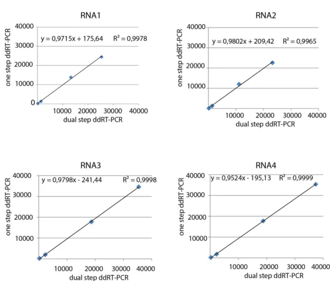
3.2 Comparison between one step and dual step ddRT-PCR for the absolute quantification of standards .. 44
3.3 Optimization of dual step ddRT-PCR for the quantification of BNYVV RNA1 to 4 ... 47
3.4 Host species and virus inoculation ... 50
3.5 Total and encapsidated RNA extraction and preparation of samples for dual step ddRT-PCR ... 51
4. Results ... 51
6. Discussion... 56
6. References ... 58
Chapter II: BNYVV genome integrity within infected cells ... 63
1. Introduction: a link between viral movement and multipartitism... 64
2. Preparation of S. oleracea protoplast and fixation ... 68
3. Individual protoplast isolation and triplex RT-qPCR ... 68
4. Discussion... 74
5. References ... 77
Chapter III: A specific heterologous RNA/RNA interaction is involved in the BNYVV infectivity ... 81
1. Introduction: regulated assembly of collective infectious units ... 82
2. Material and methods ... 85
2.1 Plasmids for Electro Mobility Shift Assay (EMSA) ... 85
2.2 Native or partially denaturating EMSA of in vitro co-transcribed transcripts... 86
2.3 Site directed mutagenesis of BNYVV full length cDNA clones ... 87
2.4 Construction of RNA5 derived replicons carrying RNA1 or RNA2 interaction domains ... 87
2.5 In vitro transcription, infection of Chenopodium quinoa protoplasts or plants and Northern blotting (high molecular weight RNAs) ... 88
3. Results ... 88
3.1 EMSAs of in vitro co-transcription of partial RNA1 and RNA2 cDNA clones ... 88
3.2 In vivo experiment of RNA1/RNA2 interaction disruption by interfering of competitive RNA5 derived replicons. ... 96
3.3 In vivo experiment of RNA1/RNA2 interaction disruption by interfering of competitive ODNs ... 97
3.4 In vivo experiment of disrupted and restored RNA1/RNA2 interaction by synonymous and compensatory mutations ... 99
4. Discussion... 101
5. References ... 103
General conclusion ... 107
2
Benyviridae family belongs to group IV of the Baltimore classification [1], together with
viruses possessing single-stranded positive-sense RNA genome (ssRNA (+)) that replicate through ssRNA(-) intermediates. The family possesses one single genus, Benyvirus, which includes four plant viral species: the type member Beet necrotic yellow vein virus (BNYVV),
beet soil-borne mosaic virus (BSBMV), burdock mottle virus (BdMV) and rice stripe
necrosis virus (RSNV) [2].
Initially assigned to Tobamovirus genus because of the virion morphology, BNYVV was included in the Furovirus genus characterized by the multipartite RNA genome species transmitted by fungal vectors [3]. Furovirus genus was then redefined in Furovirus,
Pomovirus, Pecluvirus, Hordeivirus genera and Benyvirus genus [4] later added in the newborn Benyviridae family [2]. This new classification was based on the following taxa criteria: vector of transmission; number of open reading frame (ORF) encoding replicase subunits; presence or absence 3’ end poly-A and presence or absence of triple gene block (TGB), a gene module involved in the cell-to-cell and long-distance movement in the host. As the above mentioned genera, Benyviruses are non-enveloped rigid rod-shaped particles with helical symmetry, an axial channel and a diameter of about 20 nm (Figure 1). Depending on the genomic RNA encapsidated, virion lengths could reach 390 nm representing the longest rod-shaped particle ever observed within plant viruses [5]. Genome is split in up to five RNAs that, unlike the RNAs of other multipartite rod-shaped plant viruses, have a capped 5’ end and a polyadenylated 3’ end tail [2]. Longest RNA (RNA1) is monocistronic having one large ORF. This RNA encodes for a replication-associated polyprotein that undergoes an autocatalytic clivage. This characteristic is a second criteria distinguishing Benyvirus genus from other genera whose species carry two ORFs encoding multiple replication-associated proteins [5]. Finally similarly to Pomoviruses and Pecluviruses, Benyviruses are characterized by their cell-to-cell movement function relying on a TGB and their host transmission mediated by protozoa vectors [4].
3
Figure 1 Negative contrast-stained BNYVV viral particles observed in transmission electron microscopy (a) and computer-filtered micrographs (b, c, d). Right-handed helix has a pitch of 2.6 nm and an axial repeat of four turns, involving 49 protein subunits [6]. One subunit covers four nucleotides (ibid). Bar represents 100 nm (modified from Gilmer and Ratti [2]).
2. Beet necrotic yellow vein virus
2.1 Types and Strains
BNYVV is a multipartite virus having up to five genomic RNAs required to complete its viral cycle in a natural context. Each RNA is separately encapsidated in independent particles. Viral isolates typically contain four particle species of 390, 265, 100 and 85 nm length. The presence of a fifth RNA species is described in European and Asian isolates giving a supplementary particle with a size ranging from 65 to 80 nm in length [7]. The five distinct encapsidated RNAs have been described and named RNA1 (6.8 kb), RNA2 (4.7 kb), RNA3 (1.8 kb), RNA4 (1.5 kb) and RNA5 (1.45 kb) [2]. Recent phylogenetic analyses based on CP, p25 and p31 nucleotide sequences [8,9] organized BNYVV isolates in A-I, A-II, A-III and B distinct types and in more than 10 strains [7]. Type and strain characteristics, such as CP, p25, and p31 clusters, presence or absence of RNA5 and geographical distribution are presented in table 1.
4
A-I China-H A or B I I + China, Japan, UK
A-I China-Y A or B I I + China
A-II P type A II II + France, Kazakhstan, UK, Iran
A-II Japan-D A II II + Japan
A-II Japan-O A II I or II + Japan
A-II China- B A or B II II + China, Germany
A-II China-L B II I + China
A-III Italian A type A I III - Europe, USA, Middle East
B China-X A III I or III + China
B B type B III IV -
Germany, France, Belgium, Austria, Switzerland, Czech Republic, China
Table 1 BNYVV types and derived strains. Groupings represents clusters derived from nucleotide sequence phylogenetic analyses of CP (A and B), p25 (I–III), and p31 (I–IV). + or – indicates presence of absence of RNA5 in the strain (modified from Tamada et al [7])
2.2 Vector transmission
BNYVV is persistently transmitted by the protozoan Polymyxa betae [10], a root obligate parasite of different plant species mainly belonging to Chenopodiaceae family. Four biological forms characterize the protozoan life cycle (Figure 2):
Resting spores (sporosori) are able to remain viable and viruliferous in the soil for years. They germinate in the presence of host exudates together with suitable condition of temperature, pH and humidity [11].
Primary biflagellate zoospores resulted from sporosori germination represent the main biological form of dissemination within P. betae life cycle. They reach and encyst in a rootlet cell in which their cytoplasmic content is injected through a tubular structure (Rohr) containing a dagger-like body (Stachel) [10]. BNYVV transmission in the host is thought to take place with the cytoplasmic fusion between zoospore and rootlet cell [11].
Multinucleated plasmodium derived from the sporangial phase after several cycles of mitotic nuclear division [10].
Zoosporangium, resulted from maturation of plasmodium, leads the production of secondary zoospores that can infect new roots once released in the rhizosphere. Multinucleate plasmodium can also maturate as sporogenic stage forming clusters of sporosori then released in to the soil with the senescence of infected plant rootlets [11]. Factors determining the sporangial or sporogenic cycle are still unknown since the two
5 to occur at the plasmodial level with a still unknown mechanism that determine the invagination of the plasmodial membrane around viral particles facilitated by trans-membrane motifs of minor capsid protein [12].
Figure 2 Representation of P. betae life cycle: Sporosori (A); Primary zoospore germination and movement in the rhizosphere thanks to its two whiplash flagella (B, C); Cell rootlet infection consisting in the cyst formation and cytoplasmic transfer from zoospore to the host cell (D, E); Multinucleate plasmodium formation (F); Zoosporangium evolves from sporangial plasmodium (G) and release secondary zoospore in the extracellular medium (H); Sporosori cluster (I), from sporogenic plasmodium, are released in the soil after rootlet senescence (J) (modified from Peltier et al [11]).
2.3 BNYVV host range
The natural host range of BNYVV is very narrow and limited to species from Beta genus, such as B. vulgaris and B. macrocarpa, or Spinacea genus such as S. oleracea. Initially thought to infect only species belonging to Chenopodiaceae family, relatively recently, BNYVV has been suggested to have a wider host range since together with P. betae it has been detected in 29 different plant species grown in naturally infested soils, including
Amaranthaceae, Asteraceae, Brassicaceae, Caryophyllaceae, Papaveraceae, Poaceae and Urticaceae [13]. However, detection analyses have been conducted with multiplex RT-PCR on root tissues (ibid) not considering a possible contamination by viruliferous P. betae spores. Further experiments of BNYVV infectivity are therefore necessary to confirm and validate such important host range extension.
6 Rhizomania is the most important transmitted soil-borne disease affecting the sugar beet (Beta
vulgaris var. saccharifera). In the early seventies, BNYVV has been identified as the causal
agent of the disease[14] previously described by Canova in Padan Plain (Italy) as an abnormal proliferation of lateral rootlets with consequently reduction of the tap root weight and browning of the vascular system (Figure 3) [15,16]. BNYVV infection in sugar beet is mainly restricted to the root apparatus but sometimes extend as a systemic spread in leaves, causing vein necrosis and yellowing, symptoms that were used for naming the virus [14,16].
Figure 3 Rhizomania symptoms: systemic leaf necrosis and yellowing (A); diseased plant (bottom) shows abnormal proliferation of lateral rootlets with a consequent size reduction and necrosis of the tap root compared to healthy plant (top) (B); symptoms on field (C) (modified from Delbianco[17])
2.5 Genomic organization: RNA1 and RNA2 essential combination and accessory RNAs
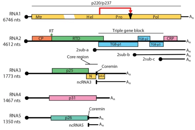
Among the five BNYVV genomic RNAs, RNA1 is essential for the replication while RNA2 ensures the functions of encapsidation, cell-to-cell movement and RNA silencing suppression. While RNA3, RNA4 and RNA5 behave as accessory species for local infection or long distance infection in S. oleracea or N. benthamiana, these viral RNAs play important, distinct and dedicated roles in the pathogenicity and the transmission of the virus within the natural host [18,19] (Figure 4). For this reason these small genomic RNA species become accessory on laboratory hosts and could be used as viral vectors of expression in the presence of RNA1 and RNA2 helper combination [20,21].
7
Figure 4 BNYVV genome organization and translational strategy. Genomic RNAs have a 5’ end cap (black circle) and a 3’ end polyA tail (An). RNA1 encodes a polyprotein that undergoes a self-cleavage (red arrow and black triangle). Methyltransferase (Mtr), helicase (Hel), protease (Pro) and RNA dependent RNA polymerase (Pol) are different domain identified on the polyprotein. RNA2 encodes for coat protein (CP) and for RT which expression depends on the suppressible UAG stop codon translational read trough (green star). CP and read-through domain (RTD) constitute RT protein. Via subgenomic RNA (sub) RNA2 encodes three triple gene block proteins (TGB-p1, TGB-p2, TGB-p3) and p14 a cysteine-rich protein (CRP) also known as the viral suppressor of RNA silencing. RNA3 encodes the p25 protein and possesses other two ORF which products N and p4.6 proteins were never detected. RNA4 encodes the p31 and RNA5 the p26. Noncoding (nc) RNA3 and ncRNA5 are produced by exoribonuclease activity [22]. The conserved coremin motif present in the „core region’ of RNA3 and in RNA5 is necessary for long distance movement in Beta species (modified from Gilmer and Ratti [2])
BNYVV RNA1 is 6,746 nts long [23] and encodes all viral factors required for the RNA replication since this RNA species is able to self replicate if transfected without the other genomic components in Chenopodium quinoa protoplasts [24]. RNA1 has one unique ORF containing two possible start codon (AUG154 and AUG496) encoding for two long
polypeptides p237 and p220 [25] having both three distinct domains associated with viral replication: methyltransferase domain (MTR), NTP-binding/helicase domain (HEL) and RNA-dependent RNA-polymerase domain (RdRp) (ibid). Papain-like protease domain is located between HEL and RdRp and is responsible for the post-translational cleavage of the p237 and p220 into p150 and p66. The RNA1-encoded protein or the clivage products interact
8 associated with the endoplasmic reticulum (ER) network [25–27]. Interestingly no major endoplasmic reticulum (ER) reorganization has been observed in infected cells suggesting that production of a huge viral factory is avoided by the virus [27] probably preventing a competition for the replication machinery between essential and shorter “non essential” RNAs.
Polycistronic RNA2 is 4,612 nts long and is characterized by having different translational strategies to express five ORFs encoding six different proteins[28]. Coat protein of 21 kDa (CP) is expressed directly by genomic RNA2 starting from its AUG145 start codon [29]. An
UAG709 stop codon is suppressed once in ten times by a read-through translation mechanism
to produce the minor capsid protein p75 (RT) (ibid). Localized at one extremity of each viral particle, RT is required for the nucleation process that probably takes place in the cytoplasmic side of the outer mitochondrial membrane [30]. Furthermore, alanine scanning mutagenesis of this protein identified a C-terminal half motif (KTER) essential for the transmission of viral particles by P. betae vector [31].
The four other ORFs are translated from three subgenomic RNAs (sgRNAs) which sequences are collinear to the 3’ half end of RNA2. These sgRNAs bring ORF2, ORF3 and ORF4, encoding TGB proteins (p42, p13 and p15), and the ORF5, required for the production of p14 closer to the 5’ terminus to allow translation. P42 is translated from 2sub-a, p13 and p15 from 2sub-b while p14 from 2sub-c [32]. The p42 has a nucleic acid binding activity and sequence motifs characteristic of superfamily I DNA or RNA helicase including a P-loop ATP/GTP binding domain [33]. The p13 protein possesses a highly conserved hydrophilic motif flanked by two hydrophobic domains able to cross ER and plasma membranes while p15, translated by a ribosomal leaking scanning of p13 start codon appears mainly hydrophobic [34]. BNYVV RNAs move cell-to-cell through plasmodesmata in a CP independent manner, an hordei-like mechanism requiring only the three TGB proteins [32,35]: The p42 proteins cooperatively interact with viral RNAs thanks to their N-terminal domain and form ribonucleoprotein complexes (RNP) delivered by the integral membrane proteins p13 and p15 to plasmodesmata following the ER network [35–37]. Hordei-like TGB proteins accumulate during infection with conserved TGB1/TGB2/TGB3 ratios of approximately 100:10:1 [38].
9 unregulated expression of BNYVV TGB proteins inhibits cell-to-cell movement [39].
The 3’end proximal ORF5 encodes a C-4 zinc finger cysteine-rich protein of 14 kDa (p14). This protein is the viral suppressor of RNA silencing (VSR) and localizes both in the cytoplasm and the nucleolus [40]. The p14 VSR activity is mainly cytoplasmic and has been associated with reduced accumulation of secondary small interfering (si) RNAs derived from endogenous RNA-dependent RNA-polymerase 6 (RDR6) pathway [40,41]. Affecting silencing transitivity of plant RNAi, p14 counteracts the restriction of systemic spread of BNYVV genomic components, playing a crucial role in the long-distance movement of the virus [40,41].
Furthermore p14 acts synergically with a viral noncoding RNA produced from genomic RNA3 (ncRNA3) for the viral long distance movement in B. macrocarpa and in Nicotiana
benthamiana infected with a BNYVV expressing an hypomorphe VSR. Together, these data suggest a link between silencing suppression and viral spread [40,41].
RNA3 is 1,774 nts long [42] and is involved in the viral long-distance movement in Beta species [18] as well as in the manifestation of rhizomania symptoms in B. vulgaris [43,44] . RNA3 contains three different ORF, encoding a protein of 25 kDa and two other proteins never detected in natural context (N and p4.6) [27]. The p25 protein localizes in the nuclear and cytoplasmic compartment independently of other viral factors [45], thanks to a N-terminal nuclear localization signal (57KRIRFR62) and a C-terminal nuclear export sequence
(169VYMVCLVNTV178 ) [45,46]. P25 is an avirulence protein [47] and has been suggested to
represent the pathogenic determinant for the rhizomania disease since transgenic Arabidopsis thaliana lines constitutively expressing the protein, display a root-branching phenotype, express high levels of auxin and low amounts of jasmonic acid derivatives [48]. Nuclear import and export motifs, together with a zinc finger domain, suggest that p25 could acts as a transcriptional factor. Such hypothesis has been corroborated in yeast one hybrid experiments in which the Gal4 or LexA DNA-binding domain-fusion with p25 is able to promote the transcription of the reporter genes. The domain responsible for transcription activation is constituted by p25 residues 103 to 146 [49]. Systemic spread in Beta species do not rely on p25 expression but on the strictly connection between “coremin” motif (located within the “core” domain) and the accumulation of ncRNA3 [18,22,50]. A 5′-3′ exoribonuclease
10 lead to the accumulation of ncRNA3 in vivo with a probable saturation of the exoribonuclease with consequences on the RNA silencing machinery [22,41]. As stated above, this could explain the synergy observed between the VSR and the accumulation of the ncRNA3 species [40,41].
Monocistronic RNA4 is 1,467 nts long [42] and encodes a cytosolic protein of 31 kDa [51] involved in the aggravation of foliar symptoms and, together with p75, in the vector transmission [52]. Experiments in N. benthamiana demonstrated a role of the p31 protein in a root specific suppression of RNA silencing [52]. Such behavior still needs to be confirmed in
Beta species.
RNA5 is 1,350 nts long and encodes a protein of 26 kDa which functional properties resemble those of the RNA3-encoded p25 protein [53,54]. As p25, p26 protein localizes both in the cytosol and nucleus and strongly activates transcription in yeast one hybrid system [55]. Transcriptional activation domain is located in the first 17 amino acid residues of the protein and is not related to the necrosis symptoms on C. quinoa, suggesting a probable avirulence behavior of p26 on such host [55,56]. When present, RNA5 increases symptomatology on B.
vulgaris roots [57] and provokes necrotic lesions on C. quinoa leaves. As RNA3, RNA5 possesses the coremin sequence, produces a noncoding RNA (ncRNA5) which is able to complement an absence of RNA3 required for the long distance movement on B. macrocarpa [58].
3. Ins and Outs of Multipartite Positive-Strand RNA Plant
Viruses: Packaging versus Systemic Spread
A Review published by:
Mattia Dall’Ara, Claudio Ratti, Salah E. Bouzoubaa and David Gilmer
Review
Ins and Outs of Multipartite Positive-Strand RNA
Plant Viruses: Packaging versus Systemic Spread
Mattia Dall’Ara1,2, Claudio Ratti2,*, Salah E. Bouzoubaa1and David Gilmer1,*
1 Institut de Biologie Moléculaire des Plantes, Integrative Virology, CNRS UPR2367, Université de Strasbourg, 12 rue du Général Zimmer, 67084 Strasbourg, France; mattia.dallara5@unibo.it (M.D’A.);
salah.bouzoubaa@ibmp-cnrs.unistra.fr (S.E.B.)
2 Dipartimento di Scienze Agrarie, Area Patologia Vegetale, Università di Bologna, Viale Fanin 40, 40127 Bologna, Italy
* Correspondence: claudio.ratti@unibo.it (C.R.); gilmer@unistra.fr (D.G.); Tel.: +39-051-2096733 (C.R.); +33-367-155362 (D.G.)
Academic Editor: Eric O. Freed
Received: 31 May 2016; Accepted: 9 August 2016; Published: 18 August 2016
Abstract:Viruses possessing a non-segmented genome require a specific recognition of their nucleic acid to ensure its protection in a capsid. A similar feature exists for viruses having a segmented genome, usually consisting of viral genomic segments joined together into one viral entity. While this appears as a rule for animal viruses, the majority of segmented plant viruses package their genomic segments individually. To ensure a productive infection, all viral particles and thereby all segments have to be present in the same cell. Progression of the virus within the plant requires as well a concerted genome preservation to avoid loss of function. In this review, we will discuss the “life aspects” of chosen phytoviruses and argue for the existence of RNA-RNA interactions that drive
the preservation of viral genome integrity while the virus progresses in the plant.
Keywords:phytovirus; segmented genome; genome integrity; systemic movement; RNA-RNA interaction
1. Introduction
Preserving genome integrity is a key challenge for any organism, and viruses with an RNA-based genome are not excluded from this basic rule. Obligate parasites that have a monopartite genome mainly face recombination events that draw viral evolution. While viruses with segmented genomes also encounter similar evolutionary traits, another constraint applies to the maintenance of genome integrity. Indeed, all genomic segments should be available within the same cell and be transmitted from one cell or one organism to another. To do so, all genomic components must be packaged within the same viral particle. This is well exemplified by Orthomyxoviridae members where all genomic negative-stranded ribonucleoprotein (RNP) complexes are selectively assembled together within one enveloped viral particle [1], or Bunyaviridae [2] where all three segments are maintained together within the envelope. Similar examples can be drawn for double-stranded RNA viruses such as Reoviridae or the well-known Cystoviridae pseudomonas phage Ø6 that possesses three genomic double-stranded (ds)RNA segments within the same viral structure [3].
Viruses that possess a positive-stranded RNA genome are found in genera that infect bacteria (e.g., Leviviridae enterobacteria phage Qß), animals and insects (e.g., Picornavirales enterovirus C—namely poliovirus—and cricket paralysis virus) and also plants (e.g., Virgaviridae, tobacco mosaic virus). To date, distinguishing features of (+)-strand RNA viruses that infect bacteria, animals or insects reside in the nature of the viral genome that is limited to either one single-stranded (ss)RNA molecule or two genomic RNAs packaged within a unique icosahedral capsid (e.g., Nodaviridae, flock house virus). However, (+)-strand RNA phytoviruses with segmented genomes range between
Viruses 2016, 8, 228 2 of 18
two to five positive-strand RNA molecules and such so-called multipartite viruses possess either an icosahedral (sometimes bacilliform) or helical shape. A large majority of these phytoviruses have distinctive features compared to the aforementioned animal (+)-strand RNA viruses, as each segment can be individually packaged as ribonucleoprotein complexes within a helical structure. Icosahedral phytoviruses with a segmented positive-sense RNA genome either package single segment species in each capsid or a combination of segments within the limits of physical size constraints. A.L.N. Rao has reviewed mechanisms driving genome packaging of spherical plant RNA viruses [4] while Solovyev and Makarov recently focused on plant viruses with helical capsids [5].
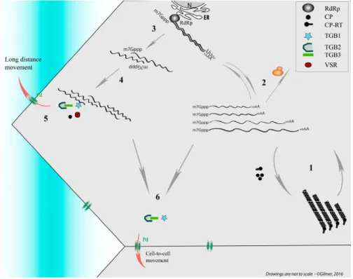
The question about the requirement for the long distance movement of the ‘virus’ of either virion (packaged RNA) or RNP complex is still open and it appears closely linked to the ability of the viral RNAs to be “packaged.” Such infection of vascular tissues is not as uniform as those of mesophyll cells where phytoviruses usually begin their journey by moving cell to cell (for review see [6]). Several boundaries constituted by the bundle sheath (BS) of V or IV class veins (for vein classification, refer to [7]) followed by the vascular parenchyma (VP) have to be passed by the viral material in order to reach companion cells (CCs) and follow the photoassimilates flow of the sieve elements (SEs). To finish the journey, the reverse route passing through class III veins to access CC and then mesophyll cells has to be pursued to start moving cell-to-cell in the distant tissues (Figure1).
‐ ‐ ‐ ‐ ‐ ‐ ‐ ‐
Figure 1.Schematic representation of the initial infection (red thunderbolt) and its progression (red lines and arrows) within a source leaf of an infected plant. Infectious material passes through plasmodesmata (Pd, double green ovals) from mesophyll (ME), bundle sheath (BS), vascular parenchyma (VP) to companion cells (CC) to access sieve elements (SE) and reach the distant tissues. A reverse route occurs in the upper leaves (sink leaf) or roots (not represented). Right panels show leaf venation and illustrate viral phloem loading and unloading through minor and major veins of source and sink leaves. X, Xyleme; I−V: vein classes.
Whatever the structural shape adopted for plant multipartite RNA viruses, their genomic segments are separated by distinct capsids. This situation raises concerns about the preservation of the viral genome integrity that requires all RNA segments to move from one cell to a neighbor or distant cell in the plant (Figure 1). This ensures the setup of a novel infection site where all viral genes need to be expressed. Here, up to three-decade old as well as new literature describing packaging, cell-to-cell and vascular movement of plant icosahedral and helical viruses has been reviewed to bring into the light our conceptual view of the nature of the systemic moving of viral material in the infected plant. Without addressing details about the RNA expression and replication mechanisms, we propose to distinguish between virions acting as storage of infectious material, ready for transmission, and moving material constituted of RNP complexes containing all RNA segments of the multipartite genome. These RNP complexes involving RNA-RNA interactions act as drivers for the “in planta” viral cycle and appear as the adequate entities for genome preservation, particularly in the long-distance journey of the virus. While this concept appears “insignificant” for monopartite RNA viruses, multipartite viruses need to preserve the expression of their entire genome in the targeted cells. After the description of chosen monopartite phytoviruses, we review and discuss the situation of multipartite RNA viruses and focus on their need to preserve the expression of their entire genome in the targeted cells. A model for benyviruses has been illustrated to present our hypotheses.
2. Lessons from Turnip Crinkle Virus, Satellite Tobacco Mosaic Virus, Groundnut Rosette Virus and Tobacco Mosaic Virus: Monopartite RNA Genomes
For some viral species, icosahedral shells are able to assemble in virus-like particles where the genome, usually dsDNA, is incorporated using energy-dependent nanomachines, extensively studied for bacterioviruses T4, P22 or Ø29. The latter uses a non-coding RNA hexamer for DNA entry. Recent insights about the genomic encapsidation of the positive-strand RNA genome of bacterioviruses MS2 revealed the importance of coat protein (CP) dimer interactions, with an estimated number of 60 hairpin RNA structures distributed around the RNA genome (packaging signals), leading to induced-fit RNA-viral protein interactions rather than electrostatic interactions. In this sense, genomic MS2 RNA constitutes a scaffold for assembly initiated or terminated by the assembly protein, following a Hamiltonian path [8–10].
2.1. Positive-Strand RNA Packaging into Icosahedral Units
2.1.1. Autonomous Turnip Crinkle Virus
Turnip crinkle virus (TCV) from the Tombusviridae family is structured by an association of 180 copies of a 38 kDa CP. Its crystallographic structure resembles the tomato bushy stunt virus. TCV viral particles can be dissociated at elevated pH where RNA is attached to six CP subunits that permit reassembly in vitro [11]. Mapping protein-RNA interactions revealed the interaction between CP and two RNA domains. A 15 base-pair (bp) long hairpin structure has been identified within the replicase gene and a 28 nucleotide (nt) bulged hairpin loop is present within a 186 nt essential element within the CP coding sequence. Viral encapsidation was also shown to be dependent on the length of the viral RNA [12], suggesting the requirement of a separation between 5′(replicase gene) and 3′ (CP gene) CP-interacting sequences, suggesting a bipartite packaging signal for this monopartite RNA viral genome. TCV CP is dispensable for the viral cell-to-cell movement but essential for the systemic spread [13] as this protein acts as a suppressor of RNA silencing [14,15].
2.1.2. Helper Virus Replication-Dependent Satellite Tobacco Mosaic Virus
Satellite tobacco mosaic virus (STMV) is an icosahedral T = 1 virus with a small genomic RNA. Atomic structure revealed 30 stem-loop RNA structures, each associated with a two-fold symmetry axis [16]. Interestingly, the RNA secondary structure of STMV analyzed in solution [17] does not correspond to the structure predicted in the T = 1 capsid. This discrepancy indicates a selection of the RNA genome into an icosahedral structure following a Hamiltonian pathway as described for MS2