Université de Montréal
Structure-function studies of class I aldolases
Exploring novel activities: mechanism, moonlighting, and
inhibition
par Paul Heron
Département de Biochimie et Médecine Moléculaire Faculté de Médecine
Thèse présentée
en vue de l’obtention du grade de Philosophiae Doctor (Ph.D.) en Biochimie et Médecine Moléculaire
option Biologie Structurale
Université de Montréal
Faculté des études supérieures et postdoctorales
Cette thèse intitulée:
Structure-function studies of class I fructose-1,6-bishosphate aldolases – exploring novel activities: mechanism, moonlighting, and inhibition
Présentée par : Paul Heron
a été évaluée par un jury composé des personnes suivantes :
Daniel Zenklusen, président-rapporteur Jurgen Sygusch, directeur de recherche Serguei Chteinberg, membre du jury Catherine Vénien-Bryan, examinatrice externe
Marc Drolet, représentant du doyen de la FES
Thèse acceptée le
Résumé
La fructose-1,6-bisphosphate aldolase de classe I est une enzyme glycolytique (EC 4.1.2.13) qui catalyse le clivage réversible du fructose-1,6-bisphosphate (FBP) en dihydroxyacétone phosphate (DHAP) et glycéraldéhyde-3-phosphate (G3P). Des années de recherche sur FBP aldolase ont permis d’identifier les résidus impliqués dans son mécanisme réactionnel, ont tracé en grande partie les coordonnées de la réaction, ont révélé de nouvelles fonctions dites « moonlighting », et ont validé l’aldolase comme une cible attrayante pour des applications anti-glycolytiques tel que le cancer. Il existe néanmoins des questions en suspens relatives à ces activités que nous avons étudiées.
Tout d'abord, la trajectoire détaillée de l'aldéhyde relatif à sa liaison au site actif allant jusqu’à la formation du lien carbone-carbone par condensation aldolique est indéfini. Pour élucider les détails moléculaires liés à ces événements, nous avons déterminé des structures cristallographiques à hautes résolution de l’aldolase de classe I chez Toxoplasma gondii, qui porte une identité de séquence élevée avec l’aldolase humaine (57%), en complexe avec l’intermédiaire ternaire de pré-condensation. Le complexe ternaire révèle un mode de liaison non-productive inhabituel pour G3P dans une configuration cis qui permet l’alignement de l'aldéhyde à proximité du nucléophile naissant. La configuration compétente pour la condensation aldolique provient d'une transposition cis-trans de l'aldéhyde qui produit une liaison hydrogène courte permettant la polarisation de l'aldéhyde et le transfert de proton au niveau de Glu-189. Nos résultats informent les chimistes synthétiques qui cherchent à développer l’aldolase comme biocatalyseur pour des réactions stéréo-contrôlées.
Le rôle présumé de l’aldolase dans la production du méthyglyoxal (MGO), un métabolite dicarbonyle hautement réactif qui génère des « advanced glycation end products » (AGES) a également été étudié structurellement et enzymatiquement. Une enquête structurelle cristallographique de MGO générée par décomposition enzymatique chez l’aldolase de classe I a révélé que, contrairement aux indications préliminaires, l'apparition hypothétique de MGO et de phosphate inorganique (Pi) résultant de la décomposition enzymatique de DHAP dans le site actif de l’aldolase est mieux interprétée par une population mixte de DHAP et de molécules d'eau. Une étude enzymatique a révélé que la décomposition spontannée des trioses-phosphate
est une source majeure de la production de MGO, alors qu’une production catalysée par l’aldolase est peu concluante. L’identification des sources de production de MGO continue d'être une priorité afin de développer des stratégies pour atténuer les manifestations cliniques de pathologies associées au MGO.
La FBP aldolase est également reconnu pour ses activités « moonlighting » - du fait qu’elle effectue plus d'une activité sans rapport avec sa fonction glycolytique. Divers partenaires de l’aldolase sont rapportés dans la littérature, y compris les adhésines de surface cellulaire chez les parasites apicomplexes, dans lequel l’aldolase exécute une fonction d'échafaudage entre le complexe actomyosine et les adhésines - une interaction qui est décisive pour la motilité et l'invasion des cellules hôte. Le mode de liaison de cette interaction a été étudié et nos résultats sont compatibles avec une liaison au site actif. Les détails précis de cette interaction ont des implications thérapeutiques, étant donné que le ciblage de celui-ci réduit l'invasion des cellules hôte par les parasites.
Enfin, l’aldolase de classe I est de plus en plus reconnu pour son potentiel comme cible anti-glycolytique dans les cellules qui sont fortement tributaires du flux anti-glycolytique, comme les cellules cancéreuses et les parasites protozoaires. Le développement de nouveaux inhibiteurs de haute affinité est donc non seulement avantageux pour des études mécanistiques, mais représente un potentiel pharmacologique sans fin. Nous avons développé une nouvelle classe d’inhibiteurs de haute affinité de type inhibition lente et avons déterminé la base moléculaire de leur inhibition grâce à des structures cristallographiques à haute résolution et par un profilage enzymatique.
Cette étude, qui combine plusieurs disciplines, y compris la cristallographie, enzymologie et chimie organique, souligne l'intérêt et l'importance d'une approche multidisciplinaire.
Mots-clés : aldolase de classe I, condensation aldolique, mécanisme catalytique,
stéréosélectivité, méthylglyoxal, métabolite toxique, Toxoplasma gondii, adhésine, conception rationnelle d’inhibiteur, cristallographie aux rayons X.
Abstract
Class I Fructose-1,6-bisphosphate aldolases are glycolytic enzymes (EC 4.1.2.13) that catalyze the reversible cleavage of fructose-1,6-bisphosphate (FBP) to dihydroxyacetone phosphate (DHAP) and glyceraldehyde-3-phosphate (G3P). Years of research on FBP aldolases has identified residues implicated in the reaction mechanism, mapped the greater part of the reaction coordinates, and revealed novel moonlighting functions. Further, FBP aldolase is recognized as an attractive target for anti-glycolytic applications such as cancer. There are nevertheless outstanding questions related to these activities that were investigated in this thesis.
First, the detailed trajectory of the reaction mechanism from aldehyde binding in the active site to carbon-carbon bond formation by aldol condensation is undefined. To elucidate the molecular details related to these events, we solved high-resolution crystallographic structures of native class I aldolase from Toxoplasma gondii, which has a high sequence identity with human aldolase (57 %), in complex with the pre-condensation ternary intermediate. The ternary complex reveals a condensation-incompetent binding mode for G3P in a cis-configuration that aligns the aldehyde alongside the nascent nucleophile. The productive aldol-competent configuration arises from a cis-trans rearrangement of the aldehyde that produces a short hydrogen bond required for polarization of the aldehyde and coincident proton transfer at Glu-189. Our results inform synthetic chemists seeking to develop aldolases for stereo-controlled reactions in biosynthetic applications.
The suspected role of aldolase in methylglyoxal (MGO) production, a highly reactive dicarbonyl metabolite that produces advanced glycation end-products (AGES) was also probed structurally and enzymatically. A crystallographic structural investigation of MGO generated by enzymatic decomposition in class I aldolase revealed that, contrary to preliminary indications, the appearance of MGO and inorganic phosphate (Pi) resulting from enzymatic decomposition of DHAP in the active site of aldolase is more appropriately modeled by a mixed population of DHAP and water molecules. Enzymatic investigation revealed triose-phosphate decomposition to be a major source of MGO production, whereas production by aldolase did not exceed assay background levels. Identifying the main sources of MGO production continues to be a priority for mitigating the clinical manifestations of MGO-derived pathologies.
FBP aldolase is also recognized for its moonlighting properties – performing more than one activity unrelated to the glycolytic function. Diverse aldolase partners are reported, including cell surface adhesins in apicomplexan parasites, in which aldolase performs a bridging function between the actomyosin complex and the cytoplasmic domain of the adhesins – an interaction that is crucial for motility and host-cell invasion. The binding mode of this interaction was investigated and our results are consistent with active site binding. The precise details of aldolase-adhesin binding has therapeutic implications, since targeting of the latter reduces host-cell invasion by parasites.
Finally, class I aldolase is gaining prominence as an anti-glycolytic target in cells that are highly dependent on glycolytic flux, such as cancer cells and protozoan parasites. Developing new high-affinity inhibitors for these enzymes is therefore not only advantageous for mechanistic studies, but has endless pharmacological potential. We developed a novel class of high-affinity aldolase inhibitors, bisphosphonates, and determined the molecular basis of their inhibition with high-resolution crystallographic structures and enzymatic profiling.
This study, which combined several disciplines, including crystallography, enzymology, and organic chemistry, underscores the interest and significance of a multidisciplinary approach.
Keywords: class I aldolase, aldol condensation, catalytic mechanism, stereoselectivity,
methylglyoxal, toxic metabolite, Toxoplasma gondii, micronemal adhesins, drug-design, X-ray crystallography.
Table of Contents
Résumé ... iv
Abstract ... vi
Table of Contents ... viii
List of Tables ...x
List of Figures ... xi
List of Schemes ... xiv
List of Abbreviations ... xv Dedication ... xvii Acknowledgments ... xviii Chapter 1 Introduction ... 1 1.1 Fructose-1,6-bisphosphate aldolase... 2 1.1.1 General aspects ... 2 1.1.2 Historical perspective ... 2 1.1.3 Classification ... 5 1.1.4 Distribution of aldolases ... 9
1.1.5 General properties of aldolase ... 11
1.2 Reaction catalyzed by Class I aldolase ... 15
1.2.1 Rabbit muscle aldolase – a model aldolase ... 15
1.2.2 Mechanism at a glance ... 16
1.2.3 Catalytic residues – from structure to function ... 20
1.2.4 Substrates ... 27
1.2.5 Methylglyoxal origins and glycolysis ... 30
1.3 Class I Aldolase as a therapeutic target ... 36
1.3.1 Targeting Aldolase – generalities ... 36
1.3.2 Classes of Inhibitors ... 42
1.4.1 General objective ... 47
1.4.2 Specific objectives ... 47
1.4.3 General methodology ... 49
1.4.4 Synopsis ... 59
Chapter 2 Stereospecific suppression of enantiomeric intermediates and rotational isomers in a key ternary complex in class I FBP aldolases ... 60
Chapter 3 Bisphosphonate inhibitors of mammalian glycolytic aldolases ... 95
Chapter 4 Aldolase and methylglyoxal production: structural and enzymatic investigation of a promiscuous aldolase activity ... 136
Chapter 5 Biophysical study of the aldolase bridging function in Toxoplasma gondii: evidence for ionic strength dependence ... 176
Chapter 6 Discussion & Perspectives ... 216
Synopsis ... 215
6.1 Investigation of novel aspects of the reaction mechanism and promiscuous catalytic activities ... 217
6.1.1 Stereoselectivity of the class I FBP aldolase addition reaction ... 217
6.1.2 Engineering of aldolase for synthetic biocatalyst applications... 223
6.1.3 Methylglyoxal formation in Aldolase ... 224
6.1.4 Future studies – Mechanism ... 226
6.2 The aldolase scaffolding activity in apicomplexan parasites ... 227
6.2.1 Future studies – Moonlighting... 231
6.3 Development and optimization of inhibitors for class I aldolase ... 232
6.3.1 Future Studies - Inhibition ... 235
Chapter 7 Conclusion ... 239
Conclusion... 240
Annex ... 241
List of Tables
Table I. Aldolases ... 6
Table II. Structures of selected competitive inhibitors ... 42
Table III. Selected irreversible inhibitors ... 44
Table IV. Selected slow-binding inhibitors ... 45
Table 2-1. Data collection and refinement statistics ... 73
Table 2-2. Progress of populations of active site species in TgALD crystals soaked with FBP. . 76
Table 2-3. Populations of the FBP cleavage product (triose-Ps) at varying local FBP concentrations in Class I aldolase. ... 78
Table 2-4. Kinetic parameters of the FBP cleavage reaction for recombinant native aldolases from Toxoplasma gondii and rabbit muscle. ... 79
Table 3-1. Data Collection and Refinement Statistics ... 101
Table 3-2. IC50 values for aldolase inhibition by 2 measured for varying pre-incubation periods. ... 110
Table 3-3. Kinetic parameters describing the slow-binding inhibition of aldolase with 2. ... 111
Table 3-4. Inhibition mode and constants for inhibitors 1 – 4. ... 112
Table 4-1. Data collection and refinement statistics. ... 149
Table 5-1. Thermodynamic binding parameters between TgALD and adhesin C-tails. ... 190
List of Supporting Tables
Table S4-1. Data collection and refinement statistics ... 172List of Figures
Figure 1-1. Glycolysis pathway. ... 4
Figure 1-2. FBP aldolase reaction. ... 7
Figure 1-3. Mechanism of FBP cleavage in Class I and II aldolases. ... 8
Figure 1-4. Phylogenetic tree of Class I and Class II FBP aldolases. ... 10
Figure 1-5. The tertiary structure of various FBP aldolases compared with TIM. ... 13
Figure 1-6. Tetrameric quaternary structure of Class I aldolase and view of active site. ... 14
Figure 1-7. Dimeric structure of Class II aldolase and view of active site. ... 14
Figure 1-8. General reaction mechanism of Class I FBP aldolases. ... 17
Figure 1-9. Active site of rabbit muscle aldolase (PDB: 1ZAH). ... 21
Figure 1-10. Snapshots of catalysis in FBP aldolase showing key intermediates. ... 24
Figure 1-11. Mechanism for stereospecific proton transfer mediated by C-terminus. ... 25
Figure 1-12. FBP condensation in Class I FBP aldolase. ... 26
Figure 1-13. FBP analogs: substrates (FBP, XBP, SBP, and SedBP) and inhibitors (RBP and TBP). ... 28
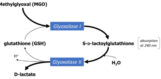
Figure 1-14. The glyoxalase system. ... 31
Figure 1-15. Pathways of methylglyoxal metabolism. ... 33
Figure 1-16. General decomposition scheme of triose-phosphates to methylglyoxal. ... 34
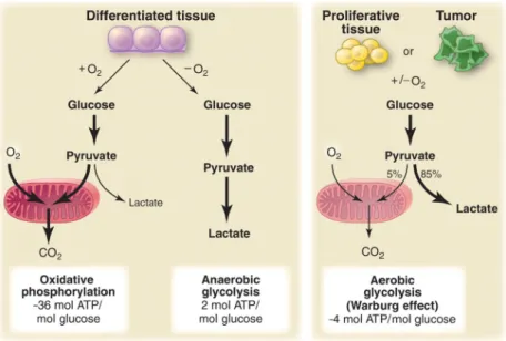
Figure 1-17. Schematic representation of the differences between oxidative phosphorylation, anaerobic glycolysis, and aerobic glycolysis (Warburg effect). ... 37
Figure 1-18. WASP and the naphthol-based inhibitor NASEP bind a unique hydrophobic pocket. (taken from St-Jean et al., 2007) ... 41
Figure 1-19. Solubility phase diagram for protein crystallization. ... 52
Figure 1-20. Enzyme-coupled assay for measuring Aldolase activity. ... 57
Figure 2-1. Progress of the aldol addition reaction in a class I aldolase... 74
Figure 2-2. pH activity profile of the FBP cleavage reaction for TgALD. ... 80
Figure 2-3. Reaction geometry associated with aldol addition implicating putative cis-trans rotational isomerization. ... 82
Figure 2-4. L-G3P model reveals basis for enantiomeric discrimination in class I aldolase. ... 83
Figure 3-1. Crystal structure of the ALDOA-1 complex. ... 102
Figure 3-2. Crystal structure of the ALDOA-2 complex. ... 105
Figure 3-3. Superposition of the ALDOA-1 and ALDOA-2 complexes and interactions at the P1 -site. ... 106
Figure 3-4. Crystal structure of ALDOA in complex with compounds 3 and 4. ... 108
Figure 3-5. Active site dynamics upon aldolase-binding of 1 and 2... 113
Figure 3-6. Cell growth inhibition assay. ... 116
Figure 4-1. Aldolase crystal structure (DHAP-frac) showing evidence of phosphate elimination. ... 143
Figure 4-2. Difference density maps of aldolase soaked with DHAP for increasing time periods. ... 150
Figure 4-3. Occupancy and Temperature factor variability as a function of DHAP soak time. ... 153
Figure 4-4. pH profile of MGO generated from FBP in the presence of aldolase. ... 155
Figure 4-5. MGO generated in the presence of aldolase and associated metabolic enzymes. ... 156
Figure 4-6. Comparison of quenching methods. ... 158
Figure 4-7. Inorganic phosphate determination from fructose-bisphosphate and triose-phosphates in the presence of aldolase. ... 160
Figure 5-1. Model for gliding motility in apicomplexans. ... 181
Figure 5-2. Conservation of amino acid residues within micronemal adhesin proteins. ... 194
Figure 6-1. Pre-aldol addition reaction trajectory in class I FBPAs (stereo view). ... 219
Figure 6-2. Superposition of intermediates from diverse aldolases (stereo view)... 221
Figure 6-3. Naphthalenic-phosphate and bisphosphonate derivatives as high affinity aldolase inhibitors. ... 232
Figure 6-4. Kinetic analysis of aldolase inhibition by UM76 showing apparent mixed inhibition (A) and slow-binding inhibition (B). ... 237
List of Supporting Figures
Figure S2-1. Discrimination of enzymatic intermediates formed with FBP in T. gondii aldolase
active site. ... 93
Figure S2-2. Configurational rearrangements during aldol addition. ... 94
Figure S3-1. Active site comparison of ALDOA-1, ALDOA-2 and ALDOA-FBP Schiff base complex. ... 132
Figure S3-2. Superposition of 3 and NASEP in ALDOA active site. ... 133
Figure S3-3. Slow-binding inhibition of aldolase by inhibitor 2. ... 134
Figure S3-4. Dual competitive inhibition of aldolase by inhibitor 3. ... 135
Figure S3-5. Competitive inhibition of ALDOA activity by inhibitor 4. ... 135
Figure S4-1. Superposition of WT-Aldolase with DHAP soaked Aldolase. ... 173
Figure S4-2. Omit maps of aldolase active sites for all soaking times. ... 174
Figure S4-3. MGO production from FBP in the presence of Aldolase as a function of time. .. 175
Figure S5-1. TgALD fold. ... 208
Figure S5-2. Superposition of WASP and LC4 peptides onto TgALD structure. ... 209
Figure S5-3. ITC binding isotherms for TgALD and MIC2 C-tails. ... 210
Figure S5-4. ITC binding isotherms for TgALD and AMA1 C-tails. ... 211
Figure S5-5. ITC binding isotherms for TgALDΔC variant and MIC2/AMA1 C-tails. ... 212
Figure S5-6. ITC binding isotherms for TgALD adhesin C-tails in varied experimental conditions. ... 213
Figure S5-7. ITC binding isotherms showing no binding for TgALD and TgALDΔC with MIC2t-25 in conditions of high ionic strength. ... 214
List of Schemes
Scheme 2-1. Intermediates of the Catalytic Mechanism in Class I Aldolases ... 65
Scheme 3-1. General mechanism for describing slow-binding inhibition. ... 120
Scheme 4-1. Decomposition of the triose-phosphates to methylglyoxal. ... 141
List of Abbreviations
AGES Advanced glycation end products
Ala Alanine
ALDOA Aldolase A
AMA1 Apical membrane antigen 1
Arg Arginine
Asp Aspartic acid
ATP Adenosine triphosphate C-terminal Carboxy-terminal
cm Centimeter
DHAP Dihydroxyacetone phosphate EDTA Ethylenediaminetetraacetic acid F-actin Actin filaments
FBP Fructose-1,6-bisphosphate HTS High throughput screen G3P Glyceraldehyde-3-phosphate
GDH Glycerol 3-phosphate dehydrogenase
Glu Glutamic acid
GLUT4 Glucose transporter 4
ITC Isothermal titration calorimetry
K Kelvin
k rate constant
Ka Association constant
kcat Turnover number
KD Dissociation constant
kDa Kilo Dalton
Ki Inhibition constant
Lys Lysine
mg Milligram
mM Millimolar
µM Micromolar
ηM Nanomolar
MGO Methylglyoxal
MGS Methylglyoxal synthase MIC2 Microneme protein 2
NADH Nicotinamide adenine dinucleotide NASEP Naphthol AS-E phosphate
N-terminal Amino-terminal
ηm Nanometer
NMR Nuclear Magnetic Resonance
PDB Protein Data Bank
RNA Ribonucleic acid
ROS Reactive oxygen species
Ser Serine
SUMO Small ubiquitin-like modifier TgALD Toxoplasma gondii aldolase
TIM Triosephosphate isomerase
Trp Tryptophan
Tyr Tyrosine
Dedication
To my family, friends, and dearest of all, my wife.
Acknowledgments
First I would like express my great appreciation to Dr. Jurgen Sygusch for his patient guidance, enthusiastic encouragement and useful critiques during my thesis. I would also like to acknowledge the members of my thesis committee, Dr. Pascale Legault and Dr. Joelle Pelletier, for their advice and assistance in keeping my progress on schedule. My special thanks are also extended to the members of my jury for accepting to correct my thesis.
There are countless people at the University who have supported my work in one way or another, including the support staff in Biochemistry who have been helpful with the administrative aspects of my thesis. I would also like to extend my thanks to the dedicated staff at the MS platform in Chemistry (Alexandra F) and the crystallography platform (Michel S). A special acknowledgement goes to Marie P in Bioinformatics for giving me the opportunity to teach undergrads for so many years. I am also grateful to Jim O for entrusting me his undergraduate crystallography lecture. I would be remiss if I didn’t acknowledge the friendly and helpful staff at BNL.
Many colleagues I have worked alongside in my laboratory have made this journey endurable and I would like to acknowledge the following for the many smiles we have shared (in chronological order): Mathieu C, Em F, Clotilde LK, Arnold DP, Octavian T, Jaan, Livia O, Joyce W, Loic, Maher, Mara, Jérémie R, Ghiles, Viridiana, Rux - and finally, the last two deserve a mention for the special comradery we have shared, Benoit J and Amira Y. Members of several other labs have been a pleasure to work alongside, including colleagues in the Baron lab, Omichinski lab, Legault lab, Ferbeyre lab and Brisson lab.
This acknowledgment would be incomplete without singling out the following colleagues with whom I’ve developed a special friendship and have shared memorable experiences: Bastien C et Julie B, Normand C, Thomas M, Mathieu V and their significant others.
My time at UdeM would not have been the same without my weekly injection of sports, including squash with TM, running with the RadicauxLibres, and soccer with my fellow biochemists – I hope to have left a lasting impression on my colleagues in regard to the importance of physical health.
My parents, grandparents, brothers, sisters, and friends are to be thanked for their continued support through prayer and encouragement.
Finally, my wife and best friend, Marissa – you bring the most joy of anyone to my life. Thank you for sharing so many adventures with me and being so supportive of me. You now have a Ph.T. (Putting Hubby Through) diploma.
Chapter 1
Introduction
1.1
Fructose-1,6-bisphosphate aldolase
1.1.1
General aspects
Proteins are complex molecules necessary for the chemical processes that occur in living organisms. As the chief actors within the cell, their mechanisms of action, regulation, structure, and interaction are the subject of intense scrutiny by a great number of researchers. One such protein that has been rigorously dissected for several decades is the glycolytic enzyme, aldolase. Nevertheless, sustained research on this ‘old dog’ continues to reveal the remarkable complexity of this understated enzyme. The goal of this thesis is to continue this tradition, and bring to light some of the lesser known facets of class I aldolases, including aspects related to its mechanism, inhibition, and moonlighting functions. The following introduction will provide a universal portrait of class I aldolases that is generally applicable to each of the subsequent chapters.
1.1.2
Historical perspective
The chemistry of life requires a bewildering array of biochemical reactions that occur simultaneously in a living cell, the sum of which is called metabolism. These reactions function coherently together to maintain fundamental processes such as tissue development, waste disposal, reproduction, and conversion of food to energy – processes that we refer to as life. One of the hallmarks of life is that these reactions do not occur randomly and spontaneously. They are tightly regulated and made possible by a phenomenon known as enzymatic catalysis, which provides the necessary acceleration that is required for an otherwise slow and un-controlled reaction. The early study of enzymes (enzymology), tightly woven with the discipline of biochemistry, evolved largely from the investigation of fermentation in the nineteenth century.
In 1860, Louis Pasteur observed that fermentation occurs in microorganisms. The inability to isolate and reproduce biochemical reactions outside living cells reinforced the largely held assumption that living cells were endowed with “vital forces” governed by special laws that enabled them to perform biochemical transformations (Kresge et al., 2005; Manchester, 1995). This theory was dispelled by a major breakthrough in 1897 when Eduard Buchner discovered that cell-free extracts that could carry out fermentation, marking the inception of biochemistry
(Buchner, 1897; Nobelprize.org). This enabled the systematic dissection of cell extracts, which led to breakthroughs in 1905 by Arthur Harden and William Young who discovered that heat-labile fractions called “zymase’ (mixture of enzymes) and “cozymase” (co-factors such as NAD+, ATP, ADP and metal ions) were both necessary for fermentation. Emil Fischer’s discovery in 1894 that glycolytic enzymes display specific activity towards stereoisomeric sugars gave birth to the notion of “lock and key mechanism” between an enzyme and its substrate – a concept still used in classrooms today when teaching enzymology (Fischer, 1894).
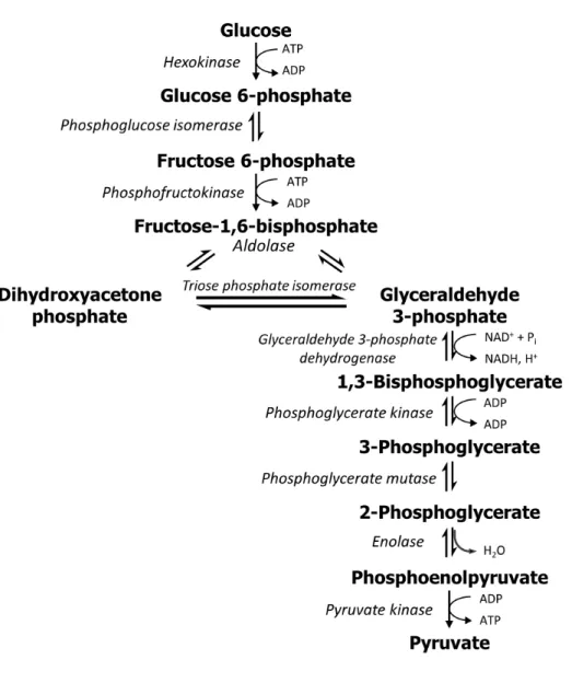
It took several decades and the effort of many investigators to piece together the complex puzzle of glycolysis (Figure 1-1). By 1940, all components were elucidated and the pathway was formally termed the Embden-Meyerhof-Parnas pathway for the major contributions made by Gustav Embden, Otto Meyerhof, and Jacob Parnas. These early investigators pioneered many of the fundamental biochemical techniques of isolation and purification of intermediates that animated many other later discoveries. The short-lived nature of the intermediates of glycolysis and their sheer number proved to be the utmost challenge.
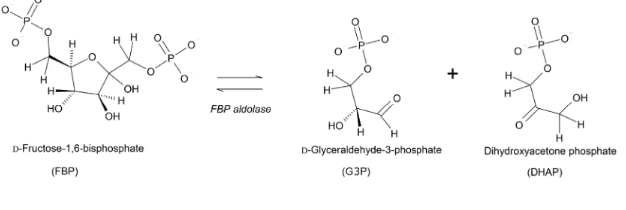
Otto Meyerhof, whose primary interest was to understand how energy is transformed during chemical reactions in the cell, was particularly instrumental in the discovery of the zymohexase (fructose-1,6-bisphosphate aldolase (FBPA)), the enzyme responsible the reversible cleavage of the hexose diphosphate (known as fructose-1,6-bisphosphate (FBP)) into two triose phosphate isomers, dihydroxyacetone phosphate (DHAP) and glyceraldehyde-3-phosphate (G3P) (Meyerhof and Junowicz-Kocholaty, 1943; Meyerhof and Lohmann, 1934).
The advent of new techniques such as chromatography, X-ray crystallography, NMR spectroscopy, mass spectrometry, and electron microscopy, has accelerated the discovery and detailed understanding of many metabolic pathways in cells. FBP aldolase has been no exception, and decades of investigation into this model enzyme have revealed intricacies regarding its activity in different species, its specificity for a variety of substrates, and its three-dimensional structure.
Figure 1-1. Glycolysis pathway.
Schematic representation of the enzymes and chemical intermediates of glycolysis. One molecule of glucose will generate a net of 2 pyruvates, 2 ATP, and 2 NADH. Pyruvate is a substrate for several metabolic pathways, including the citric acid cycle, fatty acid biosynthesis, amino acid biosynthesis, and gluconeogenesis.
1.1.3
Classification
FBP aldolases are lyases (Enzyme Commission number: EC 4), capable of cleaving bonds
by means other than hydrolysis or oxidation. In the subclass of enzymes capable of carbon-carbon bond cleavage (EC 4.1), the aldehyde-lyases catalyze the reversal of an aldol
condensation (EC 4.1.2) and are hence termed, aldolases. The members of the aldolase class of
enzymes can be distinguished by the stereospecific aldol condensation of a donor (nucleophile) to an acceptor (aldehyde) in the aldol condensation reaction. For example, as with FBP aldolase, fuculose-1-phosphate aldolase uses DHAP as a donor but the acceptor is lactaldehyde, producing fuculose-1-phosphate. Further, depending on the stereochemistry of the reaction, the same donor and acceptor can generate different products around the carbon-carbon bond. FBP aldolase produces a 3(S)-4(R) configuration in FBP because the attack of the nucleophilic donor occurs on the si-face of G3P. Conversely, tagatose-bisphosphate aldolase (TBPA) orients the
re-face of G3P to the donor DHAP affording a 3(S)-4(S) stereochemistry around the C3-C4
bond, producing tagatose-bisphosphate (TBP). Table I highlights a number of aldolases that are grouped according to their donors and acceptors. Although not exhaustive, Table I demonstrates that several aldolases share common donors (other donors include phosphoenolpyruvate and acetaldehyde (Pricer and Horecker, 1960; Ray and Benedict, 1980; Weissbach and Hurwitz, 1959)).
Table I. Aldolases1
Donor Acceptor Aldolase [a] Product
FBP-aldolase DHAP TBP-aldolase[1] Fuc-1-P-aldolase[2] Rha-1-P-aldolase[3] KDO-aldolase[4] Pyruvate KDF-aldolase[5] KDG-aldolase[5] KDGP-aldolase[5]
[a] Abbreviations: FBP, D-frutcose1,6-bisphospahte; TBP, tagatose-1,6-bisphosphate; Fuc-1-P, L
-fuculose-1-phosphate; Rha-1-P, L-rhamnulose-1--fuculose-1-phosphate; KDO, 3-deoxy-D-mannooctulosonate; KDF, 2-keto-3-deoxy-D-fuconate; KDG, 2-keto-3-deoxy-D-glucarate; KDGP, 2-keto-3-deoxy-6-phosphogluconate.
1 Table adapted from Coinçon (Coinçon, 2010) and Whitesides et al. (Whitesides and Wong, 1985)
FBP aldolase (EC 4.1.2.13) specifically catalyzes the retro-aldol condensation of FBP into
the triose-phosphates DHAP and G3P (Figure 1-2) which occurs in the catabolic direction of glucose metabolism (glycolysis). In the reverse direction (gluconeogenesis), FBP aldolase performs the aldol condensation of the aldehyde, G3P, with the ketone, DHAP, to form FBP.
Nevertheless, aldolases are not classified according to the nucleophilic donor, but are instead classified by their mechanism of action. Decades of investigation of aldolases from different sources and organisms revealed that two classes of aldolases could be distinguished by the mechanism that is used to stabilize the negative charge that develops on the reaction intermediates (Richards and Rutter, 1961; Rutter, 1964). The first mechanism, used by Class I aldolases, involves the formation of a covalent intermediate between the ε-amino group of a lysine and the ketone of the incoming substrate (FBP or DHAP) (Grazi et al., 1962b). The resulting covalent intermediate is an iminium (commonly referred to as a Schiff base). The iminium creates an electron sink at the C2 carbon of the substrate that stabilizes the formation of the nascent enolate intermediate (enamine). In contrast, Class II aldolases stabilize the negative charge on the enolate intermediate (enediolate) with a divalent metal cation – generally Zn2+, bypassing the need for a covalent intermediate. This polarizes the carbonyl group at C2, creating the electron sink required for cleavage of the carbon-carbon bond. Both strategies ultimately increase the electrophilic nature of the C2 carbon, which enables abstraction of the proton on the O4 hydroxyl group, leading to cleavage of the C3-C4 bond and formation of the enolate intermediates (Figure 1-3).
The distinction between the two classes was first reported after the observation that aldolases were differentially inhibited by chelating agents such as ethylenediaminetetraacetic acid (EDTA), whereby Class II aldolases (first shown in yeast aldolase) were strongly inhibited because of their essential dependence on divalent metal ions (Richards and Rutter, 1961). Conversely, Class I aldolases (typified by the rabbit muscle aldolase) were found to be sensitive to sodium borohydride (NaBH4) treatment, which reduces the Schiff base intermediate to a secondary amine and stabilizes the irreversible complex in the active site (Grazi et al., 1962a). This distinction meant aldolases could readily be classified based on their reactivity to either EDTA (Class II) or NaBH4 (Class I) but early investigators quickly realized that their distribution among different organisms wasn’t so apparent.
Figure 1-3. Mechanism of FBP cleavage in Class I and II aldolases.
1.1.4
Distribution of aldolases
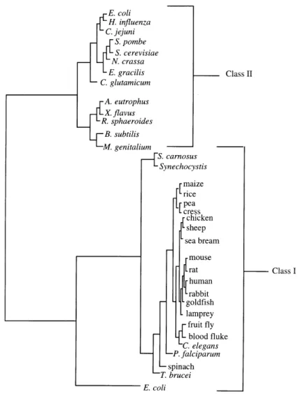
The different classes of FBP aldolases show a complex and peculiar distribution in the biosphere. Since FBP aldolase occupies a central role in sugar metabolism, it is ubiquitous in all three major kingdoms of life – archaea, bacteria, and eukaryotes. It was originally assumed that Class I aldolases were restricted to higher forms of life, including animals, plants, protozoans and green algae (Rutter and Groves, 1964) and that Class II aldolases were relegated to lower forms of life such as bacteria, fungi and blue-green algae (Lebherz and Rutter, 1969; Rutter, 1964). The distribution of aldolases in select organisms is illustrated in Figure 1-4. The striking distinction in mechanism together with other structural and functional differences led Rutter (Rutter, 1964) to postulate a convergent evolution of unrelated ancestral molecules.
Eukaryotic organisms were then discovered that produce both Class I and Class II aldolases, as first shown in Euglena (Rutter, 1964) and Clamydomonas (Russell and Gibbs, 1967). This occurrence is now equally described in prokaryotic organisms – notably Escherichia coli (E.coli) (Baldwin and Perham, 1978; Stribling and Perham, 1973; Thomson et al., 1998), and
Lactobacillus casei (London, 1974). Upon observation in E. coli that both classes of aldolase
were produced when grown on a gluconeogenic carbon source (pyruvate or lactate), but that only Class II was expressed on a glycolytic carbon source (glucose), it was suggested that in these organisms, Class I is better suited for gluconeogenesis while the Class II enzyme is constitutively expressed (Baldwin and Perham, 1978). In general however, most organisms produce only one class of aldolase. Both classes of aldolase are thus represented in each of the three primary kingdoms. Further, certain prokaryotes exclusively contain Class I aldolases, as first discovered in Micrococcus aerogenes (Lebherz and Rutter, 1973) and subsequently in
Mycobacterium smegmatis (Bai et al., 1982, 1975) Staphylococcus aureus (Götz et al., 1980)
and others. The isolation and characterization of Class I aldolases from a variety of sources have revealed distinguishing features. Eukaryotic Class I aldolases are invariably tetrameric with a subunit molecular mass of approximately 40 kDa. Sequence comparisons of the latter suggest a high degree of conservation, particularly in active site residues (Freemont et al., 1988; Kelley and Tolan, 1986). Conversely, prokaryotic Class I aldolases appear to be more diverse than their eukaryotic counterparts. Their oligomeric states range from monomeric to decameric and their
treatment, suggesting an alternative to the functional and highly conserved carboxy-terminal tyrosine residue, which is characteristic of eukaryotic Class I aldolases.
Adding to the elaborate puzzle, another sub-class of aldolases, an archaeal type Class I aldolase (Class IA aldolase) was recently identified and characterized in a hyperthermophilic organism (Siebers et al., 2001). Despite functional similarity with classical Class I aldolase in their use of a Schiff-base mechanism, this new family shows no significant sequence similarity with the latter.
Figure 1-4. Phylogenetic tree of Class I and Class II FBP aldolases.
These findings demanded a revision of the simplistic phylogenetic proposal by Rutter (Rutter, 1964). The low sequence similarity between Class I and Class II aldolases continues to suggest a convergence of function. However, the discovery of organisms expressing both types suggests the existence of a hypothetical progenote capable of produce both types. Subsequent loss of one of the classes could be explained by the functional redundancy of aldolases (Marsh and Lebherz, 1992). Notwithstanding our speculative understanding of its origins, more recent structural studies on various aldolases have shown that despite having a low sequence similarity, the different classes display a common tertiary fold: the (βα)8-(TIM)-barrel fold (TIM – triosephosphate isomerase).
1.1.5
General properties of aldolase
All FBP aldolases, regardless of their low sequence identity (~20%) (Marsh and Lebherz, 1992), adopt the TIM-barrel fold. This architectural type is the most frequently occurring protein fold (known as superfold) in the SCOP database (Structural Classification of Proteins)2 which categorizes protein structures in the Protein Data Bank (PDB) (Gerstein and Levitt, 1997). It was first observed in triosephosphate isomerase (Figure 1-5B) and consists of eight α-helices on the outside and eight parallel β-strands on the inside that alternate along the peptide backbone (Wierenga, 2001) (Figure 1-5A). Members of the superfold are nearly always enzymes and are most often involved in molecular or energy metabolism in the cell (Nagano et al., 2002). The TIM-barrel fold contains 33 superfamilies, including the aldolase superfamily in which we find the Class I and Class II FBP aldolase families. Notwithstanding the diversity of their catalytic reactions, TIM-barrel proteins share common features including the following:
1. active site residues are always found at the C-terminal end of the β-strands in the barrel and the eight loops following the β-strands (βα-loops), implying that the geometry of the active sites is dictated by composition of the βα-loops (Brändén, 1991);
2. electrostatic calculations demonstrate the existence of a positive potential at the C-terminal end of the barrel, consistent with the preference of TIM-barrel folds to bind negatively charged substrates (Copley and Bork, 2000; Raychaudhuri et al., 1997) ;
3. active sites frequently contain either phosphate-binding sites (~50% of members) or metal-ion binding sites (~66% of members) – features that are found in FBP aldolases (Nagano et al., 2002; Wierenga, 2001).
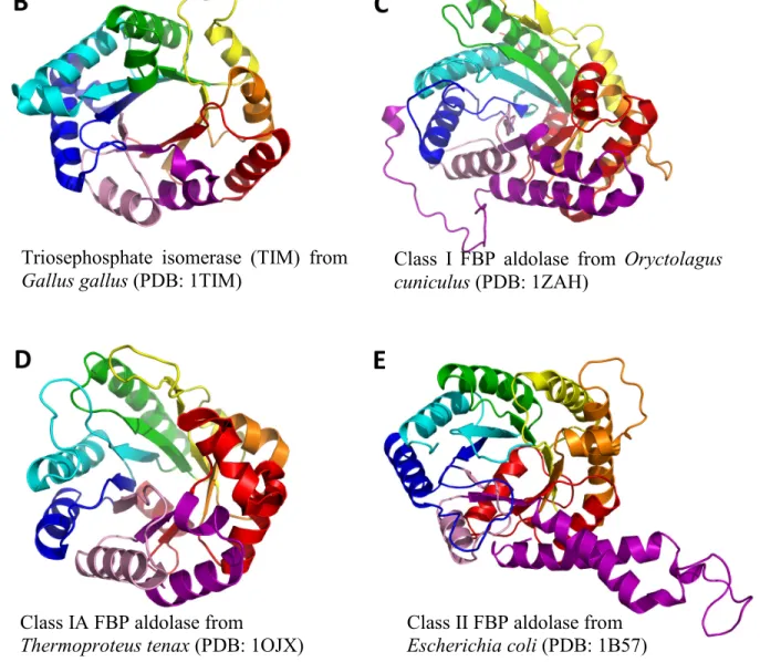
Although they ultimately perform the same reaction, Class I and Class II aldolases have distinct active site architectures. The active site of Class I aldolase is deep within the barrel involving several β-strand residues and a mobile, catalytic C-terminal helix that covers the active site (Figure 1-5C), while the active site in Class II aldolases is at the opening of the barrel and involves a catalytic loop (Figure 1-5E) (Sygusch et al., 1987).
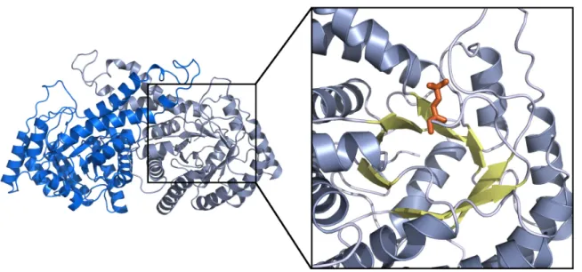
The quaternary organization of eukaryotic Class I aldolases is invariably tetrameric with a total molecular mass of 160 kDa (Figure 1-6) (Kawahara and Tanford, 1966). Prokaryotic Class I aldolases are not regular and range in size from 27 – 40 kDa, and assemble into a variety of oligomeric states from monomers to decamers. On the other hand, Class II aldolases are generally dimeric, having a total molecular mass of 80 kDa (Figure 1-7).
As the focus of my studies have been on Class I aldolases, the following sections will be primarily concerned with Class I aldolases. The next section will provide a detailed exposition of the reaction mechanism in Class I aldolase, which will help gain a fuller appreciation of the complexities within an ostensibly simple biochemical reaction.
Triosephosphate isomerase (TIM) from
Gallus gallus (PDB: 1TIM) Class I FBP aldolase from Oryctolagus cuniculus (PDB: 1ZAH)
Class IA FBP aldolase from
Thermoproteus tenax (PDB: 1OJX) Class II FBP aldolase from Escherichia coli (PDB: 1B57)
Schematic representation of the secondary structure for the archetype (βα)8-TIM-barrel fold – shown here for triosephosphate isomerase.
Figure 1-5. The tertiary structure of various FBP aldolases compared with TIM.
(5A) Representation of the secondary structure (β-sheets: arrows; α-helices: cylinders) of an archetypal TIM-barrel fold. The repeating (βα) motifs are colored individually and numbered 1-8. (5B-5E) The tertiary fold of TIM monomer and FBP aldolases monomers from each class.
Figure 1-6. Tetrameric quaternary structure of Class I aldolase and view of active site.
The structure depicts a typical Class I aldolase, here an FBP aldolase from rabbit muscle (PDB: 1ZAI) (St-Jean, 2005). The inset on the right reveals a close-up of the active site where FBP (orange) is covalently bound to the catalytic lysine. The active site is deep in the β-barrel (yellow), surrounding by the helices (grey) of the TIM-barrel fold.
Figure 1-7. Dimeric structure of Class II aldolase and view of active site.
The dimer of a Class II aldolase from E. coli is presented (PDB: 1B57) (Hall et al., 1999). On the right, the active site and an inhibitor (phosphoglycolohydroxamate – a DHAP analogue) bound at the substrate binding site. Note the position of the binding site at the cusp of the barrel.
1.2
Reaction catalyzed by Class I aldolase
1.2.1
Rabbit muscle aldolase – a model aldolase
Class I FBP aldolase has received much attention since the attribution of its function by Meyerhof (Meyerhof and Lohmann, 1934). Limited by the technical purification and isolation practices of his generation, early investigators required large amounts of sample in order to conduct experiments. Aldolase from rabbit muscle proved to be an ideal source because of its natural abundance, representing as much as 5% of the protein weight of rabbit muscle (Herbert et al., 1940; Maughan et al., 2005; Taylor et al., 1948). Its abundance and ease of isolation were the reasons that aldolase from rabbit muscle became the model enzyme for Class I aldolases. For several decades, this required the sacrificing of animals to perform isolation of pure enzyme but the advent of molecular cloning techniques and recombinant enzyme expression from bacterial hosts were eventually applied to rabbit muscle aldolase and led to significant yields that have been used since (Morris and Tolan, 1993).
Rabbit muscle expresses one of three known mammalian isoforms – aldolase A (ALDOA). Two other distinct aldolases were detected in mammals – aldolase B (ALDOB) and C (ALDOC) (Peanasky and Lardy, 1958; Penhoet et al., 1966; Warburg and Christian, 1943). They were first identified from observed differences in catalytic properties of aldolase isolated from different tissues. ALDOA is found in muscle and red blood cells; ALDOB is the predominant form in the liver; and ALDOC is found in the brain. Although hybrid tetramers can be formed, the substrate specificity of each isoform in the hybrid is conserved (Penhoet et al., 1967). The substrate specificity of each isoform concurs with the recognized metabolic role of its corresponding tissue. Specifically, the distinction can be made with respect to the enzyme’s catalytic preference for fructose-1-phosphate (F1P) or fructose-1,6-bisphosphate (FBP). ALDOA has an FBP/F1P activity ratio of approximately 50:1; the ratio is 1:1 in ALDOB; and the ratio is 25:1 in ALDOC (Penhoet et al., 1966; Rutter, 1964). ALDOB, central to fructose metabolism, occurs almost entirely in the liver and participates in glycogen biosynthesis by hydrolyzing F1P into the triose phosphates (DHAP and G3P) which feed into gluconeogenesis. The loss of activity of ALDOB from mutations is detrimental, and leads to hereditary fructose intolerance (Ali et al., 1998; Hers
and Joassin, 1961). The three isoforms can also be distinguished by their immunological characteristics, with antibody preparations showing little to no cross-reactivity among the isoforms (Penhoet et al., 1966).
The most marked difference between the isoforms occurs at the C-terminus. It was possible to recreate the catalytic fingerprint of one isoform by swapping its C-terminal region onto the core of a different isoform (Berthiaume et al., 1993). Likewise, upon carboxypeptidase treatment (C-terminal cleavage), rabbit muscle aldolase exhibits a 20-fold fall in the rate of cleavage of FBP and cleavage of F1P remains unchanged (Drechsler et al., 1959), suggesting the specificity in ALDOA is afforded by its C-terminus. Sequence homology of the non-conserved terminal residues (~20 C-terminal residues) is sufficient for proper classification of the three isoforms. Contrariwise, sequence comparisons of several Class I aldolases indicates a high degree of homology at the active site, including the three isoforms (Fothergill-Gilmore and Michels, 1993). This suggests that the core amino acids that line the active site serve to ensure a basal activity common to the three isoforms (Kitajima et al., 1990).
For this reason, rabbit muscle aldolase continues to be the workhorse model for studies on Class I aldolase. Further, the human muscle aldolase only differs from the rabbit homolog by 6 amino acids (out of 363); the 6 substitutions are conserved (physicochemical properties) and distant from the active site. The homologs were correctly predicted to have equivalent tertiary structures (Sawyer et al., 1988). This was soon confirmed when several high-resolution structures from different sources were obtained by X-ray crystallographic studies (rabbit muscle - Sygusch et al., 1987; human muscle - Gamblin et al., 1990; and drosophila - Hester et al., 1991). Most of the mechanistic studies have been performed on rabbit muscle aldolase. Accordingly, the following mechanistic considerations are mostly related to the rabbit muscle isozyme.
1.2.2
Mechanism at a glance
Much of the early work involved the use of radiolabeling, enzymatic and isotope exchange experiments. Later studies were structure-based and involved mutational studies. The use of substrate analogues and inhibitors have also been instrumental in elucidating the reaction
mechanism. Here, the general reaction mechanism will be presented from a biochemical and historical perspective, and structural review of essential residues will follow.
A simplified reaction scheme for the aldol condensation reaction in aldolases, outlined in Figure 1-8, proceeds with the formation of an iminium intermediate (Schiff base) between the ε-amino group of the active site lysine (K229 in rabbit muscle) and the carbonyl group of DHAP. The neutral carbinolamine (Figure 1-8 - species 1) is a transition to the protonated iminium form
(species 2). The iminium is activated by stereospecific abstraction of the pro(S) hydrogen at the
C3 carbon leading to enamine formation (species 3), a stable resonance form of the reactive
nucleophile. The following step is the condensation between the activated double bond of the enamine and the carbonyl on G3P, forming the C3-C4 bond and leading to a second iminium species (species 4). Hydrolysis of this iminium proceeds via another carbinolamine (species 5),
and releases FBP. In the cleavage direction, the positive charge of the iminium on species 4
creates the electron sink that is required for C3-C4 bond cleavage. Although this scheme summarizes the condensation (gluconeogenic) course of the reaction, it is worth noting that it is thermodynamically favoured, with an equilibrium constant of 104 – 105 M-1, favouring the formation of FBP (Connett, 1985; Lehninger et al., 1955; Veech et al., 1969). Regardless, both directions involve the intermediates that are shown in Figure 1-8. It was Rose in 1965 (Rose et
al., 1965) who first demonstrated that the reaction proceeds by an ordered sequence whereby G3P can only bind after formation of the covalent enamine intermediate with DHAP. The following sub-sections will describe the findings that exposed each of the intermediates in Figure 1-8.
i. Carbinolamine
The first step involves formation of a non-covalent Michaelis complex between the active site and DHAP. This was demonstrated in a crystal structure (PDB: 1ADO) in which DHAP is shown to adopt three mutually exclusive binding modes (Blom and Sygusch, 1997). One of the modes reveals the catalytic lysine, Lys-229, forming hydrogen bonds such that its’ Nζ lone pair is aligned in an incipient conformation favourable for nucleophilic attack at the C2-carbonyl carbon of DHAP. This leads to the formation of the carbinolamine intermediate (species 1). The
existence of this intermediate was confirmed by 13C NMR (nuclear magnetic resonance) experiments with a DHAP analogue, glycolaldehyde-phosphate (Ray et al., 1983). Subsequent protonation and dehydration at the C2 carbon leads to iminium formation.
ii. Iminium
Treatment of aldolase with NaBH4 after reacting the enzyme with radiolabeled DHAP (32P or 14C) inactivates the enzyme via conversion to a stable secondary amine. Degradation of the enzyme to its components reveals the covalent adduct with the incorporated radiolabel, N6
-β-glyceryl-lysine (Grazi et al., 1962a, 1962b), evidence of iminium (species 2) formation between
DHAP and an active site lysine. The existence of the iminium was also confirmed by inactivation with cyanide, which is postulated to react with the iminium (Cash and Wilson, 1966).
iii. Enamine
The enamine intermediate (species 3) is produced upon proton abstraction at DHAP C3
carbon. In reality, the enamine species (neutral) is the more stable resonance form of the incipient carbanion that forms on C3 carbon after proton abstraction. Several lines of evidence demonstrate the presence of this intermediate. First, isotopic exchange experiments with deuterium suggested that a proton is exchanged with DHAP after iminium formation (Bloom
and Topper, 1956; Rose and Rieder, 1955; Rutter and Ling, 1958) and it was next demonstrated that only the pro(S) proton at position 3 is exchanged (Rose and Rieder, 1958). Moreover, the presence of the carbanion has been demonstrated with oxidation-reduction indicators such as potassium ferricyanide [Fe(CN)6]3− and tetranitromethane which oxidize the intermediary complex to hydroxypyruvaldehyde-phosphate (Christen and Riordan, 1968; Healy and Christen, 1973). Additionally, the enamine formation was suggested from spectrophotometric data in which spectral changes were observed at 240 ηm (corresponding to enamine) after addition of DHAP to aldolase (Mehler and Bloom, 1963; Topper et al., 1957). Acid denaturation experiments in equilibrium conditions allowed trapping of intermediates and revealed that 60% of DHAP-bound complex is made up of the enamine form, while 20-24% is in the iminium/carbinolamine form, with the difference being composed of the non-covalent Michaelis complex (Kuo and Rose, 1985).
iv. Aldol condensation and iminium
The ensuing aldol condensation can occur once G3P enters the active site and is positioned appropriately. The proximity of the G3P carbonyl oxygen to an acidic residue, a conserved protonated glutamate, creates the necessary polarization at the G3P carbonyl-carbon to facilitate aldol condensation and form the C3-C4 bond. The result is another iminium intermediate (species 4) which has also been validated by NaBH4 reduction, producing glucitol-lysine after protein degradation (Avigad and Englard, 1972). The stereospecificity of this reaction was confirmed when it was observed that only one epimer is formed upon reduction (Di Iasio et al., 1977), meaning the aldol attack forming the C3-C4 bond only occurs on the si-face of the carbonyl. The precise reason for this specificity remains elusive and will be the subject of chapter 2. In the cleavage direction, the reversibility of the iminium was confirmed by 16O/18O exchange between the carbonyl oxygen of FBP and water molecules (Model et al., 1968). Liberation of FBP occurs by hydration at the iminium. The addition of the water molecule occurs on the re-face of the iminium, as it was shown that the active site ε-amino-lysyl group attacks the si-face of the C2 carbonyl on FBP to produce the 2(R)-carbinolamine (species 5) (Di Iasio
v. Cleavage direction
Prior to iminium formation in the cleavage direction, FBP must first undergo ring opening to expose the free carbonyl in the acyclic form. However, in solution, FBP exists in equilibrium between three forms: the cyclic α-anomer (~20%), the cyclic β-anomer (~80%), and the acyclic form (~2%) (Benkovic et al., 1972; Gray, 1971; Gray and Barker, 1970; Koerner et al., 1973; Swenson and Barker, 1971). Pre-steady state kinetics demonstrated that Schiff-base intermediate formation is faster than the rate of ring-opening, which itself is well documented (Midelfort et al., 1976), suggesting that aldolase preferentially binds the β-anomer and catalyzes ring-opening (Choi and Tolan, 2004). This was confirmed in a crystal structure of aldolase from
Thermoproteus tenax (PDB: 1W8S) in which the cyclic β-anomer is bound at the active site
(Lorentzen et al., 2005). A highly coordinated catalytic water was identified as the potential proton donor to the O5 oxygen on the furanose in the ring-opening mechanism.
1.2.3
Catalytic residues – from structure to function
Much of our understanding of the specific roles of active site residues in catalysis has been either confirmed or discovered in crystal structures of aldolases, which have been deposited in the Protein Data Bank (PDB). More recently, high resolution structures from St-Jean (St-Jean et al., 2005; St-Jean and Sygusch, 2007) have been instrumental in providing a full portrait of the mechanistic roles of active site residues. The role of each conserved residue, illustrated in Figure 1-9 is described below.
i. Lysine
The catalytic lysine responsible for iminium formation with either DHAP or FBP was identified by early investigators as 229 (Lai et al., 1965). Until the first crystal structures, this was the only residue whose role was unambiguous (see Figure 1-10 – panel A). A second lysine,
Lys-107, was identified in the active site by inactivation with pyridoxal-phosphate, with which it forms an iminium (Anai et al., 1973; Shapiro et al., 1968). Protection experiments with FBP and other substrates indicated that Lys-107 must be located in the active site, and likely
contributes to the binding of the P6 phosphate group in FBP. This would correspond to one of the two binding loci that were identified for inorganic phosphate (Ginsburg and Mehler, 1966), which are now ascribed to a high affinity site – the P1-phosphate site, and a lower affinity site – the P6-phosphate site. Likewise, a third lysine, Lys-146, was revealed in the active site after it was shown to react covalently with a substrate analogue, N-bromoacetylethanolamine-phosphate (Hartman and Brown, 1976). Protection with DHAP against inhibition by the analogue suggested it contributed to the binding near the P1-phosphate locus. Subsequent modification of Lys-146 by site-directed mutagenesis (K146A, K146Q, K146L, and K146H) demonstrated the importance of this residue as activity was significantly compromised (Morris and Tolan, 1994).
The linear alignment of the three lysines in the active site raised an interesting question regarding their ionisation states, as proximity of charged groups often results in electrostatic modification of pKa values. Lys-229 must maintain its’ nucleophilic character over a broad range of pHs since the enzyme preserves activity between pH 4 – 11, with a peak between 6 – 9. Structural data supports the hypothesis that Lys-229 is activated by a nearby Glu-187 during iminium formation (Maurady et al., 2002). Further, titration of the pyridoxal-phosphate
6.5 and 8.5 was suggested for Lys-146 after titration experiments with N-bromoacetylethanolamine-phosphate (Hartman and Brown, 1976). Structural data suggests that Lys-146 remains protonated in order to stabilize the developing negative charge on the C4 hydroxyl that occurs after proton transfer to Glu-187 prior to C3-C4 bond cleavage (St-Jean et al., 2005). Further, Lys-146 also plays a key mechanistic role in facilitating another proton transfer. Activation of the DHAP enamine in the condensation direction requires abstraction of the DHAP C3-pro(S) proton, which has been shown to implicate the C-terminal Tyr-363 (St-Jean and Sygusch, 2007). Lys-146 stabilizes the developing negative charge on the phenolate ion during proton abstraction. Moreover, Lys-146 hydrogen-bonds the substrate C4-hydroxyl and assists substrate cleavage by stabilizing the developing negative charge on the C4-hydroxyl during this proton abstraction. The significant inhibition of enamine formation from the iminium as well as cleavage activity in Lys-146 varints (K146M) corroborates this assignment of function (St-Jean and Sygusch, 2007).
ii. Arginine
The implication of arginines in the active site was first shown by inactivation with α-dicarbonyl reagents specific for arginines (Yankeelov et al., 1968). Protection experiments revealed the likely contribution of an arginine to binding at the P1-phosphate site. Structural data showed that the side-chain of Arg-303 exhibits a significant conformational move into the active site cleft that is triggered by substrate binding at the P1-phosphate locus. The Arg-303 guanidinium group forms a salt bridge with the P1-phosphate of all incoming substrates (Choi et al., 2001). This key function is corroborated by mutagenesis of Arg-303 to alanine (R303A) which exhibits a reduced specific activity and a notable increase in the Km (lower binding affinity) (Choi et al., 1999). The same study demonstrated the importance of two other arginines (by mutagenesis to alanine), Arg-42 and Arg-148, albeit with less impact on substrate binding than the R303A mutant. Arg-148 is implicated in P6-phosphate binding.
iii. Aspartic acid
Several key acidic residues are involved in the reaction mechanism, including 187, Glu-189, and Asp-33. A critical role for Asp-33 in the catalytic mechanism was first proposed from mutational and structural studies (Morris and Tolan, 1993). Site-directed mutagenesis of
Asp-33 mutants (DAsp-33A, DAsp-33S, DAsp-33E) led to drastic loss of activity, specifically relating to enamine formation, whereas the FBP iminium still formed. It was therefore suggested that Asp-33 was responsible for proton abstraction from the C4 hydroxyl during cleavage, a role now ascribed to Glu-187. A subsequent structure showing the Asp-33 carboxylate group in proximity to the C3-proton of DHAP led to a proposed role for Asp-33 in abstraction of the C3-pro(S) C3-proton (Choi et al., 2001). However, this is now ascribed to the C-terminal Tyr-363, whose hydroxyl was shown, from structural data, to be within 3Å of the C3 carbon in the iminium in an orientation consistent with incipient proton transfer (see Figure 1-11) (St-Jean and Sygusch, 2007).
Nonetheless, the proximity of Asp-33 between Lys-146 and Lys-107 suggests it is involved in stabilizing the cation form of the lysines. This interaction coordinates Lys-146, placing it near O4-hydroxyl. Also, hydrogen-bonding of Asp-33 with C3-hydroxyl in the DHAP enamine intermediate helps maintain co-planarity of the DHAP O3 with respect to the C2 and C3 carbons that is requisite for resonance stabilization of the enamine form. Finally, Asp-33 could serve as a stereoselectivity filter for the substrate skeleton.
iv. Glutamic acid
A combination of mutagenesis and structural studies were also instrumental in highlighting the role of glutamates in the reaction mechanism. Glu-187 mutants (E187Q and E187A) displayed reduced cleavage activity and global pH profiles of the enzyme reaction indicate an acidic inflection point at 5.1, consistent with general base catalysis involving a glutamate (Maurady et al., 2002). Structural data has supported a role for Glu-187 in proton abstraction on the C4-hydroxyl which initiates C3-C4 bond cleavage. Furthermore, multiple roles have been ascribed to Glu-187 as a result of these crystal structures including: a) activation of Lys-229 prior to iminium formation; b) hydrogen-bonding of the FBP C2-carbonyl and C4-hydroxyl, thereby aligning the FBP for nucleophilic attack by Lys-229; c) and a proton transfer role in the conversion of the carbinolamine intermediate to the iminium (St-Jean et al., 2005). The latter role is supported by structural characterization of the enzyme with a competitive inhibitor, D
-mannitol-bisphosphate (MBP), which only differs from FBP in the replacement of the 2-keto function by a hydroxyl group in the (R)-configuration, mimicking the carbinolamine intermediate (Figure 1-10 – panel B). The O2-hydroxyl of MBP hydrogen-bonds intimately with
2005). A second glutamate, Glu-189, is postulated to intervene in a proton relay system to the active site periphery, but is less critical, as demonstrated by the negligible impact of mutants (E189A) (Maurady et al., 2002).
v. C-terminus and other residues
The importance of the C-terminus is discussed in previous sections. Essentially, the involvement of the C-terminus was discovered early when carboxypeptidase-treated aldolase was found to be significantly less active (Drechsler et al., 1959). The crystallographic structure of a Lys146Met variant from our laboratory, which is defective in deprotonation at the C3 iminium, is consistent with the conserved Tyr-363 facilitating the re-face stereospecific pro(S) proton exchange with respect to the DHAP C3 carbon atom in the iminium and enamine intermediates of rabbit muscle aldolase (St-Jean and Sygusch, 2007). This corroborates the initial interpretation of biochemical data by Rose et al. (1965) that proton exchange at this step becomes rate-limiting after carboxypeptidase treatment.
Figure 1-10. Snapshots of catalysis in FBP aldolase showing key intermediates.
Panel A shows selected conserved active site residues (discussed in the text) that participate in
the reaction mechanism. FBP is covalently bound to Lys-229 via the iminium intermediate (PDB: 1ZAI). Hydrogen-bonding interactions are highlighted (see text for description). Panel B
shows the binding mode of a substrate analogue, MBP, a carbinolamine mimic (PDB: 1ZAJ). Note the distinctive hydrogen-bond formed between Glu-187 and the O2-hydroxyl of MBP.
There remaining conserved residues in the active site (see Figure 1-9) include serine residues
that mediate hydrogen-binding at the phosphate-binding sites. Ser-271 forms an unusually short hydrogen-bond with the phosphate oxyanion at the P1-phosphate site, indicating a strong active site attachment by the P1-phosphate oxyanion (Blom and Sygusch, 1997). The P6-phosphate binding is ensured by hydrogen-bonds formed with Ser-35 and Ser-38 (and Lys-146).
Figure 1-12 recapitulates the catalytic mechanism, integrating the complete enzymology and crystallography data discussed above and describing FBP condensation in class I FPBAs.
Figure 1-11. Mechanism for stereospecific proton transfer mediated by C-terminus.
The proton transfer mechanism implicates prior activation of the Tyr-363 hydroxyl via the phosphate oxyanion and a water molecule to yield the phenolate, which then abstracts the DHAP C3-pro(S) proton. Panel A reveals the crystal structure which helped elucidate this pathway
(PDB: 2QUU). Note that Tyr-363 in panel A is not from wild-type FBP aldolase but from the superposed variant K146M (PDB: 2QUT). Panel B shows the precise reaction pathway resulting
from interpretation of the structural data shown on the left. Stabilization of intermediates is procured by the Lys-107/Asp-33/Lys-146 triad. (panel B taken from St-Jean et al., 2009).
Figure 1-12. FBP condensation in Class I FBP aldolase.
The catalytic cycle begins with DHAP attachment and narrowing of the active site cleft. Glu-187 polarizes the C2-carbonyl and catalyzes iminium formation (reactions a-c). The ensuing reactions (d-e) involve activation of the Tyr-363 hydroxyl, whose developing charge is stabilized by its proximity to Lys-146. The proton transfer of the C3-pro(S) proton is ensured by the phenolate ion (reaction f), yielding the nucleophilic enamine intermediate. Glu-189 performs a proton relay to reprotonate Glu-187 (reaction g), which is then ready to polarize the carbonyl of the incoming aldehyde, G3P. The C3-carbanion of DHAP attacks the C1-carbonyl of G3P, which becomes protonated by Glu-187, and aldol condensation is completed (reaction h). The hydrolysis of FBP is not shown but can be envisaged as the reverse of reactions a-d. (adapted from St-Jean and Sygusch, 2007).
1.2.4
Substrates
Experimentation with substrates and analogues has contributed immensely to our understanding of catalysis in aldolases. Structural, enzymological, and other biophysical data can provide a great deal of information regarding active site architecture and mechanism. The interest in studying aldolases stems not only from a mechanistic perspective but also from an organic synthesis perspective. Aldolase is a synthetically useful catalyst for the stereoselective aldol condensation owing to the favourable reaction equilibrium (104 M-1) towards the condensation side. Aldolase, although specific for DHAP, can accept aldehydes having a range of structures in addition to its natural substrate, G3P (Bednarski et al., 1989). A noticeable feature inferred from these results is the importance of minimizing bulky groups on the aldehyde. Likewise, DHAP analogs helped identify minimal features required for donor substrates in the condensation reaction. The most salient feature is based on the observation that no reaction occurs if a substitution is made at position 3 in DHAP. This points to the importance of the C3-hydroxyl group of DHAP (Richards and Rutter, 1961). Structural data demonstrates the importance of the hydroxyl group in forming a key hydrogen-bond with Asp-33, stabilizing the proper orientation of the C3-pro(S) proton.
The promiscuity towards aldehyde acceptors suggests that aldolase should cleave sugars other than FBP. Indeed, this has been observed for several FBP analogs, including D
-xylulose-1,5-bisphosphate (XBP) (Mehler and Cusic, 1967), L-sorbose-1,6-bisphosphate (L-SBP)
(Richards and Rutter, 1961; Tung et al., 1954), and D-sedoheptulose-1,7-bisphosphate (SedBP)
(B L Horecker and Mehler, 1955). Conversely, other analogs such as D
-ribulose-1,5-bisphosphate (RBP) (Rose and Warms, 1985) or D-tagatose-1,6-bisphosphate (TBP) (Hartman
and Barker, 1965) are not substrates for the aldolase cleavage reaction but can readily bind the active site, acting as potent competitive inhibitors. The determining feature seems to be the stereochemistry at the C3 and C4 chiral centers where cleavage occurs. Aldolase discriminates against analogs that do not have the 3(S)-4(R) configuration, such as RBP [3(R)-4(R)] and TBP [3(S)-4(S)] (Figure 1-13).
FBP XBP
L-SBP Sed-BP
RBP
TBPFigure 1-13. FBP analogs: substrates (FBP, XBP, SBP, and SedBP) and inhibitors (RBP and TBP).
The phosphorylation of substrates and analogs, preferably bisphosphorylated, is considered essential. This was recognized by early investigators who observed that bisphosphate analog inhibitors were on average 100 times more effective as inhibitors than their monophosphorylated counterparts. This agrees with Km values for mono- vs. bisphosphorylated substrates (i.e. FBP vs F1P) (Richards and Rutter, 1961). Minimal binding requires phosphorylation at the P1-phosphate site, as DHAP stripped of its phosphate does not bind to aldolase. Also, substitution of the phosphate on DHAP for a sulfate is insufficient as its weaker basicity cannot promote the activation of Tyr-363 to the phenolate (Grazi et al., 1973). The low affinity of binding for fructose-6-phosphate compared to fructose-1-phosphate indicates the binding at the P6-phosphate site is cooperative but is not necessary whereas the P1-phosphate site is indispensable. The difference is now understood from crystal structures which show the P1-phosphate participating in five hydrogen-bonds compared to three hydrogen-bonds at the P6-phosphate site (St-Jean et al., 2005).