Key Words: AHA Scientific Statements ◼ apnea ◼ cardiopulmonary
resuscitation ◼ defibrillators ◼ delivery of health care ◼ electric countershock ◼ heart arrest ◼ life support care
Ashish R. Panchal, MD,
PhD, Chair
Jason A. Bartos, MD, PhD
José G. Cabañas, MD,
MPH
Michael W. Donnino, MD
Ian R. Drennan, ACP,
PhD(C)
Karen G. Hirsch, MD
Peter J. Kudenchuk, MD
Michael C. Kurz, MD, MS
Eric J. Lavonas, MD, MS
Peter T. Morley, MBBS
Brian J. O’Neil, MD
Mary Ann Peberdy, MD
Jon C. Rittenberger, MD,
MS
Amber J. Rodriguez, PhD
Kelly N. Sawyer, MD, MS
Katherine M. Berg, MD,
Vice Chair
On behalf of the Adult
Basic and Advanced Life
Support Writing Group
© 2020 American Heart Association, Inc.
Part 3: Adult Basic and Advanced Life
Support
2020 American Heart Association Guidelines for Cardiopulmonary
Resuscitation and Emergency Cardiovascular Care
Circulation
https://www.ahajournals.org/journal/circ
TOP 10 TAKE-HOME MESSAGES FOR ADULT
CARDIOVASCULAR LIFE SUPPORT
1. On recognition of a cardiac arrest event, a layperson should simultaneously
and promptly activate the emergency response system and initiate
cardiopul-monary resuscitation (CPR).
2. Performance of high-quality CPR includes adequate compression depth and
rate while minimizing pauses in compressions,
3. Early defibrillation with concurrent high-quality CPR is critical to survival
when sudden cardiac arrest is caused by ventricular fibrillation or pulseless
ventricular tachycardia.
4. Administration of epinephrine with concurrent high-quality CPR improves
survival, particularly in patients with nonshockable rhythms.
5. Recognition that all cardiac arrest events are not identical is critical for
opti-mal patient outcome, and specialized management is necessary for many
conditions (eg, electrolyte abnormalities, pregnancy, after cardiac surgery).
6. The opioid epidemic has resulted in an increase in opioid-associated
out-of-hospital cardiac arrest, with the mainstay of care remaining the activation of
the emergency response systems and performance of high-quality CPR.
7. Post–cardiac arrest care is a critical component of the Chain of Survival and
demands a comprehensive, structured, multidisciplinary system that requires
consistent implementation for optimal patient outcomes.
8. Prompt initiation of targeted temperature management is necessary for all
patients who do not follow commands after return of spontaneous
circula-tion to ensure optimal funccircula-tional and neurological outcome.
9. Accurate neurological prognostication in brain-injured cardiac arrest survivors
is critically important to ensure that patients with significant potential for
recovery are not destined for certain poor outcomes due to care withdrawal.
10. Recovery expectations and survivorship plans that address treatment,
surveil-lance, and rehabilitation need to be provided to cardiac arrest survivors and
their caregivers at hospital discharge to optimize transitions of care to home
and to the outpatient setting.
PREAMBLE
In 2015, approximately 350 000 adults in the United States experienced
non-traumatic out-of-hospital cardiac arrest (OHCA) attended by emergency medical
services (EMS) personnel.
1Approximately 10.4% of patients with OHCA survive
their initial hospitalization, and 8.2% survive with good functional status. The key
drivers of successful resuscitation from OHCA are lay rescuer cardiopulmonary
resuscitation (CPR) and public use of an automated
external defibrillator (AED). Despite recent gains, only
39.2% of adults receive layperson-initiated CPR, and
the general public applied an AED in only 11.9% of
cases.
1Survival rates from OHCA vary dramatically
be-tween US regions and EMS agencies.
2,3After significant
improvements, survival from OHCA has plateaued since
2012.
Approximately 1.2% of adults admitted to US
hos-pitals suffer in-hospital cardiac arrest (IHCA).
1Of these
patients, 25.8% were discharged from the hospital
alive, and 82% of survivors have good functional
sta-tus at the time of discharge. Despite steady
improve-ment in the rate of survival from IHCA, much
oppor-tunity remains.
The International Liaison Committee on
Resusci-tation (ILCOR) Formula for Survival emphasizes 3
es-sential components for good resuscitation outcomes:
guidelines based on sound resuscitation science,
ef-fective education of the lay public and resuscitation
providers, and implementation of a well-functioning
Chain of Survival.
4These guidelines contain recommendations for
ba-sic life support (BLS) and advanced life support (ALS)
for adult patients and are based on the best available
resuscitation science. The Chain of Survival, introduced
in Major Concepts, is now expanded to emphasize the
important component of survivorship during recovery
from cardiac arrest, requires coordinated efforts from
medical professionals in a variety of disciplines and, in
the case of OHCA, from lay rescuers, emergency
dis-patchers, and first responders. In addition, specific
rec-ommendations about the training of resuscitation
pro-viders are provided in “Part 6: Resuscitation Education
Science,” and recommendations about systems of care
are provided in “Part 7: Systems of Care.”
INTRODUCTION
Scope of the Guidelines
These guidelines are designed primarily for North
Amer-ican healthcare providers who are looking for an
up-to-date summary for BLS and ALS for adults as well as for
those who are seeking more in-depth information on
resuscitation science and gaps in current knowledge.
The BLS care of adolescents follows adult guidelines.
This Part of the 2020 American Heart Association (AHA)
Guidelines for CPR and Emergency Cardiovascular Care
includes recommendations for clinical care of adults
with cardiac arrest, including those with
life-threaten-ing conditions in whom cardiac arrest is imminent, and
after successful resuscitation from cardiac arrest.
Some recommendations are directly relevant to lay
rescuers who may or may not have received CPR
train-ing and who have little or no access to resuscitation
equipment. Other recommendations are relevant to
persons with more advanced resuscitation training,
functioning either with or without access to
resuscita-tion drugs and devices, working either within or outside
of a hospital. Some treatment recommendations
in-volve medical care and decision-making after return of
spontaneous circulation (ROSC) or when resuscitation
has been unsuccessful. Importantly, recommendations
are provided related to team debriefing and systematic
feedback to increase future resuscitation success.
Organization of the Writing Group
The Adult Cardiovascular Life Support Writing Group
included a diverse group of experts with backgrounds
in emergency medicine, critical care, cardiology,
toxicol-ogy, neuroltoxicol-ogy, EMS, education, research, and public
health, along with content experts, AHA staff, and the
AHA senior science editors. Each recommendation was
developed and formally approved by the writing group.
The AHA has rigorous conflict of interest policies
and procedures to minimize the risk of bias or
improp-er influence during the development of guidelines.
Be-fore appointment, writing group members disclosed
all commercial relationships and other potential
(in-cluding intellectual) conflicts. These procedures are
described more fully in “Part 2: Evidence Evaluation
and Guidelines Development.” Disclosure information
for writing group members is listed in Appendix 1.
Methodology and Evidence Review
These guidelines are based on the extensive evidence
evaluation performed in conjunction with the ILCOR and
affiliated ILCOR member councils. Three different types
of evidence reviews (systematic reviews, scoping reviews,
and evidence updates) were used in the 2020 process.
Each of these resulted in a description of the literature
that facilitated guideline development. A more
compre-hensive description of these methods is provided in “Part
2: Evidence Evaluation and Guidelines Development.”
Class of Recommendation and Level of
Evidence
As with all AHA guidelines, each 2020 recommendation
is assigned a Class of Recommendation (COR) based on
the strength and consistency of the evidence,
alterna-tive treatment options, and the impact on patients and
society (Table 1). The Level of Evidence (LOE) is based on
the quality, quantity, relevance, and consistency of the
available evidence. For each recommendation, the
writ-ing group discussed and approved specific
recommen-dation wording and the COR and LOE assignments. In
determining the COR, the writing group considered
the LOE and other factors, including systems issues,
economic factors, and ethical factors such as equity,
ac-ceptability, and feasibility. These evidence-review
meth-ods, including specific criteria used to determine COR
and LOE, are described more fully in “Part 2: Evidence
Evaluation and Guidelines Development.” The Adult
Basic and Advanced Life Support Writing Group
mem-bers had final authority over and formally approved
these recommendations.
Unfortunately, despite improvements in the design
and funding support for resuscitation research, the
overall certainty of the evidence base for
resuscita-tion science is low. Of the 250 recommendaresuscita-tions in
these guidelines, only 2 recommendations are
sup-ported by Level A evidence (high-quality evidence
from more than 1 randomized controlled trial [RCT],
or 1 or more RCT corroborated by high-quality registry
studies.) Thirty-seven recommendations are supported
by Level B-Randomized Evidence (moderate evidence
from 1 or more RCTs) and 57 by Level
B-Nonrandom-ized evidence. The majority of recommendations are
based on Level C evidence, including those based on
limited data (123 recommendations) and expert
opin-ion (31 recommendatopin-ions). Accordingly, the strength
of recommendations is weaker than optimal: 78 Class
1 (strong) recommendations, 57 Class 2a (moderate)
recommendations, and 89 Class 2b (weak)
recommen-dations are included in these guidelines. In addition, 15
recommendations are designated Class 3: No Benefit,
and 11 recommendations are Class 3: Harm. Clinical
trials in resuscitation are sorely needed.
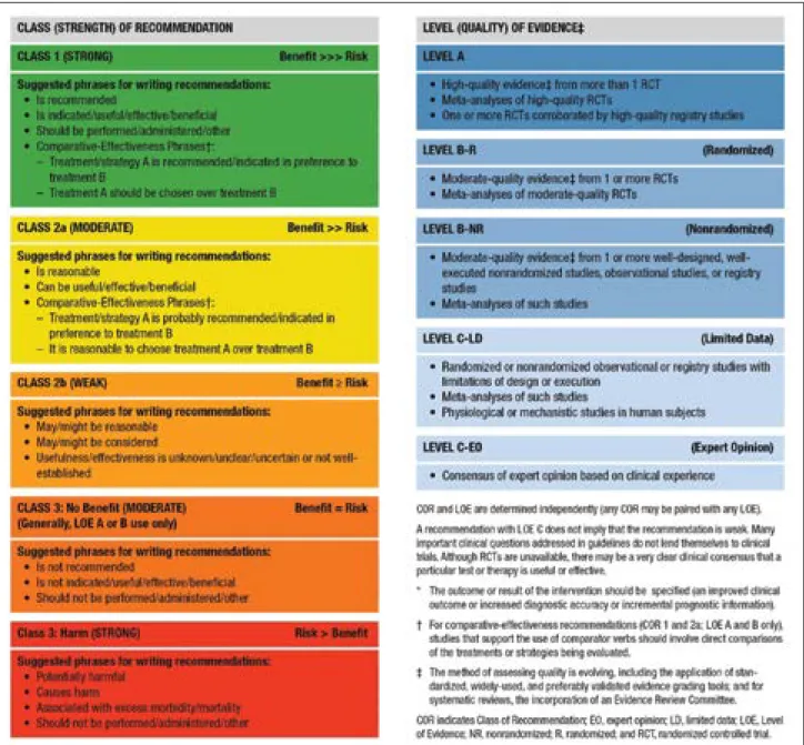
Table 1. Applying Class of Recommendation and Level of Evidence to Clinical Strategies, Interventions, Treatments, or Diagnostic Testing in Patient Care (Updated May 2019)*
This table defines the Classes of Recommendation (COR) and Levels of Evidence (LOE). COR indicates the strength the writing group assigns the recommendation, and the LOE is assigned based on the quality of the scientific evidence. The outcome or result of the intervention should be specified (an improved clinical outcome or increased diagnostic accuracy or incremental prognostic information). Classes of Recommendation COR designations include Class 1, a strong recommendation for which the potential benefit greatly outweighs the risk; Class 2a, a moderate recommendation for which benefit most likely outweighs the risk; Class 2b, a weak recommendation for which it’s unknown whether benefit will outweigh the risk; Class 3: No Benefit, a moderate recommendation signifying that there is equal likelihood of benefit and risk; and Class 3: Harm, a strong recommendation for which the risk outweighs the potential benefit. Suggested phrases for writing Class 1 recommendations include
• Is recommended • Is indicated/ useful/effective/beneficial • Should be performed/administered/other Comparative-effectiveness phrases include treatment/strategy A is recommended/indicated in preference to treatment B, and treatment A should be chosen over treatment B. Suggested phrases for writing Class 2a recommendations include • Is reasonable • Can be useful/ effective/beneficial
Comparative-effectiveness phrases include treatment/strategy A is probably recommended/ indicated in preference to treatment B, and it is reasonable to choose treatment A over treatment B. For comparative-effectiveness recommendations (COR 1 and 2a; LOE A and B only), studies that support the use of comparator verbs should involve direct comparisons of the treatments or strategies being evaluated. Suggested phrases for writing Class 2b recommendations include • May/might be reasonable
• May/might be considered
• Usefulness/ effectiveness is unknown/unclear/uncertain or not well-established Suggested phrases for writing Class 3: No Benefit recommendations (generally, LOE A or B use only) include • Is not recommended • Is not indicated/useful/effective/beneficial • Should not be performed/administered/other Suggested phrases for writing Class 3: Harm recommendations include
• Potentially harmful
• Causes harm
• Associated with excess morbidity/mortality
• Should not be performed/administered/other Levels of Evidence For LOEs, the method of assessing quality is evolving, including the application of standardized, widely-used, and preferably validated evidence grading tools; and for systematic reviews, the incorporation of an Evidence Review Committee. LOE designations include Level A, Level B-R, Level B-NR, Level C-LD, and Level C-EO. Those categorized as Level A are derived from
• High-quality evidence from more than 1 randomized clinical trial, or RCT
• Meta-analyses of high-quality RCTs
• One or more RCTs corroborated by high-quality registry studies Those categorized as Level B-R (randomized) are derived from • Moderate-quality evidence from 1 or more RCTs
• Meta-analyses of moderate-quality RCTs
Those categorized as Level B-NR (nonrandomized) are derived from • Moderate-quality evidence from 1 or more well-designed, well-executed nonrandomized studies, observational studies, or registry studies
• Meta-analyses of such studies
Those categorized as Level C-LD (limited data) are derived from • Randomized or nonrandomized observational or registry studies with limitations of design or execution
• Meta-analyses of such studies
• Physiological or mechanistic studies in human subjects
Those categorized as Level C-EO (expert opinion) are derived from • Consensus of expert opinion based on clinical experience COR and LOE are determined independently (any COR may be paired with any LOE). A recommendation with LOE C does not imply that the recommendation is weak. Many important clinical questions addressed in guidelines do not lend themselves to clinical trials. Although RCTs are unavailable, there may be a very clear clinical consensus that a particular test or therapy is useful or effective.
Guideline Structure
The 2020 Guidelines are organized into knowledge
chunks, grouped into discrete modules of information
on specific topics or management issues.
5Each modular
knowledge chunk includes a table of recommendations
that uses standard AHA nomenclature of COR and LOE.
A brief introduction or short synopsis is provided to
put the recommendations into context with important
background information and overarching management
or treatment concepts. Recommendation-specific text
clarifies the rationale and key study data supporting the
recommendations. When appropriate, flow diagrams
or additional tables are included. Hyperlinked
refer-ences are provided to facilitate quick access and review.
Document Review and Approval
Each of the 2020 Guidelines documents was submitted
for blinded peer review to 5 subject-matter experts
nominated by the AHA. Before appointment, all peer
reviewers were required to disclose relationships with
industry and any other conflicts of interest, and all
dis-closures were reviewed by AHA staff. Peer reviewer
feedback was provided for guidelines in draft format
and again in final format. All guidelines were reviewed
and approved for publication by the AHA Science
Advi-sory and Coordinating Committee and the AHA
Execu-tive Committee. Disclosure information for peer
review-ers is listed in Appendix 2.
REFERENCES
1. Virani SS, Alonso A, Benjamin EJ, Bittencourt MS, Callaway CW, Carson AP, Chamberlain AM, Chang AR, Cheng S, Delling FN, et al: on behalf of the American Heart Association Council on Epidemiology and Prevention Statistics Committee and Stroke Statistics Subcommit-tee. Heart disease and stroke statistics—2020 update: a report from the American Heart Association. Circulation. 2020;141:e139–e596. doi: 10.1161/CIR.0000000000000757
2. Okubo M, Schmicker RH, Wallace DJ, Idris AH, Nichol G, Austin MA, Grunau B, Wittwer LK, Richmond N, Morrison LJ, Kurz MC, Cheskes S, Kudenchuk PJ, Zive DM, Aufderheide TP, Wang HE, Herren H, Vaillancourt C, Davis DP, Vilke GM, Scheuermeyer FX, Weisfeldt ML, Elmer J, Colella R, Callaway CW; Resuscitation Outcomes Consortium In-vestigators. Variation in Survival After Out-of-Hospital Cardiac Arrest Be-tween Emergency Medical Services Agencies. JAMA Cardiol. 2018;3:989– 999. doi: 10.1001/jamacardio.2018.3037
3. Zive DM, Schmicker R, Daya M, Kudenchuk P, Nichol G, Rittenberger JC, Aufderheide T, Vilke GM, Christenson J, Buick JE, Kaila K, May S, Rea T, Morrison LJ; ROC Investigators. Survival and variability over time from out of hospital cardiac arrest across large geographi-cally diverse communities participating in the Resuscitation Out-comes Consortium. Resuscitation. 2018;131:74–82. doi: 10.1016/j. resuscitation.2018.07.023
4. Søreide E, Morrison L, Hillman K, Monsieurs K, Sunde K, Zideman D, Eisenberg M, Sterz F, Nadkarni VM, Soar J, Nolan JP; Utstein Formula for Survival Collaborators. The formula for survival in resuscitation. Resuscitation. 2013;84:1487–1493. doi: 10.1016/j. resuscitation.2013.07.020
5. Levine GN, O’Gara PT, Beckman JA, Al-Khatib SM, Birtcher KK, Cigarroa JE, de Las Fuentes L, Deswal A, Fleisher LA, Gentile F, Goldberger ZD, Hlatky MA, Joglar JA, Piano MR, Wijeysundera DN. Recent Innovations, Modifications, and Evolution of ACC/AHA Clinical Practice Guidelines: An Update for
Our Constituencies: A Report of the American College of Cardiology/ American Heart Association Task Force on Clinical Practice Guidelines.
Cir-culation. 2019;139:e879–e886. doi: 10.1161/CIR.0000000000000651
ACD active compression-decompression ACLS advanced cardiovascular life support ADC apparent diffusion coefficient AED automated external defibrillator AHA American Heart Association ALS advanced life support aOR adjusted odds ratio AV atrioventricular BLS basic life support COR Class of Recommendation
CoSTR International Consensus on Cardiopulmonary Resuscitation and Emergency Cardiovascular Care Science With Treatment Recommendations
CPR cardiopulmonary resuscitation
CT computed tomography
DWI diffusion-weighted imaging ECG electrocardiogram
ECPR extracorporeal cardiopulmonary resuscitation
EEG electroencephalogram
EMS emergency medical services
ETCO2 (partial pressure of) end-tidal carbon dioxide ETI endotracheal intubation
GWR gray-white ratio ICU intensive care unit IHCA in-hospital cardiac arrest
ILCOR International Liaison Committee on Resuscitation
IO intraosseous
ITD impedance threshold device
IV intravenous
LAST local anesthetic systemic toxicity LOE Level of Evidence
MAP mean arterial pressure MRI magnetic resonance imaging NSE neuron-specific enolase OHCA out-of-hospital cardiac arrest
Paco2 arterial partial pressure of carbon dioxide
PCI percutaneous coronary intervention
PE pulmonary embolism
PMCD perimortem cesarean delivery pVT pulseless ventricular tachycardia RCT randomized controlled trial ROSC return of spontaneous circulation S100B S100 calcium binding protein SGA supraglottic airway
Abbreviations
(Continued )
MAJOR CONCEPTS
Overview Concepts of Adult Cardiac
Arrest
Survival and recovery from adult cardiac arrest depend
on a complex system working together to secure the
best outcome for the victim. The main focus in adult
cardiac arrest events includes rapid recognition, prompt
provision of CPR, defibrillation of malignant shockable
rhythms, and post-ROSC supportive care and
treat-ment of underlying causes. This approach recognizes
that most sudden cardiac arrest in adults is of cardiac
cause, particularly myocardial infarction and electric
disturbances. Arrests without a primary cardiac origin
(eg, from respiratory failure, toxic ingestion, pulmonary
embolism [PE], or drowning) are also common,
how-ever, and in such cases, treatment for reversible
under-lying causes is important for the rescuer to consider.
1Some noncardiac etiologies may be particularly
com-mon in the in-hospital setting. Others, such as opioid
overdose, are sharply on the rise in the out-of-hospital
setting.
2For any cardiac arrest, rescuers are instructed
to call for help, perform CPR to restore coronary and
cerebral blood flow, and apply an AED to directly treat
ventricular fibrillation (VF) or ventricular tachycardia
(VT), if present. Although the majority of resuscitation
success is achieved by provision of high-quality CPR and
defibrillation, other specific treatments for likely
under-lying causes may be helpful in some cases.
Adult Chain of Survival
The primary focus of cardiac arrest management for
pro-viders is the optimization of all critical steps required to
improve outcomes. These include activation of the
emer-gency response, provision of high-quality CPR and early
defibrillation, ALS interventions, effective post-ROSC care
including careful prognostication, and support during
recovery and survivorship. All of these activities require
organizational infrastructures to support the education,
training, equipment, supplies, and communication that
enable each survival. Thus, we recognize that each of
these diverse aspects of care contributes to the ultimate
functional survival of the cardiac arrest victim.
Resuscitation causes, processes, and outcomes are
very different for OHCA and IHCA, which are
reflect-ed in their respective Chains of Survival (Figure 1). In
OHCA, the care of the victim depends on community
engagement and response. It is critical for community
members to recognize cardiac arrest, phone 9-1-1
(or the local emergency response number), perform CPR
SSEP somatosensory evoked potential STEMI ST-segment elevation myocardial infarction SVT supraventricular tachycardia
TCA tricyclic antidepressant TOR termination of resuscitation TTM targeted temperature management VF ventricular fibrillation
VT ventricular tachycardia
Figure 1. 2020 American Heart Association Chains of Survival for IHCA and OHCA.
CPR indicates cardiopulmonary resuscitation; IHCA, in-hospital cardiac arrest; and OHCA, out-of-hospital cardiac arrest.
2020 AHA Chains of Survival for IHCA and OHCA. (2; IHCA, OHCA) 2 horizontal chains for adults, 1 for In-Hospital Cardiac Arrest and 1 for Out-of-Hospital Cardiac Arrest. On each chain, 6 links show icons for actions to help an adult in
(including, for untrained lay rescuers, compression-only
CPR), and use an AED.
3,4Emergency medical
person-nel are then called to the scene, continue resuscitation,
and transport the patient for stabilization and definitive
management. In comparison, surveillance and
preven-tion are critical aspects of IHCA. When an arrest occurs
in the hospital, a strong multidisciplinary approach
in-cludes teams of medical professionals who respond,
provide CPR, promptly defibrillate, begin ALS measures,
and continue post-ROSC care. Outcomes from IHCA are
overall superior to those from OHCA,
5likely because of
reduced delays in initiation of effective resuscitation.
The Adult OHCA and IHCA Chains of Survival have
been updated to better highlight the evolution of
sys-tems of care and the critical role of recovery and
survi-vorship with the addition of a new link. This Recovery
link highlights the enormous recovery and survivorship
journey, from the end of acute treatment for critical
ill-ness through multimodal rehabilitation (both short- and
long-term), for both survivors and families after cardiac
arrest. This new link acknowledges the need for the
sys-tem of care to support recovery, discuss expectations,
and provide plans that address treatment, surveillance,
and rehabilitation for cardiac arrest survivors and their
caregivers as they transition care from the hospital to
home and return to role and social function.
REFERENCES
1. Lavonas EJ, Drennan IR, Gabrielli A, Heffner AC, Hoyte CO, Orkin AM, Sawyer KN, Donnino MW. Part 10: special circumstanc-es of rcircumstanc-esuscitation: 2015 American Heart Association Guidelincircumstanc-es Update for Cardiopulmonary Resuscitation and Emergency Car-diovascular Care. Circulation. 2015;132(suppl 2):S501–S518. doi: 10.1161/CIR.0000000000000264
2. Dezfulian C, Orkin AM, Maron BA, Elmer J, Girota S, Gladwin MT, Merchant RM, Panchal AR, Perman SM, Starks M, van Diepen S, Lavonas EJ; on behalf of the American Heart Association Council on Cardiopulmonary, Critical Care, Perioperative and Resuscitation; Council on Arteriosclerosis, Thrombosis and Vascular Biology; Council on Cardiovascular and Stroke Nursing; and Council on Clinical Cardiology. Opioid-associated out-of-hospital cardiac arrest: distinctive clinical features and implications for healthcare and public responses: a scientific statement from the American Heart Association. Circulation. In press.
3. Sayre MR, Berg RA, Cave DM, Page RL, Potts J, White RD; Ameri-can Heart Association Emergency Cardiovascular Care Commit-tee. Hands-only (compression-only) cardiopulmonary resuscitation: a call to action for bystander response to adults who experience out-of-hospital sudden cardiac arrest: a science advisory for the public from the American Heart Association Emergency Cardio-vascular Care Committee. Circulation. 2008;117:2162–2167. doi: 10.1161/CIRCULATIONAHA.107.189380
4. Kleinman ME, Brennan EE, Goldberger ZD, Swor RA, Terry M, Bobrow BJ, Gazmuri RJ, Travers AH, Rea T. Part 5: adult basic life support and car-diopulmonary resuscitation quality: 2015 American Heart Association Guidelines Update for Cardiopulmonary Resuscitation and Emergency Cardiovascular Care. Circulation. 2015;132(suppl 2):S414–S435. doi: 10.1161/CIR.0000000000000259
5. Virani SS, Alonso A, Benjamin EJ, Bittencourt MS, Callaway CW, Carson AP, Chamberlain AM, Chang AR, Cheng S, Delling FN, et al: on behalf of the American Heart Association Council on Epidemiology and Prevention Statistics Committee and Stroke Statistics Subcommit-tee. Heart disease and stroke statistics—2020 update: a report from the American Heart Association. Circulation. 2020;141:e139–e596. doi: 10.1161/CIR.0000000000000757
SEQUENCE OF RESUSCITATION
Recognition of Cardiac Arrest
Synopsis
Lay rescuer CPR improves survival from cardiac arrest
by 2- to 3-fold.
1The benefit of providing CPR to a
patient in cardiac arrest outweighs any potential risk
of providing chest compressions to someone who is
unconscious but not in cardiac arrest. It has been
shown that the risk of injury from CPR is low in these
patients.
2It has been shown previously that all rescuers may
have difficulty detecting a pulse, leading to delays in
CPR, or in some cases CPR not being performed at
all for patients in cardiac arrest.
3Recognition of
car-diac arrest by lay rescuers, therefore, is determined
on the basis of level of consciousness and the
respira-tory effort of the victim. Recognition of cardiac arrest
by healthcare providers includes a pulse check, but
the importance of not prolonging efforts to detect a
pulse is emphasized.
Recommendation-Specific Supportive Text
1. Agonal breathing is characterized by slow,
irregular gasping respirations that are
inef-fective for ventilation. Agonal breathing is
described by lay rescuers with a variety of
terms including, abnormal breathing, snoring
respirations, and gasping.
4Agonal
breath-ing is common, reported as bebreath-ing present
in up to 40% to 60% of victims of OHCA.
5The presence of agonal breathing is cited as
a common reason for lay rescuers to
misdiag-nose a patient as not being in cardiac arrest.
6In patients who are unresponsive, with absent
or abnormal breathing, lay rescuers should
assume the patient is in cardiac arrest, call for
help, and promptly initiate CPR. These 2
crite-ria (patient responsiveness and assessment of
breathing) have been shown to rapidly identify
a significant proportion of patients who are in
cardiac arrest, allowing for immediate initiation
of lay rescuer CPR. Further, initiation of chest
compressions in patients who are unconscious
Recommendations for Recognition of Cardiac Arrest COR LOE Recommendations
1 C-LD
1. If a victim is unconscious/unresponsive, with absent or abnormal breathing (ie, only gasping), the lay rescuer should assume the victim is in cardiac arrest.
1 C-LD
2. If a victim is unconscious/unresponsive, with absent or abnormal breathing (ie, only gasping), the healthcare provider should check for a pulse for no more than 10 s and, if no definite pulse is felt, should assume the victim is in cardiac arrest.
but not in cardiac arrest is associated with low
rates of significant adverse events.
2The adverse
events noted included pain in the area of chest
compressions (8.7%), bone fracture (ribs and
clavicle) (1.7%), and rhabdomyolysis (0.3%), with
no visceral injuries described.
22. Protracted delays in CPR can occur when
check-ing for a pulse at the outset of resuscitation
efforts as well as between successive cycles
of CPR. Healthcare providers often take too
long to check for a pulse
7,8and have difficulty
determining if a pulse is present or absent.
7–9There is no evidence, however, that checking
for breathing, coughing, or movement is
supe-rior to a pulse check for detection of
circula-tion.
10Thus, healthcare providers are directed
to quickly check for a pulse and to promptly
start compressions when a pulse is not
defini-tively palpated.
9,11This topic last received formal evidence review in 2010.
3REFERENCES
1. Sasson C, Rogers MA, Dahl J, Kellermann AL. Predictors of sur-vival from out-of-hospital cardiac arrest: a systematic review and meta-analysis. Circ Cardiovasc Qual Outcomes. 2010;3:63–81. doi: 10.1161/CIRCOUTCOMES.109.889576
2. Olasveengen TM, Mancini ME, Perkins GD, Avis S, Brooks S, Castrén M, Chung SP, Considine J, Couper K, Escalante R, et al; on behalf of the Adult Basic Life Support Collaborators. Adult basic life support: 2020 Interna-tional Consensus on Cardiopulmonary Resuscitation and Emergency Car-diovascular Care Science With Treatment Recommendations. Circulation. 2020;142(suppl 1):S41–S91. doi: 10.1161/CIR.0000000000000892 3. Berg RA, Hemphill R, Abella BS, Aufderheide TP, Cave DM, Hazinski MF,
Lerner EB, Rea TD, Sayre MR, Swor RA. Part 5: adult basic life support: 2010 American Heart Association Guidelines for Cardiopulmonary Resus-citation and Emergency Cardiovascular Care. Circulation. 2010;122(suppl 3):S685–S705. doi: 10.1161/CIRCULATIONAHA.110.970939
4. Riou M, Ball S, Williams TA, Whiteside A, Cameron P, Fatovich DM, Perkins GD, Smith K, Bray J, Inoue M, O’Halloran KL, Bailey P, Brink D, Finn J. ‘She’s sort of breathing’: What linguistic fac-tors determine call-taker recognition of agonal breathing in emergency calls for cardiac arrest? Resuscitation. 2018;122:92–98. doi: 10.1016/j. resuscitation.2017.11.058
5. Fukushima H, Imanishi M, Iwami T, Seki T, Kawai Y, Norimoto K, Urisono Y, Hata M, Nishio K, Saeki K, Kurumatani N, Okuchi K. Abnormal breath-ing of sudden cardiac arrest victims described by laypersons and its as-sociation with emergency medical service dispatcher-assisted cardiopul-monary resuscitation instruction. Emerg Med J. 2015;32:314–317. doi: 10.1136/emermed-2013-203112
6. Brinkrolf P, Metelmann B, Scharte C, Zarbock A, Hahnenkamp K, Bohn A. Bystander-witnessed cardiac arrest is associated with reported agonal breathing and leads to less frequent bystander CPR. Resuscitation. 2018;127:114–118. doi: 10.1016/j.resuscitation.2018.04.017
7. Eberle B, Dick WF, Schneider T, Wisser G, Doetsch S, Tzanova I. Check-ing the carotid pulse check: diagnostic accuracy of first responders in pa-tients with and without a pulse. Resuscitation. 1996;33:107–116. doi: 10.1016/s0300-9572(96)01016-7
8. Moule P. Checking the carotid pulse: diagnostic accuracy in students of the healthcare professions. Resuscitation. 2000;44:195–201. doi: 10.1016/s0300-9572(00)00139-8
9. Ochoa FJ, Ramalle-Gómara E, Carpintero JM, García A, Saralegui I. Com-petence of health professionals to check the carotid pulse. Resuscitation. 1998;37:173–175. doi: 10.1016/s0300-9572(98)00055-0
10. Perkins GD, Stephenson B, Hulme J, Monsieurs KG. Birmingham assess-ment of breathing study (BABS). Resuscitation. 2005;64:109–113. doi: 10.1016/j.resuscitation.2004.09.007
11. Mather C, O’Kelly S. The palpation of pulses. Anaesthesia. 1996;51:189– 191. doi: 10.1111/j.1365-2044.1996.tb07713.x
Initiation of Resuscitation
Recommendations for Initiation of Resuscitation: Lay Rescuer (Untrained or Trained)
COR LOE Recommendations
1 B-NR
1. All lay rescuers should, at minimum, provide chest compressions for victims of cardiac arrest.
1 C-LD
2. After identifying a cardiac arrest, a lone responder should activate the emergency response system first and immediately begin CPR.
1 C-LD
3. We recommend that laypersons initiate CPR for presumed cardiac arrest, because the risk of harm to the patient is low if the patient is not in cardiac arrest.
2a C-LD
4. For lay rescuers trained in CPR using chest compressions and ventilation (rescue breaths), it is reasonable to provide ventilation (rescue breaths) in addition to chest compressions for the adult in OHCA.
Synopsis
After cardiac arrest is recognized, the Chain of Survival
continues with activation of the emergency response
sys-tem and initiation of CPR. The prompt initiation of CPR
is perhaps the most important intervention to improve
survival and neurological outcomes. Ideally, activation of
the emergency response system and initiation of CPR
oc-cur simultaneously. In the oc-current era of widespread
mo-bile device usage and accessibility, a lone responder can
activate the emergency response system simultaneously
with starting CPR by dialing for help, placing the phone
on speaker mode to continue communication, and
im-mediately commencing CPR. In the rare situation when
a lone rescuer must leave the victim to dial EMS, the
pri-ority should be on prompt EMS activation followed by
immediate return to the victim to initiate CPR.
Existing evidence suggests that the potential harm
from CPR in a patient who has been incorrectly
identi-fied as having cardiac arrest is low.
1Overall, the
ben-efits of initiation of CPR in cardiac arrest outweigh the
relatively low risk of injury for patients not in cardiac
arrest. The initial phases of resuscitation once cardiac
arrest is recognized are similar between lay responders
and healthcare providers, with early CPR representing
the priority. Lay rescuers may provide chest
compres-sion–only CPR to simplify the process and encourage
CPR initiation, whereas healthcare providers may
pro-vide chest compressions and ventilation (Figures 2–4).
Recommendation-Specific Supportive Text
1. CPR is the single-most important intervention for
a patient in cardiac arrest, and chest compressions
should be provided promptly. Chest compressions
are the most critical component of CPR, and a chest
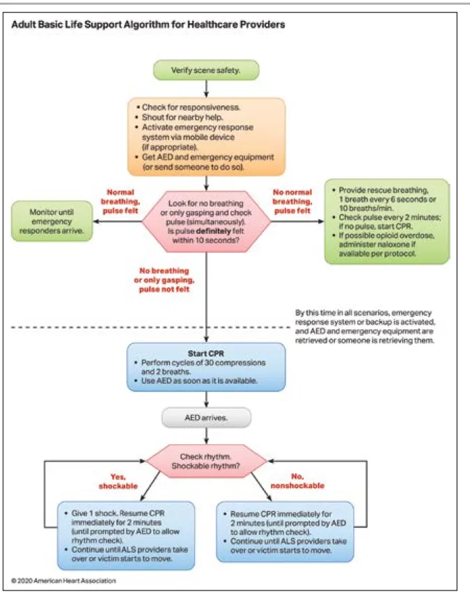
Figure 2. Adult BLS Algorithm for Healthcare Providers.
AED indicates automated external defibrillator; ALS, advanced life support; BLS, basic life support; and CPR, cardiopulmonary resuscitation.
Text in cascading boxes describes the actions that a provider should perform in sequence during an adult cardiac arrest. Arrows guide providers from one box to the next as they perform the actions. Some boxes have 2 arrows that lead outward, each to a different box depending on the outcome of the most recent action taken. Pathways are linked. Box 1 Verify scene safety. Box 2 Check for responsiveness. Shout for nearby help. Activate the emergency response system via mobile device (if appropriate). Get an AED and emergency equipment (or send someone to do so). Box 3 Look for no breathing or only gasping and check pulse (simultaneously). Is a pulse definitely felt within 10 seconds? If the person is breathing normally and has a pulse, proceed to Box 3a. If the person is not breathing normally but has a pulse, proceed to Box 3b. If the person is not breathing or is only gasping and no pulse is felt, proceed to Box 4. Box 3a
Monitor the person until emergency responders arrive. Box 3b Provide rescue breathing, 1 breath every 6 seconds or 10 breaths per minute. Check pulse every 2 minutes; if no pulse, start CPR. If it is a possible opioid overdose, administer naloxone if available per protocol. By this time in all scenarios, emergency response system or backup is activated, and AED and emergency equipment are retrieved or someone is retrieving them. Box 4 Start CPR
• Perform cycles of 30 compressions and 2 breaths.
• Use the AED as soon as it is available. Box 5 The AED arrives. Box 6 The AED checks the rhythm. Is the rhythm shockable? If Yes, the rhythm is shockable, proceed to Box 7. If No, the rhythm is nonshockable, proceed to Box 8. Box 7
• Give 1 shock. Resume CPR immediately for 2 minutes (until prompted by the AED to allow rhythm check).
• Continue until advanced life support providers take over or the victim starts to move.
Box 8
• Resume CPR immediately for 2 minutes (until prompted by the AED to allow rhythm check).
• Continue until advanced life support providers take over or the victim starts to move.
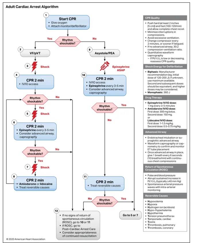
Figure 3. Adult Cardiac Arrest Algorithm.
CPR indicates cardiopulmonary resuscitation; ET, endotracheal; IO, intraosseous; IV, intravenous; PEA, pulseless electrical activity; pVT, pulseless ventricular tachycar-dia; and VF, ventricular fibrillation.
Cascading numbered boxes correspond to actions the provider should perform in sequence. Each box is separated by an arrow that signifies the pathway the provider should take. Some boxes are separated by 2 arrows that lead to different boxes, meaning that the provider should take a different pathway depending on the outcome of the previous action. Pathways are hyperlinked. Box 1 Start CPR • Give oxygen • Attach monitor/defibrillator Rhythm shockable? Yes, proceed to Box 2 for VF/pVT. No, proceed to Box 9 for Asystole/PEA. Box 2 VF/pVT Box 3 Deliver shock. Box 4 CPR 2 minutes • IV/IO access Is rhythm shockable? If Yes, proceed to Box 5. If No, proceed to Box 12. Box 5 Deliver shock. Box 6 CPR 2 minutes • Epinephrine every 3 to 5 minutes.
• Consider advanced airway, capnography.
Is rhythm shockable? If Yes, proceed to Box 7. If No, proceed to Box 12. Box 7 Deliver shock. Box 8 CPR 2 minutes • Amiodarone or lidocaine. • Treat reversible causes. Box 9 Asystole/PEA. Give Epinephrine ASAP. Box 10 CPR 2 minutes
• IV/IO access.
• Epinephrine every 3 to 5 minutes.
• Consider advanced airway, capnography.
Is rhythm shockable? If Yes, proceed to Box 5 or Box 7. If No, proceed to Box 11. Box 11 CPR 2 minutes.
• Treat reversible causes.
Is rhythm shockable? If Yes, proceed to Box 5 or Box 7. If No, proceed to Box 12. Box 12
• If no
signs of return of spontaneous circulation (ROSC), go to Box 10 or Box 11 • If ROSC, go to Post–Cardiac Arrest Care
• Consider appropriateness of continued resuscitation
Sidebar CPR Quality
• Push hard (at least 2 inches [5 cm]) and fast (100-120/min) and allow complete chest recoil.
• Minimize interruptions in compressions.
• Avoid excessive ventilation.
• Change compressor every 2 minutes, or sooner if fatigued.
• If no
advanced airway, 30 to 2 compression-ventilation ratio. • Quantitative waveform capnography
- If PETCO2 is low or decreasing, reassess CPR quality.
Shock Energy for Defibrillation •
Biphasic: Manufacturer recommendation (eg, initial dose of 120-200 Joules); if unknown, use maximum available. Second and subsequent doses should be equivalent, and higher doses may be considered. • Monophasic: 360 Joules Drug Therapy • Epinephrine IV/IO dose: 1 milligram every 3 to 5 minutes • Amiodarone IV/IO dose: First dose: 300 milligram bolus. Second dose: 150 milligram. orLidocaine IV/IO dose: First dose: 1-1.5 milligrams per kilogram. Second dose: 0.5-0.75 milligrams per kilogram. Advanced Airway • Endotracheal intubation or supraglottic advanced airway • Waveform capnography or capnometry to confirm and monitor ET tube placement
• Once advanced airway in place, give 1 breath every 6 seconds (10 breaths per minute) with
continuous chest compressions Return of Spontaneous Circulation (ROSC)
• Pulse and blood pressure
• Abrupt sustained increase in PETCO2 (typically greater than or equal to 40 millimeters of mercury)
• Spontaneous arterial pressure waves with intra-arterial monitoring Reversible Causes • Hypovolemia
• Hypoxia • Hydrogen ion (acidosis) • Hypo-/ hyperkalemia • Hypothermia • Tension pneumothorax • Tamponade, cardiac • Toxins • Thrombosis, pulmonary • Thrombosis, coronary
Figure 4. Adult Cardiac Arrest Circular Algorithm.
CPR indicates cardiopulmonary resuscitation; ET, endotracheal; IO, intraosseous; IV, intravenous; pVT, pulseless ventricular tachycardia; and VF, ventricular fibrillation.
Cascading numbered boxes and a circular pattern correspond to actions the provider should perform in sequence. Box 1 Start CPR • Give oxygen. • Attach monitor/ defibrillator. Box 2
• Check rhythm. This box starts a repetitive pattern, represented by the outside of a circle. If VF/pVT, deliver shock, followed by 2 minutes of: - Continuous CPR - Monitor CPR Quality - Continuous CPR • After 2 minutes, check rhythm again and repeat this cycle until Return of Spontaneous Circulation (ROSC), then initiate post-cardiac arrest care.
Inside the circle are listed things to perform as necessary during the resuscitation effort: Drug Therapy
• IV/IO access • Epinephrine every 3 to 5 minutes
• Amiodarone or lidocaine for refractory VF/pVT Consider Advanced Airway
• Quantitative waveform capnography
Treat Reversible Causes Sidebar CPR Quality
• Push hard (at least 2 inches [5 cm]) and fast (100-120/min) and allow complete chest recoil.
• Minimize interruptions in compressions.
• Avoid excessive ventilation.
• Change compressor every 2 minutes, or sooner if fatigued. • If no advanced airway, 30 to 2 compression-ventilation ratio.
• Quantitative waveform capnography
- If PETCO2 is low or decreasing, reassess CPR quality. Shock Energy for Defibrillation
• Biphasic: Manufacturer recommendation (eg, initial dose of 120 to 200 Joules); if unknown, use maximum
available. Second and subsequent doses should be equivalent, and higher doses may be considered. • Monophasic: 360 Joules
Drug Therapy
• Epinephrine IV/IO dose: 1 milligram every 3 to 5 minutes • Amiodarone IV/IO dose: First dose: 300 milligram bolus. Second dose: 150 milligrams. orLidocaine IV/IO dose: First dose: 1-1.5 milligrams per kilogram. Second dose: 0.5-0.75 milligrams per kilogram. Advanced Airway
• Endotracheal intubation or supraglottic advanced airway
• Waveform capnography or capnometry to confirm and monitor
ET tube placement
• Once advanced airway in place, give 1 breath every 6 seconds (10 breaths per minute) with continuous chest compressions
Return of Spontaneous Circulation (ROSC) • Pulse and blood pressure
• Abrupt sustained increase in PETCO2 (typically greater than or equal to 40 millimeters of mercury) • Spontaneous arterial pressure waves with intra-arterial monitoring Reversible Causes • Hypovolemia • Hypoxia • Hydrogen ion (acidosis) • Hypo-/hyperkalemia • Hypothermia • Tension pneumothorax • Tamponade, cardiac • Toxins • Thrombosis, pulmonary • Thrombosis, coronary
compression–only approach is appropriate if lay
rescuers are untrained or unwilling to provide
respi-rations. Beginning the CPR sequence with
compres-sions minimized time to first chest compression.
2–4Nationwide dissemination of chest compression–
only CPR for lay rescuers was associated with an
increase in the incidence of survival with favorable
neurological outcome after OHCAs in Japan, likely
due to an increase in lay rescuers providing CPR.
5Chest compressions should be provided as soon
as possible, without the need to remove the
vic-tim’s clothing first.
2. The optimal timing of CPR initiation and
emer-gency response system activation was
evalu-ated by an ILCOR systematic review in 2020.
1An
observational study of over 17 000 OHCA events
reported similar results from either a “call-first”
strategy or a “CPR-first” strategy.
6In the current
era of ubiquitous mobile devices, ideally both the
call to activate EMS and the initiation of CPR can
occur simultaneously.
3. Four observational studies
7–10reported outcomes
from patients who were not in cardiac arrest and
received CPR by lay rescuers. No serious harm from
CPR was found in patients when they were later
determined not to have been in cardiac arrest.
1This is in contrast to the significant risk of
with-holding CPR when a patient is in cardiac arrest,
making the risk:benefit ratio strongly in favor of
providing CPR for presumed cardiac arrest.
4. In some observational studies, improved outcomes
have been noted in victims of cardiac arrest who
received conventional CPR (compressions and
ventila-tion) compared with those who received chest
com-pressions only.
5,11,12Other studies have reported no
difference in outcomes for patients receiving
conven-tional versus compression-only CPR.
11,13–21Given the
potential benefit of conventional CPR, if lay rescuers
are appropriately trained, they should be encouraged
to concurrently deliver ventilation with
compres-sions. A thorough review of the data concerning the
ratio of compressions to ventilation when
perform-ing conventional CPR is discussed in Ventilation and
Compression-to-Ventilation Ratio.
These recommendations are supported by the 2020
ILCOR Consensus on CPR and Emergency Cardiovascular
Care Science With Treatment Recommendations (CoSTR).
1Recommdendations for Initiation of Resuscitation: Healthcare Provider
COR LOE Recommendations
1 C-LD
1. A lone healthcare provider should commence with chest compressions rather than with ventilation.
2a C-LD
2. It is reasonable for healthcare providers to perform chest compressions and ventilation for all adult patients in cardiac arrest from either a cardiac or noncardiac cause.
Recommendation-Specific Supportive Text
1. The 2010 Guidelines for CPR and Emergency
Cardiovascular Care included a major change
for trained rescuers, who were instructed to
begin the CPR sequence with chest
compres-sions rather than with breaths (circulation,
air-way, and breathing versus airair-way, breathing,
and circulation) to minimize the time to
initia-tion of chest compressions. This approach is
resupported by new literature, summarized in a
2020 ILCOR systematic review (Table 2).
1–4In the
recommended sequence, once chest
compres-sions have been started, a single trained rescuer
delivers rescue breaths by mouth to mask or by
bag-mask device to provide oxygenation and
ventilation. Manikin studies demonstrate that
starting with chest compressions rather than
with ventilation is associated with faster times
to chest compressions,
3,23rescue breaths,
4and
completion of the first CPR cycle.
42. Healthcare providers are trained to deliver both
compressions and ventilation. Delivery of chest
compressions without assisted ventilation for
prolonged periods could be less effective than
conventional CPR (compressions plus
ventila-tion) because arterial oxygen content decreases
as CPR duration increases. This concern is
espe-cially pertinent in the setting of asphyxial cardiac
arrest.
11Healthcare providers, with their training
and understanding, can realistically tailor the
sequence of subsequent rescue actions to the
most likely cause of arrest.
These recommendations are supported by the 2020
CoSTR for BLS.
1Table 2. Adult BLS Sequence22
Step
Lay Rescuer Not
Trained Lay Rescuer Trained
Healthcare Provider
1 Ensure scene safety. Ensure scene safety. Ensure scene safety. 2 Check for response. Check for response. Check for
response. 3 Shout for nearby
help. Phone or ask someone to phone 9-1-1 (the phone or caller with the phone remains at the victim’s side, with the phone on speaker mode).
Shout for nearby help and activate the emergency response system (9-1-1, emergency response). If someone responds, ensure that the phone is at the side of the victim if at all possible.
Shout for nearby help/activate the resuscitation team; the provider can activate the resuscitation team at this time or after checking for breathing and pulse. 4 Follow the telecommunicator’s* instructions. Check for no breathing or only gasping; if none, begin CPR with compressions. Check for no breathing or only gasping and check pulse (ideally simultaneously). Activation and retrieval of the AED/emergency equipment by the lone healthcare provider or by the second person sent by the rescuer must occur no later than immediately after the check for no normal breathing and no pulse identifies cardiac arrest. 5 Look for no breathing or only gasping, at the direction of the telecommunicator. Answer the telecommunicator’s questions, and follow the telecommunicator’s instructions.
Immediately begin CPR, and use the AED/ defibrillator when available. 6 Follow the
telecommunicator’s instructions.
Send the second person to retrieve an AED, if one is available.
When the second rescuer arrives, provide 2-rescuer CPR and use the AED/defibrillator. AED indicates automated external defibrillator; BLS, basic life support; and CPR, cardiopulmonary resuscitation.
*Telecommunicator and dispatcher are terms often used interchangeably.
REFERENCES
1. Olasveengen TM, Mancini ME, Perkins GD, Avis S, Brooks S, Castrén M, Chung SP, Considine J, Couper K, Escalante R, et al; on behalf of the Adult Basic Life Support Collaborators. Adult basic life support: 2020 Interna-tional Consensus on Cardiopulmonary Resuscitation and Emergency Car-diovascular Care Science With Treatment Recommendations. Circulation. 2020;142(suppl 1):S41–S91. doi: 10.1161/CIR.0000000000000892 2. Lubrano R, Cecchetti C, Bellelli E, Gentile I, Loayza Levano H, Orsini F,
Bertazzoni G, Messi G, Rugolotto S, Pirozzi N, Elli M. Comparison of times of intervention during pediatric CPR maneuvers using ABC and CAB se-quences: a randomized trial. Resuscitation. 2012;83:1473–1477. doi: 10.1016/j.resuscitation.2012.04.011
3. Sekiguchi H, Kondo Y, Kukita I. Verification of changes in the time taken to initiate chest compressions according to modified basic life support guidelines. Am J Emerg Med. 2013;31:1248–1250. doi: 10.1016/j.ajem.2013.02.047
4. Marsch S, Tschan F, Semmer NK, Zobrist R, Hunziker PR, Hunziker S. ABC versus CAB for cardiopulmonary resuscitation: a prospective, ran-domized simulator-based trial. Swiss Med Wkly. 2013;143:w13856. doi: 10.4414/smw.2013.13856
5. Iwami T, Kitamura T, Kiyohara K, Kawamura T. Dissemination of Chest Compression-Only Cardiopulmonary Resuscitation and Survival After Out-of-Hospital Cardiac Arrest. Circulation. 2015;132:415–422. doi: 10.1161/CIRCULATIONAHA.114.014905
6. Kamikura T, Iwasaki H, Myojo Y, Sakagami S, Takei Y, Inaba H. Advantage of CPR-first over call-first actions for out-of-hospital cardiac arrests in non-elderly patients and of noncardiac aetiology. Resuscitation. 2015;96:37– 45. doi: 10.1016/j.resuscitation.2015.06.027
7. White L, Rogers J, Bloomingdale M, Fahrenbruch C, Culley L, Subido C, Eisenberg M, Rea T. Dispatcher-assisted cardiopulmonary resuscitation: risks for patients not in cardiac arrest. Circulation. 2010;121:91–97. doi: 10.1161/CIRCULATIONAHA.109.872366
8. Haley KB, Lerner EB, Pirrallo RG, Croft H, Johnson A, Uihlein M. The fre-quency and consequences of cardiopulmonary resuscitation performed by bystanders on patients who are not in cardiac arrest. Prehosp Emerg Care. 2011;15:282–287. doi: 10.3109/10903127.2010.541981
9. Moriwaki Y, Sugiyama M, Tahara Y, Iwashita M, Kosuge T, Harunari N, Arata S, Suzuki N. Complications of bystander cardiopulmonary resuscita-tion for unconscious patients without cardiopulmonary arrest. J Emerg
Trauma Shock. 2012;5:3–6. doi: 10.4103/0974-2700.93094
10. Tanaka Y, Nishi T, Takase K, Yoshita Y, Wato Y, Taniguchi J, Hamada Y, Inaba H. Survey of a protocol to increase appropriate implementation of dispatcher-assisted cardiopulmonary resuscitation for out-of-hos-pital cardiac arrest. Circulation. 2014;129:1751–1760. doi: 10.1161/ CIRCULATIONAHA.113.004409
11. Kitamura T, Iwami T, Kawamura T, Nagao K, Tanaka H, Hiraide A; Im-plementation Working Group for All-Japan Utstein Registry of the Fire and Disaster Management Agency. Bystander-initiated rescue breath-ing for out-of-hospital cardiac arrests of noncardiac origin. Circulation. 2010;122:293–299. doi: 10.1161/CIRCULATIONAHA.109.926816 12. Ogawa T, Akahane M, Koike S, Tanabe S, Mizoguchi T, Imamura T.
Out-comes of chest compression only CPR versus conventional CPR conducted by lay people in patients with out of hospital cardiopulmonary arrest
witnessed by bystanders: nationwide population based observational study. BMJ. 2011;342:c7106. doi: 10.1136/bmj.c7106
13. Svensson L, Bohm K, Castrèn M, Pettersson H, Engerström L, Herlitz J, Rosenqvist M. Compression-only CPR or standard CPR in out-of-hos-pital cardiac arrest. N Engl J Med. 2010;363:434–442. doi: 10.1056/ NEJMoa0908991
14. Rea TD, Fahrenbruch C, Culley L, Donohoe RT, Hambly C, Innes J, Bloomingdale M, Subido C, Romines S, Eisenberg MS. CPR with chest compression alone or with rescue breathing. N Engl J Med. 2010;363:423– 433. doi: 10.1056/NEJMoa0908993
15. Iwami T, Kawamura T, Hiraide A, Berg RA, Hayashi Y, Nishiuchi T, Kajino K, Yonemoto N, Yukioka H, Sugimoto H, Kakuchi H, Sase K, Yokoyama H, Nonogi H. Effectiveness of bystander-initiated cardiac-only resuscitation for patients with out-of-hospital cardiac arrest. Circulation. 2007;116:2900– 2907. doi: 10.1161/CIRCULATIONAHA.107.723411
16. Kitamura T, Iwami T, Kawamura T, Nagao K, Tanaka H, Berg RA, Hiraide A; Implementation Working Group for All-Japan Utstein Registry of the Fire and Disaster Management Agency. Time-dependent effectiveness of chest compression-only and conventional cardiopulmonary resuscitation for out-of-hospital cardiac arrest of cardiac origin. Resuscitation. 2011;82:3– 9. doi: 10.1016/j.resuscitation.2010.09.468
17. Ong ME, Ng FS, Anushia P, Tham LP, Leong BS, Ong VY, Tiah L, Lim SH, Anantharaman V. Comparison of chest compression only and standard car-diopulmonary resuscitation for out-of-hospital cardiac arrest in Singapore.
Resuscitation. 2008;78:119–126. doi: 10.1016/j.resuscitation.2008.03.012
18. SOS-KANTO Study Group. Cardiopulmonary resuscitation by bystanders with chest compression only (SOS-KANTO): an observational study.
Lan-cet. 2007;369:920–926. doi: 10.1016/S0140-6736(07)60451–6
19. Bobrow BJ, Spaite DW, Berg RA, Stolz U, Sanders AB, Kern KB, Vadeboncoeur TF, Clark LL, Gallagher JV, Stapczynski JS, LoVecchio F, Mullins TJ, Humble WO, Ewy GA. Chest compression-only CPR by lay rescu-ers and survival from out-of-hospital cardiac arrest. JAMA. 2010;304:1447– 1454. doi: 10.1001/jama.2010.1392
20. Olasveengen TM, Wik L, Steen PA. Standard basic life support vs. continuous chest compressions only in out-of-hospital car-diac arrest. Acta Anaesthesiol Scand. 2008;52:914–919. doi: 10.1111/j.1399-6576.2008.01723.x
21. Panchal AR, Bobrow BJ, Spaite DW, Berg RA, Stolz U, Vadeboncoeur TF, Sanders AB, Kern KB, Ewy GA. Chest compression-only cardiopulmonary resuscitation performed by lay rescuers for adult out-of-hospital cardiac arrest due to non-cardiac aetiologies. Resuscitation. 2013;84:435–439. doi: 10.1016/j.resuscitation.2012.07.038
22. Kleinman ME, Brennan EE, Goldberger ZD, Swor RA, Terry M, Bobrow BJ, Gazmuri RJ, Travers AH, Rea T. Part 5: adult basic life support and cardio-pulmonary resuscitation quality: 2015 American Heart Association Guide-lines Update for Cardiopulmonary Resuscitation and Emergency Cardio-vascular Care. Circulation. 2015;132(suppl 2):S414–S435. doi: 10.1161/ CIR.0000000000000259
23. Kobayashi M, Fujiwara A, Morita H, Nishimoto Y, Mishima T, Nitta M, Hayashi T, Hotta T, Hayashi Y, Hachisuka E, Sato K. A manikin-based observa-tional study on cardiopulmonary resuscitation skills at the Osaka Senri med-ical rally. Resuscitation. 2008;78:333–339. doi: 10.1016/j.resuscitation. 2008.03.230
Opening the Airway
Introduction
A patent airway is essential to facilitate proper
ventila-tion and oxygenaventila-tion. Although there is no high-quality
evidence favoring one technique over another for
es-tablishment and maintenance of a patient’s airway,
res-cuers should be aware of the advantages and
disadvan-tages and maintain proficiency in the skills required for
each technique. Rescuers should recognize that
mul-tiple approaches may be required to establish an
ad-equate airway. Patients should be monitored constantly
to verify airway patency and adequate ventilation and
oxygenation. There are no studies comparing different
strategies of opening the airway in cardiac arrest
pa-tients. Much of the evidence examining the
effective-ness of airway strategies comes from radiographic and
cadaver studies.
Recommendations for Opening the Airway COR LOE Recommendations
1 C-EO
1. A healthcare provider should use the head tilt–chin lift maneuver to open the airway of a patient when no cervical spine injury is suspected.
1 C-EO
2. The trained lay rescuer who feels confident in performing both compressions and ventilation should open the airway using a head tilt–chin lift maneuver when no cervical spine injury is suspected.
2b C-EO
3. The use of an airway adjunct (eg, oropharyngeal and/or nasopharyngeal airway) may be reasonable in unconscious (unresponsive) patients with no cough or gag reflex to facilitate delivery of ventilation with a bag-mask device.
2a C-EO
4. In the presence of known or suspected basal skull fracture or severe coagulopathy, an oral airway is preferred compared with a nasopharyngeal airway. 3: No
Benefit C-LD
5. The routine use of cricoid pressure in adult cardiac arrest is not recommended.
Recommendation-Specific Supportive Text
1 and 2. The head tilt–chin lift has been shown to be
effective in establishing an airway in noncardiac
arrest and radiological studies.
2–5No studies have
compared head tilt–chin lift with other airway
maneuvers to establish an airway during cardiac
arrest.
3. Although there is no evidence examining the
effec-tiveness of their use during cardiac arrest,
oropha-ryngeal and nasophaoropha-ryngeal airways can be used to
maintain a patent airway and facilitate appropriate
ventilation by preventing the tongue from
occlud-ing the airway. Incorrect placement, however, can
cause an airway obstruction by displacing the
tongue to the back of the oropharynx.
6,74. The benefit of an oropharyngeal compared
with a nasopharyngeal airway in the presence
of a known or suspected basilar skull fracture
or severe coagulopathy has not been assessed
in clinical trials. However, an oral airway is
pre-ferred because of the risk of trauma with a
nasopharyngeal airway. Multiple case reports
have observed intracranial placement of
naso-pharyngeal airways in patients with basilar skull
fractures.
8,95. There is no evidence that cricoid pressure
facili-tates ventilation or reduces the risk of aspiration
in cardiac arrest patients. There is some evidence
that in non–cardiac arrest patients, cricoid
pres-sure may protect against aspiration and gastric
insufflation during bag-mask ventilation.
10–13However, cricoid pressure may also impede
venti-lation and the placement of a supraglottic airway
(SGA) or intubation,
14–20and increase the risk of
airway trauma during intubation.
21This topic last received formal evidence review in 2010.
22Recommendations for Opening the Airway After Head and Neck Trauma
COR LOE Recommendations
1 C-EO
1. In cases of suspected cervical spine injury, healthcare providers should open the airway by using a jaw thrust without head extension.
1 C-EO
2. In the setting of head and neck trauma, a head tilt–chin lift maneuver should be performed if the airway cannot be opened with a jaw thrust and airway adjunct insertion.
3: Harm C-LD
3. In the setting of head and neck trauma, lay rescuers should not use immobilization devices because their use by untrained rescuers may be harmful.