HAL Id: tel-03230734
https://tel.archives-ouvertes.fr/tel-03230734
Submitted on 20 May 2021HAL is a multi-disciplinary open access archive for the deposit and dissemination of sci-entific research documents, whether they are pub-lished or not. The documents may come from teaching and research institutions in France or abroad, or from public or private research centers.
L’archive ouverte pluridisciplinaire HAL, est destinée au dépôt et à la diffusion de documents scientifiques de niveau recherche, publiés ou non, émanant des établissements d’enseignement et de recherche français ou étrangers, des laboratoires publics ou privés.
Spatiotemporal distribution of ploidy levels and ploidy
specific transcriptome during Tomato fruit development
Edouard Tourdot
To cite this version:
Edouard Tourdot. Spatiotemporal distribution of ploidy levels and ploidy specific transcriptome during Tomato fruit development. Vegetal Biology. Université de Bordeaux, 2021. English. �NNT : 2021BORD0121�. �tel-03230734�
THÈSE PRÉSENTÉE
POUR OBTENIR LE GRADE DE
DOCTEUR DE
L’UNIVERSITÉ DE BORDEAUX
ÉCOLE DOCTORALE N°154 : SCIENCES DE LA VIE ET DE LA SANTE
SPÉCIALITÉ : BIOLOGIE VEGETALE
Par Edouard TOURDOT
Spatiotemporal distribution of ploidy levels and ploidy
specific
transcriptome
during
Tomato
fruit
development
Sous la direction de : Nathalie GONZALEZ
Soutenue le 29/03/2021
Membres du jury :
M. HERNOULD, Michel Professeur Université de Bordeaux Président du jury Mme. BAROUX, Célia Professeur University of Zürich Rapporteur M. DE VEYLDER, Lieven Directeur de Recherche VIB-UGent Center Rapporteur Mme. COOKSON-WODRICH, Sarah Jane Chargé de Recherche INRAE Examinateur M. ZOUINE, Mohamed Chargé de Recherche INRAE/INP-ENSAT Examinateur Mme. GONZALEZ, Nathalie Directrice de recherche INRAE - Directrice de Thèse
Remerciements
Before starting my acknowledgement, I would like to thank the jury members, Célia Baroux, Sarah Cookson, Lieven De Veylder, Mohamed Zouine and Michel Hernould.
J’aimerais remercier l’équipe Organogénèse du Fruit et endoreduplication (maintenant nommée : Floraison, Développement du Fruit et Contraintes Environnementales) de m’avoir accueilli comme doctorant et aux membres de l’équipe de m’avoir accepté. Merci Nathalie (G) d’avoir acceptée de travailler avec moi pendant cette thèse, d’avoir fait preuve de tant de patience et de bienveillance avec moi (bienveillance, qui sera je pense le mot qui dictera cette expérience). Merci d’avoir rendu possible tout le travail que j’ai pu faire et de m’avoir laissé une certaine liberté pendant cette thèse qui a été une très bonne expérience pour moi. Merci aussi pour tout ce que j’ai pu apprendre durant cette thèse aussi bien mes compétences nouvelles (je pense à la bioinfo) que personnellement (je pense à la bienveillance). Je n’ai aucun regret d’avoir choisi cette thèse même si dans l’ensemble tout a été chamboulé. J’espère que de ton côté tu ne regrettes pas trop ton choix de doctorant des trois dernières années et que les autres seront moins chiant que moi.
Avant d’arriver en thèse, il y a eu la licence et le master qui ont été des éléments déterminant pour en arriver là et beaucoup de gens sont intervenus et leur aide m’a été précieuse et je les remercie. Pour cela je souhaiterais particulièrement remercier plusieurs personnes, Michel Hernould, Didier Thoraval, Fred Gévaudant et Fred Delmas. Michel, je veux te remercier pour tous nos échanges toujours enrichissants et aux pires moments de la journée (souvent proche du départ pour rentrer à la maison) mais aussi pour m’avoir accepté en master et m’avoir soutenu pendant celui lorsque c’était difficile. Merci à Didier, tes cours en licences étaient géniaux et m’ont donné envie de continuer (je m’excuse de ne pas avoir choisi la génétique) vers le master et ton soutien m’a été précieux.
Bien sûr, merci aux deux Fred pour leurs expériences et connaissances qui ont toujours su prendre le temps de partager malgré leur emploi du temps overbooké. Merci encore à tous les deux d’avoir accepté lorsque l’on était en master de prendre le temps de faire plaisir à des gamins ayant envie de faire iGEM. Fred D, je te remercie de ta gentillesse et ta sympathie, c’était toujours un plaisir de faire « n’importe quoi » avec toi (je pense à la magnetofection entre autres). Merci à Fred G d’avoir toujours su me donner des conseils avisés peu importe le moment sur des manips ou des questions. Merci à Norbert pour m’avoir formé, littéralement, pendant mon M2 et le début de ma thèse car je pense sincèrement avoir énormément appris aussi bien scientifiquement qu’humainement en travaillant avec toi. Merci d’avoir été présent aussi aux moments où j’en avais besoin. C’est toujours un plaisir de passé du temps avec toi et je suis très content qu’aujourd’hui nous soyons amis !
JP (Michael ou Jean-Pascal), on ne sait jamais avec toi, merci à toi aussi d’avoir été un super collègue en plus d’être un ami. Même si au départ tu faisais peur dans ton antre au bout ou avec tes centaines de boîte, finalement tu n’es pas un si mauvais gars (haha). Merci d’avoir été là pour le soutien pendant la thèse, la reprise du sport et les soirées, journées et moment passé ensemble, toujours du bon temps. Bon courage pour ta thèse, je serais là en soutient !
Bien sûr, merci aux autres doctorants de l’équipe, Arthur et Sabri, pour votre bonne humeur. Cela a toujours été plaisant de travailler avec vous et d’échanger sur le sport, la musique, les répliques de film… vous êtes tous les deux des gars bien et je croise fort les doigts pour que votre thèse se déroule bien et que vous ayez les best résultats ! Arthur tu n’as que le fardeau de 30 ans de recherche international à conclure (:p). Spécial dédicace à toi Gabriel qui vit bien loin de nous, on ne t’oublie pas !
Enfin je souhaitais remercier mes proches, ceux qui n’ont pas travaillé avec moi mais qui ont su être un soutien moral infaillible pendant ce parcours jusqu’aujourd’hui. Un énorme merci à mon meilleur ami, Quentin, avec qui je partage tout depuis plus de 12 ans (4518 jours pour être exact, Facebook 2021).
Je voudrais remercier tout particulièrement ma moitié, Floriane, qui a été un soutien majeur sans qui je pense tout cela n’aurait pas été possible. Merci d’être la aujourd’hui et d’être la mère de ma fille, Freya !
Merci de toujours avoir été là, de me supporter à la maison quand je râle et que je suis de mauvaise humeur. Ton soutien est extrêmement important et je pense que je ne pourrais jamais te rendre l’équivalent.
Et pour finir, je remercie mes parents d’avoir permis que je choisisse ce qu’il me plait de faire et d’avoir soutenu mes choix quel qu’ils soient. Je dédie ce manuscrit à ma mère qui me manque et que j’aimerais toujours.
General abbreviation 4
Genes and Proteins abbreviations 6
Figure list 10
Chapter 1 : INTRODUCTION 13
Potential roles of endoreduplication in Plants 14
What is endoreduplication? Some generalities and definitions 14
Endoreduplication and plant development 16
A) Endoreduplication in the hypocotyl in Arabidopsis 16
B) Endoreduplication in the Arabidopsis thaliana leaf 18
a. Epidermal pavement cells 18
b. Leaf Epidermal specialized cells: the trichomes 18
C) Endoreduplication in the root 20
D) Endoreduplication in the flower 24
E) Endoreduplication in fruits and seeds 26
Endoreduplication in the model of study during the PhD: the tomato fruit pericarp 28
A) Endoreduplication during Tomato fruit development 28
B) Ploidy distribution in the pericarp and potential role of endoreduplication 32
Genetic regulation of endoreduplication in Plants 33
Dynamic of CDK-CYC complexes during cell cycle progression 33
Transition from the cell cycle to the endocycle 34
A) Regulation of CDK-CYC complexes by proteolysis 34
B) Regulation of CDK-CYC by phosphorylation 35
C) Binding of Cyclin kinase inhibitor proteins 35
D) Transcriptional regulation of CCS52 36
Maintenance and ending of the endocycle. 37
Genetic manipulation of endoreduplication in other plant than Arabidopsis 39
Effect of endoreduplication on the regulation of transcription 40
Global Effect on gene expression in polyploid plants 40
Global effect on gene expression of endoreduplication 41
Ploidy specific gene expression in endoreduplicated cells 44
A) Gene expression regulation mechanisms. 47
B) Regulation of gene expression by chromosome positioning in the nucleus. 47 C) Transcriptional regulation and chromatin accessibility 51 D) Role of transcription factors in the ploidy specific regulation of expression 54
E) DNA methylation and gene expression regulation 54
Presentation of my PhD work 55
Chapter 2: Results 57
Technological development for ploidy determination in situ and for ploidy dependent
transcriptomic 58
Introduction 58
Results and discussion 61
A) Use of spike-in to normalize gene expression in a global shift in expression context. 61 B) Optimizing the quantification of ploidy in situ from sample preparation to imaging using
Fluorescence in situ Hybridization 64
a. Optimization of the FisH protocol 64
b. Semi-automated imaging 68
c. Ploidy quantification pipeline 68
Dynamics of ploidy distribution and ploidy-dependent transcriptome during pericarp fruit
development 78
Introduction 78
Results 80
A) Ploidy specific transcriptome during early Tomato fruit development: experimental setup and
data-preprocessing 80
B) Gene expression is changing during early fruit development and based on ploidy levels. 80 C) Biological processes expressed as a function of ploidy. 87
a. Biological processes expressed in 6DPA fruits 87
b. Biological processes expressed in 9DPA fruits 90
c. Biological processes expressed in 12DPA fruits 93
D) Dynamics of ploidy specific gene expression in the course of early fruit development 95 E) Comparing ploidy specific transcriptome and laser microdissected pericarp transcriptome. 99 F) Quantification of ploidy on sorted nuclei by FisH: Assessing the efficiency of a 5s rDNA oligo
probe. 105
G) Endoreduplication occurs early during Tomato pericarp development and ploidy levels rapidly
increase in the central layers of the pericarp. 109
H) The integration of ploidy distribution with ploidy specific transcriptome reveals that ploidy specific gene expression is following specific spatio-temporal patterns. 113
Discussion 119
A) Endoreduplication in the pericarp induces differential gene expression. 119 B) Endoreduplication during early fruit development is associated with cell specialization. 119 C) Localization of ploidy levels in the pericarp can be inferred using ploidy transcriptome data and
publicly available transcriptome. 120
D) Ploidy level distribution is dynamic during early fruit development. 120
Chapter 3: Materials and methods 123
Biological materials 124
Plant materials and Cultures 124
A) Solanum lycopersicum 124
B) Nicotiana benthamiana 124
C) Bacterial stains and culture media 124
Cytological analysis 124
Isolation of tomato nuclei and flow cytometry 124
Molecular biology 125
Methods of nucleic acid manipulation and analysis 125
A) Extraction of nucleus RNA 125
B) Quality controls of RNA 125
C) Synthesis of libraries and RNA sequencing 125
Cytogenetic 125 FisH 125 CRISPR-imaging 126 Imaging 127 Image analysis 127 Bioinformatic 127 Software 127
Mapping and normalization of RNA-Seq data 127
Identification of differentially expressed genes and clustering 127
Gene ontology enrichement 128
Ploidy mapping and analysis 128
Technological development of tools for the determination of in situ ploidy levels and ploidy
specific transcriptome 130
Transcriptome related technologies 130
FisH related technologies 131
CRISPR-imaging 131
Global shift in expression upon endoreduplication. 131 A subset of genes is specifically regulated by ploidy levels in the pericarp. 132 The ploidy distribution in tomato pericarp during early development stages is already
organized as a gradient. 135
Perspective 136
How is the ploidy-specific expression regulated? 136
Spatiotemporal gene expression in the pericarp is correlated to ploidy levels. 138
Towards the identification of ploidy marker genes 138
Establishment progress and role of endoreduplication in the pericarp and Tomato fruit growth 139
Conclusion 141 References 142 ANNEXES 0 Presentation 0 Oral presentations 0 Poster presentation 0 Disciplinary trainings 0
Workshop Plant Regeneration, Transformation and genome editing, April 2018. 0
Workshop 3D FisH and image analysis, October 2018. 1
Workshop From Pictures to Numbers, September 2019 1
Bioinformatic school AVIESAN-INSERM : Initiation to processing of NGS data. 1
Transversal training 2
“Anglais scientifique 2° session Module 1 - From Research to Publication”, “Anglais scientifique 2ème session Module 4 Conference Talk” and, “Anglais scientifique 2ème session Module 5 - From text to talk”. 2
PAPIRUS (Parcours d’accompagnement pour publier et communiquer en science) 2
EDEN (Ecole des doctorants et encadrants de l’INRA 2
MOOC-INTRODUCTION A LA STATISTIQUE AVEC R -SESSION 11 2
Find a post doc abroad 2
General abbreviation
µm Micrometers
ABA Abscisic Acid
AsA Ascorbic Acid
ATAC-seq
Assay for Transposase-Accessible Chromatin with highthroughput sequencing
C Chromatids
CAM Crassulacean Acid Metabolism
ChIP-qPCR
Chromatin Immuno Precipitation quantitative Polymerase Chain Reaction
cm Centimeters
COL-0 Columbia-0
CRISPR Clustered Regularly Interspaced Short Palindromic Repeats CRISPR-GO CRISPR-Genome Organization
DAP Days After Pollination
DAPI 4',6-diamidino-2-phénylindole DE Differentially Expressed DEG Differentially Expressed Genes
DNA Deoxyribonucleic Acid
DPA Day Post Anthesis
E1, E2,
E3 Epidermal layer 1, 2, 3 EBC Epidermal Bladder Cells
FACS Fluorescence Activated Cell Sorting
FC Fold Change
FDR False Decovery Rate
FisH Fluorescence in situ Hybridization G1
phase Gap 1 phase
G2
GO Gene Ontology
h Hour
HG HomoGalacturonan
Hi-C seq Chromosome Conformation Capture Method I1, I2 Inner epidermal cell layers 1 and 2
kb kilobase
LM Laser Microdissection
M phase Mitosis phase
M.crystallinum Mesembryanthemum crystallinum M’ and M layers Mesocarp layer
mm Milimeters
mRNA Messenger RNA
NADPH Nicotinamide Adenine Dinucleotide Phosphate NCR Nodule-specific Cystein-Rich
ncRNA non-coding RNA
NOR Nucleolus Organizer Region
OE Overexpressed
ORC Origin Recognition Complex
PC1 Principal Component 1
PC2 Principal Component 2
PCA Principal Component Analysis
PCD Programmed Cell Death
PhD Philosophiæ doctor
pol Polymerase
qPCR quantity Polymerase Chain Reaction
RNA Ribonucleic Acid
RNA seq RNA sequencing
rRNA Ribosome RNA
S phase Synthesis phase scATAC-seq Single cells ATAC-seq
SE Standard error
sgRNA single guide RNA
Slxxx Solanum lycopersicum
TADs Topologically Associating Domains
TF Transcription Factor
TFBS Transcription Factor Binding Sites
tRNA Transfert RNA
WT Wild Type
WVA106 West Virginia 106
Genes and Proteins abbreviations
AAO3 ALDEHYDE OXIDASE 3
ABCG ATP-BINDING CASSETTE (ABC) TRANSPORTER G APC/C ANAPHASE PROMOTING COMPLEX/CYCLOSOME
ARF AUXIN RESPONSE FACTOR
ARP3 ACTIN RELATED PROTEIN 3
ARR ARABIDOPSIS RESPONSE REGULATOR
AtARR2 Arabidopsis thaliana TWO-COMPONENT RESPONSE REGULATOR2
AtCCS52A1 Arabidopsis thaliana CELL CYCLE SWITCH 52 A1
ATCG32, ATCG11
ATP BINDING CASSETTE G proteins 32, 11
AtCOP1 Arabidopsis thaliana CONSTITUTIVE PHOTOMORPHOGENIC 1
AtDEL1 Arabidopsis thaliana DP-E2F-like 1
AtGTL1 GT-2 LIKE
ATHB Arabidopsis thaliana HOMEOBOX-LEUCINE ZIPPER proteins
AtMED16 Arabidopsis thaliana MEDIATOR16
AtUBP14 Arabidopsis thaliana UBIQUITIN-SPECIFIC PROTEASE 14
AtWEE1 Arabidopsis thaliana WEE1
ATXR5/ATXR6 ARABIDOPSIS TRITHORAX-RELATED PROTEIN 5 or 6 Aux/IAA AUXIN/INDOLE-3-ACETIC ACID
BARD1 BREAST CANCER TYPE 1
bHLH basic HELIX-LOOP-HELIX
BRCA1 BREAST CANCER ASSOCIATED 1
CAF-1 CHROMATIN ASSEMBLY FACTOR-1
CAS9 CRISPR ASSOCIATED 9
CCS CELL CYCLE SWITCH
CDC6 CELL DIVISION CONTROL PROTEIN 6
CDK CYCLIN-DEPENDENT KINASE CDKB CYCLIN-DEPENDENT KINASE B CER1 ECERIFERUM1 CER3 ECERIFERUM3 CKI Inhibitors CDK CMT3 CHROMOMETHYLASE 3 CYC CYCLIN CYCA1 Cyclin A1 CYCB2 Cyclin-B2 CYCB3 Cyclin-B3
DEL1 Degenerin del-1
DML2 DEMETER-LIKE 2
EGL3 ENHANCER OF GLABRA3
FBL17 F BOX LIKE PROTEIN 17
G6PDH GLUCOSE-6-PHOSPHATE DEHYDROGENASE
GAPDH GLYCERALDEHYDE-3-PHOSPHATE DEHYDROGENASE
GAUT GALACTURONOSYLTRANSFERASE
GL1 GLABRA1
GL3 GLABRA3
GT8 GLYCOSYLTRANSFERASE 8 H2A, H2B HISTONE 2A, HISTONE 2B
H2AS9ph HISTONE2A phosphorylated on Lysine 9 H2Aub1 HISTONE2A Monoubiquitinated
H2Bub1 HISTONE2B Monoubiquitinated
H3, H4 HISTONE3, HISTONE4,
H3K27me3 HISTONE3 tri-methylated on lysine 27 H3K36me3 HISTONE3 tri-methylated on lysine 36 H3K4me3 HISTONE3 tri-methylated on lysine 4 H3K9ac HISTONE3 acetylation on lysine 9
HPY2 HIGH PLOIDY 2
HUB1 HISTONE MONO-UBIQUITINATION 1
ICK/KRPs INTERACTOR/INHIBITOR OF CDK/KIP-RELATED PROTEINs ILP1 INCREASE LEVEL OF POLYPLOIDY 1
IRX9-L GALACTOSYLGALACTOSYLXYLOSYLPROTEIN 3-BETA-GLUCUROSYLTRANSFERASE
KCS β-KETOACYL-COA SYNTHASE
KRP KIP RELATED PROTEIN
KRP5 Cyclin-dependent Kinase Inhibitor 5
LMI1 LATE MERISTEM IDENTITY 1
MBP3 MADS-BOX PROTEIN 3
MCM Mini Chromosome Maintenance
MET1 METHYLTRANSFERASE 1
MtCCS52A Medicago
MtDME Medicago truncatula DEMETER
NI NEUTRAL INVERTASE
nsLTP Nonspecific LIPID TRANSPORT PROTEIN
PDF2 PROTODERMAL FACTOR 2
PES1 PHYTYL ESTER SYNTHASE 1
PK PYRUVATE KINASE
POK1 PHRAGMOPLASM ORGANISATOR KINASE 1
PSY1/2 PHYTOENE SYNTHASE 1/2
RBR1 RETINOBLASTOMA RELATED PROTEIN 1 RBR1 Retinoblastoma-Related protein 1
RLK RECEPTOR LIKE KINASE
SIM SIAMESE
SIWEE1 Solanum lycopersicum WEE1
SlCCS52A (SlCCS52A1)?
SlEZ1, SlEZ2
ENHANCER OF ZESTE 1 or 2
SlGPAT4 Solanum lycopersicum GLYCEROL-3-PHOSPHATE ACYLTRANSFERASE 4
SlIAA17 Solanum lycopersicum INDOLE-3-ACETIC ACID 17
SlLAX3 Solanum lycopersicum LIKE-AUX 3
SlPIN9 Solanum lycopersicum PIN FORMED 9
SMRs SIAMESE-RELATED PROTEINs
T6PP TREHALOSE-6-PHOSPHATE PHOSPHATASE
TAGL1 TOMATO AGAMOUS LIKE 1
TCP15 TEOSINTE BRANCHED 1/CYCLOIDEA/PCF 15
TTG1 TRANSPARANT TESTA GLABRA1
UVI4 UV INSENSITIVE 4?
WSD1 Wax Ester Synthase /Acyl-CoA: Diacylglycerol Acyltransferase
XTH XYLOGLUCAN ENDOTRANSGLUCOSYLASE/HYDROLASE
XTH5 XYLOGLUCAN ENDOTRANSGLUCOSYLASE/HYDROLASE
Figure list
Figure 1. Cell cycle and endoreduplication progression and polytene chromosome illustration.
Figure 2. Endoreduplication during Arabidopsis hypocotyl.
Figure 3. Endoreduplication during Arabidopsis leaves development Figure 4. Endoreduplication and trichome development.
Figure 5. Schematic representation of root morphology and organization. Figure 6. Schematic representation of nodule tissue ontology.
Figure 7. Illustration of Arabidopsis thaliana sepals and epidermal giant cells. Figure 8. Examples of endoreduplication occurrences in fruits.
Figure 9. Endoreduplication during Tomato fruit pericarp development
Figure 10. Endoreduplication occurrence through Tomato fruit pericarp development. Figure 11. Changes of gene expression in endoreduplicated root hair cells
Figure 12. Endoreduplication can induce high expression of specialized genes in Maize, Arabidopsis and Tomato.
Figure 13. Chromosome position within the nucleus can define its transcriptional program.
Figure 14. Organization of chromosome territories in Animals and plants
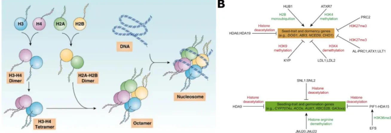
Figure 15. Chromatin structure is dependent on the nucleosome composition and histone post-translational modifications.
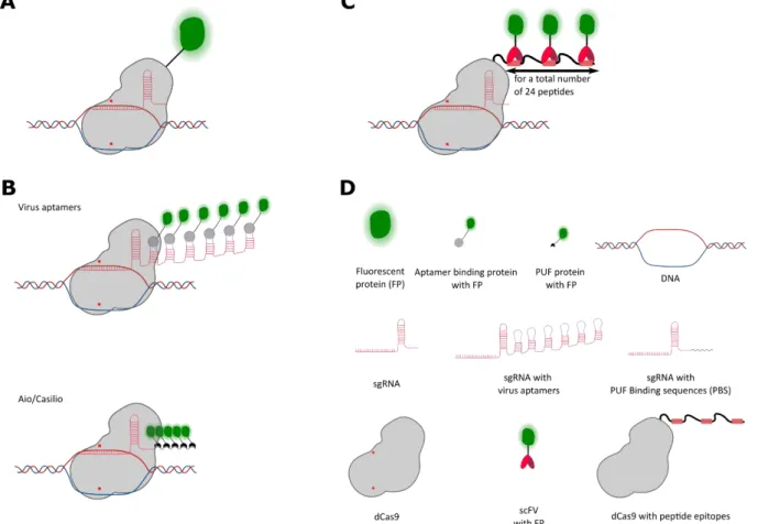
Figure 16. CRISPR/Cas9 based methods for loci visualisation in living cells.
Figure 17. Use of spike-ins in the context of global shift of expression in tomato pericarp cells.
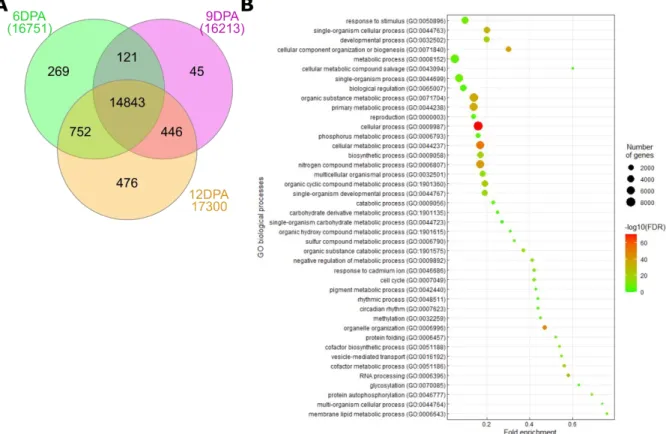
Figure 18. Venn diagram of non-differentially expressed genes found in 6-, 9- and 12DPA ploidy transcriptomes and their associated biological processes.
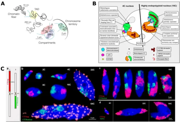
Figure 19. Repeated sequences used as targets for FisH and CRISPR imaging to quantify ploidy.
Figure20. Flowchart representation of the semi-automated procedure of ploidy quantification in the tomato pericarp cells.
Figure20bis. Illustration example of automatic nuclei assignment to cell layer for ploidy mapping
Figure 21. Flowchart representation of the steps performed by the plugin to identify and count FisH spots.
Figure 22. Example of application of the Histogram segmentation tool before and after the filtering steps.
Figure 23. Example of plugin assisted FisH spot counting and issues.
Figure 24. Illustration of CRISPR-imaging observations after transient and stable transformation.
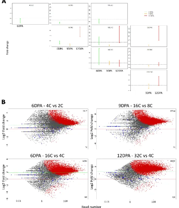
Figure 25. Ploidy sorted nuclei from 6, 9 and 12 DPA pericarp fruits show differential gene expression depending on ploidy and developmental stage.
Figure 26. Clustered normalized expression profiles of differentially expressed genes in pericarp of 6-9-12DPA fruits
Figure 27. Clustered normalized expression profiles of differentially expressed genes in pericarp of 6-9-12DPA fruits
Figure 28. Overrepresented biological processes in the different expression clusters from 6 DPA pericarp
Figure 29. Overrepresented biological processes in the different expression clusters from 9 DPA pericarp
Figure 30. Overrepresented biological processes in the different expression clusters from 12 DPA pericarp
Figure 31. Overlaps between DE genes from the different clusters of 6DPA, 9DPA and 12DPA fruits.
Figure 32. Representation of cuticle synthesis genes and their expression in relation to ploidy levels in the pericarp
Figure 33. Significant overlaps between ploidy specific transcriptome and LM pericarp transcriptome of Shirozaki et al., 2018.
Figure 34. Ploidy markers from a ploidy specific transcriptome of Arabidopsis thaliana root cortex (Bhosale et al., 2018) compared to the pericarp ploidy
transcriptome.
Figure 35. Morphology of Tomato pericarp sorted nuclei based on their ploidy and evaluation of the 5sr DNA oligo FisH probe for ploidy quantification.
Figure 36. Nuclear shape is modified with increasing ploidy levels.
Figure 37. Ploidy distribution, nuclear volumes, and cell sizes in 6, 9 and 12DPA Tomato pericarps.
Figure 38. Dynamic of nuclear volumes during early fruit development
Figure 39. Data integration of ploidy transcriptome and FisH ploidy quantification reveals a spatiotemporal ploidy specific gene expression.
Figure 40. CDKA1 and CDKB1/2 expression in relation with ploidy levels in pericarp cell layers
Figure 41. Cell cycle and endoreduplication related genes expression in relation with ploidy levels in pericarp cell layers
Figure 42. Dynamic changes of ploidy levels in the different pericarp cell layers during early fruit development from 6DPA to 12DPA
Potential roles of endoreduplication in Plants
What is endoreduplication? Some generalities and definitions
The cell cycle is a universal process, occurring in all living organisms, that results in cell division and the transfer of the genetic information (the whole copy of the genome) from a mother cell to two daughter cells (Figure 1A).
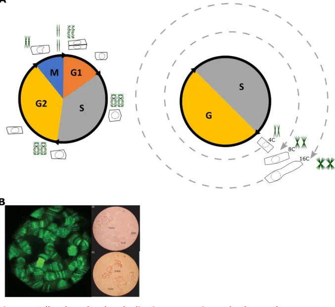
Endoreduplication is a modified cell cycle (also referred as endoreplication or endoploidization) during which cells increase their DNA content through successive rounds of nuclear genome duplication without cell division leading to increased ploidy level (Figure 1A). Ploidy is a relative measure of DNA content of a cell referring to the haploid DNA quantity of the genome, with 1C referring to 1 chromatid copy for a chromosome. Therefore, if DNA quantity of a diploid cell is 64C, this means that each chromosome has 64 copies of chromatids in this particular cell. Endoreduplication occurs without cytokinesis and the classical mitotic DNA replication events such as, DNA condensation/decondensation phases or spindle assembly (Edgar et al., 2014). In endoreduplicated cells, duplicated DNA is still attached as sister chromatids by the centromeres forming a structure called polytene chromosomes (Edgar and Orr-Weaver, 2001). These polytene chromosomes have been visualized for example in
Drosophila salivary gland cells that serve as a model for the study of endoreduplication
in insects (Figure 1B, Zhimulev and Koryakov 2009) and their existence was demonstrated in Tomato fruit pericarp cells (Bourdon et al., 2012).
Endoreduplication is a widespread phenomenon found in several clades in Eukaryotes. However, ploidy levels are highly variable between species or even between different tissues of the same individual. Moreover, it is not known if during endoreduplication DNA is always totally replicated in the different species or organs in which it occurs. In animals, endoreduplication is only found in some specific cell types such as in giant neurons of Gastropods (200000C), in salivary glands of Drosophila (2048C) and follicle cell epithelium and in Silkworm Bombyx mori (up to 20 rounds of DNA replication = 1 048 576C). In Mammals, endoreduplication also occurs in trophoblast giant cells (1024C), hepatocytes, cardiac myocytes, and cancer cells (D’Amato and Durante 2001, Orr-Weaver, 2015, Shu et al., 2018). The role of endoreduplication in animals is not fully elucidated but has been proposed to support rapid growth and strong metabolism of specialized organs such as nurse cells and salivary glands of larva in Drosophila or trophoblast in Mice (Fox and Duronio, 2013, Lee et al.,2009, Orr-Weaver, 2015). Another role for endoreduplication corresponds to body size control in ants and nematodes (Scholes, 2013).
In plants, endoreduplication is found in almost 90% of Angiosperms (Joubès and Chevalier, 2000). This phenomenon occurs in several plant families and does not seem to follow phylogenic linkage suggesting that endoreduplication results from separate evolution (homoplasy) because of specific strategies of life rather than common ancestry (Barow and Meister, 2003). For example, annual or bi-annual plants tend, more than perennials, to perform endoreduplication as a common developmental process (Barow and Meister, 2003).
Figure 1. Cell cycle and endoreduplication progression and polytene chromosome illustration.
(A) Illustration of the different phases of cell cycle (left panel) and endoreduplication progression (right panel). The evolution of cell size and chromosome morphology during the different phase is represented. G1: Gap phase 1; S :DNA Synthesis phase; G2: Gap2 phase; M: mitosis phase, G: Gap phase.
(B) Polytene chromosome visualization. Left panel: polytene chromosome within an intact larval salivary gland nucleus of Drosophila melanogaster, as revealed by green fluorescent protein (GFP) on left panel. Right panel: giant chromosomes with different degrees of polyteny visualized in light microscopy. Adapted and modified from Spradling, 2017 and Dyka et al., 2016.
Thus, endoreduplication could support rapid growth needed to complete a life cycle from seed to seed in a season (Barow and Meister 2003, Bainard et al., 2012). Within the same organism, endoreduplication can occur in several organs of the plant, at variable levels such as in Arabidopsis thaliana (Arabidopsis) leaves and sepals (up to 16C), roots (up to 8- to 16C) and trichomes (32- to 64c) or as in Tomato fruit pericarp cells (up to 512C) and leaves (up to 8C) (Cookson et al., 2006, Roeder et al., 2010, Bhosale et al., 2018, Churchman et al., 2006, Joubès et al., 1999). Recent studies suggest that endoreduplication could be involved in response to environmental stresses allowing rapid cell differentiation and strong metabolic capacities to improve tolerance to stress (Scholes and Paige 2015, Bhosale et al., 2019 and Lang and Schnittger 2020). But endoreduplication is also involved in different plant developmental processes. A more detailed description of the different roles of endoreduplication during plant development and in particular in our model, the Tomato fruit, is given below.
Endoreduplication and plant development
Endoreduplication occurs throughout plant development in many different organs and tissues: in above ground organs such as hypocotyl, leaf, and in reproductive structures such as flowers, fruits and seeds; in below ground organs such as the roots but also specialized structures like nodules. Endoreduplication occurs more frequently in organs that develop a large structure rapidly and have a need for a strong metabolic activity and storage, such as Maize endosperm cells (Leiva-Neto et al., 2004) or Medicago nodules Foucher and Kondorosi, 2000). In general, DNA quantity in a cell is positively correlated with cell size (Figure 1A, Beaulieu et al., 2008, Bourdon et al., 2010) and most of the described cases of endoreduplicated cells are larger than non-endoreduplicated ones in the same organ (Breuer et al., 2007). These observations led to link endoreduplication with a role in cell growth and organ size control (Kondorosi
et al., 2000, Larkins et al., 2001, Barow and Meister, 2003). Endoreduplication also
seems to be important for other developmental processes and hereafter, a series of organs and developmental events in which endoreduplication has a role are detailed.
Endoreduplication in the hypocotyl in Arabidopsis
In Arabidopsis, the hypocotyl is formed during embryogenesis and is composed of a fixed number of cells. Therefore, after germination most of the growth occurs through cell expansion (Gendreau et al., 1997). Endoreduplication takes place in the hypocotyl cells very early after germination and ploidy levels can reach up to 8C in light-grown plantlets (Gendreau et al., 1997). Interestingly, in this organ, light seems to inhibit endoreduplication since in the darkness, higher ploidy levels (16C) are observed (Figure 2A and 2B). The increased proportion of higher ploidy level are associated with an increase in cell length and hypocotyl length (Gendreau et al., 1998; Amijima et al., 2014). In this organ, the hypothesized role for endoreduplication is thus to be a strategy to grow more in a short time, in dark environment, supporting etiolated growth in order to reach light rapidly.
Figure 2. Endoreduplication during Arabidopsis hypocotyl.
(A) Endoreduplication patterns in the hypocotyl of light-grown Arabidopsis seedlings. (B) Endoreduplication patterns in the hypocotyl of dark-grown Arabidopsis seedlings A and B, adapted and modified from Gendreau et al., 1999.
Endoreduplication in the Arabidopsis thaliana leaf
a. Epidermal pavement cells
In Arabidopsis, leaf development is divided in two phases, a cell division phase and a cell expansion phase that overlap in time. First, the leaf primordium, emerging from the shoot apical meristem, grows by cell proliferation, that occurs in the entire leaf and produces new cells (Figure 3A). In a second phase, cell proliferation progressively stops, and cells start to expand. This cell expansion phase progresses as a front from the tip to the base of the leaf (Gonzalez et al., 2012, Figure 3A). This second growth phase is associated with the occurrence of endoreduplication (Melaragno et al., 1993) with ploidy levels reaching up to 16C in the epidermal pavement cells (Figure 3B), the outermost cell layer of the leaf. To better understand how endoreduplication is related to increasing cell size in leaves, a mathematical model was built to simulate epidermal cell size distribution as a function of endoreduplication dynamics (Kawade and Tsukaya, 2017). This model, taking into account that endoreduplication dynamics is following a Poisson law and considering that cell size is determined according to ploidy level, recapitulated experimental data showing the Gaussian distribution of cell size. However, a meta-analysis of published data linking endoreduplication with cell growth in leaves of Arabidopsis mutants showed that in most cases endoreduplication is not the only factor explaining the changes in cell size (Tsukaya 2019).
Endoreduplication in the leaf is influenced by environmental factors such as day length since the transfer of Arabidopsis plants from short-day-period to long-day-period increases the proportion of 8C and 16C nuclei accompanied with an increase in leaf size (Del Prete et al., 2019). Interestingly, this increased proportions of 8C and 16C is associated with an accumulation of the floral transition markers FLOWERING LOCUS T, CONSTANS and APETALA suggesting that endoreduplication could be involved in signaling transition from vegetative phase to reproduction (Del Prete et al., 2019). In these leaves, the increase of ploidy levels has also been associated with a complex reprogramming of metabolism, including changes in sugar metabolism, cell wall synthesis and secondary metabolism (Del Prete et al., 2019). In Arabidopsis leaf, the potential roles of endoreduplication are thus to regulate at least partially cell-size and leaf size and is also associated with flowering after changes by light conditions and metabolic changes.
b. Leaf Epidermal specialized cells: the trichomes
In Arabidopsis leaves, the epidermal pavement cells are not the only ones to undergo endoreduplication. Trichomes, large cells that differentiate from pluripotent epidermal cells called protodermal cells in young leaves, also undergo endoreduplication (Figure 4A). In Arabidopsis, trichomes have a three branched morphology dependent on ploidy levels (Figure 4B). The early differentiation of trichomes requires on a positive transcriptional signaling cascade of a transcription factor complex composed of GLABRA1 (GL1), TRANSPARENT TESTA GLABRA1 (TTG1), GLABRA3 (GL3) and ENHANCER OF GLABRA3 (EGL3) (Hülskamp et al., 1994; Fambrini and Pugliesi, 2019). One of the first consequence of this transcriptional activation is that the cell stops dividing and starts endoreduplicating and enlarging (Figure 4A). The cell expansion is mainly driven by the growth of the vacuole and occurs simultaneously with branching.
Figure 3. Endoreduplication during Arabidopsis leaves development
(A) Schematic representation of Arabidopsis developmental stages from 4 to 20 days after sowing, at rosette, leaf and cellular level. The developmental phases are represented in different colors: in green, the cell proliferation phase; in orange, the meristemoid division phase (not discussed here) and in yellow, the cell expansion phase.
(B) Representation of an epidermal cell population colored by ploidy levels in the leaf. A from Gonzalez et al., 2012. B adapted and modified from Melaragno et al., 1993
When fully grown, the trichome has reached 32C DNA content and has three branches (Figure 4B and 4C).
Generally, size and branching of the trichome follow a positive correlation with DNA quantity and mutants with increased endoreduplication levels show increased trichome branching (Figure 4D, Kasili et al., 2011, Fambrini and Pugliesi 2019). Trichome overbranching is also observed after treatment with colchicine (Figure 4E, Robinson et al., 2018), a drug able to disrupt the cell cycle, inducing whole genome duplication and thus polyploidy in plants (Robinson et al., 2018). On the other hand, inhibition of endoreduplication during trichome initiation results in dedifferentiation of the cell that finally is integrated in the epithelium as an epithelial cell, suggesting a strong link between endoreduplication and the differentiation of trichome in the Arabidopsis leaf (Bramsiepe et al., 2010). In Arabidopsis, endoreduplication in the trichome is thus proposed to have a role in regulating cell size and controlling cell fate.
Endoreduplication in the root
Endoreduplication also occurs during root development. The primary root can be divided in three zones: the meristem zone where cells are mostly dividing, a transition zone in which cells shift from the cell cycle to the endocycle, and the elongation zone where cells expand and endoreduplicate reaching up to 16C. In Arabidopsis, proper root development depends on the correct maintenance of the meristem size and a fine balance between dividing and differentiating (expanding and endoreduplicating) cells (Figure 5, Heyman et al., 2017; Bhosale et al., 2018). In Arabidopsis root, the transition between division and endoreduplication is regulated by hormones, and more specifically by auxin and cytokinin. A gradient of auxin is present in the root as a maximum in the meristem zone to a minimum in the transition zone (Figure 5, Breuer
et al., 2014; Mambro et al., 2017). This auxin minimum is located at the place where
cytokinin signaling takes place and where the transition between the cell cycle and endoreduplication is occurring (4C-8C boundary in Figure 5). The phytohormones auxin and cytokinin play an important role in this regulation. It has been shown that cytokinin controls the shift from the mitotic cycle to the endocycle in the root (Takahashi et al., 2013). Plants with reduced response to cytokinin have lower endoreduplication activity in roots (Takahashi et al., 2013). A screen of ethyl methanesulfonate for larger or smaller organs and change in ploidy levels in treated Arabidopsis seeds resulted in the identification of a mutant named high ploidy 2 (hpy2) (Ishida et al., 2009). hpy2 seedlings have higher ploidy levels in the root, very short primary root and a reduced root meristem area composed of larger cells compared to WT (Ishida et al., 2009). Thus, in the primary root, endoreduplication seems to regulate growth by allowing cell expansion after the transition zone.
Some epidermal cells of the root can differentiate into root hairs cells that are long tubular shaped cells. Root hairs cells can be as long as 1 mm or more. They greatly increase the rooting area helping in nutrient acquisition, anchorage, and microbe interactions (Grierson et al., 2014). It has been reported that root hair cells undergo several rounds of endoreduplication and reach 16C ploidy level when fully developed (Sliwinska et al., 2012).
Figure 4. Endoreduplication and trichome development.
(A) Development of a trichome of Arabidopsis wild type. Trichome development starts with the specification of single cells within the epidermal layer of the leaf (cell fate/initiation). After this stage, the cell will start doing three rounds of endoreduplication and enlarge (outgrowth), attaining ten-times the epidermal cell size. Then, branching initiates and simultaneously cell is continuing to enlarge and undergo one more endoreduplication round (Cell growth and expansion). Finally, the trichome will eventually continue to grow until being fully grown (Maturation).
(B) Scanning electron micrograph of wild-type (Col-0) trichome showing the three branches morphology. Bar = 50µm. Adapted from Kasili et al., 2011.
(C) Flow cytometry analysis representing the ploidy levels present in wild-type Col-0 leaves. Adapted from Heyman et al., 2017.
(D) Flow cytometry analysis representing ploidy levels present in mutant line uvi-4 having increased ploidy levels in leaves (left). Adapted from Heyman et al., 2017. Scanning electron micrograph of uvi-4 mutant trichome showing over branched phenotype (right). Bar = 300µm. Adapted from Heyman et al., 2011
(E) Trichome phenotype observed in plants treated with colchicine. Colchicine is a drug perturbing the cell cycle and leading to the production of plants with whole genome duplication. Robinson et al., 2018
It was shown that root hair cells undergo one more round of endoreduplication compared to their neighbor non hair epidermal cells (16C vs 8C, Bhosale et al., 2018) indicating a potential role of endoreduplication in the specification of these root hair cells. In the hyp7 mutant, mutated in the RHL1 protein having an essential role in the DNA topoisomerase VI complex, showing a decreased of endoreduplication results in leaf epidermal cells, trichomes and root hair cells that are smaller (Sugimoto-Shirasu
et al., 2005). However, in in several hair mutants, the differentiation of root hair cells
in the collet and root regions is impaired when there is less endoreduplication while r the size of these cells is less or not affected by the decreased ploidy level, suggesting that endoreduplication might be necessary for the differentiation but that in these root hair cells, growth is independent of ploidy levels (Sliwinska et al., 2015). Endoreduplication in root hair cells thus seems to play a role in the establishment of their identity (Sliwinska et al., 2015; Bhosale et al., 2018).
Other specialized root structures also undergo several rounds of endoreduplication such as the nodules in Medicago (Medicago truncatula) or Lotus (Lotus Japonica) (Foucher and Kondorosi, 2000, Suzaki et al., 2014). Nodules are specialized structures formed after symbiotic infection of roots of leguminous plants by soil bacteria responsible of atmospheric nitrogen fixation. Nodules can be divided in four separated areas of different functions. First, a meristem zone produces new cells, through cell division, that will later be forming the different nodule cell layers (Figure 6A and 6B). The invasion zone is composed of endoreduplicated cells that can reach 16C and that can be infected by rhizobia. The next area is an interzone that contains symbiotic cells that are larger than in the meristem area or the invasion zone. In the interzone cells are endoreduplicated and can reach 16C to 32C. The last zone is the nitrogen-fixing zone where symbiotic cells are the largest with large vacuoles and ploidy levels reaching 64C. The areas containing the largest cells display higher ploidy levels indicating a positive relation between endoreduplication and cell size (Figure 6B, Nagymihály et al., 2017).It has been shown using plant with impaired endoreduplication process that a proper transition from the cell cycle to the endoreduplication is necessary for correct the differentiation and functionality of the nodule structures showing that endoreduplication is an important process for the differentiation of nitrogen fixing cells (Vinardell et al., 2003; Cebolla et al., 1999).
Figure 5. Schematic representation of root morphology and organization.
The dash lines represent in yellow the area considered as root meristem where cells divide, in rose the transition zone where cells enter endocycle and in green the elongation and differentiation zone where cells continue to endoreduplicate and start to grow and differentiates. The black dash lines represent an artificial frontier where cells in each side have different ploidy levels, for example the 2C-4C boundary represent areas where on the left cells are mostly 2C and on the right 4C. The root colorized areas represent in yellow the auxin gradient that is diminishing when progressing toward the transition zone (local minimum of auxin concentration) and in blue the area where cytokinin is found (transition zone).
Figure modified and adapted from Breuer et al., 2014 and Bhosale et al., 2018.
Figure 6. Schematic representation of nodule tissue ontology.
(A) Indeterminate nodule primordium with cell ontology that is indicated by the colored layers that will be involved in nodule formation. C3, C4 and C5 layers are the third, fourth and fifth layers away from epidermis.
(B) Differentiated nodule with primordium cell ontology that is indicated with same color than color used for undifferentiated primordium.
Between dashed lines are areas where indicated ploidy levels are observed. Double arrows indicate areas of nodules implicated in the nodule formation and differentiation.
Endoreduplication in the flower
Endoreduplication takes place in flowers of many different species such as Orchids (Phalaenopsis, Ho et al., 2016), Cabbage (Brassica oleracea L., Kudo and Kimura, 2002) or Red Clover (Trifolium pratense, Kocová et al., 2016).
In Arabidopsis developing flowers, endoreduplication is found in sepals that correspond to the green leaf like structures of the flower (Figure 7A). The role of sepals in is to be protective by enclosing the developing reproductive organs. Both abaxial and adaxial epidermis cells of the Arabidopsis sepals undergo endoreduplication reaching up to 16C and rarely 32C (Robinson et al., 2018). These cells are referred to as giant cells because of their length that is nearly a fifth of the sepal length (360µm long, Figure 7B and 7C, Roeder et al., 2012). In this organ, non endoreduplicated small cells (10µm long) co-exist with these endoreduplicated giant cells. Giant cell identity is determined before endoreduplication starts. However, it seems that endoreduplication is inhibiting small cell identity since transgenic plants with increased endoreduplication have a reduced number of cells expressing small cell identity markers (Roeder et al., 2012). Increase or decrease of the proportion of giant cells results in change of sepal curvature in an outward or inward manner, respectively, whereas wild type Arabidopsis sepals have a flat shape (Roeder et al., 2010). Thus, endoreduplication is influencing the morphology of sepals. Moreover, transgenic plants with increased ploidy levels in sepal epidermal cells results present an increased proportion and size of giant cells without any alteration of sepal length or growth rate (Roeder et al., 2012). In transgenics plants harboring more giant cells and thus higher ploidy levels in the sepals, due to the specific overexpression of the LGO/SMR1 gene in the sepal epidermis, an increased expression of defense genes has been observed including the up regulation of Glucosinolate biosynthesis genes (Schwarz and Roeder 2016). Glucosinolate is a secondary metabolite stored for being used as a repulsive compound against herbivores insects. The upregulation of glucosinolate biosynthesis is also upregulated in mature trichomes (Jakoby et al., 2008), thus suggesting that endoreduplication could have a role in the regulation of secondary metabolism implicated in defense. Based on these data, in the Arabidopsis sepals, endoreduplication could have a role in regulating organ curvature, cell size, cell fate and maybe defense response pathways.
Figure 7. Illustration of Arabidopsis thaliana sepals and epidermal giant cells.
(A) Wild-type Arabidopsis flower. Sepals are indicated (s)
(B, C) Scanning electron micrograph of a mature Arabidopsis sepal. The typical giant cells of the sepals are colored in pink.
Endoreduplication in fruits and seeds
Regarding fruits and seeds, endoreduplication has also been reported in 28 species among 12 families of angiosperms for fruits and in 47 species among 27 families for seeds (D’Amato, 1984; Barow and Meister, 2003; Bourdon et al., 2010). For example, in the capsule of the Foxtail Lily (Eremurus robustus), in cells from the pericarp tissue developing from the ovary wall, polyploid nuclei have been observed as well as polytene chromosome structures associated with increased nuclear size (Figure 8A and 8B, H Lauber 1946). Endoreduplicating cells have also been reported in fleshy fruit species such as cucurbits (Coombe, 1976; Barow, 2006) or Tomato (Joubès and Chevalier 2000). In several fruits, endoreduplication is negatively correlated with the duration of the growth period. Indeed, it was observed that fruits developing in less than 13 weeks undergo 3 to 8 rounds of endoreduplication whereas endoreduplication is absent in most of the studied fruit species with longer developmental timing (Figure 8C Bourdon et al., 2010). For example, tomato developing in less than 10 weeks performs 8 endocycles whereas apple developing in a bit more than 20 weeks is only composed of non endoreduplicated cells (Figure 8C)
Endoreduplication has also been reported, for more than 60 species, in the endosperm, a differentiated structure of the seed specialized in the storage and nutrition of the developing embryo. This tissue presents a large panel of ploidy levels, up to 3072C in wild cucumber (Echinocystis lobata, D’Amato 1984). Maize is the most studied species regarding endoreduplication as a potential regulator of cell size, kernel size and starch content (Leiva-Neto et al., 2004; Sabelli et al., 2009; Sabelli et al., 2013). Endosperm is formed after the double fertilization. First, one of the sperm nuclei fuses to the egg cell nucleus and forms the embryo. The second sperm nucleus fuses to the polar nucleus of the binucleate central cell resulting in the formation of a 3C cell, the primary endosperm. The endosperm then develops by several divisions and undergo up to several rounds of endoreduplication until full development (Sabelli et al., 2009). The onset of endoreduplication in the Maize endosperm is associated with the production of the zein protein that accumulates as a storage of nitrogen, and with a reduction of chromatin condensation (Sabelli and Larkins 2009).
In Teosinte, the Maize wild relative and ancestor, the endosperm has been studied to understand, at the cellular level, the domesticated characters that permitted to obtain a larger endosperm in Maize (Figure 8D). Teosinte produces highly endoreduplicated nuclei in the endosperm, till 96C (Figure 8E), whereas Maize is producing nuclei with one more endoreduplication round (192C, Figure 8F, Vilhar et al., 2002). However, the major difference in endoreduplication between Teosinte and Maize is the spatial distribution of highly endoreduplicated nuclei in the endosperm. In Maize, endoreduplicated nuclei are localized in the upper part of the endosperm while in Teosinte endoreduplicated nuclei are localized in all the central endosperm (Figure 8F). It was hypothesized that this modified distribution of highly endoreduplicated cells might have been selected by domestication and allowed a more efficient starch accumulation in Maize than in Teosinte (Figure 8F, Dermastia et al., 2009).
Figure 8. Examples of endoreduplication occurrences in fruits.
(A) Drawing of the cells composing the pericarp of Ememurus robustus fruit showing the cellular organization and cell size diversity.
(B) Drawing of nuclei observed in Ememurus robustus at different ploidy stages. The top left nuclei correspond to a polyploidy nucleus (big nucleus) and a diploid nucleus (small nucleus). On the right and at the bottom are represented chromosomes structures observed in polyploid and tetraploid nuclei, on the right and at the bottom, respectively.
(C) Occurrence of endoreduplication in fleshy fruits. The maximal number of endocycles determined in fully ripened fleshy fruits was plotted against the duration of fruit growth until ripening.
(D)Photographs of teosinte (maize ancestor, left) and cultivated maize (right) caryopses.
(E) Ploidy map of teosinte caryopsis at 11 days after pollination (F) Ploidy map of maize caryopsis at 16 days after pollination
A and B adapted from Lauber, 1947. C from Chevalier et al., 2011. D and E from Dermastia et al., 2009. F adapted from Vilhar et al., 2002.
In conclusion, endoreduplication in the endosperm is frequent and studies of Maize and its ancestor Teosinte shows that this process is important for kernel development and might be linked to starch accumulation in endosperm.
In conclusion, endoreduplication seems to have different roles in different tissues and organs. High endoreduplication levels are often associated with large cell size and organ size in leaves or fruits. The analysis of mutants with reduced endoreduplication allowed to observe that some mutants are able to compensate this reduction by increasing cell number to preserve the organ size (Massonnet et al., 2011). Endoreduplication might have a role in enhancing/potentiating cell expansion rather than driving it because cell expansion can occur at a similar level even in absence of increased ploidy levels. Cell differentiation is also another event in which endoreduplication has a role because important impairment of endoreduplication limits differentiation in trichomes and nodules, for example. Finally, endoreduplication could have a role differentiation and regulation of metabolism in Maize kernel for starch and protein accumulation both occurring in large endoreduplicated cells.
Endoreduplication in the model of study during the PhD: the
tomato fruit pericarp
Because Tomato fruit develops rapidly and can present large variation in size and form, it is used as a model of study to understand which roles endoreduplication and its spatial distribution could have for fleshy fruit development. Hereafter I will describe what is known about where and when endoreduplication is occurring during Tomato fruit development.
Endoreduplication during Tomato fruit development
In Tomato (Solanum lycopersicum), the fruit is a berry formed by the differentiation of the ovary. It is composed of a pericarp that derives from the walls of, at least, two fused carpels, of a jelly locular tissue surrounding the seeds and of a placenta maintaining seeds attached to a central columella (Figure 9A).
At the end of Tomato fruit development, the pericarp, the locular tissue and columella to a lesser extend are composed of cells that have undergone several rounds of endoreduplication (Figure 9B, Joubès et al., 1999) and the ploidy levels varies between cultivars suggesting that the number of endoreduplication rounds is influenced by the genotype (Cheniclet et al., 2005).
In Tomato, endoreduplication already occurs in the ovary wall during flower development (Cheniclet et al., 2005, Renaudin et al., 2017) that starts with the formation of the floral meristem and finishes with the anthesis that corresponds to the pollination and fertilization of the fully functional flower.
Before fertilization, the ovary wall grows mainly by cell division and is composed of seven to nine cell layers between stage 11 and 16 (4- and 8 mm flowers in Solanum
Figure 9. Endoreduplication during Tomato fruit pericarp development
(A) Anatomy of a Tomato (Solanum lycopersicum cv. WVA106) fruit. Equatorial section. Scale bar = 1cm.
(B) Flow cytometry histograms of DAPI stained nuclei extracted from different tissues of Tomato fruit at breaker stage.
(C and D) Structure of the pericarp of the cherry tomato WVA106 at anthesis (C) and 4DPA (D). Blue circles indicate vascular bundles, yellow arrows new cell walls and red indicates extracellular spaces delimitating the M cell layers.
(E) Illustration of different developmental stage of tomato fruit growth and the associated Phase according to Gillaspy et al., 1993
Figures modified and adapted from Joubès et al., 1999 (B) and Renaudin et al., 2017 (C and D). (E) from Jean-Philippe Mauxion (personal communication).
At stage 18, before anthesis, 43% of the nuclei are endoreduplicated, with around 2-3% of 8C and almost 40% of 4C, considered as a first round of endoreduplication due to the absence of division structures, such as condensed chromosome of metaphase to telophase and division planes (Renaudin et al., 2017). After fertilization and during the whole fruit development, the pericarp volume represents nearly half of total fruit volume and the growth rate of the fruit is the same as the growth rate of the pericarp (Renaudin et al., 2017). This is indicating that the pericarp is mainly representing the tomato fruit growth, thus the next paragraph will mostly be focusing on the cellular events and endoreduplication dynamic during pericarp development.
The ovary wall and the derived structure, the pericarp, is divided in three areas (Figure 9C and 9D). First, an outer-epidermis contains three cell layers named E1 (most outer layer), E2 and E3 (the two-sub outer-epidermal layers, Figure 9C). Second, an inner part, formed after anthesis is composed of multiple layers grouped under the name of M’ layers. M’ cells mostly originate from layers E2 and E3 after periclinal cell divisions (Figure 9D). Third, four cell layers formed before anthesis are composing the M layer (Figure 9C). The M and M’ layers form the mesocarp. Finally, the inner part of the pericarp in contact with the locular tissue is corresponding to two cell layers with the inner-epidermis layer I1 and its sub-epidermal layer I2 (Figure 9C).
Fruit growth and development is divided in three phases according to Gillaspy (1993). The first phase corresponds to the fruit set during which internal signals, corresponding to hormonal signal of gibberellins, auxins and downstream regulators, are inducing growth (Ariizumi et al., 2013). This phase starts if the fertilization succeeded and lasts for nearly 20h (Figure 9D; Cheniclet et al., 2005). The following phase lasts between 7 to 10 days depending on the genotype (Gillaspy et al., 1993). During this period, the pericarp is characterized by the occurrence of high cell division activity. In the pericarp, cell divisions start the first day after anthesis and are mostly localized in the three epidermal layers (E1, E2 and E3, Figure 10A and 10B) and two inner-epidermal layers (I1 and I2, Figure 10E and 10F). The mitotic activity in the outer epidermis generates new cell layers whose number varies in function of the genotype (Figure 10G). However, during this period, cell expansion occurs already and increases greatly the volume of the M layer cells by 100 times compared to anthesis (Figure 10C). The increase in M layers volume participates to half the increase of the fruit volume (Cheniclet et al., 2005, Renaudin et al., 2017). Concomitantly, ploidy levels increase in the pericarp from a mean C value (sum of the C value weighted by their frequency) of 3.2 to 7 from anthesis to 10 days post fertilization. This change in mean ploidy levels is due to the disappearance of 2C nuclei and the appearance of new classes of ploidy (8C and 16C, Figure 10H) from anthesis to 10DPA (Days Post-Anthesis, Cheniclet et al., 2005). At the end of this phase, in the pericarp, only the outer epidermis continues to divide.
Figure 10. Endoreduplication occurrence through Tomato fruit pericarp development.
(A to F) Evolution of cell number and cell volume in the different pericarp cell layers during fruit development (WVA106). Variables are expressed as relative values with reference to the value of 1 at anthesis (dot line).
(G) Illustration of the morphological differences between three fruits and pericarp sections of different Tomato cultivars at breaker stage. From left to right is a fruit of Grosse de Gros, a fruit of Ferum 26 and a fruit of WVA106. Scale bars: left = 1cm, right = 1mm
(H) Developmental dynamic of ploidy levels in Wva106 pericarp. Top panel: evolution of 2C, 4C, 8C, and 16C ploidy levels. Bottom panel: evolution of 32C, 64C, 128C and 256C ploidy levels.
(I) Ploidy mapping in a mature green tomato pericarp.
The top picture is showing the pericarp structure of WVA106 at mature green stage. Scale bar = 500 μm.
At the bottom is represented the ploidy map showing the frequency of nuclei by cell layer in the pericarp.
Figures modified and adapted from Cheniclet et al., 2005 (A and H), Bourdon et al., 2011 (I) and Renaudin et al., 2017 (B to G)
The last phase starts when the fruit growth is mostly due to cell expansion in the mesocarp. Pericarp cell number does not change much from this stage. At the end of this phase the pericarp volume is ten times more important than at the start of this phase. This growth is mainly driven by cell expansion of the M’ cells participating to 50% of the increase in size (Renaudin et al., 2017). This cell expansion occurs simultaneously with a drastic increase in ploidy levels due to the occurrence of 2 to 3 rounds of endoreduplication (64C, 128C and 256C, Figure 10H). At the end of this phase corresponding to the mature green stage the fruit has reached its final size and the mean ploidy levels increased to 24 in cherry tomato and can be higher, up to 70, in other genotypes (Cheniclet et al.,2005). After this period of growth, the fruit will start the ripening phase.
Ploidy distribution in the pericarp and potential role of endoreduplication
At mature green stage, the establishment of a ploidy map in the pericarp allowed to get a better view of the distribution of the ploidy levels inside this tissue (Figure 10I; Bourdon et al., 2011). At this stage, the epidermal layers E1 and E2 are the only ones without endoreduplicated cells. In E1, 2C cells represent more than 60% of the cells whereas the other 40% are 4C cells (Figure 10I). In E2, 2C cells represent less than 10% of the cell population, 4C cells represent nearly 40% and the other 50% are half 8C and half 16C (Figure 10I). The last outer-epidermal layer, E3, is composed of 4C to 32C cells and 8C cells represent the most represented group. In the inner epidermis, I1 is composed of 8C to 32C cells and I2 of 16C to 256C cells (Figure 10I). The absence of 2C and 4C cells in the inner epidermis indicates that dividing activity has disappeared from these layers. Most of the cells in I1 layer are 16C whereas ploidy levels are nearly equally represented in I2 layer. Finally, M and M’ layers are composed of the highest ploidy levels. In M’, ploidy levels range from 8C to 256C but 8C cells are found only in the first M’ layer and 16C only in the two first M’ layers (Figure 10I). In addition, the measurement confirmed a positive correlation between ploidy and cell size (Bourdon
et al., 2011). Although this correlation is not perfect. It suggests that endoreduplication
is more likely favoring the potential of growth by increasing the range of possible growth rather than driving cell growth (Bourdon et al., 2011, Nafati et al., 2011). Endoreduplication supporting rapid growth is also suggested by the presence of 8C nuclei at anthesis and rapid increase of 8C cells during the first days of development (Renaudin et al., 2017). 8C cells before anthesis are hypothesized to be in the M layer because M layer attains higher ploidy levels at the end of the development (Renaudin
et al., 2017). The role of endoreduplication in Tomato fruit thus seems to be an
enhancer of cell expansion since it is present before cell expansion starts, at anthesis, and both cell expansion and ploidy rise rapidly after anthesis in the mesocarp.
Finally, we have seen that before anthesis some cells in the ovary are already endoreduplicated that might be supporting the rapid cell expansion of mesocarp cells. In Tomato fruits, central mesocarp cells are large and highly endoreduplicated cells, are also accumulating a lot of soluble sugars and organic acids in vacuoles (Beauvoit et
al.,2014), thus endoreduplication could have a role in regulating metabolism of
Genetic regulation of endoreduplication in Plants
Dynamic of CDK-CYC complexes during cell cycle progression
The endocycle is a modified version of the classical mitotic cell cycle. The cell cycle in Eukaryotes is divided in four phases, Gap1 (G1), S (Synthesis), Gap2 (G2) and M (Mitosis) phases (Figure 1A). During the G1 phase the cell decides to progress into the cell cycle or not in function of environmental and internal signals. Favorable signals, such as temperature or water and hormones or sucrose allow the synthesis of the mRNA and proteins necessary for the subsequent cell cycle phases. After the G1 checkpoint and requirements are passed, the cell enters the S phase, during which the whole genome is duplicated through the production of an extra copy of each chromosome pair, thus passing from 2n to 4n (n is referring to the number of each chromosome). The S phase is followed by the G2 phase during which a verification of DNA integrity after duplication, named G2/M checkpoint, occurs and necessary components for the next step are produced. The M phase is the last step of the cell cycle and corresponds to the equivalent segregation of the chromosomes. The M phase ends with the cytokinesis leading to the formation of two daughter cells that will contain the same genetic information. The endocycle is only composed of a succession of G and S phases without M phase, resulting in an increase in cell ploidy levels and production of polytene chromosomes (Figure 1B, Vlieghe et al., 2007).
During the cell cycle, the correct progression of each phase requires the action of heterodimeric complexes composed of a catalytic subunit referred to as CYCLIN-DEPENDENT KINASE (CDK) and a regulatory subunit, the CYCLIN (CYC). In CDK-CYC complexes, the CDK assures phosphorylation activities whereas the CYC defines the substrate specificity and the localization within the cell (Inzé and De Veylder, 2006). The high number of CDKs and CYCs proteins and the diversity of CDK-CYC complexes are crucial for the molecular regulation of the cell cycle and the endocycle. In Arabidopsis, the CDKs belong to a conserved family of serine/threonine protein kinases containing 12 members classified from A to F and CDKA and CDKB have been implicated to direct the regulation of the cell cycle (Gutierrez 2009) while CDKD and CDKF are CDK-activating kinases regulating small RNA processes and help to preserve mitotic activity in Arabidopsis (Hajheidari et al., 2012, Takatsuka et al., 2015) and CDKG are regulating mRNA splicing (Zheng et al., 2014). In Arabidopsis, the CYC family is a large family of around 30 members categorized in four sub-families, CYCLINS A, B, D and H, generally involved in G1/S and S phase of the cell cycle (Gutierrez 2009) but the role of each cyclin has not clearly been established. Some have been associated to mitosis regulation such as CYCB1;1, CYCB1;2, CYCA2;3, CYCA3;1 and CYCD3;1 (De Veylder et al., 2011, Gutierrez 2009) or to meiosis regulation such as for example CYCA1;2, CYCA2;1, CYCA3;2 and CYCB3;1 (Bulankova et al., 2013).
CDK-CYC complexes have an essential role in the control and the maintenance of the cell cycle preventing endocycle. Consequently, the repression of their function in the cell is essential for the shift from the cell cycle to the endocycle (Edgar et al., 2014).