HAL Id: tel-01192592
https://tel.archives-ouvertes.fr/tel-01192592
Submitted on 3 Sep 2015HAL is a multi-disciplinary open access archive for the deposit and dissemination of sci-entific research documents, whether they are pub-lished or not. The documents may come from teaching and research institutions in France or abroad, or from public or private research centers.
L’archive ouverte pluridisciplinaire HAL, est destinée au dépôt et à la diffusion de documents scientifiques de niveau recherche, publiés ou non, émanant des établissements d’enseignement et de recherche français ou étrangers, des laboratoires publics ou privés.
Characterization of Foxp2 functions in the mouse cortex
Vera Medvedeva
To cite this version:
Vera Medvedeva. Characterization of Foxp2 functions in the mouse cortex. Neurons and Cognition [q-bio.NC]. Université Pierre et Marie Curie - Paris VI, 2015. English. �NNT : 2015PA066118�. �tel-01192592�
Université Pierre et Marie Curie
Ecole doctorale Cerveau Cognition Comportement
Institut du Fer à Moulin / Neurodevelopmental disorders
Characterization of Foxp2 functions in the mouse cortex
Par
Vera Pavlovna MEDVEDEVA
Thèse de doctorat de Neurogenetics
Dirigée par
Matthias Groszer, CR1, Inserm
Présentée et soutenue publiquement le 17 Juin 2015Devant un jury composé de :
Pr. Ann LOHOF Présidente Dr. Markus WOEHR Rapporteur Dr. Pierre BILLUART Rapporteur Pr. Josef PRILLER Examinateur Dr. Carlos PARRAS Examinateur
Dédicace To my grandfather Leonid Michailovich Kolmak Леониду Михайловичу Колмаку
Acknowledgements
First of all I would like to thank my supervisor Matthias Groszer who has kindly given me the opportunity to work in his laboratory and pursue his and sometimes my own scientific ideas. The scientific and, even more, life experiences in his laboratory were truly exceptional and rather determining for me, due to the very special environment established there.
I have to thank the members of our team for the indispensable support and warm friendship. Thanks to Cedric Mombereau, a brilliant scientist and a teacher, who largely formed my neuroscientific thinking and introduced me to behavioral neuroscience; I could easily call Cedric my second supervisor. Thanks to Corentin Le Magueresse for his very helpful suggestions during the preparation of this thesis. Thanks to Alfredo
Cabrera Socorro, a man of infinite energy, who masters in perfection the art of having
fun - an essential quality during not-so-easy times.
My sincerest gratitude to the whole Institute du Fer à Moulin and in particular to
Jean-Antoine Girault: for his support and for the creation of a place with such a friendly
and highly collaborative atmosphere. It is thanks to the cooperative and highly professional researchers at IFM that it was possible to realize the majority of the experimental part of this thesis (and beyond). Special thanks to Laurence Goutebroze,
Aude Muzurelle, Patricia Gaspar, Imane Moutkine, Tanay Ghosh and Frank Julius Meye for sharing their experience in biochemistry, histology, molecular biology and
stereotaxic surgery as well as for their immediate help and constant presence throughout my experimental life. Special thanks to Alessia Usardi at College de France for demonstration and advices on establishing BacTRAP.
Very special thanks to Cataldo Schietroma - my best friend and the greatest supporter of a vital importance throughout thesis writing and preparation. More than that, your scientific feedback is not at all a minor one: critical reading and corrections of the manuscript and profitable discussions throughout all the 5 years dedicated to this project had the greatest impact on this work.
Many thanks to all my friends outside the lab. Antonina Kurtova and Julia
Korchagina, you girls are my doubles in many senses, including the PhD experience we
went through in parallel and together, having many thoughts and emotions to share. Thanks to the many friends I met through ENP network and to the people I became close at IFM. Long conversations alongside drinks and outdoor activities together made my life richer and helped to enjoy more fully the Parisian life-style.
Finally, my family played not the least role in this work: thank you, dear parents and
grandmother, for your constant presence, support and understanding. That means a lot
Table of Contents
List of abbreviations
List of figures
Preface
1Introduction
FOXP2 deficiency causes a complex speech and language disorder 4
Foxp2 as an entry point to study molecular and neural networks contributing to
cognitive aspects of speech and language 10
The study of animal models provides insights into conserved FoxP2 functions 14
The role of FoxP2 in development 14
Activity dependent function of FoxP2 in mature brain: evidence for a role in social
behaviour and vocalizations 16
FoxP2 cellular functions 20
Mouse models in Foxp2 research: motor learning and ultrasound vocalizations 22
Foxp2 in the mouse cortex 28
Aims
40
Materials and Methods
Mice 41
Histological analysis 42
Analysis of projections: brain stereotaxic injections 43
Expression analysis 45
Behavioral tests 46
Ultrasonic vocalizations (USV) 50
Cell type–specific mRNA purification by translating ribosome affinity purification
(BacTRAP) 53
Results
1. Generation and characterization of Foxp2 cortex-specific homozygous knockout
mice 56
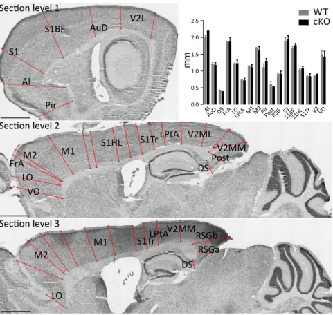
1.1. Cortical Foxp2 ablation does not affect gross cortical morphology 56
1.2. Foxp2 cKO animals do not show gross projection abnormalities 64
1.3. Postnatal development of Foxp2 cKO mice and WT littermates is
1.4. The role of cortical Foxp2 in DA signaling related behavior 66
1.5. Social interaction defects in Foxp2 cortical knockout mice 71
1.6. The role of cortical Foxp2 in modulating ultrasonic vocalizations (USVs) 76
2. Molecular profiling of lower cortical neurons in Foxp2+/- mice 82
3. Autism related gene-Mint2- is downregulated in the cortex of Foxp2 cKO mice 92
Discussion
Cortical cytoarchitecture in Foxp2 mutant mice 95
Prolonged cocaine administration alters locomotor responses of Foxp2 cortical
knockout mice 97
Reduced social approach behaviors in animals with Foxp2 cortical deficiency 99 Modulation of vocal communication in mice carrying a cortical Foxp2 deletion 101 RNA profiling results suggest deregulation of genes involved in social cognition and
behavior 104
General conclusions 106
Future directions
Morphological studies 107
Behavioral studies 108
Publications supporting the thesis
109Altered social behavior in mice carrying a cortical Foxp2 deletion 110
Foxp2 in the nucleus accumbens regulates reward signaling and social behavior 111
List of abbreviations
ADHD attention-deficit/hyperactivity disorder
ASD autism spectrum disorders
cKO Foxp2 cortical knockout mice, line NEX-Cre Foxp2lox/lox
ChIP chromatin immunoprecipitation
CSEA cell-type-specific expression analysis tool
D1R dopamine receptor type 1
DA dopamine
DVD developmental verbal dyspraxia
FTLD frontotemporal lobar degeneration
GO Gene Ontology
IP immunoprecipitation
IPC intermediate progenitor cells
m.o. months old
mdThal mediodolsal thamamus
mPFC medial prefrontal cortex
NuAc nucleus accumbens
RG radial glial progenitors
SLI speech and language impairment
SNP single nucleotide polymorphism
USV ultrasonic vocalizations
VTA ventral tegmental area
Nomenclature: The gene and mRNA are referred to as FOXP2 in humans, Foxp2 in rodents, and FoxP2 in other species (italicised) – for the encoded proteins, these same symbols are used, but they are not italicised (Fisher, 2007).
List of figures
Figure 1. KE family pedigree. 5
Figure 2. FoxP2 evolution in vertebrates. 11
Figure 3. Foxp2 mRNA expression in the embryonic mouse brain at E16.5 and in the newborn
human. 15
Figure 4. Brain circuitries involved in vocalizations in vocal-learning species and mice. 18 Figure 5. Vocalizations structure and social behaviors impaired in FoxP2 deficient animals. 19 Figure 6. Gene Ontology categories of FOXP2 regulated genes in embryonic human frontal
cortex and basal ganglia. 21
Figure 7. Overview of Foxp2 expression in the adult mouse brain. 23
Figure 8. Foxp2 in cortical neurogenesis. 31
Figure 9. Foxp2 is expressed in the deep layers of the cortex. 32
Figure 10. Foxp2 in NuAc is involved in social behavior and reward processing. 37 Figure 11. ICY Colocalizer protocol adapted for Tbr1-Foxp2 colocalization. 44 Figure 12. Experimental procedure for male-male social interaction analysis. 49 Figure 13. Classification of calls by frequency jumps according to Holy & Guo (2005). 51
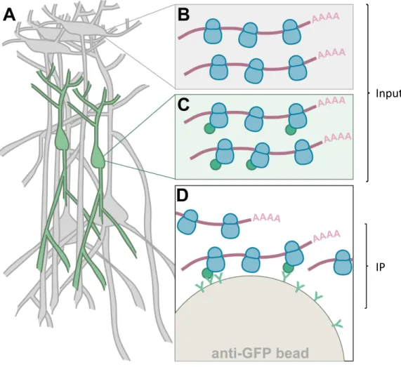
Figure 14. The translating ribosome affinity purification (TRAP). 54
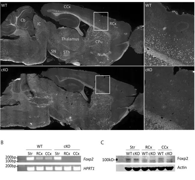
Figure 15. Cortex-specific Foxp2 deletion in the Nex-Cre; Foxp2lox/lox mouse (cKO) line 57
Figure 17. Cells counting. 60
Figure 18. Tbr1 immunostaining and quantification. 61
Figure 19. Tbr1and Foxp2 colocalization in the cortical layer 6. 62
Figure 20. Ctip2 immunostaining and quantification. 63
Figure 21. Foxp2 cKO projection from prefrontal cortex to mediodorsal thalamus appears
indistinguishable from WT littermates. 65
Figure 22. Normal weight gain of cKO animals throughout postnatal development. 67
Figure 23. Sensitization to cocaine. 72
Figure 24. Social behavior alterations in Foxp2 cortical knockout mice in male-male interaction
test. 74
Figure 25. Transitional behavioral graphs. 75
Figure 26. Ultrasound vocalization analysis. 78
Figure 27. Courtship song abnormalities of Foxp2 cKO males. 79
Figure 28. USV abnormalities of Foxp2 cKO animals, detected during same-sex interactions. 81
Figure 29.Validation of the Ntsr1 BacTRAP line. 83
Figure 30. GO categories of differentially expressed genes in the cortex of Foxp2+/- and WT
mice. 89
Figure 31. Cell type specific expression analysis (CSEA) of differentially expressed genes in
Foxp2+/- vs WT cortices. 90
Preface
While animals can develop rather sophisticated communicative systems and cognitive capabilities, human language and accompanying mental abilities are profoundly different from animals and constitute a uniquely human trait. Current hypotheses on human language and cognition imply the existence of dissociable mechanisms, which underlie human linguistic competence. Human language must therefore have evolved, similarly to other complex abilities, through qualitative and quantitative modifications of morphological traits and molecular networks that existed in our ancestors and were the object of natural selection (Pinker and Bloom, 1990; Fitch et al., 2010; Scharff and Petri, 2011).
According to the hypothesis of Chomsky, Fitch and Hauser (2002) the ‘faculty of language’ is based on three basic components: 1) a sensory-motor system which grossly underlies signaling and perception and in linguistics falls into the phonological domain, i.e. the organization of sound system of language 2) a conceptual-intentional system underlying concept formation, expression and interpretation, or in linguistic terms, semantics, which deals with meaning; and 3) a structure-generating system, or syntax, which provides the map between signals (phonology) and concepts (semantics). It is essentially the third component that allows inherent human communicative system, language, to be generative, recursive and virtually limitless and unique. The structure-generating system, however, may have its roots in abstract computational mechanisms and may have evolved for reasons other than language and separately from communicative systems (Chomsky, 1965).
A critical question is whether these components correspond to distinct brain circuits. Based on converging evidence from aphasic and imaging studies, Sakai et al. (2001) proposed a modular specialization of cortical Wernicke’s area, angular gyrus (Brodmann’s area) and Broca’s area which are directly related to phonological, semantic, and syntactic processing. There is further evidence for the involvement of other cortical areas (ex., inferior frontal regions or the left middle temporal regions) in language production, but imaging neuroscience has been unable so far to further discriminate linguistic components from other cognitive elements. Clearly, ‘the language system does
not stand alone, but interacts with other systems of perception, memory, and consciousness, as well as with the speech output system’ (Sakai et al., 2001).
Furthermore communication, at least at the level of the first two components, signals and semantics, implies a strong social component. Social abilities indispensable for language acquisition include a capacity for imitation and vocal learning for the signaling and perception component (phonology), and mind-reading (theory-of-mind) abilities for concepts interpretation, i.e. semantics. These mechanisms are thought to exist in other species and a number of studies have already provided promising insights into the mechanisms of human social cognition (Fitch, 2009; Fitch et al., 2010).
Thus, interdisciplinary cooperation between linguists, evolutionary biologists and neuroscientists is required for understanding the faculty of language (Fitch et al., 2005; Poeppel et al., 2008).
FOXP2 was the first gene to be implicated in a speech and language disorder in humans. Efforts to characterize FOXP2 functional roles at different levels reflect the challenges of investigating biological substrates for speech and language development and evolution. Clinically, FOXP2 mutations were first associated with ‘language grammar’ deficiencies as well as higher cognitive and basic motor abnormalities in humans. Yet FOXP2 expression is not exclusive to humans and is widely present in numerous brain regions and in peripheral organs during development and throughout adulthood. Hence, it is not specifically a ‘speech’ gene, but may have ancestral functions and may even have been selected for reasons other than language. As a transcription factor it controls the expression of a large number of targets. This complexity is increased by the existence of numerous splice variants which, as emerging evidence suggests, might be regulated at the translational level in an activity dependent fashion. Therefore FOXP2 most likely subserves multiple complex roles in brain development and function. Despite the complexity of FOXP2 phenotypic manifestations, different recent lines of research have highlighted the role of FOXP2 in brain structures underlying learning and communication: functional studies on patients carrying mutations in the gene, as described above; association of FOXP2 transcriptional targets with pathologies characterized by strong language components, as for instance Cntnap2, involved in
speech development and ASD; and, characterization of Foxp2 functions in vocal learning in animal models, notably songbirds.
The aim of the present work is to identify the functions of FOXP2 in mouse cortical structures. Such functions may underlie ancestral mechanisms recruited and adapted to human speech and language during evolution.
In the Introduction I present studies supporting the hypothesis that FOXP2 is a molecular entry point for the study of the biological basis of speech and language development and evolution. Further emphasis is put on FOXP2 cortical and cognitive functions.
The first chapter is dedicated to the description of human clinical conditions associated with FOXP2 mutation, as well as mental disorders closely associated with disturbances of FOXP2 controlled molecular networks.
The second chapter describes the potential role of FOXP2 in human evolution and reinforces the idea of FOXP2 being a molecular window into speech, language and cognition.
The third chapter focuses on the function of FoxP2 at the circuitry level, and argues why muroids along with songbirds constitute a valid neurobiological model for studying aspects of FoxP2-related phenotypes. This chapter also describes aspects of cortical morphology in relation to Foxp2-associated cognitive functions.
In the Context I summarize work done in our laboratory to elucidate the role of Foxp2 in dopamine regulated circuits in the Nucleus Accumbens, which could be relevant for the regulation of specific social behaviours.
Introduction
FOXP2 deficiency causes a complex speech and language disorder
The KE family comprised of three generations in which severe speech and language impairment (developmental verbal dyspraxia, DVD) segregated with an autosomal dominant mutation in the transcription factor FOXP2. The mutation is an arginine-to-histidine substitution (R553H) located in the forkhead DNA-binding domain, which disrupts DNA-binding and transactivation properties (Lai et al., 2001; Vernes et al., 2006; Mizutani et al., 2007), (Fig.1).
Although the monogenetic inheritance pattern of the disorder was evident at the time of its first description (Hurst et al., 1990), its nature and clinical diagnosis became a matter of polemics (Fletcher et al., 1990; Gopnik, 1990b), as the manifestation of the dysfunction and the exact language defect appeared to be rather complex.
Linguist M. Gopnik defined the disorder as a dysphasia – i.e. disturbed ability to communicate - due to inability of patients to infer general syntactic rules, like the generation of plurals, tenses, genders. This lack of general grammar rules was especially evident in spontaneous speech, writing or grammatical judgement; however, despite disturbed ability in usage of grammatical constructs, the affected individuals seemed to be able to assign their meaning correctly. Gopnik suggested that the defect was not a consequence of a general cognitive problem, but was specific to grammar domain ‘because the language skills that are not impaired are at least as complex as those which are’ (Gopnik, 1990a).
Investigations of the KE family by Vargha-Khadem et al. concluded that the most prominent phenotype of the disorder is a deficit in the language production-processing system rather than in grammar-processing or morphosyntactic rule formation. The disorder was defined as speech dyspraxia (from ‘praxis’- greek for process, activity), with a central defect in oro-facial motor movements; the extent of the deficit is such that a speech of some affected individuals is unintelligible for the naïve listener. These articulation problems are reflected in a relative immobility of the lower face and mouth and consequently affect oral and facial movements when motor actions are performed
Figure 1. KE family pedigree: filled shapes - affected members, open shapes - unaffected
members, circles-females, squares-males, / - deceased. Difference in cortical activation patterns of affected vs. unaffected family members during covert verb generation task, red arrow points to Broca’s area. FOXP2 protein structure and location of causative mutation R553H: polyQ, polyglutamine tract; ZnF, zinc finger, LeuZ, leucine zipper; FOX, forkhead-box, Acidic, C-terminal acidic tail region domains. Modified from Watkins et al. (2002a), Liegeois et al. (2003) and Vernes et al. (2006).
Pedigree of the KE family
R553H
polyQ ZnF LeuZ FOX Acidic
N C
FOXP2
simultaneously and sequentially (Vargha-Khadem et al., 1995). Accordingly the orofacial dyspraxia can be robustly detected using three tests: word repetition, non-word (i.e. meaningless words) repetition, and simultaneous and sequential oro-facial movements – all three essentially representing a measure of orofacial motor coordination (Vargha-Khadem et al., 1998; Watkins et al., 2002a). The motor phenotypes were instrumental to the precise assignment of affected and unaffected family members for genetic mapping and identification of the underlying FOXP2 mutation. The final confirmation of the causative genomic region came from an individual with a chromosomal translocation unrelated to the family (Lai et al., 2001).
However, intellectual and linguistic deficits are considered part of the condition; examination of affected KE family members using a battery of linguistic tests (13 tasks examining general language functions and 4 specifically addressing grammar) showed impairments in all but one: object naming. Impairments in language tasks included word and sentence repetitions, lexical decision (to define if the word is real), phoneme deletion and addition from words and non-words, non-word spelling and reading, rhyme production; grammatical tests addressed the ability to process inflections (i.e. knowledge of morphosyntactic rules), tense production and receptive knowledge of sentence embedding in the form of relative clauses (Vargha-Khadem et al., 2005).
Moreover, in addition to the pronounced language problems, the authors reported lower IQ scores in the KE family, and concluded that cognitive impairments are not confined to morphosyntax, but rather extend to verbal and non-verbal domain generally (Vargha-Khadem et al., 1995). Permanent lack of appropriate communication endured from early childhood might contribute to a slightly lowered IQ, a secondary effect to DVD. This idea is supported by Watkins et al. (2002a) stating that ‘a disorder may adversely affect the development of intelligence or of the skills required to maintain a given level of intelligence’
Given the potential presence of a cognitive component in the KE family disorder, it was relevant to dissociate it from the motor-articulation deficits, and to identify which brain structures were affected (Liegeois et al., 2003). Therefore a covert verb generation task was used: the participants were asked to generate the verb to a given noun in their
thoughts, without pronouncing it or performing any movement. While performing this task under functional magnetic resonance imaging (fMRI) monitoring, affected KE family members showed underactivation of Broca’s area (classically associated with syntactic aspects of speech) and in Brodmann’s area (semantic aspects), together with atypical patterns of overall cortical activation: higher and diffused activation of the right hemisphere as compared to the left, and posterior compared to anterior (Fig.1). In this task underactivation was also detected in striatal structures (right putamen/globus pallidus), interpreted as reflecting articulation planning impairments. Interestingly, the overt (spoken) verb tasks – generation and repetition – yielded largely similar misactivation patterns, suggesting that at least in these tests the phenotype largely relied on upstream cortical and striatal abnormalities (Liegeois et al., 2003). Consistently with fMRI observations, grey matter volume measurements in structural imaging studies revealed alterations in motor related structures with the most striking effects in striatal areas (caudate nucleus, putamen and globus pallidus), but also in cerebellum (ventral and posterior lobe); several cortical areas were affected morphologically as well: Brodmann’s and Broca’s areas, cingulate, sensorimotor, motor, posterior temporal, anterior insular, medial occipitoparietal and inferior frontal cortices (Vargha-Khadem et al., 1998; Watkins et al., 2002b; Belton et al., 2003). Comparison of affected KE-family members to patients with acquired Broca’s aphasia (i.e. a collection of language disorders caused by damage to the brain, in this case a left hemisphere stroke) was employed to partially discriminate cortical-driven and striatal aspects of the performance: the difference between the two conditions was detected only within the tests of verbal fluency and lexical decisions, but surprisingly did not seem to lie within oral praxis or receptive and expressive language deficits (Watkins et al., 2002a).
Ackermann (2014) discussed fundamental differences in the profiles of speech motor deficits in verbal dyspraxia associated with FOXP2 mutations versus Parkinsonian motor speech disorder, caused by basal ganglia dysfunctions. Acquired impairments in speech performance due to striatal damage are characterized by execution of orofacial movements with ‘undershooting’ gestures and disruptions of prosodic aspects of verbal utterances (i.e. rhythm, stress, and intonation), while communication disorders due to
fronto-opercular cortex or anterior insula damages in the language-dominant hemisphere resemble FOXP2 phenotypes to much greater extent (Ackermann et al., 2014).
Hence, although it is difficult to conclude which language deficit in FOXP2 deficient patients arises primarily, misarticulation or linguistic, at least in adults the primary problem may lie upstream of the motor system, and has a significant contribution from cortical dysfunctions.
Characterization of the KE family was followed by reports of other patients showing similar phenotypes with FOXP2 loss-of-function mutations and disruptions of genomic regions residing in the vicinity of the gene (O'Brien et al., 2003; MacDermot et al., 2005; Lennon et al., 2007; Tomblin et al., 2009; Palka et al., 2012).
Disruptions of FOXP2 chromosomal region (7q31) have been linked to a spectrum of neurodevelopmental disorders, such as autism and schizophrenia (as well as major depression, dyslexia, FTLD, ADHD); these pathologies exhibit some level of association with FOXP2 polymorphisms, often in parallel with the expressivity of language endophenotypes (Gong et al., 2004; Li et al., 2005; Sanjuan et al., 2005; Sanjuan et al., 2006; Padovani et al., 2010; Tolosa et al., 2010; Schaaf et al., 2011; Spaniel et al., 2011; Ribases et al., 2012; Wilcke et al., 2012; Li et al., 2013; Corominas et al., 2014; McCarthy-Jones et al., 2014). However, data on the direct genetic association between FOXP2 and many of the aforementioned disorders remain controversial (Newbury et al., 2002; Gauthier et al., 2003; Laroche et al., 2008).
A molecular link between FOXP2 and mental disorders has been established in functional genomics studies through a number of genes and pathways involved in a several distinct mental pathologies (Spiteri et al., 2007; Vernes et al., 2007; Konopka et al., 2009; Vernes et al., 2011). Specifically, among the best described genes directly regulated by FOXP2 are:
CNTNAP2 – a gene substantially enriched in frontal gray matter and associated with autism, Tourette’s syndrome and severe recessive disorder involving cortical dysplasia and focal epilepsy (Vernes et al., 2008)
MET - component gene involved in human temporal lobe development, associated with autism (Mukamel et al., 2011)
DISC1 - a leading candidate susceptibility gene for schizophrenia, bipolar disorder and recurrent major depression, which has been implicated in other psychiatric illnesses of neurodevelopmental origin, including autism (Walker et al., 2012).
SPRX2 - a gene responsible for speech dyspraxia and mental retardation which accompany a form of sylvian epilepsy (Roll et al., 2006), which independently links SPRX2 to speech and language
uPAR - an interaction partner of SPRX2 which is associated with autism spectrum disorder (ASD) (Eagleson et al., 2010; Roll et al., 2010).
FOXP2 may be linked to autism and intellectual disability via interacting partners such as FOXP1 (Bacon and Rappold, 2012) and TBR1. The latter is a recently described autism candidate gene (Deriziotis et al., 2014) and protein-interaction studies of ASD susceptibility genes have shown a direct interaction with FOXP2(Sakai et al., 2011; Corominas et al., 2014).
Thus, several lines of evidence strongly suggest that FOXP2 is part of larger molecular networks underlying distinct language and social cognitive dysfunctions
To summarize, human studies on FOXP2 gene function suggest that it is involved in complex motor phenotypes of speech and language processing. In addition, FOXP2 role in both semantic and syntactic aspects of language is supported by pronounced defects in grammar rules formation and problems in lexical decision-making, in agreement with morphological abnormalities in the cortex including Broca’s and Brodmann’s area. Evidence for FOXP2 role in phonological domain of language derives from recent studies on healthy individuals. FOXP2 polymorphisms were shown to contribute to the normal inter-individual variability in hemispheric asymmetries and frontal cortex activation patterns for speech and language audio (dichotic listening task), and also visual (reading tasks) perception (Pinel et al., 2012; Ocklenburg et al., 2013). Cortical lateralization impairments, characteristic of FOXP2 dysfunction, are well described in language and literacy per se and often diagnosed in autism and speech and language disorders (SLI) (De Fosse et al., 2004; Bishop, 2013). Co-morbidity of FOXP2-based language and molecular phenotypes with a variety of mental disorders, ASD in particular, hints to FOXP2 involvement in more complex cognitive functions than simple processing of
certain aspects of language. In this respect Corballis (2004) suggested a link between FOXP2 and the mirror neuron system, which underlies many critical symptoms of autism. ‘Thus, while studies of FOXP2 can offer insights into relevant neural pathways, it is not a ‘gene for speech’.’- Fisher (2007).
Foxp2 as an entry point to study molecular and neural networks contributing to cognitive aspects of speech and language
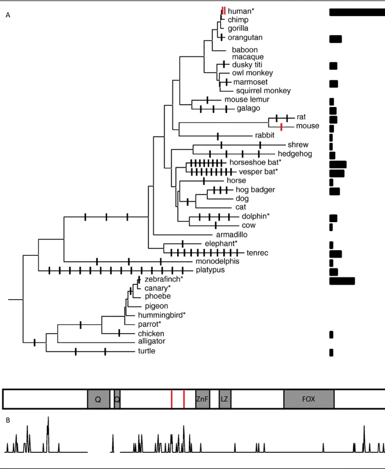
The discovery of FOXP2 as the first gene involved in speech and language development led to studies on its role in human evolution. Using comparative analysis of human, chimpanzee, orangutan and mouse DNA sequences, two independent groups established that FOXP2 is a remarkably conserved gene that was the object a selective sweep in the human lineage. Positive selection in the coding sequence is thought to have converged on two amino acids within exon 7 that distinguish humans from other primates, while only three amino acids substitutions differentiate mouse Foxp2 from humans (Enard et al., 2002; Zhang et al., 2002) (Fig. 2). Further investigations suggested that selection pressure may have been exerted beyond the two human specific substitutions of the coding FOXP2 region, on sites located near the gene (Ptak et al., 2009; Maricic et al., 2013). Genome-wide scans for positively selected genes during mammalian evolution identified only 5 transcription factors in the hominid branch with a selection rate comparable to FOXP2 and none of them was associated with brain function, but with spermatogenesis and the immune system (Kosiol et al., 2008; Enard, 2011).
Enard proposed that a murine model, carrying two ‘human’ amino acid substitutions within Foxp2, could provide valuable information of the evolution of mammal circuits underlying speech and language in humans (Enard, 2011). Therefore, he and his colleagues have introduced the two human specific substitutions in the orthologous mouse locus, generating a homozygous knock-in Foxp2 ‘humanized’ mouse (Foxp2-Hum) (Table 1) (Enard et al., 2009). Although Foxp2 is expressed in vitally important organs such as heart, lung and guts (Shu et al., 2001), the exhaustive screen for almost 300 different phenotypic parameters did not produce any evidence for effects of the
Figure 2. FoxP2 evolution in vertebrates. A, Amino acid changes outside the polyglutamine tracts
(Q) are mapped on the phylogeny of FoxP2 sequences from vertebrates. The bars on the right site depict the ratio of amino acid changes to the length of the terminal branches. Asterics indicate species with evidence for vocal learning. B, Amino acid changes in the tree are plotted for each
position of the human FOXP2 protein sequence. ZnF, zinc finger; LZ, leucine zipper; FOX, forkhead-box domains, the two human amino acid changes are shown as red lines. Adapted from Enard (2011).
FOX ZnF LZ
A
Foxp2Hum allele in any organ system except the central nervous system. Here, anomalies were revealed in behavioral, electrophysiological, histological and molecular parameters within the brain. Furthermore, such anomalies were most prominent within cortico-striatal circuits (Enard et al., 2009; Reimers-Kipping et al., 2011; Schreiweis et al., 2014).
These findings are in agreement with the cortico-striatal core of phenotypes in affected KE-family members. The authors suggested that human-specific mutations in Foxp2 in mice could model aspects of speech and language evolution in humans (Enard et al., 2009).
The neural circuits underlying uniquely human functions, involving abstract thinking and the cognitive mechanisms of language processing (as, for example, syntax), are located in the cortex. This brain structure expanded significantly in the human lineage relative to other primates, developing new regions in the frontal and parieto-temporal lobes. Cortical volume expansion is mechanistically based on increases in interecellular space and cell number, due to higher cortical neurogenesis, enhanced elaboration of dendritic trees and expansion of neuropil areas, especially in the prefrontal cortex (Geschwind and Rakic, 2013).
As discussed below (in Foxp2 cellular functions, Foxp2 in the mouse cortex), FoxP2 is strongly engaged in processes of neurite outgrowth and cortical neurogenesis (Spiteri et al., 2007; Vernes et al., 2007; Vernes et al., 2011; Tsui et al., 2013; Chiu et al., 2014); in this light it is interesting to examine how FOXP2 role in cortical evolution is gradually emerging from comparative genomic studies:
When addressing evolution of the coding genome at the single nucleotide level it is accepted that brain-expressed genes are not specific for positive selection in human lineage (Wang et al., 2007; Geschwind and Rakic, 2013). However, FOXP2 appears to be one of the salient exceptions, as inferred from Enard’s studies described above, as well as from the analysis of Neanderthal and Denisovan genomes sharing the human-derived form of FoxP2 right before the lineages split (Krause et al., 2007; Meyer et al., 2012). Many human brain-specific evolutionary accelerated changes lie within non-coding nucleotide stretches, i.e. enhancers and promoters (Capra et al., 2013). Visel et al. in their comparative study of conserved non coding regions relevant to telencephalic evolution,
found a dozen of elements in close and distant proximity of FOXP2, as well as many more close to the genes constituting FOXP2 molecular network. Among these is the highly conserved transcription factor POU3F2, which has been suggested as candidate for having caused a recent selective sweep of FOXP2 in the human lineage (Maricic et al., 2013; Visel et al., 2013). In a separate study using comparative epigenetic profiling of human, mouse and macaque corticogenesis to identify enhancers which gained activity in human evolution, FOXP2, although not identified as one of the hub genes, showed substantial connectivity to gain-enriched modules of co-expressed genes(Reilly et al., 2015).
Konopka et al. performed comparative analyses of transcriptomes from the telencephalon of human, chimpanzee and macaque telencephalon. They demonstrated a striking increase in transcriptional complexity specific to the human lineage in the frontal lobe, while genes expression in the caudate nucleus was conserved. In particular, the human prefrontal cortex is enriched in alternatively spliced genes involved in neuron projections, neurotransmitter transport, synapses, axons, and dendrites, as well as genes implicated in schizophrenia. Among the most differentially connected genes, this module contained FOXP2, and 13 genes that overlap with previously identified FOXP2 targets, as well as its dimerization partner - FOXP1 (Konopka et al., 2012).
Taken together comparative genomic data support the hypothesis that FOXP2 was actively recruited in the formation of the most evolved structures of the human cortex during hominin evolution.
If the human telencephalon is a structure, providing unique capacities, how could we approach its origins? Insights might come from the application of the concept of ‘deep homology’, stating that new structures do not arise de novo. Instead there is a continuum of events which follows an evolutionary constraint imposed by pre-existing genetic regulatory circuits, which were initially established in early metazoans. Classical examples of deep homology are the re-appearance of eyes in widely divergent organisms, based on similar genetic components such as ciliary phototransduction and melanogenic pathways, or tetrapod limbs and fish fins which are both based on deeper homology in the network of Hox genes (Shubin et al., 2009). When evolutionary constraints are further restrained by the structure, composition and dynamics of the developmental system (the
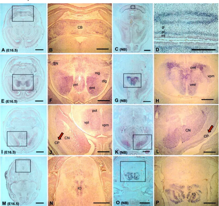
fusion of these two fields is also called ‘evo-devo’ concept) there is even less room for deep phenotypic variability. The concepts of ‘deep homology’ and ‘evo-devo’ are classically applied to evolution of structures and forms, but could be adapted to neural circuits and behaviours (Robinson et al., 2008; Scharff and Petri, 2011). In the case of FoxP2, genomic sequence, developmental expression pattern and molecular network are conserved in distant mammals (such as human and mice) (Lai et al., 2003)(Fig.3). Within this framework mouse Foxp2 could serve as an entry point to study ancestral substrates underpinning complex aspects of FOXP2-dependent human cognition.
The study of animal models provides insights into conserved FoxP2 functions
The role of FoxP2 in development
The speech and language deficits in individuals carrying genetic aberrations of FOXP2 clinically manifest already in childhood (Hurst et al., 1990; Vargha-Khadem et al., 1995). The presence of developmental vocal learning deficits is supported by performance differences in tests of repetition of words with known meaning between adult affected KE family members and patients with acquired aphasia: the latter, who had a normal cognitive and linguistic development prior to the trauma leading to aphasia, performed significantly better than the former (Watkins et al., 2002a; Vargha-Khadem et al., 2005).
Histological examinations of postmortem brains support Foxp2 role in nervous system formation, in human and mice, and the patterns of expression are highly conserved throughout development in both species (Fig. 3). Functional genomics data on Foxp2 targets obtained on embryonic human and mouse tissue supports and further elucidates mechanisms of Foxp2 role in CNS formation (discussed in more detail below in FoxP2 cellular functions) (Spiteri et al., 2007; Vernes et al., 2007; Vernes et al., 2011).
Foxp2 specifically plays a role in cortical development, affecting distinct stages of neuronal maturation (Clovis et al., 2012; Tsui et al., 2013; Chiu et al., 2014)(Foxp2 in the mouse cortex, Fig.7). Complete deletion of Foxp2 in mice is lethal: homozygous Foxp2 mutant mice display reduced weight gain, aberrant manifestation of developmental reflexes and die within 4 weeks after birth (Groszer et al., 2008), Table 1.
Figure 3. Foxp2 mRNA expression in the embryonic mouse brain at E16.5 and in the newborn
human. Sequential transverse sections, from anterior to posterior. Cortical plate (CP) Foxp2 distribution is marked with a red arrow. Adapted from Lai et al. (2003).
FoxP2 conserved role in the ontogenesis of nervous system is illustrated by its expression in zebrafish, with a well preserved pattern in pallium and subpallium - telenecephalic structures (Bonkowsky and Chien, 2005). In songbirds, a model which is extensively used in FoxP2 research due to its vocal learning abilities, FoxP2 is expressed in pallium, striatal and thalamic-like structures throughout development, with a prominent expression in Area X – a structure which intermingles striatal and pallidal components and is actively involved in vocal performance (Teramitsu et al., 2004). In Area X FoxP2 mRNA show increased expression during a particular segment of time when young birds actively learn how to imitate song from a tutor (Haesler et al., 2004), decreases upon brain maturation as song acquires more stereotypy (Thompson et al., 2013) and is necessary for adequate song acquisition (Haesler et al., 2007).
Comparing FoxP2 expression patterns in vocal-learners and non-learners in-between avian, reptilian and mammalians, Haesler et al (2004) proposed that FoxP2 is an ancient transcription factor involved in shaping of cerebral architecture of striatal sensory and sensory-motor circuits to generate a permissive environment for vocal communication evolution and development of vocal learning. This hypothesis is in accordance with data arising from primate research and from a variety of human language disorders, suggesting that striatal control underlies the entrainment and automatization of speech motor patterns during speech acquisition, while mature verbal communication requires less striatal processing capacities and relies on the left-hemisphere peri- or subsylvian cortex (Ackermann et al., 2014).
Activity dependent function of FoxP2 in mature brain: evidence for a role in social behaviour and vocalizations
FoxP2 expression persists throughout development into the mature adult brain (Ferland et al., 2003), suggesting activity-dependent function. Evidence for the role of FoxP2 in the adult brain is primarily coming from the study of songbirds and mice, and it appears to be tightly linked to the ability to learn and to modify the accuracy of socially-driven vocalizations upon involvement of the dopaminergic system (Murugan et al., 2013).
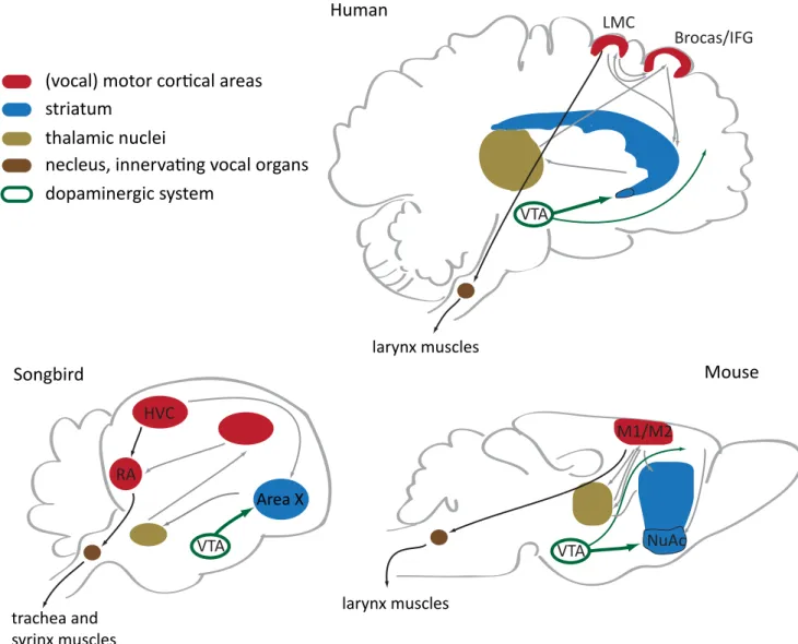
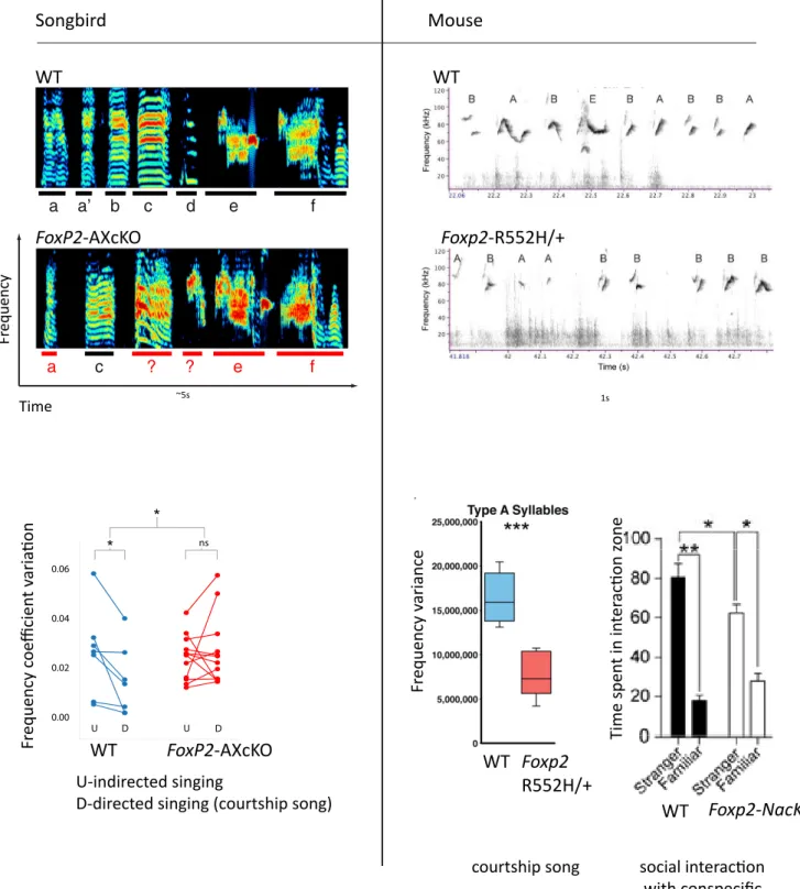
In zebra finches song learning and modulation is mediated by loops involving a set of nuclei homologous to cortex(pallium)/striatum/thalamus - like nuclei, which share many characteristics with mammalian cortico-striatal circuitry (Fisher and Scharff, 2009) (Fig.4). The organization of the precise timing signal for motor control during singing is thought to be governed by premotor (HVC) and motor (RA) pallial nuclei. RA establishes a direct connection with vocal motor neurons in the brainstem. This direct motor cortex connections may enable voluntary vocal control and vocal-learning abilities in particular species in distinct animal clades (Fig.2); it is absent in monkeys, but present in humans and have recently been described in mice (Jurgens, 2002, 2009; Arriaga and Jarvis, 2013). Before being transferred to downstream motor structures, the timing signal undergoes a social context-dependent modulation in the divergent striatum-thalamo-cortical loop (Fig. 4), where Area X is a prominent hub. Area X shows lower Foxp2 mRNA and protein levels in the absence of social context (Miller et al., 2008). For zebra finch males the social context is defined by the presence of the female to whom structured and well organized song is presented (directed song), while greater variability (i.e. the song is more enriched in motifs, syllable types and frequency range) is observed when males are singing alone (undirected song). FoxP2 knockdown in Area X of adult birds abolishes refinement of the directed song (Fig. 5) by rendering the timing signal insensitive to modulation by dopamine receptor 1 (D1R) and dramatically decreasing the levels of dopamine-regulated neuronal phosphoprotein DARPP-32, a key dopamine signaling regulator (Teramitsu and White, 2006; Murugan et al., 2013).
In mice, the first evidence for Foxp2 activity-dependent expression came from studies of plasticity of auditory thalamic nuclei (MGN) , where Foxp2 is upregulated after a high volume white noise stimulation (Horng et al., 2009). Analysis of four major neuromodulators in Foxp2 mutant mice showed that dopamine (DA) levels are dramatically affected in cortico-striatal structures, and serotonin levels in nucleus accumbens (NuAc) (Enard et al., 2009). In striatum and cortex nearly all Foxp2+ neurons co-express DARPP-32 (Hisaoka et al., 2010; Vernes et al., 2011). With Cedric Mombereau in our team (Mombereau et al., submitted) we assessed the potential contribution of Foxp2 to DA signaling using pharmacological stimulation with cocaine, a
Figure 4. Brain circuitries involved in vocalizations in vocal-learning species and mice. Remarkably, all the depicted forebrain structures express FoxP2. Black arrows indicate cortical projections that within one synaptic switch innervate vocal organs – this direct motor connection is thought to be exclusive for vocal learning species. LMC, laryngeal motor cortex; IFG, inferior frontal gyrus; VTA, ventral tegmental area; M1, primary motor cortex; M2, secondary motor cortex. Figure is modified from Arriaga et al. (2012).
RA Area X HVC VTA trachea and syrinx muscles M1/M2 larynx muscles VTA NuAc VTA LMC Brocas/IFG larynx muscles Human Songbird Mouse dopaminergic system thalamic nuclei striatum
(vocal) motor corical areas
Figure 5. Vocalizations structure and social behaviors impaired in FoxP2 deficient animals. Upper
panels depict typical sonograms obtained from animals under normal conditions and after FoxP2 disruption. Bottom graphs illustrate frequency alterations in a courtship song of both species as well as diminished social interest revealed in 3-chamber test on NuAc knockout (Foxp2-NacKO) male mice. FoxP2-AXcKO, FoxP2 shRNA knockdown in Area X; Foxp2-R552H/+, heterozygous mutant, carrying Foxp2 an amino acid substitution identical to the one found in the KE family. Adapted from Arriaga (2011),Mombereau et al. (sumbitted) and Murugan et al. (2013).
Songbird WT FoxP2-AXcKO Time Fr equency Mouse WT Foxp2-R552H/+ ~5s 1s 0.00 0.02 0.04 0.06 U D U D ns * * U-indirected singing
D-directed singing (courtship song)
Fr equency c oe fficien t v aria ion FoxP2-AXcKO WT courtship song Fr equency v ariance social interacion with conspecific Time spen t in in ter acion z one WT Foxp2 R552H/+ WT Foxp2-NacKO
drug which recruits dopaminergic pathways. After stimulation of Foxp2 heterozygous mice, we localized a specific underactivation pattern to D1R+ neurons of NuAc (ventral striatum nuclei). Foxp2 protein levels in WT NuAc showed significant downregulation after one cocaine injection, correlating with cocaine-induced locomotor behavior attenuation in conditional Foxp2-NuAc knockdown mice. As NuAc is a structure particularly implicated in reward-associated learning and Foxp2-NuAc knockdown mice showed a decreased preference specifically for social situations (time spent in the chamber with conspecific WT) (Fig. 5, Fig. 10 and Context), our study suggests that Foxp2 regulates DA-mediated social reward signaling.
Some evidence exists for speech and language modulation by DA in humans. Neurological and psychiatric studies indicate that dopamine receptor antagonists disrupt vocal motor control and lead to the development of uncontrolled laryngeal spasms. Voice, articulation, phonological processing and syntactic complexity deterioration in Parkinsons disease patients - a disorder of dopaminergic neurons loss - is the most striking example of DA-dependent voice and speech problems. Similarly, alterations in DA levels or in nigrostriatal dopamine release are linked to speech related defects as diverse as vocal tics in Tourette’s syndrome, auditory hallucinations in schizophrenia and stuttering (Simonyan et al., 2012).
FoxP2 cellular functions
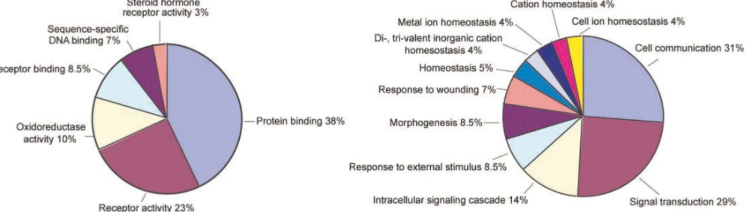
The identification of FOXP2 transcriptional targets has provided valuable insights into regulated cellular processes. High-throughput approaches such as FoxP2 chromatin-immunoprecipitation, coupled with gene expression profiling in cell cultures transfected with FoxP2 variants, or mouse and human embryonic material have been used (Spiteri et al., 2007; Vernes et al., 2007; Konopka et al., 2009). Differentially expressed direct and indirect targets have been functionally annotated using ontology categories defining specific biological processes. Gene Ontology (GO) categories characteristic for developmental FOXP2 targets in humans (Fig. 6) largely overlap with mouse GO categories. Detailed analysis of mouse data complements with the categories of neurogenesis, neuron projection morphogenesis, cell localisation functions as well as synaptic plasticity and spine formation (Vernes et al., 2011). These functions are
Figure 6. Gene Ontology categories of FOXP2 regulated genes in embryonic human frontal cortex
and basal ganglia. Adapted from Spiteri et al. (2007).
supported by studies of Foxp2 mutant mice in vivo with pronounced affects on development and function of cortico-striatal circuits (Groszer et al., 2008; Enard et al., 2009; Schulz et al., 2010; Chiu et al., 2014).
Foxp2 role in cortical neurogenesis is well described in mice, where Foxp2 ectopic misexpression in cortical progenitors impairs in vivo radial migration, neurite maturation and synaptogenesis (Clovis et al., 2012; Sia et al., 2013), and affects progenitors type transitions during neuronal differentiation (Tsui et al., 2013; Chiu et al., 2014)(Fig.8). FoxP2 specific role in neurogenesis is further supported by studies in chicken and zebra finch (Rousso et al., 2012; Thompson et al., 2013).
In zebra finches, the major functional modules associated with singing and regulated by FoxP2 relate to synaptic plasticity (more specifically, the suppression of postsynaptic plasticity) via FoxP2 interconnection with genes linked to MAPKK and NMDA receptors function, actin cytoskeleton regulation, and tyrosine phosphatase (Hilliard et al., 2012).
Cocaine stimulation of heterozygous mice (Foxp2-R552H/+-Enu, Table1) revealed neuronal activity alterations in NuAc mediated by genes involved in calcium signaling, among which voltage-dependent calcium channel alpha 1G (Cacna1g) has been genetically associated with ASD, providing further support for the role of Foxp2 in social behavior (Mombereau et al., submitted).
Mouse models in Foxp2 research: motor learning and ultrasound vocalizations Foxp2 expression pattern is highly conserved across four species of muroid rodents: two species of singing mice (Scotinomys teguina and S. xerampelinus), their close relative the deer mouse (Peromyscus maniculatus), and more distantly related laboratory mouse Mus musculus (Campbell et al., 2009).
Foxp2 distribution in the mouse brain is rather broad and extends to lower cortical layers, striatum, mesolimbic and nigrostriatal dopaminergic systems, thalamic somatosensory areas, Purkinje cells of cerebellum and inferior olivary complex, olfactory system and ascending auditory and visual relays (Fig. 7) (Ferland et al., 2003; Campbell et al., 2009). This expression pattern suggests that Foxp2 is unlikely to regulate circuits selectively governing verbal or vocal functions, but rather specifically interacts with genetic factors that define relevant circuits and pathways. The relevant functional
Figure 7. Overview of Foxp2 expression in the adult mouse brain. Highlighted structures
comprise the circuitry of striatal modulation of fine motor response, suggesting a role in motivational and integrative circuits, rather than in the direct regulation of motor output. AOB, accessory olfactory bulb; BST, bed nucleus of stria terminalis; Cb, cerebellum; CPu, caudate putamen; Ctx, cortex; IO, inferior olivary complex; LS, lateral septum; MeA, medial amygdala; MOB, main olfactory bulb; MPA, medial preoptic area; NAcc, nucleus accumbens; SNc, substantia nigra pars compacta; STh, subthalamic nucleus; Tu, olfactory tubercle; VTA, ventral tegmental area. Adapted from Campbell et al. (2009).
circuitry in adult mice has been suggested to lie within networks controlling fine motor output, multimodal sensory processing and sensorimotor integration (Campbell et al., 2009).
A concise overview of the existent genetically modified Foxp2 mouse models and their basic phenotypes has been provided in a recent review by French and Fisher (2014),
Table 1.
Motor learning
Because of the known relevance of cortico-striatal motor circuitry for auditory guided vocal communication (Wohlgemuth et al., 2014), supported by high conservation of striatal circuitries among vertebrates, most attempts to experimentally access Foxp2 functions in mice have used motor learning paradigms. The heterozygous loss-of-function model, bearing KE family etiological mutation: Foxp2-R552H/+-Enu (Table 1), manifests motor learning impairments in three distinct assays: voluntary-controlled running wheel, automatically accelerated rotarod and auditory-motor associations learning (accessing auditory-motor integration impairments potentially relevant for speech acquisition); the latter approach also revealed a worse learning curve in another heterozygous model - Foxp2-S321X/+. Striatal electrophysiological abnormalities were observed in Foxp2-R552H/+ mice, including reduced cortico-striatal long-term depression (LTD) in brain slices, and abnormally high ongoing striatal activity along with dramatic alterations of striatal plasticity during motor skill acquisition in vivo (Groszer et al., 2008; French et al., 2012; Kurt et al., 2012).
Foxp2 humanized mice (Foxp2-Hum) a model which effectively manifests gain-of-function profile, show enhanced striatal LTD compared to WT and Foxp2 heterozygote mice (Enard et al., 2009; Reimers-Kipping et al., 2011). Furthermore, humanized mice demonstrate faster proceduralization of action sequences in conditional T-maze paradigm with spatial cues. This learning paradigm accesses declarative (place-based) learning transitions to procedural (response-based) learning, and reveals the speed of information transfer from dorso-medial to dorso-lateral striatum (Schreiweis et al., 2014).
Table1. Genetically modified mouse lines (modified from French and Fisher (2014)).
Mouse line Disruption Basic phenotypes
Foxp2-KO
Removed the FOX domain yielding knockout mice.
Homozygotes die by 3 weeks of age. Heterozygotes show mild developmental delay, but normal performance in Morris water maze memory and learning paradigm.
Foxp2-R552H-KI
Knockin point mutation in an Arg-to-His substitution in the FOX domain of the encoded protein. This
substitution is found in affected members of the KE family (R553H).
Homozygotes die by 3 weeks of age. Some heterozygotes show mild-moderate developmental delay.
Foxp2-R552H-Enu
Mice with Arg-to-His substitution seen in the KE family, isolated from an ENU-mutagenesis screen.
Homozygotes die at 3-4 weeks of age. Heterozygotes are overtly normal.
Foxp2-S321X
Mice with a point mutation, isolated from an ENU-mutagenesis screen. Results in a premature stop codon, shown to be equivalent to a null allele (no protein). This mutation is found in a second family segregating SLI.
Homozygotes die at 3-4 weeks of age. Heterozygotes are overtly normal.
Foxp2-N549K
Mice with a point mutation isolated from an ENU-mutagenesis screen. Results in an Asn-to-Lys substitution in the FOX domain.
Homozygotes survive into adulthood (3-5 months age) with severe motor problems. Heterozygotes are overtly normal.
Foxp2-Flox
LoxP sites inserted around exons 12-14 to facilitate Cre-mediated removal of the FOX domain.
When crossed to the global Sox2-Cre line, homozygotes die at 3-4 weeks of age and heterozygotes are overtly normal.
Foxp2-Hum
Knockin strategy used to modify exon 7 and to introduce flanking LoxP sites. Results in 2 changes (T302N and N324S), where the amino acids found in the mouse are substituted for the orthologous human amino acids, partially humanizing the encoded protein.
Both homozygous and heterozygous humanized mice are overtly normal. Removal of exon 7 using a global Cre line results in a knockout phenotype. Homozygous knockouts die postnatally and heterozygous knockouts are overtly normal.
Interestingly, genetic manipulation of single drosophila ortholog - FoxP - seems to be responsible for operant self-learning abnormalities in this species, pointing to deeply ancient FoxP protein family mechanisms, underlying clue-based motor sequence acquisitions (Mendoza et al., 2014).
Ultrasound vocalizations
Campbell et al (2009) compared Foxp2 brain distribution in two species of singing mice, characterized by highly complex and structured social vocalizations which require tight control of facial musculature, and related species, including lab mice. In this study the authors were looking for the morphological parameters which could explain the particular vocalization capabilities of singing mice , however no evident differences between species were detected, but only minor random fluctuations within limbic forebrain and cortex. Thus, the function of Foxp2 in vocal communication does not seem to extend to mouse species differences in articulatory and acoustic complexity and is fundamentally conserved.
Mus musculus elicits two types of calls: audible, which often signals a distress situation, and ultrasonic vocalizations (USVs). USVs study is a rapidly developing field and a promising tool for analysis of communicative and social behaviour alterations in laboratory mice (Ey et al., 2011; Ey et al., 2013). Holy and Guo (2005) demonstrated that a mouse USV can be not only recognised as a song enriched with multiple syllables types, but also has non-random structure of syllable sequences, organised into motifs and phrases. Although mouse songs are not as elaborate as birds ones (Fig 5), it is generally accepted that mice are able to modify their song structure according to the social context to the extent that it becomes possible to talk about ‘mouse syntax’ and ‘prosody’ (Guo and Holy, 2007; Lahvis et al., 2011; Chabout et al., 2012; Chabout et al., 2015).
Whether mice are able to imitate and learn specific vocal patterns like songbirds and humans is a matter of debate (Arriaga and Jarvis, 2013; Portfors and Perkel, 2014). It is clear, however, that pup vocalizations are innate since newborn are deaf for half of their maturation period, and likely represent an isolation signal in order to elicit maternal care. The structure of this calls changes through development, changing frequency
characteristics, call length and phrasal structure and overall becoming more complex (Musolf and Penn, 2012).
Speech and language impairment in the KE family is a neurodevelopmental disorder. Therefore, initial studies in Foxp2 mouse models focused on pups vocalizations. Complete absence of functional Foxp2 in homozygous mice correlated with dramatic decrease in pup vocalizations along with loss of weight, severe motor abnormalities, lack of spontaneous activities, and delay of developmental reflexes maturation. Thus, the absence of vocalizations could have been due to somatic weakness and general arousal problems. Indeed, when homozygous pups were stimulated by tail lifting - a stressful condition - both WT and mutants emitted comparable number of calls (audible clicks and USV). Heterozygous Foxp2-R552H-Enu pups exhibit normal USV rates with normal acoustical parameters (call duration, power, minimal and maximal frequencies) along with overall normal developmental parameters; heterozygous Foxp2-R552H-KI and -KO mice showed reduced rate of calls accompanied with weight reduction and developmental delays. Thus, the general conclusion is that Foxp2 partial loss-of-function does not affect innate vocalization production (Shu et al., 2005; Fujita et al., 2008; Groszer et al., 2008; Gaub et al., 2010).
Homozygous humanised mouse pups were studied through several developmental stages, which also covered later stages than those studied for heterozygous mutants. An initial analysis showed that normal basic call parameters, such as number of calls and interval duration, were unaffected. However, further analyses were based on structural classification in syllables types, distinguishing short and long-lasting calls with no frequency jumps (i.e. ‘simple’ calls, the prevalent types) and calls with frequency jumps (‘complex’ calls). All call types showed differences in various acoustic parameters (including mean, minimum and maximum of the frequency), however in opposite directions: while simple calls scored lower, calls with frequency jumps were higher in Foxp2-Hum versus WT. In addition, complex calls duration was significantly increased in humanised mice (Enard et al., 2009).
Altogether, it is difficult to compare results obtained in different Foxp2 models due to varying experimental designs and confounding factors of developmental delays. Enard’s study, however, suggests that Foxp2 gain-of-function mutation might have changed fine
acoustic parameters in mouse vocalisations. However, it is difficult to conclude whether these changes relate to innate vocalisations or to learned and modifiable ones, since the analysis in the study covered a rather long developmental period. The ecological significance of these changes remains unknown.
To date, the role of Foxp2 in adult mouse vocalizations is less well studied. Arriaga analyzed Foxp2-R552H male mice singing in response to female urine, sometimes designated as courtship song (Arriaga, 2011). The song analysis in this study distinguished simple calls with no frequency jumps (type A), and eleven more types of calls with varying frequency jump architecture. Call rate, repertoire and fractions of calls did not change in comparison to WT littermates. However, when the focus was shifted to spectral characteristics of the most abundant type A syllables, multiple frequency parameters appeared to be altered: starting, minimum, ¼ calls frequencies were significantly higher in heterozygous animals (however, mean and maximum frequency did not differ); in addition, a number of characteristics effectively reflecting frequency range distribution were decreased in heterozygotes. The frequency alterations were opposite to those observed in Foxp2-Hum pups, which is in concordance with previous studies (Enard et al., 2009).
The presence of frequency range alterations in Foxp2 mouse mutants resembles songbird striatal FoxP2 knockdown in Area X during directed singing, although, in opposite direction (Fig. 5).
Notably, FoxP knockdown in drosophila alters courtship behaviour and renders courtship male ‘pulse song’ (i.e. a vibration of a wing for production of trains of pulses) faster with more variable inter-pulse intervals, longer pulse song bouts and generally reduced percentage of time spent singing. These alterations occur together with motor coordination problems. Nevertheless, the findings suggest a deep functional homology with vertebrates in courtship communication behaviour (Lawton et al., 2014).
Foxp2 in the mouse cortex
The mammalian neocortex consists of six main layers of neurons which differ in cellular morphology, connectivity and physiological properties. Upper layers (L2/L3), contain pyramidal neurons which project intracortically; layer 4 contains pyramidal and
granule neurons and serves as sensory information relay as it receives a bulk of thalamocortical axons; layer 5 is an output layer with a variety of projection targets, including corticobulbar and corticospinal projections; cortical layer 6 contains the largest diversity of morphological cell types with not well defined functionality, receiving and sending projection to the thalamus and within the cortex (Briggs, 2010; Kwan et al., 2012). Layer 6 is remarkably enriched in Foxp2 through all the areas of the cortex, in both human and mice; Foxp2 is sparsely present in layer 5 as well (Ferland et al., 2003; Campbell et al., 2009; Hisaoka et al., 2010). In the developing cortex, Foxp2 is expressed in cortical progenitor cells throughout neurogenesis (Ferland et al., 2003; Takahashi et al., 2003; Tsui et al., 2013).
Cortical development occurs in inside-out fashion, with lower cortical layer arriving first and upper cortical neurons gradually migrating through the lower ones. Maturation of cortical neurons starts after embryonic developmental day 11 (E11) in mice from multiple classes of neuronal progenitors. These are collectively classified into Radial Glial progenitors (RGs) and Intermediate Progenitor Cells (IPCs). RGs are located in the lower cortical compartment – the ventricular zone (VZ) - and through asymmetrical cell divisions give rise to postmitotic neurons, astrocytes and oligodendrocytes, as well as to IPCs. IPCs reside in the adjacent upper compartment - subventricular zone (SVZ) – and through symmetrical divisions IPCs generate postmitotic neurons (prevalently of upper layers). Early postmitotic neurons form a layer of cortical plate (CP) which will subsequently differentiate into lower cortical neurons, followed by incoming later-born upper cortical neurons. The formation of six-layered cortex is roughly over at E17 and at birth (~E20) the major classes of cells in the cortex are established (Guillemot et al., 2006; Molnar et al., 2006; Molyneaux et al., 2007; Merot et al., 2009; Shoemaker and Arlotta, 2010; Kwan et al., 2012; Sun and Hevner, 2014).
Foxp2 expression in cortical progenitors is low in comparison to postmitotic neurons. Foxp2 loss-of-function in RGs has been shown to inhibit their transition to IPCs, partially by enhancing RGs proliferation, which leads to inhibited production of neurons. Ectopic expression of gain-of-function Foxp2-Hum gene demonstrates reversed phenotype with enhanced genesis of IPCs and neurons. Remarkably, overexpression of endogenous Foxp2 in mouse progenitors does not impair their subtype specification, but it does alter