HAL Id: tel-01693315
https://tel.archives-ouvertes.fr/tel-01693315
Submitted on 26 Jan 2018
HAL is a multi-disciplinary open access
archive for the deposit and dissemination of sci-entific research documents, whether they are pub-lished or not. The documents may come from teaching and research institutions in France or abroad, or from public or private research centers.
L’archive ouverte pluridisciplinaire HAL, est destinée au dépôt et à la diffusion de documents scientifiques de niveau recherche, publiés ou non, émanant des établissements d’enseignement et de recherche français ou étrangers, des laboratoires publics ou privés.
Characterization of the immune response to a
TLR4-based adjuvant in murine models
Natasha Dubois
To cite this version:
Natasha Dubois. Characterization of the immune response to a TLR4-based adjuvant in murine models. Immunology. Université Paris-Saclay, 2016. English. �NNT : 2016SACLS131�. �tel-01693315�
NNT :2016SACLS131
T
HÈSE DE DOCTORAT
DE
L’U
NIVERSITÉ
P
ARIS
-S
ACLAY
P
RÉPARÉE À
I
NFECTIOUS
D
ISEASE
R
ESEARCH
I
NSTITUTE
ÉCOLE DOCTORALE N°569
Innovation thérapeutique : du fondamental à l'appliqué
Discipline: Microbiologie et Thérapeutiques anti-infectieuses
Par
Natasha Dubois Cauwelaert
Caractérisation de la réponse immune induite par un adjuvant comprenant
un agoniste du TLR4 dans des modèles murins
Characterization of the immune response to a TLR4-based adjuvant in murine
models
Thèse soutenue à Châtenay-Malabry le 6 Juin 2016 Composition du Jury :
Pr. Imad Kansau PU, Université Paris-Sud President du Jury Pr Anne Collignon PU-PH, Université Paris-Sud Directeur de thèse Pr Rhea Coler PhD, Infectious Disease Research Institute Directeur de thèse Pr. Emmanuelle Cambau PU-PH, Université Paris Diderot Rapporteur Dr. Josette Raymond, MCU-PH, Université Paris Descartes Rapporteur Pr. Sardia Kerdine- Römer PU, Université Paris-Sud Examinateur Dr. Anne-Judith Waligora-Dupriet MCF, Université Paris Descartes Examinateur Pr. Jean-Louis Hermann, PU-PH, Université Versailles Examinateur
2
Acknowledgments
I wish to express my sincere appreciation to those who have contributed to this thesis and supported me in one way or the other during this long journey.
First and foremost, I would like to thanks my advisor and mentor Pr. Rhea Coler who always believed in me and in my scientific capacities and gave me the freedom to explore my own ideas and the guidance to avoid getting lost in the exploration.
Thanks also to Pr. Anne Collignon, who kindly took me under her wing and without whom this unusual arrangement between Paris and Seattle would have been a failure. Thank you for your kindness, responsiveness and dedication to this student residing 5000miles away.
I am also very grateful to Dr. Josette Raymond, without whom this thesis would not have even begun, for her time and dedication.
I would also like to thank the other members of my thesis committee, Pr Emmanuelle Cambau, Pr Jean-Louis Hermann, Pr. Saadia Kerdine-Römer, Pr. Imad Kansau, and Dr. Anne-Judith Waligora for generously offering their time and support.
I will forever be thankful to Dr. Mark Orr for his guidance, support and mentoring. His motivation, scientific rigor, constant challenge and enthusiasm were key to my scientific “blossoming”.
I also thank Dr. Tony Desbien, whose scientific input and friendship were invaluable during the roughest days, who was always there to challenge me when needed and whose enthusiasm was dearly missed upon his departure. Thanks for all your selfless attention to my work.
Similar profound gratitude to Kim Hofmeyer for her mentorship, friendship, support, all those volunteering times pouring beers, always having an answer to my questions, being that patient ear when it was most needed and guiding me through the PhD accomplishment, networking and future planning labyrinths.
Thanks to Emily Gage for being my partner in despair during those long days where nothing seemed achievable.
I thank Dr. Sue Baldwin for her collaboration and help during our long journey towards publication.
3
Profound gratitude to my “mom” Hong for taking care of my wellbeing and well eating! Thanks to Elise Beebe for all always being there to question my protocols and my multiple countries attachments.
All my gratitude to Dave Argilla and Dean Huang whose sat with me during endless experiment and never lost their smiles and positive attitude.
Arriving to a new city where you don’t know anyone can be difficult but thanks to the great friends I met here in Seattle it was on the contrary a great experience. Thanks to all of you. I especially thank my partner Grégoire, for always been there to celebrate my successes and minimize my failures, knowing how to crack a smile out of me in the most stressful times and always believing in me.
Last but not least, my family, Mama, Papa, hermanos, hermanas, soy el resultado de su apoyo
4
T
ABLE OFC
ONTENTSAbbreviations ... 6
RESUME EN FRANCAIS ... 8
INTRODUCTION ... 17
SECTION I: LITTERATURE REVIEW ... 20
I. Tuberculosis ... 21 A. History ... 21 B. Epidemiology ... 21 C. Pathogenesis ... 23 1. Primary infection ... 23 2. Post-primary Tuberculosis ... 23 3. Recurrent Tuberculosis ... 23
D. Host immune response to Tuberculosis infection ... 24
1. Innate response ... 24
2. Adaptive response ... 25
3. The tuberculous granuloma ... 27
E. Treatment and Multi-Drug-Resistance ... 27
II. Vaccines against Tuberculosis ... 29
A. BCG vaccine ... 29
B. Correlates of protection ... 29
1. The central dogma of protective immunity ... 29
2. Moving beyond the central dogma ... 31
C. The Tuberculosis vaccines pipeline ... 34
III. Sub-Unit vaccines ... 36
A. Why we need adjuvants ... 36
B. Pathogens recognition by the host ... 37
1. Danger Associated Molecular Patterns ... 37
2. Pathogen-Associated Molecular Patterns and Toll-like receptors ... 37
C. Adjuvants currently approved for use in humans ... 41
D. Adjuvants in Tuberculosis candidate vaccines ... 41
IV. ID93/GLA-SE ... 42
A. The vaccine ... 42
1. ID93 fusion protein ... 42
2. GLA-SE ... 44
3. Stable Emulsion ... 46
B. ID93/GLA-SE and Tuberculosis ... 46
C. What we know about GLA-SE adjuvanticity ... 47
SECTION II: EXPERIMENTAL RESULTS ... 50
5
A. GLA-SE adjuvanticity mechanisms of action ... 51
B. ID93/GLA-SE as an immunotherapeutic vaccine... 52
Article 1: The TLR4 Agonist Vaccine Adjuvant, GLA-SE, Requires Canonical and Atypical Mechanisms of Action for TH1 Induction ... 54
Presentation ... 54
Principal results ... 54
Article 2: IL-18 and subcapsular lymph node macrophages are essential for enhanced B cell responses with the TLR4 agonist adjuvant GLA-SE ... 70
Presentation ... 70
Principal results ... 70
Article 3: Antigen presentation by B cells guides TLR-4 mediated programming of memory CD4 T cell responses ... 98
Presentation ... 98
Principal results ... 98
ID93/GLA-SE as an immunotherapeutic vaccine... 123
A. Introduction ... 123
B. Material and methods ... 124
C. Results ... 126
D. Discussion and conclusion ... 130
DISCUSSION AND PERSPECTIVES ... 132
A. GLA-SE adjuvanticity mechanisms of action ... 133
B. ID93/GLA-SE as an immunotherapeutic vaccine... 137
GENERAL CONCLUSION ... 139
6
A
BBREVIATIONSAPC: Antigen Presenting Cell BCG: Bacillus Calmette-Guérin CpG: Cytosine Phosphate Guanine
DAMP: Damage-Associated Molecular Pattern DC: Dendritic Cell
GLA: Glucopyranosyl Lipid A
IDRI: Infectious Disease Research Institute INH: Isoniazid
KLRG1: Receptor Killer-cell lectin like Receptor G1 LN: Lymph node
LPS: Lipopolysaccharide
MDR-TB: multi-drug resistant TB MPEC: Memory Precursor Effector Cells MPL: Monophosphorylated Lipid A
Mtb: Mycobacterium tuberculosis
MVA85A: Modified Vaccinia Ankara 85A MyD88: Myeloid Differentiation factor 88 NF: Nuclear Factor
NHP: Non-Human Primates NK: Natural Killer
PAMP: Pathogen Associated Molecular Patterns PD-1: Programmed cell Death protein 1
PRR: Pattern Recognition Receptors QS21: Quillaja saponaria
7
SCMφ: Subcapsular Macrophages SE: Stable Emulsion
TB: Tuberculosis
TRIF: TIR domain-containing adaptor protein inducing interferon-β µMT-/-: B cell deficient mice
WHO: World Health Organization WT: Wild-Type
8
RÉSUMÉ EN FRANÇAIS
9 Introduction
La Tuberculose (TB) est l’une des maladies les plus anciennes de l’humanité. Des signes de TB ont été observés dans des ossements appartenant à des hommes du Néolithique et dans la colonne vertébrale de momies égyptiennes, attestant des ravages causés par ce mal dès 5000 avant JC. Ce n’est qu’en 1882 que Robert Koch identifia Mycobacterium tuberculosis (Mtb) comme agent causal de la TB. La mortalité associée à la TB a commencé à diminuer avec l’amélioration du niveau de vie (logement, nutrition et revenus) au début du 20ème siècle, bien avant
l’avènement des antibiotiques antituberculeux. En 1921, avec le développement du premier vaccin contre la TB, le BCG, et la découverte d’un certain nombre d’antibiotiques efficaces tels que la streptomycine en 1944, l’isoniazide en 1952 et la rifampicine en 1963, l’espoir d’une potentielle éradication de la TB avant la fin du XXème siècle a émergé. Malheureusement, avec 9,6 millions de nouveaux cas par an et 1,5 millions de décès, représentant la principale cause de décès par maladie infectieuse dans le monde devant le VIH en 2014 , il faut reconnaître que l’éradication de la TB est loin d’être atteinte.
D’importants efforts sont investis dans le développement d’un nouveau vaccin contre la TB. La modélisation mathématique prédit qu’un vaccin efficace à 60% résulterait en une baisse de 80% de l’incidence de la maladie d’ici 2050. Aujourd’hui, 16 candidats vaccins sont dans diverses phases d’essais cliniques avec comme objectif principal soit, de booster la vaccination BCG, soit de pouvoir être utilisé de façon autonome chez les individus immunodéprimés pour lesquels une vaccination BCG est déconseillée. En outre, certains vaccins ont également pour but de raccourcir le traitement et/ou le taux de rechute post-thérapeutique lorsqu’ils sont utilisés comme compléments immunothérapeutiques au traitement antibiotique antituberculeux actuel.
Dans le cadre de cette thèse nous nous intéresserons à l’un de ces vaccins candidats en particulier : ID93/GLA-SE. Ce candidat a été développé par l’Infectious Disease Research Institute (IDRI, Seattle, WA, USA) et est aujourd’hui en essai clinique de phase IIa dans le but d’évaluer son innocuité et son immunogénicité chez des patients adultes atteints de tuberculose pulmonaire et ayant suivi un traitement antibiotique efficace. ID93/GLA-SE est un vaccin sous-unitaire associant la protéine ID93, qui résulte de la fusion de quatre protéines de Mtb, avec un agoniste du TLR4, le Glucopyranosyl lipide A (GLA) formulé dans une émulsion stable squalène huile dans l’eau (SE).
10
L’adjuvant GLA-SE, favorise une réponse CD4 TH1 importante, considérée comme centrale dans la
protection contre la TB, et la production d’IgG2 par les lymphocytes B contre l’antigène utilisé. Cet adjuvant est aujourd’hui évalué dans plusieurs essais cliniques en association avec d’autres antigènes qu’ID93, y compris LEISHF3 contre la Leishmaniose, sm14 contre la Schistosomiase, P27A contre le Paludisme et les particules pseudo-virales H5 contre le Virus Influenza. Néanmoins, les mécanismes d’action de cet adjuvant sont encore peu connus.
Le principal objectif de cette thèse est de mieux comprendre l’effet adjuvant médié par GLA-SE. On sait aujourd’hui que le système immunitaire inné joue un rôle primordial dans le déclenchement et l’élaboration de la réponse adaptative aux vaccins. Nous essayons donc de caractériser les principaux acteurs (cellules immunitaires, cytokines, facteurs de transcription, voies de signalisation …) de la réponse innée à GLA-SE qui coordonnent la réponse adaptative subséquente, à savoir la réponse CD4 TH1 et la production d’IgG2c par les lymphocytes B. Trois
hypothèses de recherche principales sont investiguées :
1. Des facteurs spécifiques à la réponse TH1, à savoir le facteur de transcription T-bet et la cytokine IL-12, sont essentiels à la réponse adaptative à GLA-SE. De plus, les IFN de type I, importants dans l’initiation de la réponse à GLA-SE, sont également nécessaires pour l’induction des réponses TH1.
2. Les macrophages sous-capsulaires du ganglion lymphatique drainant interagissent directement avec GLA-SE et sont importants dans la réponse adaptative ultérieure.
3. La fonction de présentation d’antigène des lymphocytes B a un rôle non redondant dans l’initiation de la réponse à GLA-SE.
Un objectif secondaire est d’établir un modèle murin de rechute de TB après traitement et d’évaluer l’utilisation d’ID93/GLA-SE en tant que vaccin immuno-thérapeutique dans ce modèle. Nous postulons qu’ID93/GLA-SE réduira le taux de rechute dans un modèle murin de traitement antibiotique sous-optimal.
Les résultats de cette thèse sont discutés dans trois publications, plus une partie complémentaire non soumise à publication; tous les quatre sont résumés ci-dessous :
11 Première publication : L’adjuvant vaccin GLA-SE agoniste du TLR4 requiert des mécanismes d’action canoniques et atypiques pour l’induction de la réponse TH1.
Dubois-Cauwelaert N et al. PLoS One. 2016; 11(1): e0146372.
Le glucopyranosyl lipide A en émulsion squalène-eau (GLA-SE) est un adjuvant qui favorise une réponse immunitaire lymphocytaire CD4 TH1 importante et la production d’IgG2 par les
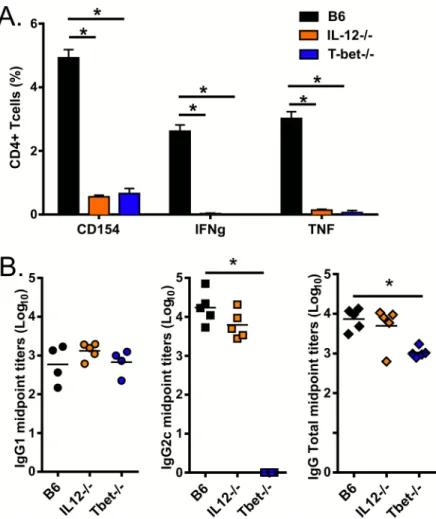
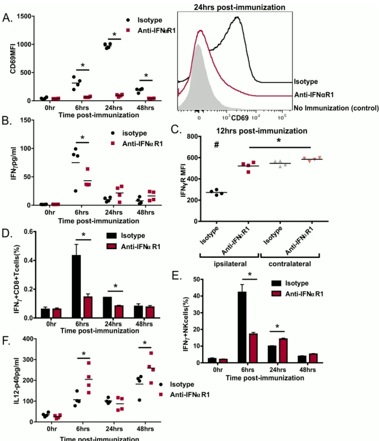
lymphocytes B. Cette immunité renforcée est suffisante pour assurer une protection contre de nombreuses maladies, y compris la Tuberculose et la Leishmaniose. Néanmoins le mode d’action de GLA-SE est encore mal compris. Afin de mieux caractériser l’action de cet adjuvant, il est important de comprendre comment les différentes cytokines et facteurs de transcription contribuent à l’initiation de la réponse immunitaire. Dans cette étude, nous avons évalué la contribution de T-bet, de l'IL-12 et de la signalisation par le récepteur aux interférons de type I (IFNαR1) dans ces réponses en utilisant des souris T-bet-/- et IL12-/- et un anticorps monoclonal bloquant anti-IFNαR1.
Conformément aux résultats de précédentes études sur différents adjuvants, nous avons démontré que l’induction par GLA-SE d'une réponse TH1 et la production d’IgG2 est dépendante
de T-bet, un facteur de transcription clé pour la production d’IFNγ et la différentiation des lymphocytes T en TH1. De plus, un déficit en IL-12, une cytokine canonique dans l’induction de la
réponse TH1 a également inhibé le développement de lymphocytes CD4 TH1 ; la production
d’IgG2 n’a en revanche pas été altérée. Finalement, nous démontrons que la réponse immunitaire innée vis-à-vis de GLA-SE, y compris la rapide production d’IFNγ par les lymphocytes CD8 mémoires et les cellules NK et l’expression de CD69 par les cellules des ganglions lymphatiques drainants, est conditionnée par les interférons de type I produits rapidement après l’immunisation. Ces cytokines contribuent par conséquent également à la réponse adaptative vis-à-vis de GLA-SE.
D'après ces résultats, nous proposons un modèle dans lequel GLA-SE induit la production d’IFNα à travers l’expression de CD69, pour piéger dans les ganglions lymphatiques les cellules nécessaires à la réponse à GLA-SE. La production d’IFNα permettrait aussi la production, , d’IFNγ par les lymphocytes CD8 mémoires et les cellules NK pendant la phase innée de la réponse immunitaire. En parallèle, GLA induit la production d’IL-12 qui, en synergie avec IFNα, promeut l’expression de T-bet et l’engagement des lymphocytes CD4 dans la voie TH1.
12
La compréhension du mécanisme par lequel les adjuvants incitent la réponse immunitaire revêt une importance cruciale dans le développement de vaccins. Les résultats obtenus lors de cette étude suggèrent que l’induction précoce des cytokines IFN de type I et II pourraient être les signatures de la réponse innée optimale à GLA-SE et prédire la réponse TH1 résultante. Des
investigations supplémentaires seront nécessaires pour déterminer si cette production précoce d’IFN de type I peut être utilisée dans une stratégie de sélection dans le développement de nouveaux vaccins ou comme signature d’adjuvant dans les essais cliniques chez l’homme.
Seconde publication : L'IL-18 et les macrophages sous-capsulaires du ganglion lymphatique sont essentiels pour l’induction des réponses lymphocytes B avec l’adjuvant GLA-SE agoniste du TLR4.
Desbien AL, Dubois Cauwelaert N et al. (Soumise à publication)
La compréhension des événements cellulaires et moléculaires qui relient les réponses immunitaires innées et adaptatives est cruciale dans la conception de nouveaux adjuvants. GLA-SE augmente les réponses cellulaires et humorales aux antigènes vaccinaux. L’induction de lymphocytes B et la production subséquente d’anticorps sont des facteurs clés dans le développement de vaccins efficaces. Dans le contexte actuel de nouvelles pandémies, de libération intentionnelle d’agents biologiques dans un but terroriste et de vaccins « à la demande » pour les voyageurs, l’initiation d’une réponse humorale rapide avec des approches vaccinales pragmatiques est hautement souhaitable.
Dans cette étude nous démontrons que peu de temps après l'injection, de l'adjuvant GLA-SE, et comparativement à l'alum, l’émulsion à base de squalène (SE) sans GLA ou de GLA sans SE, induit significativement plus de lymphocytes B spécifiques à l’antigène, des titres d’anticorps supérieurs, un plus grand nombre de lymphocytes T CD4 folliculaires (TFH) et une réponse TH1
plus importante. GLA-SE augmente la différenciation des lymphocytes B spécifiques à l’antigène cible en lymphocytes B du centre germinatif, en lymphocytes B précurseurs de lymphocytes mémoires et en pré-plasmablastes qui sécréteront rapidement des anticorps. De plus, nous montrons qu’après immunisation, les macrophages médullaires sous-capsulaires CD169+ SIGNR1+ sont les premières cellules immunitaires du ganglion lymphatique à capturer GLA-SE et sont critiques pour la réponse innée, y compris la production rapide d’IL-18 induite par GLA-SE.
13
La déplétion des macrophages sous-capsulaires (dont les macrophages médullaires font partie) ou l’abrogation de la signalisation à travers le récepteur de IL-18, altèrent de façon importante la production de lymphocytes B spécifiques à l’antigène et d’anticorps induite par GLA-SE. La déplétion des macrophages sous-capsulaires réduit aussi considérablement la réponse TH1 mais
pas la réponse TFH. Ainsi l’adjuvant GLA-SE agit en interagissant avec des macrophages
sous-capsulaires qui produisent IL-18, permettant alors l’induction de l’expansion et de la différentiation des lymphocytes B, de la sécrétion d’anticorps et l’induction d’une réponse TH1.
Cependant, la réponse TFH semble être, elle, indépendante de l’action des macrophages
sous-capsulaires.
Troisième publication : La présentation d’antigène par les lymphocytes B guide la programmation médiée par TLR4 des lymphocytes CD4 mémoires.
Dubois Cauwelaert N et al. (Soumise à publication)
Dans cette étude, nous avons cherché à déterminer le rôle potentiel des lymphocytes B, au-delà de leur capacité de production d’anticorps, dans la réponse immunitaire vis-à-vis de GLA-SE et nous nous sommes particulièrement intéressés à l’induction de la réponse CD4 TH1. Pour cela
nous avons utilisé des souris déficientes en lymphocytes B (µMT-/-).
En utilisant l’antigène ID93 en association avec GLA-SE, nous avons montré que la réponse TH1
mémoire (6 semaines après la dernière immunisation) induite par GLA-SE et mesurée par la production de cytokines TH1 par les lymphocytes CD4, est fortement altérée dans les souris
µMT-/-. Cependant, une semaine après immunisation, cette même réponse TH1 semblent peu ou pas
affectée chez ces mêmes souris. Ce phénomène avait été préalablement observé suite à l’infection de souris µMT-/- par le virus de la chorioméningite lymphocytaire (VCML).
Toutefois, même si les lymphocytes CD4 induits peu de temps après immunisation en absence de lymphocytes B semblent, en effet, être fonctionnellement similaires à ceux induits dans des souris non-déficientes, en particulier dans leur capacité à produire des cytokines TH1, ils sont
néanmoins phénotypiquement différents. En effet, les souris µMT-/- sont déficientes dans leur capacité à produire des lymphocytes CD4 effecteurs précurseurs de cellules mémoires (CEPM), préalablement définis dans d’autres études comme étant soit PD1+/KLRG1-, soit Ly6Clo/T-betlo.
14
Ces résultats démontrent que les lymphocytes B sont nécessaires pour l’initiation d’une réponse TH1 qui pourra être maintenue dans le temps.
Par la suite, en transférant les lymphocytes CD4 générés une semaine après immunisation dans les souris µMT-/- dans des souris non-déficientes, nous avons pu démontrer que ces cellules perdaient leur fonctionnalité (capacité à produire des cytokines TH1) avec le temps. Ces données
confirment nos résultats précédents, démontrant que les lymphocytes B sont nécessaires à la génération de CD4 CEPM après immunisation. D’autres expériences seront nécessaires pour déterminer si les lymphocytes B sont également nécessaires pour le maintien de la réponse TH1.
Finalement, en transférant aux souris µMT-/- des lymphocytes B sauvages ou n’exprimant pas le CMHII et donc incapables de présenter des antigènes, nous avons montré que cette présentation d’antigènes par les lymphocytes B était nécessaire pour leur rôle dans la génération de CD4 CEPM. Il faut noter que ce point est encore aujourd’hui fortement débattu dans la littérature. En conclusion, ces résultats soulignent l’importance d’un système immunitaire à multiples facettes travaillant de concert pour la génération d’une immunité à médiation cellulaire. Collectivement, les progrès récents dans la compréhension du rôle des lymphocytes B, dans la réponse humorale mais aussi dans la réponse cellulaire, auront un impact important sur le développement de vaccins contre plusieurs pathogènes et en particulier ceux nécessitant une réponse TH1. De plus, ils pourront bénéficier aux médecins qui utilisent des thérapies qui
éliminent les lymphocytes B chez les patients souffrant de troubles liés aux lymphocytes B.
ID93/GLA-SE, un vaccin thérapeutique ?
Le traitement contre la Tuberculose est long et complexe, la thérapie de première ligne implique 6 à 9 mois de traitement avec une association d’antibiotiques (isoniazide, rifampicine, pyrazinamide et éthambutol). De plus, l’émergence de résistances à ces antibiotiques, principalement causée par la difficulté qu’ont les patients à suivre une si longue thérapie, a d’autant plus complexifié ce traitement. En effet l'apparition de souches de Mtb résistantes aux antibiotiques de première ligne nécessite des traitements de durée plus longue (pouvant aller jusqu’à deux ans) avec des associations d’antibiotiques de seconde et troisième lignes. Ainsi le non-respect du traitement antibiotique dans son intégralité entraîne l’augmentation du risque d’échec du traitement, résultant en une récidive de la maladie. Dans ce contexte, le
15
développement de nouveaux schémas thérapeutiques permettant de réduire la durée du traitement antituberculeux (et donc d’améliorer l'observance) et/ou permettant de diminuer le risque de rechute après traitement est nécessaire. Il a été démontré qu’ID93/GLA-SE pourrait potentiellement être utilisé en association avec le traitement antibiotique classique pour améliorer son efficacité et diminuer la durée du traitement. Outre la réduction de la durée du traitement, il serait intéressant de déterminer si l’utilisation de ID93/GLA-SE pourrait également permettre de diminuer les taux de rechute post-traitement.
L’une des limites de l’étude de rechute post-traitement dans le modèle murin est la durée de ce genre d’expérience qui inclue 3 à 6 mois de thérapie, ce qui est extrêmement coûteux en temps et en argent.
Nous avons donc cherché à utiliser un modèle murin de thérapie et rechute qui soit moins complexe. Pour cela nous avons adapté un modèle développé dans un autre laboratoire qui nécessite une thérapie beaucoup moins longue. Grâce à ce modèle, nous avons pu démontrer qu’ID93/GLA-SE réduisait les taux de rechutes dans trois différentes lignées de souris et augmentait le taux de survie des souris SWR, une souche de souris extrêmement susceptible à la Tuberculose. Ces résultats montrent la possibilité d’utiliser ID93/GLA-SE en tant que vaccin thérapeutique et en association avec le traitement antibiotique actuel pour améliorer son efficacité. Une meilleure compréhension des capacités thérapeutiques d’ID93/GLA-SE est cruciale puisque ce vaccin fait actuellement l’objet d’un essai clinique de phase IIa ayant pour but d’évaluer son innocuité et immunogénicité lorsqu’il est administré aux patients atteint de tuberculose pulmonaire, après traitement antibiotique, en vue d’une future phase IIb qui permettrait d’évaluer une potentielle amélioration du taux de rechute chez ces patients.
Conclusion
En conclusion, l‘ensemble de ce travail nous a permis de mieux définir les caractéristiques du mécanisme d'action de GLA-SE et d'améliorer la compréhension des différents acteurs et des étapes nécessaires à la réponse adaptative induite par GLA-SE. Cependant, des travaux complémentaires seront nécessaires pour pleinement comprendre le mécanisme d'action et pour caractériser tous les intervenants de ces interactions complexes induites par l'association de GLA, agoniste de TLR4 et de l'émulsion squalène-eau.
17
INTRODUCTION
18
Tuberculosis (TB) is one of humanity’s oldest diseases. Evidence of TB has been recovered from Neolithic man bones and the spine of Egyptian mummies attesting of the devastation caused by this disease as early as 5000 BC (1). It was not, although, until 1882 that Robert Koch identified
Mycobacterium tuberculosis (Mtb) as the causative agent (2). The death toll from TB began to
decrease as living standards (housing, nutrition, and income) improved early in the 20th century, well before the advent of antituberculosis drugs. With the development of BCG, the first vaccines against TB, in 1921 and the discovery of a number of efficient antibiotics (1944-1965), the hope of TB eradication by the end of the 20th century emerged. Unfortunately with 9.6 million new TB
cases and 1.5 million TB deaths every year, even surpassing HIV as the leading cause of death by infectious disease worldwide in 2014 (3), we must acknowledge that TB eradication is far from being reached.
Major efforts are focused in the development of a new vaccine against TB as mathematical modeling of the impact that could have a hypothetical new vaccine against TB with 60% efficacy predicts an 80% drop in incidence by 2050 (4). There are 17 vaccine candidates in various clinical trial phases for which main objective is to either boost the current BCG vaccine or be used as a stand-alone vaccine in immuno-compromised individuals for which BCG immunization is not recommended. In addition some vaccines could also be used as immunotherapeutic adjuncts to the standard TB antibiotic therapy with the aim of shortening the therapy and/or reduced relapse rates after treatment completion.
One of those vaccines, ID93/GLA-SE, was developed by the Infectious Disease Research Institute (IDRI) and is today entering a phase II clinical trial to evaluate its safety and immunogenicity when administered to adult pulmonary TB patients, following successful completion of TB treatment with confirmed bacteriologic cure, in preparation for a future Phase 2b prevention of TB recurrence trial in the same population (5). ID93/GLA-SE is a sub-unit vaccine composed of a fusion of four Mtb proteins formulated with GLA-SE a Toll-like receptor 4 (TLR4) agonist Glucopyranosyl Lipid A (GLA) in a squalene oil-in-water emulsion (SE) (6, 7).
GLA-SE, IDRI’s leading candidate adjuvant, promotes strong TH1 CD4 T cells, which is thought to
be central for protection against TB, and IgG2-skewed B cell responses to protein vaccine antigens. Beyond ID93/GLA-SE, GLA-SE can be found in several vaccine candidates today in clinical trial against a variety of diseases, including LEISH-F3 GLA-SE against the Leishmania parasite (8), sm14/GLA-SE against Shistosomiasis (9), P27A/GLA-SE against Malaria (10), and
19
H5 virus-like-particle/GLA-SE against Flu (11). Nevertheless still very little is known about this adjuvant its mechanism of actions.
The main objective of this thesis was to broaden the understanding of GLA-SE mediated adjuvanticity and characterize the main actors i.e. immune cells, cytokines, transcription factors and pathways, of the innate response to GLA-SE that trigger and shape the strong adaptative response linked to this adjuvant. Our three main hypotheses for this work were:
1. Canonical TH1 shaping factors, i.e. the transcription factor T-bet and IL-12, will be essential to the adaptive response to GLA-SE. Type I IFN, previously shown to be important for the innate response to GLA-SE (12) would be also important for the subsequent adaptive response.
2. Lymph node (LN) sub-capsular macrophages will take-up GLA-SE and be critical for its adjuvanticity.
3. B cells antigen-presenting function will have a non-redundant role in the shaping to the TH1 response to GLA-SE.
An additional work pursued during this thesis was to establish a mouse model of TB relapse and evaluate the use of ID93/GLA-SE as immunotherapeutic vaccine to be used in combination with chemotherapy. We hypothesize that ID93/GLA-SE would reduce relapse rates in a suboptimal drug treatment model.
The first section of this thesis is a literature review on TB, the search of correlates of protection against it and development of candidate vaccines; we will pay particular attention to sub-unit vaccines in general and ID93/GLA-SE in particular. In a second section we will present the results of the different studies conducted during this thesis. Finally we will do a general discussion of that work, outline the various resultant perspectives that are currently being pursued and close with a general conclusion on the work that was performed.
20
SECTION I: LITTERATURE
21
I.
T
UBERCULOSISA. HISTORY
TB has had various names throughout history, “consumption”, “phthisis”, “Pott’s disease” and “white plague”; all refer to the same scourge that has been known to mankind since ancient times. Bones recovered from different part of the world showed evidence of TB among Neolithic men and in the spine of mummies from ancient Egypt. These data indicate that the humans suffered from TB as early as 5000 BC (1).
It reached epidemic proportions in Europe and North America during the 18th and 19th centuries
earning the appellation of “Captain Among these Men of Death”. The death toll from TB began to decrease as living standards (housing, nutrition, and income) improved early in the 20th century, well before the advent of antituberculosis drugs.
Different forms of TB were thought to be different diseases - pulmonary consumption for example was used to label pulmonary TB, scrofula was the term used to describe TB of the LNs and Lupus vulgaris described TB of the skin. At the beginning of the 19th century Laennec
advanced the idea of a single cause for the different forms of TB. He did not, however, realize that the condition was infectious (13). It was not until 1865 that Villemin convincingly demonstrated the infectious nature of TB (2). Later, in 1882, Robert Koch, in his famous presentation “Die
Aetiologie der Tuberculose”, identified the tubercle bacillus Mycobacterium tuberculosis (Mtb) as
the causative agent of TB (2). B. EPIDEMIOLOGY
TB is a major global health problem. It even surpassed HIV as the leading cause of death by a single infectious agent worldwide in 2014 (3). A third of the world’s population is thought to be infected with Mtb and in 2014, there were an estimated 9.6 million new TB cases and 1.5 million TB deaths (1.1 million among HIV-negative people and 0.4 million among HIV-positive people (3). The number of incident TB cases relative to population size (the incidence rate) varies widely among countries. TB is a disease of poverty that is inextricably associated with overcrowding and under-nutrition and affects mostly young adult in their most productive years. Thus, more than 80% of all TB cases are found in 22 low- and middle-income countries (14). The highest prevalence of active TBper capita is found in Africa with over 300 new cases per 100,000
22
population, driven primarily by the devastating effects of the HIV epidemic. The absolute number of cases is highest in South-East Asia, with India and China having the greatest burden of disease globally (Figure 1) (3). In the United States of America and most Western European countries, the majority of cases occur in foreign-born residents and recent immigrants from countries in which TB is endemic (15, 16).
Left untreated, each person with active TB disease will infect on average between 10 and 15 people every year and this continues the TB transmission. Overall a small proportion (5-15%) of the estimated 2- 3 billion people infected with Mtb will develop TB disease during their lifetime. However, the probability of developing TB is much higher among HIV infected people. The risk for developing TB disease is also higher in persons with diabetes, other chronic debilitating diseases leading to immune-deficiency, poor living conditions or tobacco smokers (14).
Figure 1. Global distribution of estimated TB incidence by rate and absolute number, 2014. The size of each bubble is proportional to the size of the country’s population. High-burden countries are shown in red. WHO regions: African (AFR), Americas (AMR), Eastern Mediterranean (EMR), European (EUR), South-East Asia (SEAR) and Western Pacific (WPR).
23
C. Pathogenesis
TB is transmitted through the air by aerosol.. The most important source of infection is a patient with pulmonary TB and who is coughing. Coughing produces tiny infectious droplets nuclei that are spread into the air and can remain suspended in it for long periods. The World Health Organization (WHO) divides TB infection into primary and post primary infection (17).
1. PRIMARY INFECTION
Primary infection occurs in people who have not had any previous exposure to tubercle bacilli. Droplets nuclei, which are inhaled into the lungs, are so small that they avoid the defense of the bronchi and penetrate into the terminal alveoli of the lungs, where multiplication and infection begin. Lymphatics drain the bacilli to the hilar LNs. The immune response develops about 4-6 weeks after the primary infection. The size of the infecting dose and the strength of the immune response determine the outcome. In most cases (90% of non-HIV infected people), the immune response stops the multiplication of bacilli but a few latent bacilli may persist. Infected individuals will not have any clinical symptoms nor be contagious, even though the bacteria persist in latent state in the infected organ (18). A positive tuberculin skin test would be the only evidence of latent infection. If the immune response is not strong enough to prevent multiplication of bacilli, disease will occur within a few months (17).
2. POST-PRIMARY TUBERCULOSIS
Post-primary TB occurs after a latent period of months or years following primary infection. It may occur by reactivation where the dormant tubercle bacilli acquired during a primary infection starts to multiply generally in response to a trigger such as weakening of the immune system by HIV infection or ageing. Post primary TB can also be caused by reinfection with a new strain in a person who already had a latent primary infection (17).
3. RECURRENT TUBERCULOSIS
Recurrent TB has been defined as a serial episode of active TB disease occurring after treatment success. Recurrent TB can be categorized as relapse of disease from the original infecting strain or reinfection with a new strain of Mtb based on genotyping of the isolates (19). Relapse rates depend on treatment efficacy, program quality, and the patients’ compliance with therapy.
24
The rate of disease due to reinfection, on the other hand, will be determined mainly by the level of TB exposure and transmission in the population (20, 21). The technique of restriction fragment-length polymorphism analysis now allows us to type Mtb strains and consequently distinguish between reinfection and relapse as the cause of recurrence. Using this technique it has been shown that in developing countries a high proportion of recurrences are due to reinfections which is consistent with the high incidence of TB in these populations (19, 22, 23). Recurrence of TB after treatment is a serious problem. On a patient level, recurrent TB requires another round of treatment with a regimen that, in many parts of the world , is more toxic, takes longer to complete, and may amplify drug resistance (24). On a public health level it may account for 10-30% of all cases within some weaker TB control programs, particularly those that do not use at least 6 months of rifampicin (RIF) treatment (20), and contributes to ongoing transmission of infections (25).
D. HOST IMMUNE RESPONSE TO TUBERCULOSIS INFECTION
1. INNATE RESPONSE
Mtb is inhaled through carrier droplets and the innate response develops upon encounter of the
microbe and the alveolar macrophages in the lower airways. The establishment of a successful infection depends on this initial encounter; the infection may remain locally limited within the engulfing cells of the innate immune system, or will continue to spread, causing the individual to become a clinical active TB patient. Mtb is as intracellular bacterium, and although it can infect different cells types, alveolar macrophages are its favorite niche. The initial stages of infection are characterized by innate immune responses that involve the recruitment of inflammatory cells to the lungs (26). The principal effectors of innate immunity are neutrophils, tissue macrophages (derived from blood-borne monocytes), and natural killer (NK) cells. Neutrophils are generally the first recruited cells to arrive to the infection site and are known to play an important role against TB. Rather than direct bacterial killing, they are believed to play a important role in facilitating the adaptive immune response through cytokine and chemokines signaling (27). Blood monocytes are less abundant than neutrophils in circulating blood, but are a critical part of the innate immune response to TB. They are recruited through chemokine signals produced by infected alveolar macrophages, and migrate rapidly across the blood vessels to the site of
25
infection. Within the tissue, they differentiate into macrophages with the ability to ingest and kill the bacteria. The interaction between macrophages and T cells (and in particular, the activation of macrophages by IFN-γ secreted by T cells) is considered central in the elimination of Mtb (28, 29). Although alveolar macrophages are thought to be an effective barrier to contain pathogens,
Mtb has evolved various mechanisms to evade the host immune response and survive in these
cells. Several Mtb survival mechanisms have been described, including: (i) phagosome–lysosome fusion inhibition by interfering with lipid-mediated signaling, by producing the host-like signaling kinase PknG and by hijacking the calcineurin pathway (reviewed in (30)), (ii) Mtb acidification reduction of the Mtb-containing phagosome by selectively excluding the proton-ATPase from the phagosome (31), and (iii) protection against nitric oxide through the rearrangement of the actin cytoskeleton which might lead to a reduction in the local concentration of nitric oxide (32) and through the mycobacterial proteasome machinery which provides protection against killing (33).
NK cells are large granular lymphocytes found in circulating blood; their in vivo role in controlling Mtb is not yet well understood. They have been shown to lyse Mtb-infected macrophages in vitro and to facilitate an adaptive immune response by producing IFNγ and stimulating macrophages to produce inflammatory cytokines (34).
2. ADAPTIVE RESPONSE
In most cases innate immunity is not sufficient to control Mtb. As antigen concentration increases, the adaptive response is activated and it is generally accepted that the long-term outcome of the primary infection is determined by the efficiency of its mobilization.
The induction of an adaptive response occurs after the dissemination of Mtb to draining LNs. In the LNs, presentation of bacterial antigens by dendritic cells (DC) leads to T cells priming. This process is quite slow; studies in various animal models suggest that priming of T cells does not occur until 12-21 days post-infection (35). At this time point the infection is already well established consequently, the inflammatory site where the acquired response will be has already been initiated and modulated by the bacterium. This delay is due to two main components. First, the delayed transport of Mtb from the lungs to the LN caused by the retention of Mtb within the alveolar macrophages which are not a migratory population. It is not until around 8 days post infection that Mtb, that has been replicating within the alveolar macrophages, will escape and be
26
taken up by phagocytes recruited to the infection site (neutrophils, DCs, inflammatory monocytes) (36). Then, even after its transport to the LN Mtb further slows the initiation of the adaptive immunity by promoting the induction of regulatory T cells that act to restrict T cell effector priming (37). The resulting delay represents a critical bottleneck to Mtb’s control, and possibly to its eradication by adaptive immunity (Figure 2) (38, 39). Once primed Ag-specific T cells will expand and differentiate from naïve T cells into effector T cells which then migrate to the infected lung and, in combination with other leucocytes, stimulate the formation of granulomas (40).
Figure 2. Delayed adaptive response to TB. Low-dose aerosol infection, which approximates the natural delivery route for induction of TB, results in low numbers of Mtb (red) being deposited in the lower airways and the alveolar tissue. Bacteria do not disseminate from the lung until 9 days post infection, when they can be detected in the draining lymph nodes. This dissemination coincides with the first activation of naive T cells (purple). Activation of naive T cells occurs in the presence of live bacteria, and effector cells develop with expected kinetics. The effector cell phenotype will depend on the availability of specific cytokines. These effector cells migrate to the lung in response to inflammation and mediate protection by activating infected phagocytes (pale red). The response takes 18–20 days to reach an effective level and thereby to stop bacterial growth (38).
27
3. THE TUBERCULOUS GRANULOMA
Granulomas are organized structures that contain macrophages, lymphocytes and fibroblasts. Within the granuloma, macrophages are activated, for example by CD4+ T cells secreted IFNγ, which is thought to restrict the dispersal and replication of Mtb (40). The granuloma is the hallmark of TB, and functions as a niche in which Mtb can grow and persist and as an immunologic microenvironment in which immune cells interact to control and prevent dissemination of infection.
Granulomas are observed in active, latent and recurrent TB, thus its mere formation is insufficient for control of infection, and rather the granuloma must be functioning properly. Indeed, the granuloma must be carefully balanced in terms of immune responses to provide sufficient immune cell activation to inhibit the growth of Mtb, yet modulate the inflammation to prevent pathology (40). Many different chemokines are involved in the granuloma formation. Some are produced by the epithelial cells of the respiratory tract, and others are produced by the immune cells themselves. In particular, the chemokines binding to the CCR2 receptor (CCL2/MCP-1, CCL12, and CCL13) are important for the early recruitment of macrophages to chronic inflammatory sites to initiate granuloma formation (41). CCL19 and, possibly, CCL21 are involved in the recruitment and priming of IFNγ-producing T cells. CXCL13 is involved in B-cell recruitment and the formation of follicular structures (42, 43). Although in the first instance the granuloma acts to constrain the infection, some bacilli can actually survive inside these structures for a long time in a nonreplicating hypometabolic state, as an adaptation to the unfavorable milieu. This altered physiologic state, termed latent TB infection, can endure for the lifetime of the infected individual, but for some reasons, which are still unclear, the bacilli will reactivate in 10% of the latently infected individuals, escape the granuloma and spread throughout the body, thus giving rise to clinical disease, and are finally disseminated throughout the environment (43).
E. TREATMENT AND MULTI-DRUG-RESISTANCE
Before the advent of chemotherapy, TB was one of the major causes of death in both Western and also several non-Western countries (44). Studies from the pre-chemotherapy era found that
28
about 70% of people with sputum smear-positive pulmonary TB died within 10 years, and that this figure was 20% among culture-positive but smear-negative cases of pulmonary TB (patients with a lower bacterial load) (44).
Nowadays, standard drug regimen for effective treatment of active TB involves a combination therapy with first- and/or second-line drugs for 6-8 months. The requirement for combination therapy is a central tenet of TB treatment mainly to avoid the high probability of developing drug resistance. Recommended first line anti-TB drugs includes RIF, isoniazid (INH), pyrazinamide (PZA) and ethambutol for 2 months followed by RIF plus INH for 4 months. Treatment success rates of 85% or more for new cases are regularly reported to WHO by its Member States (3).
Antibiotic resistance is an increasing problem in TB and the WHO estimates that each year there are half a million cases of multi-drug resistant TB (MDR-TB) (2), defined as a strain of Mtb that is resistant to at least INH and RIF (the two most powerful anti-TB drugs) (45). Of even greater concern are “totally drug-resistant TB” strains, which are resistant to all first- and second-line drugs. Treatment for MDR-TB is longer, and requires more expensive and more toxic drugs (combinations of second- and third-line antibiotics added to the regimen including capreomycin, ethionamide and streptomycin). For most patients with MDR-TB, the current regimens recommended by WHO last 20 months, giving the Mtb pathogen time to adapt to the drug regimen (46). The development of new drugs is currently the leading solution to the increasing incidence of resistance to the drugs currently employed for TB therapy. Several new drugs are in development but it is not clear when they will receive regulatory approval for their use in new drug regimens. Moreover, since even drug sensitive TB is treated with combination therapy, it seems naïve to assume drug resistant strains will be effectively treated with a single new drug, and there is no reason to believe that resistance will not eventually develop to new antibiotics. In addition to the need for new antibiotics to treat drug resistant isolates, there is an urgent need for treatment regimens which can shorten therapy for Mtb (45). Options for improved treatment require new strategies that take advantage of developments in vaccine design and drug delivery that have occurred in the last 50 years, including novel combination regimens in which oral or inhaled agents can be taken for shorter periods of time. To this end, there are today a small number of immunotherapeutic vaccines that are under development for use against Mtb (47).
29
II.
V
ACCINES AGAINSTT
UBERCULOSISA. BCG VACCINE
The high mortality associated with Mtb infection occurs despite the widespread use of a live, attenuated TB vaccine Mycobacterium bovis, bacillus Calmette-Guérin (BCG). BCG is effective at preventing disease in newborns and toddlers, but not pulmonary TB in adults (48). More specifically, numerous efficacy trials and epidemiological studies conducted over several decades indicate that BCG has 60-80% efficacy against severe form of TB in children, particularly meningitis (49, 50), and that its efficacy against pulmonary disease varies geographically (51, 52). Furthermore, revaccinating with BCG during adolescence in a population vaccinated with BCG at birth does not improve protective efficacy as shown in a large, randomized controlled trial in Brazil (53). Importantly, when given to people already infected or sensitized to environmental mycobacteria BCG has not shown any protection against disease, which could explain the geographical efficacy variations (54, 55). Additionally, the live attenuated BCG vaccine is unsafe for administration to HIV-positive or other immunocompromised individuals due to the possibility of developing regional BCG infection (BCG-itis) or disseminated BCG (BCG-osis) (56-59). Mathematical modeling of the impact that a hypothetical new vaccine against TB with 60% efficacy could have predicts an 80% decrease in incidence by 2050 (4). Thus there is an urgent need for a new TB vaccine to either boost immunity primed by BCG or to replace BCG.
B. CORRELATES OF PROTECTION
TB vaccine research is confounded by a conundrum: a candidate biomarker for protective immunity can only be validated in the clinical trial of an effective vaccine. However, clinical trials of an effective vaccine may not be feasible without a validated correlate of protection for the selection of the most promising candidates as for determining dose and schedule of vaccination.
1. THE CENTRAL DOGMA OF PROTECTIVE IMMUNITY
During the past 40 years a combination of studies have led to the TB central dogma that production of IFNγ by CD4 T cells is the major driver of immunity against TB.
In 1970 it was found that T cells, and not antibodies, are required for host resistance to TB (60). Later, in the 1980’s, CD4+ T cells that produce IFNγ were described as the dominant T cell subset
30
that participates in the immune response to Mtb (61). In 2001, it was shown that mice devoid of CD4-mediated immunity (MHCII-/-) or CD4+ depleted were not able to control Mtb infection and would subsequently succumb early-on (62). This experiment established a crucial role for CD4+ T cells mediated immunity in TB control. Finally, the discovery that HIV infection, which is strongly associated with TB, infects and kills CD4+ T cells supported a central role for CD4+ T cells in immunity against TB (63).
IFNγ’s, a cytokine involved in the response against viruses and intracellular bacteria, key role in immunity against TB is based on the extreme susceptibility of IFNγ-deficient mice (64). More arguments supporting this hypothesis include nontuberculous mycobacterial infection or BCG-osis in partial or complete IFNγ-deficient patients (65, 66) and IFNγ depression in whole-blood cultures from advanced TB patients (67). IFNγ activates macrophages to kill intracellular bacteria through antimicrobial effector pathways such as inducible nitric oxide synthase, IFNγ inducible GTPases, phagosomal maturation and acidification, autophagy and vitamin D receptor signaling (40). Furthermore, in humans suffering from recurrent nontuberculous mycobacterial infections, deleterious genetic mutations in the genes encoding IL-12p40 and IL-12R have been identified (68-70). IL-12 has a crucial role in the induction of IFNγ production (71), accordingly these patients display a reduced capacity to produce IFNγ (72), highlighting the potential central role of IFNγ in TB protection.
These discoveries helped to define the central dogma of protective immunity against TB, namely, that the production of IFNγ by CD4+ T cells activates macrophages to kill Mtb (Figure 3). Today, detection of IFNγ produced by T cells is still the most widely used for monitoring immune response following TB infection (QuantiFERON®-TB test) or vaccination.
31
Figure 3. Protective immunity to TB: the central dogma and a revised view of T cell mediated immunity. A. The central dogma of protective immunity to TB is that CD4+ T cells produce IFNγ which synergizes with TNF (produced by the T cell or the macrophage), and together these activate macrophage antimicrobial activity capable of restricting M. TB growth. Two pathways activated by IFNγ that are capable of killing Mtb are nitric oxide (NO) production and phagolysosome fusion, which acidifies the bacterial phagosome. B. “A revised view of protective T cell immunity” incorporates additional T cell subsets (CD4+, CD8+, and unconventional T cells – γδ T cells, Mucosal-Associated Invariant T (MAIT) cells and CD1-restricted T cells), and includes additional mechanisms by which T cells mediate killing of Mtb.
2. MOVING BEYOND THE CENTRAL DOGMA
Although essential for host resistance to Mtb, IFN-γ responses notoriously do not correlate with resistance to TB. More T cells that secrete IFNγ, or greater IFNγ levels, have not been found to correlate with protection (73). Further, a study in 2011 found that patients whose T cells produce greater amounts of IFNγ are more likely to progress to active disease than patients with weaker responses (74), supporting the idea that IFNγ levels actually correlates better with bacterial burden than with disease control (26). Similar conclusions can be taken from vaccination studies. BCG vaccination elicits protective T cells in animals, but IFNγ production by these T cells is not predictive of vaccine induced protection (75, 76). Further, one study in South-African infants vaccinated with BCG found no correlation between the number of BCG-elicited T cells that produce IFNγ and protection against TB (77). Finally some recent studies in murine models suggest that even though TH1 CD4 T cells might be essential for protection against TB,
this mechanism might be IFNγ-independent (78, 79). It seems likely then that even though IFNγ production by CD4+ T cells might be a critical component of immunity against Mtb it may not
32
necessary translate into an immune correlate of protection against TB. There is now a wide appreciation for the importance of extending our understanding of anti-TB CD4 T cell effector molecules beyond IFN-γ.
Other immune cells could potentially correlate with protection, such as CD8+ T cells, TH17 T cells,
NK cells, γδ T cells and CD4- CD8- T cells (Figure 3 B.) (26, 80).
a) CD4+T CELLS POLYFUNCTIONALITY
In addition to T cells that predominantly produce IFN-γ, recent studies have suggested that multifunctional CD4+ or CD8+ T cells that produce multiple cytokines, especially IFN-γ, tumor
necrosis factor alpha (TNF-α), and interleukin-2 (IL-2), could be important in host resistance. For example HIV non progressors expressed a high frequency of such polyfunctional CD4+ T cells (81) and similarly Ag-specific polyfunctional CD4+ T cells were found to be protective against
Leishmania infection (82). Consistent with these results, protection in mice against Mtb infection
correlates with a high frequency of polyfunctional CD4+ T cells (83). In humans active TB disease is associated with TNF producing monofunctional CD4 T cells compared to the polyfunctional T cells found in latent Mtb infection (84). The Modified Vaccinia Ankara (MVA85A), a modified vaccinia virus Ankara expressing the major Mtb antigen Ag85A, was the first TB candidate vaccine to complete an efficacy trial (after BCG). A higher frequency of polyfunctional CD4+ T cells were elicited in BCG vaccinated-MVA85A boosted individuals than that for BCG alone (85). However, disappointingly, the results obtained during the phase 2b in infants indicated no enhancement in protection, suggesting that Ag85A-specifc polyfunctional CD4+ T cells may play a necessary but not sufficient role in protection against TB (86) in that population under the conditions evaluated.
b) CD4+T CELLS PHENOTYPES
The discrepancy between the fact that TH1 CD4 T cells are critical in TB immunity, yet higher
numbers of these cells do not necessary confer protection, could potentially be explained if different subsets of TH1 CD4+ T cells differ in their ability to control Mtb. Reiley et al. identified
two functionally distinct subsets of effector CD4+cells produced during TB infection that could be distinguished by their expression of the programmed cell death protein 1 (PD-1) and the co-inhibitory receptor killer-cell lectin like receptor G1 (KLRG1). PD-1 expressing activated effector
33
cells with a higher proliferative and self-renewing capacity than the KLRG1-expressing cells, but a lower capacity of cytokine production, and the capacity of differentiating into KLRG1+ cells. KLRG1+ cells are terminally differentiated, proliferate poorly, maintain their KLRG1+ phenotype and are short-lived (87). There is growing evidence that PD-1+ CD4+ T cells mediate superior
protection against TB than terminally differentiated KLRG1+. Indeed, protection against Mtb
induced by BCG immunization in mice is lost as the infection enters the chronic phase, and this waning of immunity is coincident with an increase of KLRG1+ terminally differentiated T cells
(88). Furthermore, immunizations that confer superior longer lasting immunity preferentially expand Mtb-specific CD4+ T cells that are KLRG1-, produce IL-2 and are associated to central memory (88, 89). In addition, it was shown that CD4+ T cells must make direct contact with infected antigen presenting cells (APC) to induce control of Mtb infection (90). Thus, to be protective, Mtb-specific CD4+ T cells need to exit the circulation and enter the lung parenchyma where the Mtb infection occurs. Meanwhile, it was shown that, upon infection with Mtb, Mtb-specific CD4+ T cells residing in the lung vasculature where mainly KLRG1+ PD1- and the ones in
the infected lung parenchyma where almost exclusively PD1+ KLRG1- (91). Furthermore,
adoptive transfer of those PD1+ KLRG1- cells conferred greater protection against Mtb challenge (91, 92). Thus, CD4 T cells with a more central memory polyfunctional phenotype and ability to enter the lung parenchyma might be essential for protection against TB and those functions correlates, at least in mice, with a KLRG1- PD1+ phenotype (Figure 4).
34
Figure 4. Properties of protective Mtb-specific CD4+ T cells. Pulmonary Mtb-specific Th1 cells display two major phenotypes based on their localization in the lung parenchyma or lung-associated blood vasculature of infected mice. CD4+ T cells that can efficiently enter the lung parenchyma (in red) are almost exclusively PD1+ KLRG1-, secrete low amounts of IFNγ and are highly protective against Mtb. CD4+ T cells that reside in the blood vessels (in blue), are terminally differentiated, secrete high amounts of IFNγ and are weakly protective agiants Mtb.
C. THE TUBERCULOSIS VACCINES PIPELINE
Due to the complex nature of Mtb and resulting pathology within the human host (discussed above), multiple vaccine development strategies are being pursued: (i). prevention of infection (vaccines that are given prior to exposure in order to prevent initial infection), (ii) prevention of disease (vaccines that can be administered after exposure, to infected people that might be asymptomatic and at risk of developing disease in the future), (iii) prevention of recurrence (vaccines that can be administered after infection and during or after drug treatment, to prevent reactivation) (3). Although there is some pessimism about whether a vaccine that averts infection can be developed, the general consensus is that a vaccine that prevents progression to
35
active disease could reduce the prevalence of pulmonary TB and ultimately break the cycle of transmission.
Efforts are currently underway to develop vaccines against TB that can boost the current BCG vaccine or can be used as a stand-alone prophylactic vaccine in immune-compromised individuals where BCG immunization is not recommended. In addition, immunotherapeutic vaccine are also being developed for individuals with active TB in conjunction with TB drug therapy, with the aim of shortening the duration of the therapy and/or reducing recurrence rates after completion of treatment. There are 17 vaccine candidates in various phases of clinical trials, including five protein or adjuvant vaccines, four viral-vectored vaccines, three mycobacterial whole cell or extract vaccines, one recombinant live and one attenuated Mtb vaccine (
Figure 5) (reviewed in (3) and (93)). The two most advanced candidates are M. vaccae and M72/AS01. M. vaccae is a whole heat-killed agent currently licensed by the China Food and Drug administration as an immuno-therapeutic agent to help shorten TB treatment for patients with drug susceptible TB after it shown improved sputum reconversion and X-ray appearances when used in combination with chemotherapy in patients with untreated TB (94). This candidate vaccine is currently in Phase III trial to assess its efficacy and safety in preventing TB disease in people with latent TB (3). M72/AS01 is a recombinant fusion protein of the Mtb antigens (Ags) 32A and 39A in combination with the AS01 adjuvant, a liposomal formulation containing the monophosphorylated lipid A (MPL) (TLR4 agonist Cf. IV.A.2.b)) and Quillaja saponaria (QS21) a purified plant extract. This candidate vaccine was shown to have a clinically acceptable safety profile and to be highly immunogenic in Mtb-infected and uninfected adults (95) and is currently in a large randomized placebo-controlled Phase 2b trial to assess efficacy of two doses of M72/AS01 against pulmonary TB disease (3).
36
Figure 5. 2015 TB vaccine candidates’ pipeline.
III.
S
UB-U
NIT VACCINESA. WHY WE NEED ADJUVANTS
Vaccines made from live-attenuated or inactivated pathogens can elicit robust protective immune responses because those vaccines contain naturally occurring adjuvants. Vaccination with an antigen alone has advantages in terms of safety and cost-effectiveness, however when separated from their microbial constituents many of these vaccines, such as the acellular pertussis vaccine, are insufficiently immunogenic on their own. Adjuvants become critical to help these proteins become effective vaccines by inducing strong and long lasting immune responses. Adjuvants from the Latin word adjuvare, meaning “to help or aid”, were first described by Ramon in 1924 as “substances used in combination with a specific antigen that produced a more robust immune response than the antigen alone” (96). Most specifically, several important benefits can be found in adjuvants such as enhancement of vaccine efficacy in infants, elders and immunocompromised people, increase of functional antibody titers, induction of more and long-lasting responses, induction of robust cell-mediated immunity, broader protection through cross-reactivity to other strains or pathogens, facilitation of mucosal immunity, overcoming of antigen
37
competition in combination vaccines, reduction of the dose of antigen needed (dose sparing) and/or reduction of the number of vaccines doses needed (97, 98).
B. PATHOGENS RECOGNITION BY THE HOST
1. DANGER ASSOCIATED MOLECULAR PATTERNS
In some circumstances host inflammatory responses can cause host cell death leading to tissue injury, and the release of host cellular components to the extracellular environment. These self-molecules considered as “messengers” for danger or Damage-Associated Molecular Patterns (DAMP) shape the immune response. Host lipids, sugars, metabolites (e.g. uric acid or ATP), and nucleic acids such as RNA and DNA all contain DAMPs which are recognized when the molecules accumulate in non-steady state locations such as cytosolic DNA or extracellular ATP (99). This leads to activation of inflammasomes. Inflammasome activation leads to caspase-1 activation, which causes the maturation cleavage of pro-IL-1β and pro-IL-18 into their mature forms IL-1β and IL-18 which are secreted and shape the innate and adaptive responses to pathogens and adjuvants (100, 101).
2. PATHOGEN-ASSOCIATED MOLECULAR PATTERNS AND TOLL-LIKE RECEPTORS
Host cells are equipped with numerous types of receptors to discriminate self from non-self constituants. Pathogens are first recognized by pattern recognition receptors (PRR) expressed by many cell types including professional APCs such as macrophages and DCs, which prime T cell responses. PRR including TLR, RIG-I like receptors, C-type lectin receptors and Nucleotide oligomerization domain (NOD)-like receptors recognize a diverse array of pathogen associated molecular patterns (PAMP).
TLR are among the best characterized PRR and were first identified in the early 1980s in
Drosophila where their function are required for responses to fungal and Gram-positive bacterial
infection (102). TLRs are the canonical PRR that link innate and adaptive immunity: they sense infection though the recognition of PAMPs and induce appropriately tailored innate and adaptive immune response (103). A summary of known mammalian TLRs and their ligands is shown in Figure 6.
38
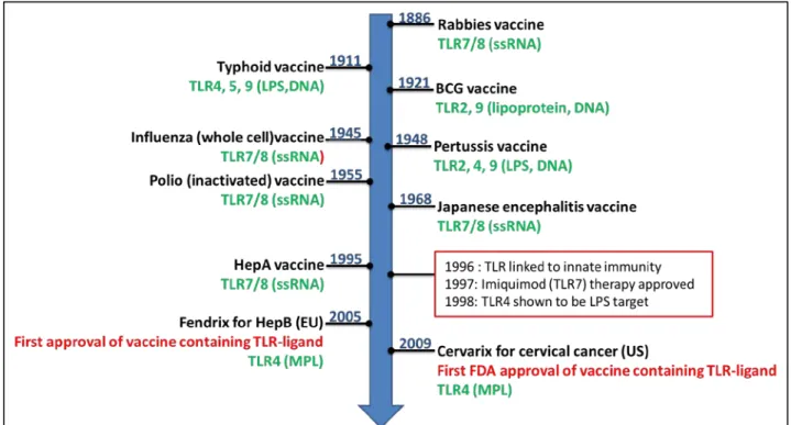
Figure 6. MammalianTLRs and their ligands.
Binding and TLR homo- or heterodimerization brings the Toll-Interleukin Receptor domains of adjacent TLRs together, providing a conformational change necessary to trigger signaling. Binding of additional adaptor proteins, including the myeloid differentiation factor 88 (MyD88) the MyD88 adaptor-like protein, the TIR domain-containing adaptor protein inducing interferon-β (TRIF) and the TRIF-related adaptor molecule TRAM, is essential for intracellular cascades (104-107) (Figure 7).
39
Figure 7. TLR signaling. Extracellular TLR homodimers (TLR4 and TLR5) are represented in black; heterodimers of TLR2 and TLR1, TLR6 or TLR10 are indicated in black/green. Intracellular homodimers (TLR3, TLR7, TLR8 and TLR9) are indicated in gray.
TLRs have been shown to be sufficient to tailor an adaptive immune response characterized by TH1 induction, IgG2c production, CD8 T cells induction and protection from reinfection to an
array of pathogens (Gram-positive and negative bacteria, DNA and RNA viruses, fungi and protozoa) (108, 109). Importantly they have also been shown to be critical for the induction of a strong adaptive immune response to various immunizations (98, 110). They control this responses at multiple levels including: (i) control of antigen uptake by transiently stimulating antigen macropinocytosis and coordinating the redeployment of actin to sustained endocytosis (111) (ii) control of antigen selection for presentation in DCs as efficiency of antigen presentation from phagocyted cargo is dependent on the presence of TLR ligands within the cargo and the generation of peptide-MHCII complexes is also controlled by TLRs (112), (iii) control of DC migration from the infection site to the draining LN through downregulation of CCR6, an inflammatory chemokine receptor, and upregulation of CCR7, a receptor for lymphoid chemokines (113, 114), (iv) control of DC maturation characterized by the upregulation of the co-stimulatory molecules CD40, CD80 and CD86 and production of the pro-inflammatory cytokines Il-12 and TNF (115), (v) direct co-stimulation of T cell subsets to enhance effector function such as IL-2 and IFNγ production and T cell proliferation (116), and (vi) control of B cells responses to T-dependent and T-independent antigens thought B cell intrinsic TLR signaling
40
that regulates antibody responses (117). Due to their important immunomodulatory capacities TLR are prime- candidates for adjuvant development. In reality, most first generation vaccines, including those consisting of inactivated or attenuated virus or bacteria contain inherent adjuvant activity by possessing molecules that can bind these receptors (Figure 8). The discovery of TLRs and their role in modulation of innate and adaptive immunity has led to exploitation of their ligands as immune modulators. The addition of TLR agonists is not a new concept, since combined vaccine adjuvants have been evaluated since the 1930s, when whole bacterial cells were added to water-in-oil emulsions to create Freunds’ complete adjuvant. Today, several well-defined and characterized TLR agonists are being selectively developed with the primary purpose to serve as adjuvant components within vaccines. A summary of those TLR based adjuvants and the disease models tested in experimental and clinical trials and human vaccines can be found in Toussi and Massari 2014 paper (107). In particular, TLR3, TLR4 and TLR9 agonists have been shown to improve a number of vaccines, for example against Hepatitis B Virus (118, 119), influenza (120-122), malaria (123-126) and Leishmania (8, 127), as well as some types of cancer (128-132).