HAL Id: tel-01711460
https://tel.archives-ouvertes.fr/tel-01711460v2
Submitted on 26 Feb 2018HAL is a multi-disciplinary open access archive for the deposit and dissemination of sci-entific research documents, whether they are pub-lished or not. The documents may come from teaching and research institutions in France or abroad, or from public or private research centers.
L’archive ouverte pluridisciplinaire HAL, est destinée au dépôt et à la diffusion de documents scientifiques de niveau recherche, publiés ou non, émanant des établissements d’enseignement et de recherche français ou étrangers, des laboratoires publics ou privés.
Silvia Lopez-Lastra
To cite this version:
Silvia Lopez-Lastra. Humanized Mice as Models to study Human Innate Immunity and Immunother-apies. Innate immunity. Université Paris-Saclay, 2017. English. �NNT : 2017SACLS039�. �tel-01711460v2�
NNT : 2017SACLS039
Thèse de doctorat de
L’Université Paris-Saclay préparée à
L’UNIVERSITÉ PARIS-SUD
E
COLED
OCTORALEn°577 :
Structure et dynamique des systèmes vivants (SDSV)
S
PÉCIALITÉ DE DOCTORAT: Sciences de la Vie et de la Santé
Par
SILVIA LOPEZ-LASTRA
Dirigée par James P. Di SantoHUMANIZED MICE AS MODELS TO STUDY HUMAN INNATE
IMMUNITY AND IMMUNOTHERAPIES
Paris, le 17 février 2017 JURY
M. Karim Benihoud M. Francesco Colucci
M. Miguel Lopez-Botet Mme. Nadine Cerf-Bensussan M. James P. Di Santo
Mme. Nathalie Sauvonnet
Professeur (Université Paris Saclay)
Directeur de recherche (University of Cambridge) Professeur (Universitat Pompeu Fabra)
Directrice de recherche (INSERM) Professeur (Institut Pasteur)
Chargé de recherche (Institut Pasteur)
Président du jury Rapporteur Rapporteur Examinatrice Directeur de thèse Co-directrice de thèse
Table of contents --- 4 Acknowledgements --- 8 Abstract/Résumé --- 11 List of figures --- 15 List of abbreviations --- 19 Introduction --- 25
I. The Human Immune System --- 29
Early hematopoiesis --- 29
Adaptive immunity --- 31
Innate immunity --- 33
i. Dendritic cells --- 35
ii. Innate Lymphoid Cells --- 42
iii. Gamma-delta T cells --- 57
II. Human Immune System Mice --- 61
Early development of recipient strains --- 61
Current HIS mouse models --- 65
i. Hu-PBL-SCID --- 65
ii. NSG and BRGS --- 65
iii. BLT (bone-marrow, liver, thymus) --- 66
iv. MITRG and MISTRG --- 66
Harnessing Immunity to Battle Tumors: lessons from HIS mice --- 67
i. Modeling cancer in HIS mice --- 67
ii. Targeting Natural Killer cells in HIS mice --- 69
iii. Adoptive NK cell transfer --- 70
iv. Novel NK cell sources --- 72
Improving the NK cell compartment in his mice --- 74
Specific aims of the thesis --- 77
Results --- 81
PAPER I: --- 83
PAPER II: --- 123
Systemic human ILC precursors provide a substrate for tissue ILC differentiation --- 123
PAPER III: --- 173
In vivo efficacy of umbilical cord blood stem cell derived NK cells in the treatment of metastatic colorectal cancer --- 173
PAPER IV: --- 201
A nanobody based bispecific targeting approach to leverage the potent and widely applicable tumor cytolytic capacity of monomorphic Vγ9Vδ2-T cells --- 201
Discussion --- 237
References --- 249
ANNEXES --- 273
Synthesis (french) --- 330
Along the long and winding road that led me here I’ve had the opportunity to meet many people who, each one in his or her own way, have contributed to the person I am today and will likely shape my future. I want to thank everyone who taught me, encouraged me and helped me remain standing with generosity and benevolence.
Animal models have extensively contributed to our understanding of human immunobiology and to uncover the underlying pathological mechanisms occurring in the development of the disease. However, mouse models do not always reproduce the genetic complexity inherent in human disease conditions. Human immune system (HIS) mouse models that are susceptible to human pathogens and can recapitulate human hematopoiesis provide one means to bridge the interspecies gap.
Severely immunodeficient host mice support life-long, high level human hematolymphoid development after engraftment with human hematopoietic stem cells (HSC). However, the differentiation and function of some blood cell types, including innate lymphoid cells (ILCs), is poorly characterized in current HIS mice. Here we describe the development of a novel HIS mouse model, named BRGSF (BALB/c
Rag2tm1Fwa
Il2rgtm1Cgn
Flk2tm1lrl
SirpaNOD
), which demonstrate enhanced maturation, function and homeostasis of human natural killer (NK) cells and other ILCs.
Furthermore, the BRGSF-based HIS mouse model recapitulated the developmental stages of human ILCs. We could identify for the first time an ILC precursor (ILCP) population that is present both in HIS mice and in human peripheral blood as well as in several lymphoid and non-lymphoid human tissues. This circulating human ILCP population may provide a substrate to generate mature ILCs in tissues in response to environmental stressors, inflammation and infection.
In a second part of the thesis we used BRGS (BALB/cRag2tm1Fwa
Il2rgtm1Cgnl
SirpaNOD
) immunodeficient mice to assess two innate lymphocyte-based immunotherapeutic approaches for treating EGFR-expressing KRAS-mutated colorectal carcinoma in vivo. The first model used a combination of umbilical cord blood (UCB)-derived NK cells and the monoclonal antibody cetuximab to promote antibody dependent cell cytotoxicity (ADCC) against the tumors. In a second model, we evaluated the therapeutic suitability of novel bispecific VHH constructs that combine inhibition of the EGFR with the target-specific activation of effector Vγ9Vδ2-T cells.
These studies highlight the utility for HIS-based mouse models to understand human innate lymphocyte development and to harness these potent effectors for anti-tumor therapies.
Les modèles animaux ont largement contribué à notre compréhension de l’immunologie humaine et des mécanismes pathologiques associés au développement des maladies. Cependant, les modèles murins ne permettent pas de reproduire toute la complexité des pathologies humaines. Les souris à système immunitaire humain (HIS), par leur capacité à récapituler l’hématopoïèse humaine et à être infectées par des pathogènes humains, constituent une solution de choix pour combler ce fossé inter-espèce.
Après greffe de cellules souches hématopoïétiques humaines, des souris hôtes sévèrement immunodéprimées permettent un haut niveau de développement du système hémato-lymphoïde humain tout au long de leur vie. Cependant, certains types cellulaires, comme les cellules lymphoïdes innées, ne parviennent pas à se différencier et à fonctionner normalement dans les modèles murins HIS actuels. Ici, nous décrivons le développement d’un modèle souris HIS original, nommé BRGSF (BALB/c Rag2tm1Fwa
Il2rgtm1Cgn
Flk2tm1lrl
SirpaNOD
), montrant une amélioration de la maturation, de la fonction et de l’homéostasie des cellules natural killer (NK) humaines et des autres ILCS.
De plus, en récapitulant les différentes étapes du développement des ILCs humaines, ce modèle souris BRGSF nous a permis d’identifier pour la première fois un précurseur d’ILC (ILCP) présent à la fois dans notre modèle HIS ainsi que dans le sang périphérique et plusieurs organes lymphoïdes et non-lymphoïdes humains. Cette population circulante d’ILCPs pourrait constituer un substrat pour la production d’ILCs matures dans les tissus périphériques en réponse à des stress environnementaux, inflammatoires et/ou infectieux.
Dans une seconde partie de ce travail de thèse, nous avons utilisé ces souris BRGS afin de tester l’efficacité de deux immunothérapies reposant sur les lymphocytes innés pour le traitement d’un carcinome colorectal exprimant EGFR et muté pour KRAS. La première approche a consisté en la co-administration des cellules NK dérivées de sang de cordon ombilical et d'anticorps monoclonal cetuximab afin de promouvoir le mécanisme de cytotoxicité cellulaire dépendante des anticorps (ADCC) contre la tumeur. La seconde stratégie a reposé sur l’injection de nanobodies VHH combinant l’inhibition de l’EGFR et l’activation spécifique du récepteur Vγ9Vδ2 des cellules T effectrices.
Les résultats de cette étude soulignent l’importance des modèles murins HIS pour la compréhension du développement des lymphocytes innés humains et pour mieux les mettre à profit dans les thérapies anti-tumeurs
Figure 1: Models of human hematopoiesis. --- 30
Figure 2: TLR recognition of conserved molecular structures in bacteria, viruses, fungi and parasites. --- 34
Figure 3: Schematic view of human dendritic cell development. --- 41
Figure 5: Identification of human peripheral blood NK cells subsets. --- 44
Figure 6: Models of MHC-I mediated education of NK cells. --- 45
Figure 7: Key activating and inhibitory receptors of human NK cells. --- 47
Figure 8: Model of early NK cell development. --- 48
Figure 9: ILC subsets and their signature cytokines. --- 51
Figure 10: Roles for ILCs in inflammation and tissue repair. --- 55
Figure 11: Receptor-ligand interactions mediating tumor cell recognition by γδT cells. --- 58
Figure 12: Antitumor versus protumor roles of αβ-T cells in the mouse. --- 59
Figure 13: Improving homeostasis and function of human immune cells in HIS mice. --- 62
Figure 14: Approaches to improve xenograft tolerance in HIS mice. --- 63
Figure 15: White blood cell composition in human blood versus humanized mice --- 64
Table 1: Main functions of toll like receptor in humans. ... 39
ADCC Antibody dependen cellular cytotoxicity
Ag Antigen
ALL Acute lymphoblastic leukemia AML Acute myeloid leukemia AMP Antimicrobial peptide APC Antigen presenting cell
BAC Bacterial artificial chromosome BLT Bone marrow-Liver-Thymus
BM Bone Marrow
BRG BALB/c Rag2tm1Fwa
Il2rgtm1Cgn BRGS BALB/c Rag2tm1Fwa
Il2rgtm1Cgn
SirpaNOD
CAR Chimeric antigen receptors CCL (C-C motif)-chemokine ligand CCR (C-C motif)-chemokine receptor cDC Classical/conventional dendritic cell CDP Dendritic cell progenitor
CLP Common lymphoid progenior CLR C-type lectin receptor
CMP Common myeloid progenior CMV Cytomegalovirus
COPD Chronic obstructive pulmonary disease CRTH2 Prostaglandin D2 receptor 2
CSC Cancer stem cell
CTGF Connective tissue growth factor CTL Cytotoxic T lymphocyte
CTLA-4 Cytotoxic T-lymphocyte-associated protein 4 CXCL (C-X-C motif)-chemokine ligand
CXCR (C-X-C motif)-chemokine receptor DAMP Damage-associated molecular pattern DC Dendritic cell
DETC Dendritic epidermal T cells DNAM DNAX Accessory Molecule-1 EBV Epstein–Barr virus
ESC Embryonic stem cell FL Fetal liver
Flt3 Fms-like tyrosine kinase 3 ligand FoxP3 Forkhead box P3
GM-CSF Granulocyte-macrophage colony stimulating factor GVHD Graft-versus-host disease
HA Hemagglutinin
HER Human epidermal growth factor receptor 2 HIS Human immune system
HIV Human immunodeficiency virus HLA Human leukocyte antigen
hpre-cDC Human convential dendritic cell precursor HSC Hematopoietic stem cell
HSCT Hematopoietic stem cell transplantation ICOSL Inducible T-cell costimulator
IDO Indoleamine 2,3-dioxygenase IFN Interferon
Ig Immunoglobulin
IL Interleukin
ILC Innate lymphoid cell infDC Inflammatory dendritic cell IPP Isopentyl pyrophosphate iPS Induced pluripotent stem cell IRF Interferon regulatory factor
ITAM Immunoreceptor tyrosine-based activation motif ITIM Immunoreceptor tyrosine-based inhibition motif KIR Killer-cell immunoglobulin-like receptors LFA Lymphocyte function-associated antigen LMPP Lymphoid-primed multipotent progenitor LPS Lipopolysaccharide
LTβR Lymphotoxin-beta receptor LTi Lymphoid tissue inducer MAIT Mucosal associated invariant T MAMP Microbial-associated patterns MAPK Mitogen-activated protein kinase MDP Monocyte-DC progenitor
MDSC Myeloid-derived suppressor cells MEP Megakaryocyte-erythrocyte progenitor MHC Major histocompatibility complex MIP Macrophage inflammatory protein
MISTRG C;129S4-Rag2tm1.1FlvCsf1tm1(CSF1)Flv Csf2/Il3tm1.1(CSF2,IL3)FlvThpotm1.1(TPO)Flv Il2rgtm1.1Flv (SIRPA)Tg1Flv/J
MITRG C;129S4-Rag2tm1.1FlvCsf1tm1(CSF1)Flv Csf2/Il3tm1.1(CSF2,IL3)FlvThpotm1.1(TPO)Flv Il2rgtm1.1Flv
MLR Mixed lymphocyte reactions moDC Monocyte derived dendritic cell MPP Multipotent progenitors
NCR Natural cytotoxicity receptor
NF-κβ Nuclear factor kappa-light-chain-enhancer NKG2 Natural killer Group 2
NKP Natural killer precursor NKR Natural killer receptor NKT Natural killer T NLR NOD-like receptor
NOD Non-obese diabetic NOG NOD.Cg-Prkdcscid
Il2rgtm1Sug /Jic NSG NOD/.Cg-Prkdcscid IL2rtm1wj /Szj PB Peripheral blood
PBMC Peripheral blood mononuclear cell PD-1 Programmed cell death protein 1 pDC Plasmacytoid dendritic cell PDX Patient derived xenograft PGE2 Prostaglandin E2
PILR Paired immunoglobulin-like type 2 PRR Pattern recognition receptor
PSC Pluripotent stem cell
RAG Recombination activating gene
RANTES Regulated on activation, normal T cell expressed and secreted RLR RIG-1 like receptors
RORg RAR-related orphan receptor gamma S1PR1 Sphingosine-1-phosphate receptor 1 SCID Severe combined immunodeficiency SIRPα Signal regulatory protein alpha SLO Secondary lymphoid organ SRR SLAM-related receptor
STAT Signal transducer and activator of transcription TCF T-cell factor
TCR T-cell receptor
TGFβ Transforming growth factor beta
Th T helper
TL1A TNF-like protein 1A TLR Toll-like receptor TNF Tumor necrosis factor
TPO Thrombopoietin
TRAIL TNF-related apoptosis-inducing ligand Treg Regulatory T cell
TSLP Thymic stromal lymphopoietin UCB Umbilical cord blood
VEGF Vascular endothelial growth factor XCL Chemokine (C motif) ligand XCR Chemokine (C motif) receptor
Biomedical research has benefited from mouse experimentation to better understand mammalian biology since the 16th century, and it was after the generation of the first inbred mouse strains in the early 20th century that they were established as model organisms giving birth to many of the biological discoveries of the last 120 years. In many cases, this led to the development of successful treatments for previously untreatable diseases, for example acute promyelocytic leukemia 1
. Although other model organisms are genetically closer to humans (such as dogs, pigs or non-human primates), mice are easier to maintain and breed, have short generation times and thanks to robust gene-modification approaches, provide an endless variety of mutants in several inbred backgrounds.
We know today that Jacques Monod and Francois Jacob’s aphorism “Anything found to be true of E. Coli must be true of elephants” turns out to be too reductionist. Moreover, M.
musculus and H. sapiens have been evolving divergently for 85 million years, adapting to
very different environments thus undergoing selection for many traits, from the circadian rhythm to our body size 2. Thanks to the decoding of mouse and human genomes in the early 2000, we now appreciate this independent evolution led to a difference in one-fifth of our genes. Not surprisingly, this part of the genome contains genes that suffered the highest selection pressure due to environmental contact such as human-microbe interactions. Indeed, the divergent sequences are involved in transcription regulation and chromatin organization with enrichment in the regions related to the immune system, metabolic processes and stress response 3
.
It is therefore not unexpected that only 8% of the cancer studies in animal models reach clinical trials and that more than 80% of these eventually fail when tested in humans 4
. The increasing knowledge of the molecular differences between mice and humans should allow us to evaluate the degree in which animal models may be suitable for translational research and when this is not the case, to then search for better systems.
Several reviews have carefully examined the differences between the immune systems of mice and humans 5,6
and highlighted the major importance of immune homeostasis for mounting an efficient immune response. To give some examples, the expression and sensitivity to TLR mediated challenge, the difference in the interferon-gamma (IFN-γ) signaling cascade, the composition of granule constituents in neutrophils and the different phenotype and molecules expressed by macrophages 7
differ in fundamental ways between mice and men, suggesting that the predictive value of mice disease models might be limited.
Should we abandon mouse research as a discovery tool for understanding human disease? Or can we adapt the mouse model to make it more relevant for human immunobiology? Chimeric xenografted mice provide an interesting opportunity to study some aspects of human immunity in a small animal model. Mice can be modified by introducing human genes or genomic regions and/or by transferring human cells. Throughout this thesis, I will use the term “humanized mice” for immunodeficient hosts that have been engrafted with human blood cells or blood-forming cells and organs that can partially recapitulate human hemato-lymphoid system development and function. These so called ‘HIS’ (Human Immune System) mice are an emerging tool to decipher human immune responses and novel immunotherapies.
I. T
HE HUMAN IMMUNE SYSTEM
E
ARLY HEMATOPOIESISI could use today the same words Alexander Maximow used in 1909 to define hematopoiesis: “an organized cellular hierarchy derived from a common precursor, the hematopoietic stem cell (HSC)”. HSCs are responsible for the trillions of blood cells formed each day in adults. On the one hand, HSCs have the capacity to give rise to daughter HSCs without differentiation; on the other hand, HSCs can enter into a dynamic and well-orchestrated differentiation process that generates other progenitors 8
. This involves a gradual loss of differentiation potential modulated by the expression of transcription factors that ultimately guides the specialization of diverse hematopoietic lineages 9
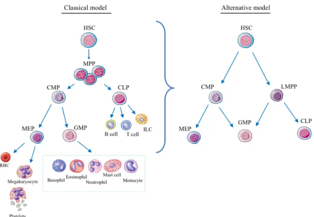
. Different models have been proposed defining specification branch points and timing of the generation of downstream progenitors (Figure 1). While the classical model splits differentiation into myeloid and lymphoid branches 10,11, an alternative model proposes a three axes panorama derived from a common myeloid progenitor (CMP) and a lymphoid-primed multipotent progenitor (LMPP) (Figure 1) 12
. The LMPP gives rise to the common lymphoid progenitor (CLP) that differentiates in T-, B-lymphocytes and innate lymphoid cells while the megakaryocyte-erythrocyte progenitor (MEP), derived from the CMP, produce platelets and erythrocytes. The novelty of this revisited model of hematopoiesis is that both the myeloid primed (CMP) and the lymphoid primed (LMPP) progenitors maintain the capacity to switch to the granulocyte-macrophage progenitor (GMP) that generates monocytes and macrophages, granulocytes and dendritic cells (DC).
Figure 1: Models of human hematopoiesis hierarchy. A) Classical model of the hematopoietic hierarchy with a strict separation between the myeloid and lymphoid branches as the first step in lineage commitment downstream of the hematopoietic stem cell. (B) Alternative model as proposed by Adolfsson et al, incorporating the identification of LMPPs.
A
DAPTIVE IMMUNITYThe adaptive immune system is formed by two broad sets of antigen-responsive cells: B (bursal or bone marrow–derived) lymphocytes and T (thymus-derived) lymphocytes, which in the blood and secondary lymphoid organs can be identified as CD19+
and CD3+ cells, respectively. In steady state, naïve B and T cells harbor a highly diverse repertoire of antigen (Ag) specificities. Upon encounter with cognate antigen, they proliferate massively generating antigen-specific ‘clones’ that differentiate into the effectors of immunity. Upon antigen clearance, a fraction of ‘memory’ cells remain that is the basis for the antigen-specific recall responses, which has been classically considered a hallmark of adaptive immunity 14,15
. In the case of B cells the antigen specificities come from immunoglobulin receptors (Ig) that after engaging with an antigen, they trigger cell activation, clonal expansion and differentiation into antibody producing plasma cells. Antibodies are capable of recognizing three-dimensional structures and interact with pathogens leading to its neutralization. In contrast, recognition of antigen by T cells involves T cell receptor complex (TCR) engagement of peptides presented by class I and II major histocompatibility complex (MHC) molecules. These peptide/MHC complexes are expressed on the surface of antigen presenting cells (APCs), especially dendritic cells (DCs) 16
. Accordingly, DC are the ‘gatekeepers’ of immunity and they dictate immune response initiation and coordinate innate and adaptive immune activation.
Thymic selection generates two types of T cells: those expressing a TCR receptor formed by αβ chains and those carrying γδ-TCR. γδ-T cells are much less understood and they are peculiar in that they have characteristics that place them in the border between innate and adaptive immunity. Their repertoire heterogeneity is poorer and they localize in very precise sites such as skin and some mucosal surfaces. The biology of γδ-T cells will be further developed below.
On the basis of the co-receptor expressed αβ-T cells are divided into CD8+ cytotoxic T cells and CD4+ helper T cells. Ag-activated helper T cells will differentiate into different phenotypes, depending on the stimulus and cytokines in the local environment, which can be characterized by their cytokine profile and by transcription factors. Th1 cells produce IFN-γ and interleukin-2 (IL-2) and express T-bet. Th2 cells produce IL-4, IL-5 and IL-13 and express GATA-3. Th17 cells produce IL-17 and IL-22 and express RORγt. A fifth type of “conventional” T cells, responsible for the maintenance of peripheral tolerance and down-modulation of immune responses, are called regulatory T cells (Treg). These can be divided
into different subsets based on the expression of FoxP3 and/or the production of IL-10, TGF-β and IL-35. Recently a new equilibrium model of immunity has proposed where Tregs can differentiate into specialized type 1, 2 or 3 subsets depending on the associate effector axis triggering the action 17–20
.
I
NNATE IMMUNITYUnlike the adaptive immunity that appeared later in evolution, the innate arm of the immune system developed already in the first multicellular organisms. Intimately related to their phylogeny is the mechanism these two systems use to recognize their target. By contrast to the specificity shown earlier for T and B-lymphocytes, innate effectors have been classically seen as “broad responders”. Innate effector cells bear ‘sensors’ that are capable of recognizing conserved structures shared by large groups of pathogens (termed microbial-associated patterns (MAMPs) 21,22
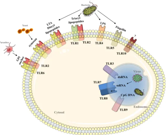
such as the lipopolysaccharide (LPS) that is present in all gram-negative bacteria). As such, the innate sensors are referred to as pattern recognition receptors (PRRs). The four PRR families include Toll-like receptors (TLR), nucleotide oligomerisation receptors (NLR), C-type lectin receptors (CLR) and RIG-1 like receptors (RLR). In humans, TLRs include 10 members, each of them with a broad range of specificities (Figure 2) 23
. Engagement with TLR ligands triggers a signaling cascade that concludes with the activation either of the MAP kinase, the NFkB or the IRF pathway. Eventually TLR stimulation leads to the production of pro- or anti-inflammatory cytokines, type I IFNs, chemokines and chemokine receptors, anti-microbial and co-stimulatory molecules and the enhance of the antigen uptake and presentation (Table 1) 24
. Also non-infectious endogenous biomolecules called damage-associated molecular patterns (DAMPs) or alarmins can bind to TLRs promoting or exacerbating the inflammatory response in a context of stress. The fundamental role of TLRs has been evidenced by several studies that compared the disease susceptibility of individuals carrying different polymorphisms in genes that participate in TLR signaling (Figure 2). These included sepsis, immunodeficiencies, atherosclerosis and asthma, among others, suggesting a great therapeutic potential in the manipulation of these receptors 25
. Indeed, recent investigations have exploited TLRs as adjuvants in vaccines and as tumor immunotherapeutics and have also assessed the potential of TLR agonists in autoimmune and inflammatory diseases 26,27
. Like TLRs, CLRs are found in contact with the extracellular space surveying for the presence of microbial ligands. This greatly heterogeneous family shares the characteristic C-type lectin-like domain and its two best characterized members are Dectin-1 and Dectin-2. These receptors signal through ITAM-like domains in myeloid cells including DCs, macrophages, neutrophils and monocytes. They are fundamental in the recognition of fungal β-glucans and α-mannans, such as in Candida albicans, as well as patterns of Listeira and Mycobacterium 28
to the previous PRR families, NLRs and RLRs are located in the cytoplasm where they patrol for the presence of intracellular pathogens. NOD1 and NOD2 are the prototypical members of the NLR family and they both detect components of the bacterial outer membrane or cell walls29,30
. After binding to those ligands, a signaling complex is assembled leading to the activation of NF-κβ and MAPK pathways. The importance of this family of receptors is evidenced in Crohn’s disease patients that express a NOD2 variant with impaired responsiveness and incorporation of bacteria within the autophagosomes. Finally, RLRs are a group of three helicases that detect the presence of foreign RNA within the cytosol. They do so by recognizing features common to viral genomes and replication intermediates, such as the poly-U region in the HCV31,32
. Their role is critical in the immune defense against RNA virus but they also participate in responses against DNA virus and bacteria pathogens.
Figure 2: TLR recognition of conserved molecular structures in bacteria, viruses, fungi and parasites.
Elie Metchnikoff identified the first innate immune effector cell, the macrophage, as a key player in cellular defense. Many others came after and today myeloid cells can be morphologically divided in two types: mononuclear and polymorphonuclear cells (or
granulocytes composed by neutrophils, eosinophils, mast cells and basophils) that provide protection against bacteria and parasitic infections via release of toxic and inflammatory molecules 33
. The mononuclear phagocytes include the macrophages and their precursors, the monocytes, that reside allover the body regulating fibrosis and repair and contributing to immune surveillance and inflammation in a tissue-specific manner 34,35
. Also, the rich and essential group of dendritic cells (DCs) belongs to this myeloid family. While myeloid cells have long been considered essential for innate immunity, we realize now that cells of lymphoid origin, such as innate lymphoid cells (ILCs) and two peculiar populations of antigen-rearranged lymphocytes, γδT cells and B-1 cells, are likewise innate effectors due to their rapid and non-Ag specific activation. Given the importance of DCs and ILCs in innate immune responses, their biology will be more deeply explored in the following sections.
i. D
ENDRITIC CELLS
In 1976 Steinman & Cohn described a novel cell type in peripheral lymphoid organs with characteristic morphological ‘dendrites’ that they baptized as the ‘dendritic’ cell or DC 36
. Little did they know at that time that these “rare” DCs would be so critical for the proper functioning of the immune system and that DC biology would become a magnet for decades of intense immunology research. The description of other cells sharing immunogenic properties, phenotypic characteristics and morphology with DCs followed, building up the heterogeneous family that we know today (Figure 3). DCs are the professional antigen presenting cells (APCs) of the immune system that patrol secondary lymphoid tissues, most peripheral tissues and non-lymphoid organs. They are equipped with a molecular machinery to capture and process antigens, present them to T lymphocytes and provide co-stimulatory signals that shape immune responses. Numerous mouse studies have shown that DCs comprise a heterogeneous family with diverse ontogeny, locations, migration patterns and roles in immunity (Figure 3). The biology of human DCs, particularly in tissues, is less well understood and most of our knowledge on human DCs derives from blood studies. Some of the recent observations showed a high differentiation capacity of some DCs in circulation suggesting that within these there are precursors or immature DCs with different phenotype from tissue DCs 37–39
.
Myeloid DC-restricted precursors that exit the BM populate different tissues where they undergo differentiation in situ to give rise to two type of mature DCs: resident DCs that
will populate lymphoid tissues, and migratory DCs that will move from non-lymphoid tissues to the lymph nodes. The latter DC subset concomitantly up-regulates maturation markers upon migration and present tissue-derived antigens to T cells 40,41
.
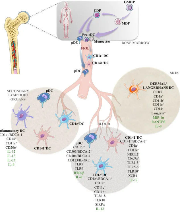
Three subsets of DCs can be found in blood, spleen and tonsils within the lineage negative HLA-DR+
population: plasmacytoid DCs (pDCs, with plasma-like morphology instead of the typical dendritic shape) and two populations of ‘classical’ DC (cDCs) with abundant dendrites 42–44
.
PLASMACYTOID DCS express a cluster of markers including CD123 (IL-3R), CD303
(BDCA-2) and CD304 (BDCA-4 or Neuropilin-1) (Figure 3). Recent work has revealed a rather multifaced identity of pDCs, being immunogenic or tolerogenic depending on the context 45
. pDCs play fundamental roles in the defense against viral infections through their potent type I interferon activity 46
. They can detect nucleic acids coming from virus, bacteria or death cells through TLR7 and TLR9 triggering the signaling cascade that ends up with the production of IFN-α, IFN-β and also IFN-λ 47–49
.
In the steady state, thymic pDCs have a tolerogenic role by priming Tregs to produce IL-10 and TGF-β and regulating their proliferation and survival 50,51
. This function is accompanied by a low CD4+
stimulation capacity and MHC-II expression. Recent work showed that this type of tolerance involves a CCR9-dependent transport of peripheral Ags and subsequent deletion of Ag-reactive thymocytes 52
. The tolerogenic machinery of pDCs has been reported as well in pathological conditions via TLR-independent activation (alternative stimulation). This results in the expression of indoleamine 2,3-dioxygenase (IDO) and inducible T cell co-stimulator ligand (ICOSL) that leads to induction of Treg expansion and to an increase in IL-10 levels. 53–55
. This non-canonical pathway has been studied in the context of cancer, and is associated with a poor prognosis.
In contrast, pDCs become great drivers of both innate and adaptive responses following activation where they increase the expression HLA-DR and co-stimulatory molecules (CD40, CD80, CD86) and undergo a morphological change that converts them in functional APCs capable of presenting Ag to naïve CD4+
T cells and cross-priming CTLs 47 . Furthermore, activated pDCs production of IFN-α, IL-12 and IL-18 results in a massive activation of NK cells and CD8+
cells as well as Th1 polarization. Interestingly, some viral infections like HIV-1 lead to chronic immune activation that impairs pDC mediated IFN-α
production via enhanced CD40-CD40L interactions, evidencing the importance of this immune mechanism in antiviral response.
Moreover, pDCs can secrete other pro-inflammatory cytokines (such as IL-6 that can drive Th17 cell commitment) and chemokines (such as CXC-chemokine ligand 8 (CXCL8), CXCL10, CC-chemokine ligand 3 (CCL3) and CCL4) that attract other immune cells to the sites of infection or inflammation. Given the importance of migration for the activity of these cells, it is not surprising that their function relies on the expression of several chemokine receptors, such as CXCR4 (development on the BM and migration to the spleen and LNs) and CCR2/CCR5 (blood recirculation) 56,57
.
CONVENTIONAL OR CLASSIC DCS are predominant in human blood and have been also
reported in LNs and spleen as well as in non-lymphoid tissues including skin, liver, lung and gut. Two major cDC subsets can be identified by surface markers (Figure 3): CD1c+
DCs that co-express CD11b and high levels of CD11c and CD141+ cDCs that are lower for CD11c expression and negative for CD11b but express DNGR-1 (Clec9a). These cDC subsets differ in their TLR expression patterns: CD1c+
DCs express all TLRs except for TLR9 while CD141+
DCs have low expression of TLR1-2, TLR6 and TLR8 and high expression of TLR3 and TLR10 58,59
.
In view of this TLR expression pattern, it is not surprising that ex vivo isolated blood CD1c+
cDCs showed a broader cytokine expression capacity and in some cases also a more potent cytokine production (for IL-12, for example). Additionally it has been reported that TLR6 mediated CD1c+
DC production of IL-23 and shaping of Th17 response has a protective role on asthma 60
. By using human lung tissues and humanized mouse models, researchers found that specifically CD1c+
DCs are capable of driving CD103 expression on CD8+
cells and promote their accumulation in lung epithelia 61
. In contrast, some unique properties have been attributed to CD141+ DCs, such as their expression of the chemokine receptor XCR1 that allows them to migrate in response to the XCL1, secreted by NK cells and CD8+
T cells 62 .
Progress in the field of human DC research has been boosted in recent years thanks to in-depth proteomic and transcriptomic analyses and to the development of humanized mouse models that can be used to study the biology of these cells in vivo. Despite their phenotypic and functional differences, the two cDC subsets share characteristics including the capacity to uptake, process and present peptides to naïve CD4+
or CD8+
terms of MHC-I expression 63
and in stimulatory capacity of CD4+
T cells in mixed lymphocyte reactions (MLR). Naïve T cell differentiation and polarization is highly dependent on cDCs as evidenced by the immunodeficiency observed in individuals with IRF8 mutations (a transcription factor necessary for the development of mononuclear phagocytes) 64
. When referring to T-cell polarization the activity of the different subsets depends greatly on the TLRs and the cytokines implicated in the reaction. Many studies have focused on comparing the two cDC subsets in terms of capacity to elicit individual cytokine production under stimuli of different nature in distinct tissues. That detailed analysis is beyond the scope of this manuscript and has been reviewed elsewhere 41
. In brief, both cDC subpopulations have been associated to Th1, Th2 and Th17 CD4+
polarization in various organs (LNs, lung and liver) with variable intensity depending on the nature of the stimulus.
The presentation of exogenous antigens on MHC-I molecules to CD8+
T cells that turns out in the initiation of the immune response is another fundamental characteristic of cDCs. In vivo CD1c+ DCs, CD141+ DCs and pDCs show equal intrinsic cross-presentation capacity although some may be specialized in certain types of antigens, like CD141+
DCs with necrotic cell-associated antigens 62,63
.
MONOCYTE-DERIVED DCS AND INFLAMMATORY DCS
Monocytes account for the majority of the phagocytic cells in human blood. They can be distinguished from DCs by the expression of CD14 but they share some of the other markers, like CD11c (Figure 3). Based on their expression of the low affinity Fc receptor CD16, they are classified in three subtypes: (1) classical CD14++
CD16−
(circulating blood guard that migrate to tissues upon inflammation), (2) intermediate CD14++
CD16+
(main producer of inflammatory TNF-α in situ), and (3) non-classical CD14+
CD16++
monocytes (IL-12, TNF-α and IL-1β producers) 65
. Under homeostatic conditions monocytes patrol the body complementing the tissue surveillance of DCs but when inflammation occurs monocytes can differentiate into macrophages and dendritic cells, the so called monocyte-derived DCs or inflammatory DCs. These processes add more difficulties to the classification and distinction of “plastic” populations and discrete subsets. In vitro-generated moDCs using GM-CSF have been used as a model to study this cells 66
; however in vivo this “plastic” events seem to happen in situ. In mice, infDCs have demonstrated similar capacities as DCs, including production of pro-inflammatory cytokines, T cell priming and polarization of Th responses. However, the study of these cells in humans has been challenging and only a few
reports claim the presence of infDCs with similar characteristics than those observed in mice in the context of atopic dermatitis, rheumatoid arthritis, and tumor ascites 67–69
. As such, our understanding of infDC function in humans is limited; humanized mice may provide a meaningful in vivo system to further study infDC biology.
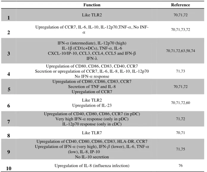
Table 1: Main functions of toll like receptors in humans.
DENDRITIC CELL DEVELOPMENT
As previously mentioned, DCs derive from GMP in the bone marrow, circulate in the blood and home to the tissues throughout the body. However, the identification of human precursors restricted to one or several DC/monocytes/macrophage subsets and the understanding of the relationships between these precursors has been challenging due to the lack of suitable culture and in vivo systems. It has been suggested that the higher proliferation
4 Function Reference
1
1 Like TLR2 70,71,72
2 2
Upregulation of CCR7, IL-6, IL-10, IL-12p70,TNF-α, No
INF-α 70,71,73,72
3 3
IFN-α (intermediate), IL-12p70 (high) IL-1β (CD1c+DCs), TNF-α, IL-6 CXCL-10/IP-10, CCL3, CCL4, CCL5 and IFN-β
IFN-λ
70,71,72,63,58,74
4 4
Upregulation of CD80, CD86, CD83, CD40, CCR7 Secretion or upregulation of CCR7, IL-6, IL-8, IL-10, IL-12p70
No IFN-α response
71,73
5 5
Upregulation of CD80, CD86, CD83, CCR7 Secretion of TNF and IL-8
Upregulation of CCR7 70,71,72 6 6 Like TLR2 Upregulation of IL-23 70,71,72,60 7 7 Upregulation of CD40, CD80, CD86, CCR7 (in pDC) Very high IFN-α response (only in pDC)
IL-12p70 response (only in cDC)
71,72 8 8 Like TLR7 70,71 9 9 Upregulation of CD40, CD80, CD86, CD83, HLA-DR, CCR7 Upregulation of IFN-α (very high), IFN-β (lower), IL-6, TNF-α
(low), IL-8, IP-10 No IL-10 secretion
71,75
É
capacity and lower expression of some maturation markers in circulating DCs indicated that those are, indeed, the precursors of DCs 38
. Recently, two studies described, both at bulk and single cell level, the hierarchy of myeloid precursors and proposed a developmental relationship between monocytes, cDCs and pDCs. Using a novel in vitro system and HIS mice, they proposed that GMP give rise to a monocyte-DC progenitor (MDP), which then differentiates, into a common DC progenitor (CDP) that produces the three main DC subsets. Furthermore, they report the identification of a circulating immediate DC precursor (hpre-cDC) that is restricted to the cDC subsets and expands in response to Flt3L (Figure 3) 39,77
. The differentiation of HSCs via DC precursors to mature DCs is regulated by the environmental signals that includes cytokines, soluble factors and cell-to-cell contacts. Early attempts to generate DCs from hematopoietic precursors in vitro revealed that two cytokines were pivotal: GM-CSF and Flt3L. However, while the first gave rise to “monocyte-like” DCs, the second was unique in driving the generation of cDCs and pDCs.
The cytokine Flt3L (fms-like tyrosine kinase 3 ligand) is ubiquitously produced by stromal cells, endothelial cells and activated T cells. Its receptor, Flt3 (also called CD135 and Flk2) is strongly expressed by early hematopoietic precursors (GMP, CLP) and is maintained on dendritic cells lineage restricted precursors and mature pDC and cDCs (Figure 3) 78
. Accordingly, ablation of Flt3 is correlated with loss of DC differentiation potential 79
. On the other hand, Flt3L administrated in mice and in humans leads to a drastic systemic expansion of both pDCs and cDCs 80,81
. Therefore Flt3L is not only important for the development but also the homeostasis (survival, proliferation) of peripheral DCs 78
Figure 3: Schematic view of human dendritic cell development The main surface markers for each subset are shown as well as the cytokines they produce (in green). GMDP: granulocyte, monocyte and dendritic cell (DC) progenitor; CDP: common DC progenitor; MDP: monocyte-‐DC progenitor; pre-‐cDC: commited precursor of classical DCs.
ii. I
NNATEL
YMPHOIDC
ELLSInnate lymphoid cells are a recently identified family of innate effectors. Two of its founding members: the natural killer cell (NK cell) and the lymphoid tissue inducer cell (LTi cell) have been known and studied for many years but the identification of new subsets with similar characteristics prompted their “clustering” as a distinct but heterogeneous effector lineage. ILCs lack recombination activating gene (RAG)-dependent rearranged antigen receptors and share a lymphoid cell morphology. They can be identified by the lack of lineage markers (CD3/CD19/CD14/CD33…) but express CD7 and CD127 (except for CD56dim
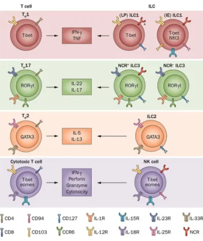
NK cells). Interestingly ILC effector functions and transcription factor dependency mirror those observed in T cell subsets prompting the speculation that ILCs are ‘innate’ counterparts of differentiated T helper and cytotoxic lymphocytes (Figure 4). Accordingly, we can distinguish three broad groups of ILCs paralleling the three major axis of T cell specialization. ILC1s depend on the transcription factor T-BET and can be subdivided into cytotoxic ILC1 or NK cells (like CTLs) and ILC1s that preferentially produce type 1 cytokines (like Th1 cells). Likewise, ILC2s and ILC3s are reminiscent of Th2 and Th17/Th22 cells in that they depended on
GATA-3 or RORγt,
respectively, and they elicit the corresponding cytokine response (see in detail below). NK cells appear distinct from other ‘helper’ ILCs in that they have different requirements for cytokines and transcription factors (reviewed in 82
).
Figure 4: Innate lymphoid cells closely reseamble T cel subsets.
TYPE 1ILCS
For almost forty years, NK cells have been considered the prototypic (and in some cases only) innate source of type 1 cytokines during the early phases of the immune response. Several studies in the last decade, first in mice and then in humans, have identified additional phenotypically diverse ILC1 subsets (particularly inside the tissues) that provide innate sources for IFN-γ. Before discussing these different ILC1 subsets, I will briefly present some of the general features that characterize human NK cells.
Natural Killer Cells
Natural killer cells owe their name to the unique ability to kill transformed or virus infected cells without prior sensitization 83–85
. Although originally described by this natural capacity to kill, NK cells are also major sources of type 1 cytokines, notably IFN-γ, TNF-α and GM-CSF, and to a lesser extent other cytokines and chemokines (MIP-1α, MIP-1β and RANTES) 86
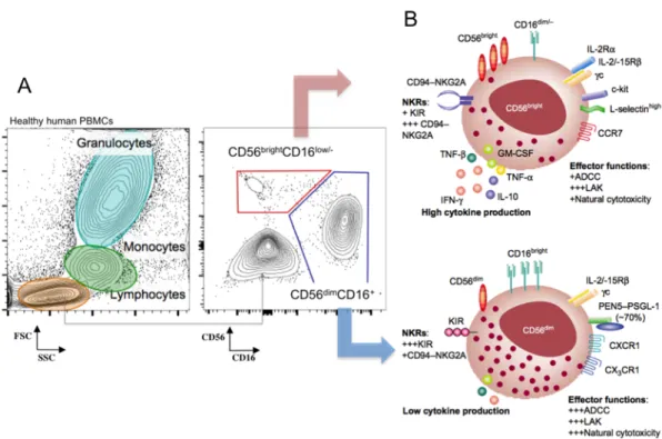
. Even before the discovery of additional ILC1 subsets, a high degree of heterogeneity was already appreciated among NK cells by virtue of the distinct lineage differentiation stages, the variety of effector functions and their tissue localization. In humans two major subsets can be distinguished in the circulation by their expression of the adhesion molecule CD56, namely CD56bright
and CD56dim
subsets (Figure 5). CD56bright
cells are considered to give rise to the fully mature CD56dim
cells, based on the order of appearance in HSC transplantation experiments and cytokine driven models of in vitro NK cell differentiation, and also to the length of the telomeres observed in the two subsets 87,88
. CD56dim
cells express the low affinity Fc receptor CD16 (responsible for the antibody-mediated cell cytotoxicity, ADCC) and account for 90% of the peripheral blood NK cells. This subset is highly cytotoxic thanks to the cargo of effector molecules, granzymes, perforin and Fas ligand, contained in secretory lysosomes that provide them with a characteristic granular morphology 89
. The CD56bright
subset, contrarily, has a low-density expression of CD16 but exhibits a high potential for cytokine production under monokine or target cell stimulation. These cells are also hyper-responsive to IL-2 due to the expression of the high-affinity heterotrimeric IL-2 receptor thus showing a higher proliferative potential and the capacity to acquire potent lytic function. CD56bright
are better at penetrating the secondary lymphoid tissues thus accounting for the majority of the NK cells in lymph nodes or decidua, whereas in lung NK cells have a very differentiated profile.
Figure 5: Identification of human peripheral blood NK cells by flow cytometry (A) and general phenotype and effector funciton of CD56bright and CD56dim NK cell subsets (From Cooper et al, 2001) (B).
Given the modulation of CD56 and CD16 expression after activation, this dual classification of NK cells may be inaccurate in the context of tissues. For example, in the decidual tissue NK cells are CD56superbright
accounting for around 70% of the lymphocytes during the first months of the pregnancy where they interact with the trophoblast and other immune cells regulating arterial remodeling and placenta development 90
. These cells, called uterine NK cells (uNK) are considered a distinct NK population as they display some other characteristics different from pNK cells like a particular pattern of KIR expression and the over-expression of CD9, galectin, alpha-1 integrin, as well as other adhesion molecules 91,92
. Interestingly, these NK cells are able to induce Treg cells in vitro indicating a role in maternal
tolerance 93,94
. Even within the CD56dim
subset one can find diverse “stages” with particular phenotypic and functional properties throughout their final differentiation 95,96
. This progression from CD56dim
to terminal differentiated cells is accompanied by the progressive loss of the proliferation capacity and the acquisition of more efficient cytolytic activity. Phenotypically, down-regulation of CD94 and NKG2A and up-regulation of CD16, KIR and perforin expression characterize this process that terminates with a CD57-expressing highly cytotoxic mature population 97
. Interestingly, cytomegalovirus infection (CMV) leads to selective expansion of this terminal mature subset, the drastic up-regulation of NKG2C expression 98,99
and the display of certain hallmarks of adaptive immunity 100
Despite their rapid and potent response upon viral infection or transformation, NK cells are efficiently rendered ‘self-tolerant’ thereby avoiding potential autoimmunity. This is achieved by an ‘education’ process generating an array of activating and inhibitory receptors that maintain the NK cells on alert while tolerating “self” 101
. Additionally, NK cells constitutively express monokine receptors that allow them to respond strongly to cytokines such as IFN-α, IL-2, IL-12, IL-15 and IL-18, which are produced by surrounding cells like monocytes and dendritic cells 102
. It is therefore the combined effects of cytokines and receptor-ligand engagement that condition NK cell responses 103
.
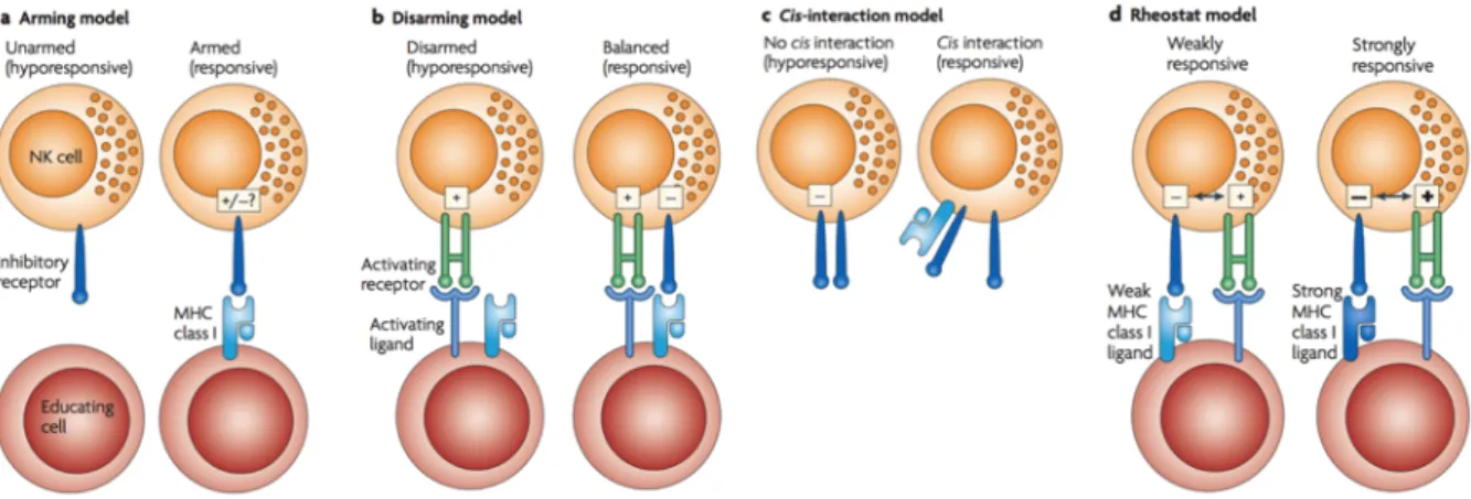
The training of a self-tolerant NK cell is dependent on the signaling through ITIM bearing inhibitory receptors 104–106
and takes place by a mechanism not yet agreed by the experts. Four main mechanisms have been suggested (Figure 6): “the arming” hypothesis establishes that NK cells are initially unresponsive and the encounter of MHC-I by inhibitory receptors during development licenses or “arms” them (Figure 6a) 104
. The opposite possibility has also been suggested, that is the “disarming” hypothesis, stating that initially responsive NK cells would become anergic after chronical stimulation unless engaged by MHC-I specific to its inhibitory receptors (Figure 6b) 107
. A third mechanism refers to as “cis-interaction model” relies on the ability of KIR receptors to bind to MHC-I molecules in cis and is based on the observation that KIR receptors can transmit inhibitory signals even in the absence of ligand interaction, but it is unclear whether this model can be applied to all KIR receptors (Figure 6c) 108
. Finally the “rheostat model” proposes that NK cell reactivity is tuned by the number and degree of affinity of self-MHC-I inhibitory receptors carried by a cell (Figure 6d) 107,109
. As such, NK cells experience a quantitative functional adaptation that generates MHC-I educated NK cells responding efficiently to stimulation and responding as well to aberrant cells that have lost MHC-I (missing-self recognition) 110
.
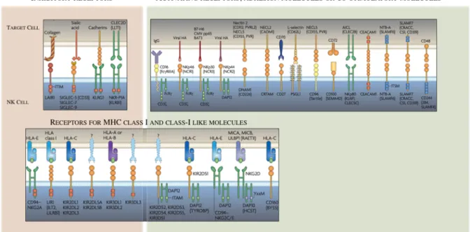
Other ITIM bearing receptors contribute to maintaining NK cells on control, including NKG2A that binds the non-classical MHC-I molecule HLA-E, and LIR-1 that engages both classical MHC-I and the non-classical HLA-G 111,112
. Additionally, non MHC-I ligands can bind ITIM-containing receptors, this is the case of KLRG1 that binds cadherins and participate in tumor invasion and metastasis detection 113,114
, and CD161 that recognizes LLT1 (osteoclast inhibitory lectin or CLEC2D, expressed by DCs and B cells) thus regulating NK cell activation (Figure 7) 115
.
Unlike T cells, allogeneic NK cell transplantation leads to graft-versus-leukemia responses without causing graft-versus-host disease (GVHD) 116
. This suggests that NK cells are not pre-wired to kill in mismatched contexts but other positive stimuli contribute to triggering the response. This positive recognition of « altered-self » or pathogen derived molecules is carried out by activating receptors that express or not ITAM-carrying adaptors. Among those that signal through ITAM are the activating KIRs, such as KIR2DS and KIR3DS, as well as the heterodimers CD94-NKG2C and CD94-NKG2E 117,118
. Intriguingly, both inhibitory NKG2A and activating NKG2C heterodimers bind to the same ligand suggesting that a more sophisticated mechanism of NK regulation might exist. ITAM-independent receptors include NKG2D whose ligands are MICA-B and ULBPs, induced in stressed cells (transformed or virus infected) 119,120
and also the family of SLAM-related receptor (SRRs), notably 2B4 greatly contribute to the regulation of NK cell function when binding CD48-expressing cells 121
(Figure 7).
An additional family of activating receptors, the natural cytotoxicity receptors (NCRs) –NKp46 (NCR1), NKp44 (NCR2), NKp30 (NCR3)- are expressed in activated and resting NK cells (Figure 7). The role of this family of receptors in human NK cell-mediated killing was demonstrated by Moretta and colleagues in culture experiments containing human NK clones and NK-susceptible tumor cells. Addition of anti-NCR monoclonal antibodies directed against individual NCRs resulted in partial inhibition of cytotoxicity whereas the combined use of all three mAbs strongly abrogated cytolysis 122
. This work suggested that NCRs cooperate in target cell recognition and killing and that the extent of that cooperation depends on the density of the ligands on target cells. Furthermore, experiments in mouse showing the reduction of the tumor cell lysis by NK cells lacking one or more NCRs reflect their high importance in in vivo tumor surveillance 123
. Several studies have also reported exogenous ligands for NCRs (reviewed in 124
NKp44 and NKp46 further evidencing the protagonist role of these receptors in NK cell function 125–127
The outcome of the receptor-ligand binding not only depends on the identity of the receptor itself but also by the adaptor proteins associated to the cytoplasmic domains. As such, the 2B4 NK receptor (also called CD244) behaves as a multifunctional receptor and the outcome of its engagement seems to depend on the stage of NK cell maturation and activation. Ligation of 2B4 with a specific antibody or with CD48-expressing cells lead to NK activation while in patients deficient for SAP (a 2B4 adaptor protein) that engagement results in inhibition of NK cell function 115,128
Tens of other activating NK receptors have been identified as well as co-stimulatory receptors that act synergistically ensuring the activation of NK cells upon “unhealthy” circumstances and not otherwise and modulating the intensity and type of immune response. These include DNAM-1, NKR-P1, PILR, LFA-1, CD2 and others depicted in Figure 7. A more exhaustive description of these receptors can be found in several excellent reviews published over the last decades 124,129,130
.
Figure 7: Activating and inhibitory receptors of human NK cells. (Adapted from Nature Reviews Poster, Vivier&Ugolini)
Surface NK cell receptors serve as landmarks that dissect the stepwise differentiation of HSCs to mature NK cells. Human NK cells can be detected in fetal liver from week 6 of gestation and in BM, lymph nodes, spleen, lung and intestine during the second trimester 131
. In adults, early stages of NK cell development take place in the BM whereas some
multipotent precursors may exit to extramedullary tissues where molecular signals drive their final differentiation 132,133
. This process is regulated by the integrated influence of soluble factors and transcriptional regulators that modulate lineage-specific gene expression134,87,135
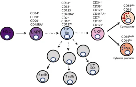
. Restricted NK precursors, including pro-NK and pre-NK (CD34+
CD45RA+
CD117+ CD94−
CD122+
) cells retain some multi-lineage potential, suggesting that these populations were not fully NK cell committed. Recently, a refined NK-restricted progenitor (rNKP) downstream of CLP has been defined with exclusive potential to the NK cell linage after in
vitro single-cell and in vivo transfer experiments (Figure 8)136
. These cells, identified by the cell surface expression profile Lin−
CD34+ CD38+ CD123− CD45RA+ CD7+ CD10+ CD127− are localized in fetal liver (FL), BM, cord blood and adult tonsils. Other upstream NK precursors that retain the capacity to differentiate not only in NK cells but also in helper ILCs have been proposed recently and will be discussed later.
Figure 8: Model of early NK cell development. (From Renoux et al., 2015)
NK cells are present in most compartments of the human body, from primary and secondary lymphoid tissues to peripheral organs. These NK cells have two origins, some of them are PB-NK cells that patrol the circulation and get recruited to the tissues, particularly upon pathologic insults to exert locally an immune response. In contrast, a second NK cell subset resides permanently in tissues, and shows phenotypic and function specialization shaped by the local environment. This tissue resident NK cell (trNK) subpopulations have been associated to the uterus (discussed earlier), skin, thymus, liver, intestines and salivary gland (at least in mice). In addition to their capacity to respond more rapidly, trNK share some attributes previously described for human and mouse resident-T cells. trNK express
CD69, which inhibits surface expression of sphingosine-1-phosphate receptor 1 (S1PR1) thereby promoting their retention in tissues 137
; CD69 is absent from PB-NK cells but is expressed by skin, liver, uterus, lymph nodes and intestine human NK cells 138–142
. Additionally, other hallmarks for tissue residency, including the expression of CD103 (that binds E-cadherin in epithelial cells) and/or CD49a (that promotes homing to non-lymphoid tissues), the tissue homing receptors CXCR6 or CCR5, and the lack of the adhesion molecule CD62L have been ascribed to some human NK cells in those organs 143
. With the establishment of ILCs as a family that distinguishes between cytotoxic (mostly recirculating) NK cells and helper (mostly resident in steady-state) ILCs some of the trNK cells have been re-classified as non-NK ILC1s 144
. Helper ILC1s
The discovery in 2006 of IFN-γ-producing cells that, unlike NK cells, depend on the transcription factor Gata-3 and express IL-7 receptor (CD127) in the thymus suggested diversity in innate lymphoid developmental pathways. We know now that these two characteristics are shared by ‘helper’ ILC subsets thus placing thymic NK cells as a group 1 ILC member 145,146
.
To date four different ILC1 populations have been identified in mice by virtue of their phenotype and dependency on T-bet and Eomes transcription factors. However these subsets have overlapping yet distinct phenotypes and functions (in an organ-dependent fashion) which makes an unambiguous ILC1 classification a challenge. The limited access to organs and the lack of in vivo genetic tracing or modifying technologies in humans further complicates this endeavor. As such, human ILC1 are largely defined based the absence of other known markers for NK cells, ILC2s or ILC3s. These include the high expression of CD127 and CD161 and the absence of CD56, CD94, NKp44, NKp46, c-kit, granzymes and perforin. Human ILC1 produce IFN-γ but not IL-13, IL-17 or IL-22 and are T-BET-positive but lack RORγt, GATA-3 or RORα 147. Given their localization, the post-birth emergence and the fact that they expand in the context of Crohn’s disease these ILC1s are likely involved in the early immune response and maintenance of homeostasis at mucosal barriers (Figure 10). Indeed, mouse models of innate inflammation showed that Ab-blockade of IFN-γ production ameliorated the local and systemic inflammation related pathology 148
.
In the interface between the phenotypic definition of ILC1 and NK cells, intraepithelial ILC1 patrol the gastrointestinal mucosa watching out for danger signal from epithelial and myeloid cells. Fuchs and colleagues described this IEL population both in the
human tonsils and intestine (NKp44+ CD103+ ) and in mice (NKp46+ NK1.1+ CD160+ ), where they develop independently of IL-15 149
. These ILC1s express low levels of CD127, CD56, NKp44 and CD94 and produce IFN-γ in response to IL-12 and IL-15 (Figure 9). Interestingly, they showed several hallmarks of TGF-β imprinting, such as expression of CD103 and CD9, characteristics also observed in uNKs. Like cNK cells they depend on T-BET and EOMES but develop independently of IL-15Rα (al least in mouse) pointing to a dependency on other cytokines such as IL-7 or IL-2 149
. Recently, a study on the PB-ILC repertoire reported CD127+
CRTh2
-CD117
cells expressing some T cell markers (cytoplasmic CD3, CD4, CD8, CD27 and CD28) but lacking αβ-TCR, γδ-TCR in the cytoplasm and CD3-TCR complexes in the cell surface 150
. Single-cell RNA sequencing of tonsil CD127+ ILCs was also able to detect this population albeit with a low T-BET expression and high expression of some T cell-associated genes, raising the need for further studies that corroborate the real identity of this intriguing population 151
.
The identification in mice of a IFN-γ-producing population in liver distinct from NK cells that displays some adaptive-like features prompted the search for a human counterpart 152
. As such, CD49a+
ILC1s that depend on T-BET but not on EOMES were found in human liver but not in the hepatic venous or in peripheral blood. Moreover, they express KIR and NKG2C and are CD56bright
but are low for CD16, CD57 and perforin. Consequently, they can express high levels of pro-inflammatory cytokines but degranulate poorly 139
. However, the highly phenotypic similarity with conventional NK cells and their lack of IL-7R (common to all the other hILCs) suggests that these cells may be more like NK cells than ‘helper’ ILC1. A recent report has argued the existence of this population and has proposed instead a novel subpopulation of CD49e
liver resident NK cells by using cytometry by time-of-flight (CyTOF) and humanized mice.
In mice an additional ILC1 population has been described in salivary glands that resembles liver ILC1 in the expression of CD49a and TRAIL but, like cNK cells, are DX5+ and rely on Eomes. Additionally, SG ILC1s are poor producers of IFNγ contrasting with ILC1 hallmark characteristics 153,154
. It remains to be investigated the presence and physiological importance of this population in man. A very recent report described for the first time in human lung the presence of ILC1 but further phenotyping and functional studies are needed to confirm their identity 155
. Likewise, an uterine ILC1 population has been mentioned recently capable of producing IFN-γ but different of the aforementioned uNK cell
in that they do not express EOMES and they are CD49a-positive, thus resembling the cells mentioned earlier in mucosal tissues 156
.
It is essential for the accurate identification of these populations to keep in mind that some of the aforementioned markers maybe modulated in the context of inflammation or activation, like in the case of CD49a 157
. Also, fate-mapping experiments in mice and in vitro assays in humans revealed transdifferentiation of ILC2s and ILC3s into IFN-γ-producing cells by the upregulation of T-bet and downregulation of Gata-3 or RORγt further complicating the in vivo identification of ILC subsets in humans 158–162
.
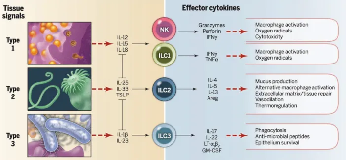
Figure 9: ILC subsets and their signature cytokines. ILCs are activated by injured or infected cell signals and respond by releasing signature cytokines that promote important effector and regulatory responses. (From
Eberl et al. 2015)
TYPE 2ILCS
ILC2s are defined by their innate capacity to produce type 2 cytokines, mainly IL-13, IL-5, IL-9 and IL-4, as well as amphiregulin (Figure 9). These soluble factors have a dual effect being either protective to the host or capable of exerting pathogenic activity. IL-13 promotes resistance to large extracellular parasites, such as helminthes, by increasing mucus production and muscle contractility, favoring epithelial cell turnover and macrophage activation. Furthermore, type 2 immunity has proven protective in a range of autoimmune diseases (arthritis, multiple sclerosis or Crohn’s disease) by the suppression of type-1 driven