HAL Id: tel-02310699
https://tel.archives-ouvertes.fr/tel-02310699
Submitted on 10 Oct 2019HAL is a multi-disciplinary open access archive for the deposit and dissemination of sci-entific research documents, whether they are pub-lished or not. The documents may come from teaching and research institutions in France or abroad, or from public or private research centers.
L’archive ouverte pluridisciplinaire HAL, est destinée au dépôt et à la diffusion de documents scientifiques de niveau recherche, publiés ou non, émanant des établissements d’enseignement et de recherche français ou étrangers, des laboratoires publics ou privés.
Potential of omega-3 EPA/DHA 6/1 to ameliorate
ageing-related endothelial dysfunction
Muhammad Akmal Farooq
To cite this version:
Muhammad Akmal Farooq. Potential of omega-3 EPA/DHA 6/1 to ameliorate ageing-related en-dothelial dysfunction. Cardiology and cardiovascular system. Université de Strasbourg, 2018. English. �NNT : 2018STRAJ107�. �tel-02310699�
UNIVERSITÉ DE STRASBOURG
ÉCOLE DOCTORALE DES SCIENCES DE LA VIE ET DE LA SANTE
Regenerative Nanomedicine – INSERM UMR 1260
THÈSE
présentée par :Muhammad Akmal FAROOQ
soutenue le : 17 Octobre 2018
pour obtenir le grade de : Docteur de l’université de Strasbourg Discipline/ Spécialité
: Sciences Pharmaceutiques -
Pharmacologie-Pharmacocinétique
Potential of omega-3 EPA:DHA 6:1 to
ameliorate ageing-related endothelial
dysfunction
THÈSE dirigée par :
Dr Cyril AUGER Université de Strasbourg
RAPPORTEURS :
Pr Thierry COUFFINHAL Université de Bordeaux
Pr Laurent MARTINY Université de Reims Champagne Ardennes
AUTRES MEMBRES DU JURY :
A
CKNOWLEDGEMENTS
Many people have helped me in the completion of this project and I want to thank them all in my humble acknowledgement.
First and foremost, I would like to express my gratitude to my mentors Dr Cyril AUGER and Prof. Valerie Schini-Kerth for their valuable and continuous support during the last three years. Specially, Dr Cyril AUGER for his kind suggestions, encouragement, constructive criticism and aid in paper work which helped me immensely during the project and enabled me to develop the skills needed through this journey.
I am very thankful to Prof. Florence TOTI for her scientific support in the lab and being the guarantor of my residence. I would also like to thank our ex-technician Mme Briggitte POLLET who helped me exceptionally in learning vascular reactivity studies on mesenteric artery and made the work environment enjoyable.
Sincere thanks to my seniors Faraj ZGHEEL, Amisi SAID, Fuong NAGA, Sonia Khemais and specially Zahid Rasool NIAZI for helping me in learning the techniques. I want to thank ex and current post docs Antonio Leon GONZALEZ, Malak ABBAS and Eugenia BELCASTRO. I also want to thank all my lab mates Hira HASAN, Abdul Wahid QURESHI, Hyunho LEE, Sinhee PARK, Christophe BRUKERT, Ahmed CHAKER, Lamia REMILA, Raed ALTAMIMI, Sebastein GAERTNER and Lamia AMOURA for all their direct or indirect help in my project, without which it would have not been possible to complete it. I also would like to thank all my Pakistani friends for their moral support and making me feel like at home during my stay in France.
I also like to acknowledge the Higher Education Commission of Pakistan and Campus France for providing me the financial support to pursue my studies in France.
Finally, I would like to express my deepest gratitude to my family members: my parents, brother, sisters and my beloved wife for their unconditional love, care, support and encouragement throughout my life. In the end, I dedicate this work to my parents and my Uncle Abdul GHAFFAR for being an inspiration for my achievements.
MUHAMMAD AKMAL FAROOQ
ii
Table of contents
Acknowledgements
i
Table of content
ii
List of figures and tables
v
List of abbreviations
vi
Abstract / Résumé
1
Abstract
2
Résumé
6
Scientific production
11
Introduction
14
1- The physiology of the endothelium
15
1.1 General anatomy of blood vessels 15 1.2 The role of the endothelium in the regulation of the vascular tone 16
1.2.1 Endothelium-derived relaxing factors (EDRF) 16
1.2.1.1 Nitric Oxide (NO) 16
1.2.1.2 Prostacyclin (PGI2) 20
1.2.1.3 Endothelium-dependent hyperpolarization (EDH) 21
1.2.2 Endothelium-Derived Contractile Factors (EDCF) 23
1.2.2.1 Reactive Oxygen Species (ROS) and Oxidative Stress 23
1.2.2.2Thromboxane (TXA2) and Prostacyclin (PGI2) 24
1.2.2.3 Angiotensin II (Ang II) 27
1.2.2.4 Endothelin-1 (ET-1) 28
2. AGE AND AGE-RELATED CARDIOVASCULAR RISK
30
2.1 Increase in cardiovascular risk with Age 31 2.2 The age-related endothelial dysfunction 32
2.2.1 Increased ROS and oxidative stress 33
2.2.2 Decreased NO bioavailability 35 2.2.2.1 Decrease production of NO 35 2.2.2.2 Increased inactivation of NO 36 2.2.3 Decreased EDH 37 2.2.4 Increased EDCF 38 2.2.4.1 COX-derived prostanoids 38
iii
2.2.4.2 Renin-angiotensin system 39
2.2.4.3 Endothelin-1 40
2.2.5 Role of inflammation in age-related endothelial dysfunction 42 2.2.6 Role of senescence in age-related endothelial dysfunction 43
3. OMEGA-3 PUFAs AND THEIR ROLE IN VASCULAR HEALTH 45
3.1 Lipids 46
3.2 Fatty Acids 46
3.2.1 Saturated fatty acids 46
3.2.2 Monounsaturated fatty acids 47
3.2.3 Polyunsaturated fatty acids 48
3.2.4 Trans-fatty acids 48
3.3 Omega-3 or n-3 PUFAs 49
3.3.1 Synthesis and bioconversion 49
3.3.2 Dietary sources and intake 50
3.4 Effects of Omega-3 PUFAs on Molecular Level 51
3.4.1 Alteration of cell membrane structure and function 51
3.4.2 Alteration of ion channel function 52
3.4.3 Alteration of nuclear factors and transcription factors 53
3.4.4 Omega-3 PUFAs derived eicosanoids 53
3.5 Risk Factor Reduction for CVDs 55
3.5.1 Blood Pressure 56 3.5.2 Plasma Lipids 57 3.5.3 Heart rate 58 3.5.4 Arrhythmia 58 3.5.5 Thrombosis 59 3.5.6 Inflammation 59 3.5.7 Endothelial function 60
3.6 Clinical studies on Omega-3 PUFAs 61
3.6.1 DART 1989 61
3.6.2 GISSI-Prevention 1999 61
3.6.3 GISSI-HF 2004 61
3.6.4 JELIS 2007 62
4. AIM OF THE STUDY
64
5. RESULTS
67
iv
Results and conclusion 105
6. DISCUSSION AND PERSPECTIVES
107
6. GENERAL DISCUSSION 108
6.1 The age-related endothelial dysfunction 108 6.2 Beneficial effects of omega-3 PUFAs 113
6.3 Conclusion and perspectives 118
7. Discussion et perspectives 120
7. DISCUSSION GENERALE
121
7.1 La dysfonction endothéliale liée à l’âge 121 7.2 Effets bénéfiques des acides gras polyinsaturés oméga-3 126
7.3 Conclusion et perspectives 130
v
List of Figures:
Figure 1 Structure of blood vessels 15
Figure 2 Vaso-protective effects of NO 17 Figure 3 Activation and inhibitory sites of eNOS 19 Figure 4 Nitric oxide mediated vaso-relaxation pathway 20 Figure 5 Endothelium derived hyperpolarization-mediated vasorelaxation 22 Figure 6 Cellular sources of reactive oxygen species 24 Figure 7 Cyclooxygenase-derived prostanoids and their respective receptors 25 Figure 8 Major EDCF-mediated signaling pathways 26 Figure 9 Angiotensin type-1 receptor-mediated actions 28 Figure 10 Prevalence of CVDs with increasing age 31 Figure 11 Age-related endothelial dysfunction 33 Figure 12 Oxidative stress can affect gene expression by modification of
transcription factors
34 Figure 13 Role of NOS in age-related endothelial dysfunction 37 Figure 14 Age-related change in the production of different prostanoids 39 Figure 15 Role of angiotensin II signaling in age-related endothelial dysfunction 40 Figure 16 Factors and diseases responsible for ET-1 pathway activation 41 Figure 17 Role of age-related pro-inflammatory environment in endothelial
dysfunction
43 Figure 18 Important poly-unsaturated fatty acids 48 Figure 19 Bioconversion of linoleic acid and α-linoleic acid 50 Figure 20 Molecular level effects of Omega-3 PUFAs 52 Figure 21 Mechanisms involved in risk reduction of cardiovascular diseases 55 Figure 22 Association between consumption of sea food and risk of coronary
heart disease
56 Figure 23 Short term oral intake of omega-3 PUFA EPA:DHA 6:1 improves the
age-related endothelial dysfunction in rats
116 Figure 24 Perspectives in pre-clinical studies 119
Figure 24 Clinical perspectives 119
List of Tables:
vi
L
IST OF ABBREVIATIONS
AA
Arachidonic acid
ABC
ATP binding cassette transporter
ACE
Angiotensin converting enzyme
ACE2
Angiotensin converting enzyme 2
ACh
Acetylcholine
ADMA
Asymmetric dimethyl arginine
AF
Arterial fibrillation
AGE
Advance Glycation End-products
Akt
Protein kinase B
ALA
Alpha linoleic acid
AMPK
Adenosine monophosphate activated protein
kinase
Ang I
Angiotensin I
AP-1
Activator protein-1
Apo CIII
Apo-lipoprotein C3
AT1R
Angiotensin type-1 receptor
BH
2Dihydrobiopterin
BH
4Tetrahydrobiopterin
BK
Bradykinin
BK
CaLarge conductance Ca
2+-activated potassium
channels
BP
Blood pressure
Ca
2+Calcium
CaM
Calmodulin
CaM Kinase II
Calcium/calmodulin-dependent protein kinase II
Ca
VVoltage-gated calcium channels
cGMP
Cyclic guanosine monophosphate
CHD
Coronary heart disease
vii
COX
Cyclo-oxygenase
COX-1
Cyclo-oxygenase-1
COX-2
Cyclo-oxygenase-2
CVD
Cardiovascular disease
Cyp-450
Cytochrome p-450
DART
Diet and reinfarction trial
DHA
Docosahexaenoic acid
DHE
Dihydroethidium
DNA
Deoxyribonucleic acid
DP
D-prostanoid receptor
ECs
Endothelial cells
EDCF
Endothelium-derived contractile factors
EDH
Endothelium-dependent hyperpolarization
EDRF
Endothelium-derived relaxing factors
EET
Epoxy-eicosaicosatrienoic acid
EETs
Epoxy-eicosatrienoic acid derivatives
EGRF
Epidermal growth factor
eNOS
Endothelial nitric oxide synthase
EP
E-prostanoid receptor
EPA
Eicosapentaenoic acid
E
pacCyclic AMP-activated exchange protein
ER
Endoplasmic reticulum
ERK
Extracellular signal regulated kinase
ET-1
Endothelin-1
ET-2
Endothelin-2
ET-3
Endothelin-3
ET
AEndothelin-1 receptor A
ET
BEndothelin-1 receptor B
FAD
Flavin-adenine dinucleotide
FMD
Flavin mononucleotide
viii
GPR120
G-protein coupled receptor 120
GTP
Guanosine trisphosphate
H
Histamine
H
2O
2Hydrogen peroxide
H
2S
Dihydrogen sulfide
HDL
High density lipoprotein
HETE
Hydroxy eicosatetraenoic acid
HSP90
Heat shock protein 90
ICAM
Intercellular adhesion molecule
IDL
Intermediate density lipoprotein
i-ƙB
Inhibitor of ƙB
IK
caIntermediate conductance Calcium channel
IL-12
Interleukin-12
IL-1β
Interleukin-1β
IL-2
Interleukin-2
IL-6
Interleukin-6
iNOS
Inducible nitric oxide synthase
IP
I-prostanoid receptor
IP
3Inositol trisphosphate
JAKs
Janus kinases
JELIS
Japan eicosapentaenoic acid lipid intervention
study
K
caCalcium activated potassium channel
K
irInwardly rectifying potassium channel
K
VVoltage-gated potassium channel
LA
Linoleic acid
LCFA
Long chain fatty acids
LDL
Low-density lipoproteins
L-NA
L-nitroarginine
LNAME
L-nitroarginine methyl ester
LOX
Lipo-oxygenase
ix
LPL
Lipoprotein lipase
LTB
4Leukotriene B
4LTB
5Leukotriene B
5LVH
Left ventricular hypertrophy
MCP-1
Monocyte chemoattractant protein-1
MGJs
Myoendothelial gap junctions
MUFA
Monounsaturated fatty acid
Na
+Sodium
Na
+/Ca
2+Sodium/calcium pump
NF-ƙB
Nuclear factor kappa-B
nNOS
Neuronal nitric oxide synthase
NO
Nitric oxide
NOS
Nitric oxide synthase
NOX
NADPH oxidase
O
2-.Superoxide anion
OH
•Hydroxyl radical
Omega-3 PUFA
Omega-3 polyunsaturated fatty acids
OONO
-Peroxynitrite anion
p16
Cyclin-dependent kinase inhibitor 2A
p21
Cyclin-dependent kinase inhibitor-1
p22
phoxMembranal subunit of NADPH oxidase
p47
phoxCytosolic subunit of NADPH oxidase
p53
Tumor protein p53
PGD
2Prostaglandin D
2PGE
2Prostaglandin E
2PGE
3Prostaglandin E
3PGF
2αProstaglandin F
2αPGH
2Prostaglandin H
2PGI
2Prostacyclin
PGI
3Prostaglandin I
3PI3K
Phosphatidyl inositol 3 kinase
x
PKC
Protein kinase C
PKG
Protein kinase G
PLA
2Phospholipase A
2PLC
Phospholipase C
PLD
Phospholipase D
PPAR
Peroxisome proliferator activated receptor
PPARα
Peroxisome proliferator activated receptor-α
PUFA
Polyunsaturated fatty acid
PUFAs
Polyunsaturated fatty acids
RAS
Renin angiotensin system
RNA
Ribonucleic acid
RNS
Reactive nitrogen species
ROS
Reactive oxygen species
R
yR
sRyanodine receptors
SA-β-Gal
Senescence associated β-galactosidase
SC-560
Cyclo-oxygenase-1 inhibitor
SFA
Saturated fatty acid
sGC
Soluble guanylyl cyclase
SHR
Spontaneously hypertensive rats
SK
caSmall conductance Ca
2+activated K
+channels
SMC
Smooth muscle cell
SOC
Store-operated channels
SOD
Superoxide dismutase
SR
Sarcoplasmic reticulum
TF
Tissue factor
TG
Triglycerides
TNF-α
Tissue necrosis factor-α
TP
Thromboxane prostanoid receptor
TXA
2Thromboxane A2
TXA
3Thromboxane A3
TXB
2Thromboxane B2
xi
VEGF
Vascular endothelial growth factor
VLDL
Very low-density lipoprotein
VSMC
Vascular smooth muscle cell
1
2
Abstract
Cardiovascular diseases are the leading cause of death worldwide, both in developed and developing countries. As the incidence of cardiovascular diseases in increasing with age, the ageing of the population will be associated with an increase in cardiovascular diseases in the future, accounting, at least in part, for the World Health Organization prediction of cardiovascular diseases remaining the leading cause of death worldwide.
The endothelium, the monocellular layer lining all blood vessels, is a pivotal organ in maintenance of the vascular homeostasis. Indeed, endothelial cells contribute to the fine regulation of the vascular tone and to the protection against thrombosis and vascular remodeling, mainly through the release of potent vasoprotective factors such as nitric oxide (NO), the endothelium-dependent hyperpolarization (EDH), or the prostacyclin PGI2.
Many preclinical and clinical studies have shown that development of cardiovascular diseases and risk factors, such as ageing, are associated early with an endothelial dysfunction, characterized by a decreased formation of vasoprotective factors and an increased formation of vasoconstricting factors, resulting in an imbalance leading towards the accelerated development of vascular pathologies. Indeed, it has been shown that physiological ageing is associated with a progressive decrease in endothelium-dependent vasodilatation. Thus the age-related endothelial dysfunction could be an early event promoting the development of cardiovascular diseases, as endothelial dysfunction is involved in the mechanisms underlying the development of atherosclerotic lesions, including up-regulation of adhesion molecules, increased chemokine secretion and leukocyte adherence, increased cell permeability, enhanced low-density lipoprotein oxidation, platelet activation, cytokine elaboration, and vascular smooth muscle cell proliferation and migration.
In addition, several studies have demonstrated that endothelial dysfunction and cardiovascular diseases development are also associated with an increased vascular oxidative stress and an up-regulation of the local angiotensin system that also contributes to the development of cardiovascular diseases through the induction of oxidative stress by up-regulating NADPH oxidase, the main producer of reactive oxygen species (ROS) in the vascular wall. Moreover, recent studies have suggested that the premature induction of senescence in endothelial cells could be an early event in the development of the endothelial dysfunction.
3
Over the last decades, numerous epidemiological studies have reported that diets could play a role in the development of cardiovascular diseases. For example, a recent interventional study has shown that compared to a western diet rich in saturated fat, a Mediterranean diet rich in unsaturated fat from olive oil or nuts could decrease the incidence of a primary cardiovascular adverse event (myocardial infarction or stroke) by 30 % in subjects with high cardiovascular risk factors. Moreover, several epidemiological studies and clinical trials have shown that dietary intake of fish, fish oil or omega-3 polyunsaturated fatty acids (n-3 PUFAs) could reduce secondary events of coronary heart disease and stroke, and could also decrease hypertension. The dietary consumption of the major omega-3 PUFAs, namely eicosapentaenoic acid (EPA) and docosahexaenoic acid (DHA), has also been related to a reduced risk of cardiovascular disease morbidity/mortality. The omega-3 PUFAs could exert their beneficial effects on the cardiovascular system by modulation of the lipids metabolism and their anti-inflammatory properties due to the decrease in pro-inflammatory cytokine production and the formation of anti-inflammatory metabolites including resolvins, protectins and maresins.
In addition, the omega-3 PUFAs could protect the cardiovascular system, at least in part, by exerting a protective effect on the endothelial function. Indeed, EPA and DHA are able to induce the activation of the endothelial nitric oxide synthase (eNOS), the enzyme responsible of the formation of NO, the most potent endothelium-derived vasoprotective factor.
Our research team previously show that the stimulation of the endothelial function by n-3 PUFAs is dependent on both the purity and the ratio of EPA and DHA. Indeed, we tested several EPA:DHA ratio (from 9:1 to 1:9) and shown that the EPA:DHA 6:1 formulation is a potent stimulator of the endothelial formation of NO, and to a lesser extent, of an increased endothelium-dependent hyperpolarization (EDH) response. The induction of the endothelial formation of NO by omega-3 fatty acids is mediated by redox-sensitive activation of the Src/PI3-kinase/Akt and MAPKs pathways leading to eNOS activation, which is dependent on the ratio and amount of the EPA:DHA in the formulation.
Moreover, our team demonstrated in a subsequent study that the chronic oral intake of the EPA:DHA 6:1 formulation was able to prevent the hypertension and endothelial dysfunction induced by angiotensin II infusion in rats. The beneficial effect of EPA:DHA 6:1 was mediated by an improvement of both the NO- and the EDH-mediated relaxations and a reduction of endothelium-dependent contractile response, most likely by preventing the oxidative stress induced by the up-regulation of the local angiotensin system.
4
As several clinical studies have shown that omega-3 PUFAs are able to decrease cardiovascular risks in patients with documented first cardiovascular events, we were looking to assess the potency of the EPA:DHA 6:1 formulation to improve the endothelial function in a pathophysiological situation associated with an established endothelial dysfunction.
The aim of the present study was to determine whether a short-term chronic oral intake of the optimized EPA:DHA 6:1 formulation is able to improve the ageing-related endothelial dysfunction in rats, and if so, to determine the underlying mechanisms.
For this study, 20 month-old male Wistar rats received by daily gavage 500 mg/kg BW of either corn oil, EPA:DHA 6:1 formulation or water (control) for 2 weeks. After 2 weeks of gavage, the animals were euthanized and the organ were collected. The main mesenteric artery was used for vascular reactivity studies, and main mesenteric artery and aorta were used for immunofluorescence and fluorescence histochemistry studies on frozen section, and for western blot analysis of protein expression.
The major results of our study show that, compared to young rats (12 weeks-old), ageing was associated with an endothelial dysfunction characterized by a blunted NO-mediated component, an abolished endothelium-dependent hyperpolarization (EDH)-mediated component, and also by the induction of endothelium-dependent contractile responses (EDCF, in the presence of NO and EDH inhibitors) sensitive to indomethacin (a cyclooxygenase inhibitor) in response to acetylcholine. Intake of EPA:DHA 6:1 for 2 weeks was able to improve the NO-mediated relaxations and to decrease the endothelium-dependent contractile responses, but had no effect on the EDH-mediated component. Intake of corn oil for 2 weeks had no effect on the ageing-related endothelial dysfunction.
To better characterize the molecular mechanisms involved in the protective effects of EPA:DHA 6:1 intake, we performed quantitative analysis of protein expression in the mesenteric artery and aorta by immunofluorescence. Firstly, we studied the expression of eNOS (NO component of relaxation), and cyclooxygenases (COXs, involved in EDCFs). Compared to the young rats, ageing is significantly associated with an increased expression of eNOS, COX-2, the inducible isoform of COXs, while down-regulating the expression of COX-1, the constitutive isoform of COXs in the arterial wall of the mesenteric artery. The
5
intake of EPA:DHA 6:1 normalized the expression levels of eNOS, COX-1, and COX-2, whereas corn oil had no significant effect.
As endothelial dysfunction is associated with a vascular oxidative stress, we measured the level of oxidative stress in the vascular wall of the mesenteric artery and aorta using the redox-sensitive fluorescent probe dihydroethidium (DHE). Compared to young rats, old rats show significant increases of DHE fluorescence throughout the vascular wall, which was significantly prevented by the EPA:DHA 6:1 intake. Moreover, the age-related increased vascular oxidative stress was due, at least in part, to the activities of uncoupled eNOS, COX-1 and COX-2, NADPH oxidase, and mitochondrial respiratory chain in the vascular wall of the aorta.
As the increased vascular oxidative stress has been attributed, at least in part, NADPH oxidase activity, we determined the expression level of NADPH oxidase subunits p22phox and p47phox. Compared to young control rats, the aortic wall of aged rats exhibit a significantly
increased expression level of p22phox and p47phox, and the EPA:DHA 6:1 treatment
normalized the expression of both NADPH oxidase sub-units.
Finally, we assess the level of premature endothelial senescence in the aortic wall. The expression levels of the senescence markers p53, p21 and p16 were significantly increased in aged rats compared to young rats. The intake of EPA:DHA 6:1 for 2 weeks normalized the expression levels of senescence markers.
Altogether, the present findings indicate that intake of EPA:DHA 6:1 significantly improved the ageing-related endothelial dysfunction in rats. The beneficial effect involves an improvement of both the NO- and the EDH-mediated relaxations as well as a reduction of endothelium-dependent contractile response most likely by preventing vascular oxidative stress and premature endothelial senescence.
6
Résumé
Les maladies cardiovasculaires représentent la première cause de mortalité dans le monde, que cela soit dans les pays développés ou ceux en cours de développement. L’incidence des maladies cardiovasculaires augmentant avec l’âge, le vieillissement des populations est associée à une augmentation des maladies cardiovasculaires dans futur participant, du moins partiellement, à expliquer les prédictions de l’Organisation Mondiale de la Santé montrant que les maladies cardiovasculaires resteront la première cause de mortalité dans la monde. L’endothélium, la monocouche cellulaire tapissant les vaisseaux sanguins, est un organe central dans la régulation de l’homéostasie vasculaire. En effet, les cellules endothéliales ont un rôle clé dans le maintien du tonus vasculaire et dans la protection contre la thrombose et le remodelage vasculaire, principalement grâce à la formation et libération de puissants facteurs vasoprotecteurs tels que le monoxyde d’azote (NO), l’hyperpolarisation dépendante de l’endothélium (EDH), ou la prostacycline PGI2.
De nombreuses études expérimentale et cliniques ont montré que le développement des maladies cardiovasculaires et de leurs facteurs de risque, dont le vieillissement physiologique, étaient associés de façon précoce avec l’apparition d’une dysfonction endothéliale caractérisée par une diminution de la formation des facteurs protecteurs et une augmentation de la formation des facteurs vasoconstricteurs, le tout engendrant un déséquilibre menant au développement accéléré des pathologies vasculaires. En effet, il a été démontré que le vieillissement physiologique était associé à une diminution progressive de la vasodilatation dépendante de l’endothélium. Ainsi, la dysfonction endothéliale liée à l’âge pourrait être un évènement précoce favorisant le développement des maladies cardiovasculaires du fait que la dysfonction endothéliale est impliquée dans la formation de lésions athéromateuses en favorisant les mécanismes sous-jacents au développement de l’athérosclérose, notamment l’augmentation de l’expression des molécules d’adhésion, de l’adhésion des leucocytes, de l’oxydation des LDL, de l’activation plaquettaires, et de la prolifération et de la migration des cellules musculaires lisses vasculaires.
De plus, plusieurs études ont montré que la dysfonction endothéliale et le développement des maladies cardiovasculaires sont associés à une augmentation du stress oxydant vasculaire et à une surexpression du système angiotensine local qui contribue également au développement des maladies cardiovasculaires de par l’augmentation du stress oxydant vasculaire induit par la surexpression de la NAPDH oxydase, la principale source des espèces réactives de l’oxygène dans la paroi vasculaire. Par ailleurs, des études récentes ont suggéré que
7
l’induction d’une senescence prématurée des cellules endothéliales constitue un évènement précoce dans le développement de la dysfonction endothéliale.
Au cours des dernières décennies, de nombreuses études épidémiologiques ont montré que l’alimentation pouvait jouer un rôle dans le développement des maladies cardiovasculaires. Ainsi, une récente étude interventionnelle a démontré que, par rapport au régime occidental riche en graisses saturées, un régime Méditerranéen riche en graisse non-saturées issu de l’huile d’olive ou des fruits secs à coques permettait de réduire de 30% l’incidence d’un premier évènement cardiovasculaire aigu (infarctus du myocarde ou accident vasculaire cérébral ischémique) chez des sujets à haut risque cardiovasculaire. De plus, plusieurs études épidémiologiques ou d’intervention ont montré que la consommation alimentaire de poisson, d’huile de poisson ou d’acides gras polyinsaturés omega-3 (n-3 PUFAs) pouvait diminuer les risques de récidive de la maladie coronarienne ou d’accident vasculaire cérébraux, et diminuait la pression artérielle chez les hypertendus. La consommation des acides gras omega-3 PUFAs majeurs, à savoir l’acide eicosapentaénoïque (EPA) et l’acide docosahexaénoïque (DHA), a aussi été associée à une réduction de la morbi-mortalité cardiovasculaire. Les n-3 PUFAs pourrait avoir un effet bénéfique sur le système cardiovasculaire en modulant le métabolisme lipidique et du fait de leur propriétés anti-inflammatoires passant par une diminution de la production de cytokines pro-anti-inflammatoires et une formation de métabolites anti-inflammatoires dont les résolvines, les marésines et les protectines.
De plus, les omega-3 PUFAs pourrait protéger le système cardiovasculaire, du moins en partie, en protégeant la fonction endothéliale. En effet, l’EPA et le DHA sont capables d’induire l’activation de la NO synthase endothéliale (eNOS), l’enzyme produisant le NO, le plus puissant des facteurs vasoprotecteurs issus de l’endothélium.
Notre équipe de recherche a récemment démontré que la stimulation de la fonction endothéliale par les omega-3 PUFAs dépend à la fois du ratio et du degré de pureté de la formulation en EPA et DHA. En effet, nous avons évalué la capacité de plusieurs formulation EPA:DHA (allant de 1:9 à 9:1) et nous avons montré que la formulation EPA:DHA 6:1 est un puissant activateur de la formation endothéliale de NO, et de façon moindre de l’augmentation de la réponse d’hyperpolarisation dépendante de l’endothélium (EDH). L’induction par les omega-3 PUFAs de la formation endothéliale de NO due à l’activation de
8
la eNOS via les voies de signalisation redox-sensibles Src/PI3-kinase/Akt et MAPKs, est dépendante du ratio et de la quantité de EPA et DHA dans la formulation.
De plus, note équipe a récemment montré dans une étude suivante que consommation orale de la formulation EPA:DHA 6:1 prévenait la survenu de l’augmentation de pression artérielle et de la dysfonction endothéliale induite par l’infusion d’Angiotensine II chez des rats. L’effet bénéfique de l’EPA:DHA 6:1 passait par une amélioration des relaxations dépendante du NO et de l’EDH et par une diminution des réponses contractiles dépendantes de l’endothélium, probablement dues à la prévention du stress oxydant vasculaire induit par l’activation du système angiotensine local.
Du fait que les études cliniques montrent que les omega-3 PUFAs sont capables de diminuer les risques cardiovasculaires chez des patients avec un premier évènement cardiovasculaire documenté, nous avons cherché à évaluer le potentiel de la formulation EPA:DHA 6:1 a améliorer la fonction endothéliale dans une situation physiopathologique associée à une dysfonction endothéliale installée.
L’objectif de la présente étude est de déterminer si une consommation chronique à court terme de la formulation optimisée EPA:DHA 6:1 est capable d’améliorer la dysfonction endothéliale liée à l’âge chez le rat, et le cas échéant, de déterminer les mécanismes sous-jacents.
Pour cette étude, des rats Wistar males de 20 mois ont reçu quotidiennement pendant 2 semaines par gavage 500 mg/kg soit d’huile de maïs, soit de la formulation EPA:DHA 6:1, soit d’eau (contrôle). Après deux semaines de traitement, les rats ont été euthanasiés et les organes sont prélevés. L’artère mésentérique a été utilisée pour l’étude de la réactivité vasculaire, et l’artère mésentérique principale et l’aorte pour des études en immunofluorescence et histochimie fluorescente sur coupes congelées.
Les principaux résultats de notre étude indiquent que, par rapport à des rats jeunes de 12 semaines, le vieillissement physiologique était associé à une dysfonction endothéliale caractérisée à la fois par une diminution de la composantes NO de relaxation en réponse à l’acétylcholine, une abolition de la composante EDH de la relaxation, et aussi par une augmentation de réponses contractiles dépendantes de l’endothélium (EDCFs) sensible à l’indométacine (un inhibiteur des cyclooxygénases) en réponse à l’acétylcholine. La
9
consommation chronique de EPA:DHA 6:1 pendant 2 semaines améliore significativement les relaxations dépendantes du NO et les EDCFs, mais n’a pas d’effet sur la composante EDH de la relaxation. La consommation d’huile de maïs pendant 2 semaines n’a pas eu d’effet sur la dysfonction endothéliale liée à l’âge.
Afin de mieux caractériser les mécanismes moléculaires impliqués dans l’effet protecteur de la formulation EPA:DHA 6:1, des analyses quantitatives des niveaux d’expression de protéines ont été effectués dans l’artère mésentérique et l’aorte par immunofluorescence sur coupes congelées. Dans un premier temps, nous avons étudié l’expression de la eNOS (composante NO de la relaxation) et des cyclooxygénases (COXs, impliquées dans les réponse EDCFs).
Par rapport aux animaux jeunes, le vieillissement est associé à une augmentation significative de l’expression de la eNOS et de COX-2, la forme inductible des COXs, et une diminution significative de l’expression de COX-1 dans la paroi vasculaire de l’artère mésentérique. La prise chronique de EPA:DHA 6:1 normalise l’expression de la eNOS, de 1 et de COX-2, alors que la prise d’huile de maïs n’a pas eu d’effet significatif.
Comme la dysfonction endothéliale est associée à un stress oxydant vasculaire, nous avons évalué le niveau de stress oxydant la paroi vasculaire de l’artère mésentérique et de l’aorte à l’aide de la sonde fluorescente redox-sensible dihydroethidium (DHE). Par rapport aux rats jeunes, le vieillissement est associé à une augmentation significative de la fluorescence dans l’ensemble de la paroi vasculaire, et cette augmentation est significativement prévenue par la prise chronique de la formulation EPA:DHA 6:1 mais pas par l’huile de maïs. De plus, l’augmentation du stress oxydant vasculaire liée à l’âge dans la paroi aortique est due, au moins partiellement, à l’activité de la eNOS découplée, de la COX-1 et de la COX-2, de la NADPH oxydase et de la chaine respiratoire mitochondriale.
Du fait que l’augmentation de stress oxydant est due, du moins partiellement, à une surexpression de la NAPDH oxydase, les niveaux d’expression des sous-unités p22phox et p47phox de la NADPH oxydase ont été déterminés. En comparaison des rats jeunes, les rats âgés montrent une augmentation significative des niveaux d’expression des sous-unités p22phox et p47phox dans la paroi de l’aorte, et le traitement avec la formulation EPA:DHA 6:1 normalise l’expression des sous-unités de la NADPH oxydase.
De plus, le niveau de senescence des cellules endothéliale a été évalué dans la paroi aortique. Le niveau d’expression des marqueurs de sénescence p53, p16 et p21est significativement augmenté dans l’endothélium des rats âgés, comparativement aux rats jeunes. Le prise orale
10
de la formulation EPA:DHA 6:1 pendant 2 semaines a permis de normaliser le niveau d’expression de ces markers de sénescence.
L’ensemble des résultats obtenus indique que la prise chronique de la formulation optimisée EPA:DHA 6:1 pendant 2 semaines améliore significativement la dysfonction endothéliale liée à l’âge chez le rat. Les effets bénéfique de la consommation chronique de la formulation EPA:DHA 6:1 impliquent une amélioration des composantes de relaxations NO, une diminution des réponses contractiles dépendantes de l’endothélium, probablement via la diminution du stress oxydant vasculaire et de la senescence endothéliale.
11
Scientific production
Publications:
The omega-3 EPA:DHA 6:1 formulation improves ageing-related blunted endothelium-dependent relaxations and increased contractile responses in the mesenteric artery: role of oxidative stress and cyclooxygenases. M.A. Farooq, L. Amoura, S. Gaertner, Z.R. Niazi, S. Park, A.W. Qureshi, M.H Oak, F. Toti, V.B. Schini-Kerth, C. Auger. Under preparation.
EPA:DHA 6:1 prevents age-related endothelial dysfunction linked to cyclooxygenase pathway in rat femoral vessels: a way to prevent venous endothelial dysfunction mediated thrombosis? S. Gaertner, M.A. Farooq, B. Pollet, J. Graaf, S. Khemais-Benkhiat, S. Schrevens, Z.R. Niazi, A.W. Qureshi, S. Park, F. Toti, D. Stephan, N. Boehm, C. Auger, V. B. Schini-Kerth. Under preparation.
Activation of thromboxane prostanoïd receptors mediates age-related endothelial dysfunction in the rat femoral vein: a potential mechanism increasing the risk of venous thromboembolism? S. Gaertner, C. Auger, M.A. Farooq, B. Pollet, J. Graaf, S. Khemais-Benkhiat, L. Amoura, S. Park, F. Toti, D. Stephan, N. Boehm, V. B. Schini-Kerth. Under preparation.
Femoral vessels premature aging and venous endothelial dysfunction in young ZSF-1 rats: key role of cyclooxygenases and potential risk for venous thrombosis induced by cumulative cardiovascular risk factors for atherosclerosis. S. Gaertner, M. A. Farooq, B. Pollet, J. Graaf, S. Khemais-Benkhiat, L. Amoura, S. Park, F. Toti, C. Auger, D. Stephan, N. Boehm, V. B. Schini-Kerth. Under preparation.
12
Empagliflozin, a sodium-glucose cotransporter inhibitor, improved heart remodeling and mesenteric artery endothelial function in the metabolic syndrome with HFpEF ZSF1 rat: role of cyclooxygenases. S. Park, M.A. Farooq, S. Gaertner, C. Brucker, A.W. Qureshi, H. Lee, D. Benrahla, B. Pollet, D. Stephan, P. Ohlmann, E. Mayoux, C. Auger, O. Morel, V.B. Schini-Kerth. Under preparation.
Treatment of rats with the omega fatty acid 3 formulation EPA:DHA 6:1 decreases the leukocyte microparticles-induced endothelial pro-inflammatory responses and senescence. A. Qureshi, R. Altamimy, A. El Habhab, L. Amoura, M. Kassem, S. Khemais, M. Farooq, H. Hasan, P. Sin-Hee, F. El-Ghazouani, C. Auger, L. Kessler, V. Schini-Kerth, F. Toti. Under preparation.
Thrombin promotes premature atrial endothelial cell senescence leading to the induction of pro-infiltrative and pro-fibrotic responses: Role of the local angiotensin II/AT1 receptor system. H. Hasan, S.H. Park, E. Belcastro, M. Abbas, A.W. Qureshi, M.A. Farooq, P. Ohlmann, F. Toti, C. Auger, V. Schini-Kerth, O. Morel, L. Jesel. Under preparation.
Oral communications:
The omega-3 EPA:DHA 6:1 formulation improves ageing-related blunted endothelium-dependent relaxations and increased contractile responses in the mesenteric artery: role of oxidative stress and cyclooxygenases. M.A. Farooq, L. Amoura, S. Gaertner, Z.R. Niazi, S. Park, A.W. Qureshi, M.H Oak, F. Toti, V.B. Schini-Kerth, C. Auger. NutRedOx Cost action, 2017, Strasbourg, France
The omega-3 EPA:DHA 6:1 formulation improves ageing-related blunted endothelium-dependent relaxations and increased contractile responses in the mesenteric artery: role of oxidative stress and cyclooxygenases. M.A. Farooq, L. Amoura, S. Gaertner, Z.R. Niazi, S.
13
Park, A.W. Qureshi, M.H Oak, F. Toti, V.B. Schini-Kerth, C. Auger. ICMAN/IUPHAR Joint meeting on natural products, 2017, Aberdeen, UK
Communications affichées :
Printemps de Cardiologie (ESC) 2017, Nantes, France, poster commenté Journee Campus d’Illkirch (JCI) 2017, Illkirch, France, poster commenté
European Symposium on Vascular Biomaterials (ESVB) 2017, Strasbourg, France ED days 2017, Strasbourg, France
14
The physiology of the
endothelium
15
1 - THE PHYSIOLOGY OF THE ENDOTHELIUM
General anatomy of blood vessels
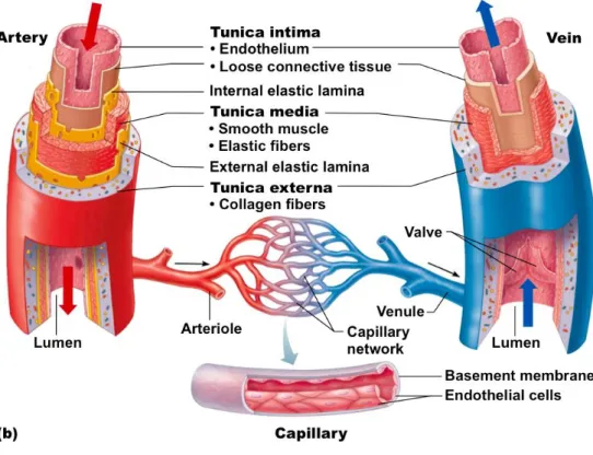
The circulatory system comprises the heart and all the blood vessels including arteries, veins, arterioles, capillaries and venules. Generally, the structure of blood vessels can be characterized into three distinct layers that are respectively the intima, media and adventitia (Figure 1). The intima, also called tunica interna, is the inner most layer of a blood vessel and is characterized by the presence of the endothelium supported by the presence of connective tissue and the internal elastic lamina. The media, also called tunica media, is a bulk of smooth muscle cells (SMC) with layers of elastic fibers that increase in thickness with the size of the vessel. The adventitia, also called tunica externa or tunica adventitia, is the external protective layer of the vessel containing elastin, collagen, fibroblasts, macrophages, nerve endings, vasa vasorum and protective fibers (Mulvany and Aalkjaer 1990).
The thickness and structure of each layer differs with the size and location of blood vessels. Large arteries contain a higher number of elastic fibers in the media while muscular arteries contain a higher number of smooth muscle cells. The capillaries contains only the endothelial layer and a basal membrane with connective tissue (Pais, Meiselman et al. 2010).
16
The role of the endothelium in the regulation of the vascular
tone
Endothelium is a monolayer of endothelial cells (ECs) which lines the entire luminal surface of the circulatory system and acts as a selective barrier between blood and the surrounding tissues. The endothelium plays an important role in the regulation of blood flow and homeostasis by releasing bioactive molecules including vasoconstricting (angiotensin II, reactive oxygen species, endothelin, vasoconstrictor prostanoids) and vasodilating (nitric oxide, prostacyclin, endothelium derived hyperpolarization) factors in response to various neurotransmitters, hormones, vasoactive compounds and mechanical stimuluses (Sandoo, van Zanten et al. 2010). Endothelium also controls vascular smooth muscle cells (VSMC) proliferation and exchange of molecules between plasma and the interstitial fluid, playing a key role in pro- and anticoagulant mechanisms (Galley and Webster 2004). Lack of endothelial regulation of the vascular homeostasis characterize the endothelial dysfunction which result in a favorable environment for the development of vascular complications ultimately leading to cardiovascular diseases (CVDs) (Lerman and Zeiher 2005).
Endothelium-derived relaxing factors (EDRF)
Nitric Oxide (NO)
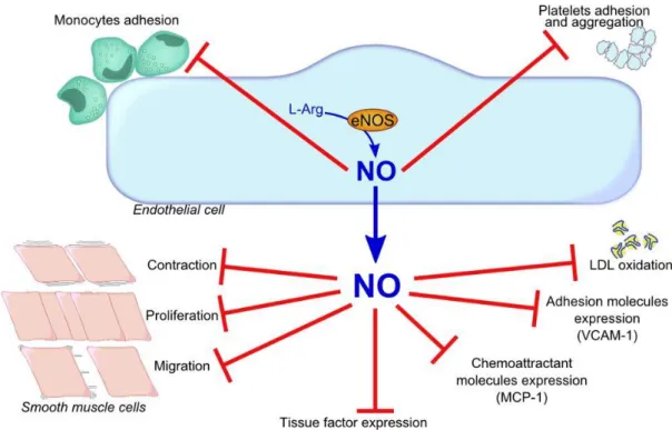
In 1980, Furchgott and Zawadzki discovered that in response to acetylcholine (ACh) the endothelium releases a substance responsible for smooth muscle relaxation in the vascular wall (Furchgott and Zawadzki 1980). This endothelium-derived relaxing factor (EDRF) was later identified as nitric oxide (NO), the first identified gaseous signaling molecule. NO not only have an important role in the regulation of vascular tone but it also plays a key role in vascular health through its vaso-protective mechanisms like the inhibition of leukocyte adhesion, platelet aggregation and the prevention of the expression of numerous pro-inflammatory and pro-thrombotic mediators such as monocyte chemoattractant protein (MCP-1), tissue factor (TF) and adhesion molecules. Thus, NO maintains the fluidity of blood by preventing platelet aggregation and monocyte adhesion (Gewaltig and Kojda 2002) (Figure 2).
NO is generated from L-Arginine by the action of NO synthase (NOS). NOS according to its location or type of function can be divided into three isoforms, namely the neuronal
17
NOS (nNOS, NOS I or bNOS) in the nervous tissue, the cytokine-inducible NOS (iNOS or NOS II) and the endothelial NOS (eNOS or NOS III). The eNOS ad nNOS isoforms are constitutively express in their respective tissues, while iNOS is generally not expressed in unstimulated cells. The eNOS isofoms is responsible for the major portion of NO produced in the vascular system in physiological situation. For the formation of NO from arginine, eNOS form homodimers that need to bind an essential cofactor, the tetrahydrobiopterin (BH4)
(Forstermann and Sessa 2012). A reduced bioavailability of the co-factor BH4, mainly
through its oxidation in BH2, as well as a lack of the substrate L-arginine, could results in the
“uncoupling” of NOS leading to the formation of superoxide anion and hydrogen peroxide (Fleming 2010). The NOS are multi-domain enzymes containing a C-terminal reductase domain and N-terminal oxygenase domain. The oxygenase domain contains the binding sites for haem, BH4 and L-arginine whereas the reductase domain contains the binding sites for
NADPH, flavin adenine dinucleotide (FAD), flavin mono nucleotide (FMD) and CaM. Binding of CaM to its binding site facilitate the transfers of electrons from NADPH at reductase domain towards haem at oxygenase-domain via FAD and FMD, enabling the haem to bind with O2 and cause its activation via reduction, which then oxidize arginine to
L-citrulline and NO (Fleming 2010, Forstermann and Sessa 2012).
Figure 2: Vaso-protective effects of NO (VCAM-1, vascular cell adhesion molecule-1; MCP-1, monocyte chemoattractant protein-molecule-1; LDL, low density lipoproteins)
18
During pre-activation phase eNOS is localized in the caveolae (small invaginations of cell membrane) bound to caveolin-1 protein. The dissociation of eNOS from the caveolin-1 delocalize eNOS to the cytosol where it is activated by cofactors, phosphorylation by post-translational enzymes and association with heat shock protein 90 (Dessy, Feron et al. 2010). Under calcium (Ca2+)-dependent activation, in response to increases in intracellular Ca2+, calmodulin detaches eNOS from caveolin-1 protein rendering the enzyme active. Ca2+ mobilizing agonists such as acetylcholine (ACh), bradykinin (BK) and histamine (H) activate the eNOS in the same manner. (Vanhoutte, Zhao et al. 2016). The other pathway of eNOS activation also called Ca2+-independent pathway is through phosphorylation of eNOS by the action of different kinases. Indeed, cyclic AMP-dependent protein kinase A (PKA), protein kinase B (Akt), AMP-activated protein kinase A (AMPK), protein kinase G (PKG), Ca2+/calmodulin-dependent protein kinase II (CaM kinase II) cause activation of eNOS by
phosphorylation of the Ser1177 residue. However, the phosphorylation on the different residue can have opposing effects as phosphorylation of Ser1177, Ser635 and Ser617 leads to eNOS activation whereas phosphorylation of Thr495 and Ser116 have an inhibitory effect (Bauer, Fulton et al. 2003, Fleming 2010) (Figure 3). Mechanical forces (shear stress), vascular endothelial growth factor (VEGF) and hormones (estrogen, insulin) cause activation of eNOS by phosphorylation via PI3K/Akt pathway (Dudzinski and Michel 2007, Forstermann and Sessa 2012).
19
Figure 3: Activation and inhibitory sites of eNOS (Numbers refer to human sequence) (Fleming 2010)
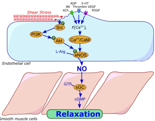
After its formation in the endothelium, NO could diffuse freely into the adjacent SMC’s where it activates the soluble guanylyl cyclase (sGC) catalyzing the conversion of 5’-guanylyl triphosphate (GTP) to cyclic 3’,5’-5’-guanylyl monophosphate (cGMP) (Figure 4). The resulting cGMP activates the protein kinase G (PKG) which decreases free cytosolic Ca2+ by inhibiting inositol trisphosphate (IP3)-activated release from sarcoplasmic reticulum (SR) and
its influx from extracellular spaces through activation of large conductance calcium-activated potassium channels (Furchgott and Vanhoutte 1989). PKG also facilitates the uptake of Ca2+
into SR through phosphorylation of phospholamban (a protein in the membrane of SR) which in turn activates SR calcium-ATPase (Gao 2010). This depletion of cytosolic Ca2+ in SR
decreases the activity of calcium-dependent myosin light chain kinase resulting in interruption of contractile process (Zhao, Vanhoutte et al. 2015).
20
Figure 4: Nitric oxide-mediated vasorelaxation pathway (ACh, acetylcholine; BK, bradykinin; ADP, adenosine diphosphate; 5-HT, serotonin; PDGF, platelet derived growth factor; VEGF, vascular endothelial growth factor; Src, sarcoma-family kinases; PI3K, phosphoinositide 3-kinase; Akt, protein kinase B; L-Arg, L-arginin; Ca2+/CaM,
calcium calmodulin; eNOS, endothelial nitric oxide synthase; NO, Nitric oxide; sGC, soluble guanylyl cyclase; GTP, guanosine 5-trisphosphate; cGMP, cyclic guanosine 3-5 monophosphate)
Prostacyclin (PGI2)
Prostacyclin is regarded as a potent vasodilator and inhibitor of platelet aggregation playing a vasoprotective role (Coleman, Smith et al. 1994). Prostacyclin is a member of prostanoids which are metabolites of arachidonic acid (AA). Arachidonic acid is produced from the hydrolysis of membrane phospholipids catalyzed by phospholipase A2 (Funk 2001).
Biosynthesis of prostacyclin in endothelial cells involve conversion of AA to endoperoxides by cyclooxygenases (COXs), which are subsequently converted to prostacyclin by the enzyme prostacyclin synthase (Tang and Vanhoutte 2009).
After its production in the endothelial cell, PGI2 is released via the ATP-binding
21
its vasodilatory and anti-aggregatory effect by acting on the IP1 receptors which are
membrane G-protein coupled receptors. IP1 activates the enzyme adenylyl cyclase converting
ATP to cAMP, which subsequently activates protein kinase A (PKA). PKA reduces availability of free Ca2+ by inhibiting its release and promoting its uptake by the sarcoplasmic reticulum (SR) in the SMCs favoring vasorelaxation (Mitchell, Ahmetaj-Shala et al. 2014). cAMP also activates an exchange protein (Epac) which, together with PKA, inhibits the proliferation of SMCs (Hewer, Sala-Newby et al. 2011). Moreover, PGI2 also facilitates the
release of NO from ECs hence promoting the vasodilatory action of NO (Shimokawa, Flavahan et al. 1988).
Endothelium-dependent hyperpolarization (EDH)
In addition to NO and prostacyclin (PGI2), EDH is an important contributor for the
endothelium-dependent vasorelaxations. EDH induces vasorelaxation which is resistant to the inhibitors L-nitroarginine methyl ester (LNAME) and indomethacin (inhibitors of eNOS and cyclooxygenases, respectively) (Takaki, Morikawa et al. 2008). EDH is not a single agent, but rather it’s a group of diffusible factors (K+ ions, H
2O2, H2S, CO, ROS, different peptides
and derivatives of COX, LOX and cytochrome p-450 pathway) influencing the action of different K+ channels causing hyperpolarization of SMCs (Figure 5) (El Assar, Angulo et al. 2012) . Chemical nature and the potency of EDH depends on the location and size of different arteries mainly influencing the resistant arteries and smaller vessels (Ozkor and Quyyumi 2011). Indeed, the contribution of EDH to the regulation of the vascular tone increases as the diameter of the artery decreases (Leung and Vanhoutte 2015).
EDH works mainly through two pathways, diffusible and contact-mediated. Diffusible mechanism is characterized by diffusion of different agents (K+ ions, epoxyeicosatrienoic acids (EETs), C-type natriuretic peptide, H2O2 and H2S) from ECs to the adjacent SMCs
causing the activation of large conductance Ca2+ -activated K+ channels (BKCa) rendering the
SMCs hyperpolarized (Goto, Ohtsubo et al. 2018).
Whereas the contact-mediated mechanism of hyperpolarization involves the activation of small conductance calcium-activated potassium channels (SKCa) and intermediate
conductance calcium-activated potassiumchannels (IKCa) in response to rise in intracellular
22
myoendothelial gap junctions (MGJs) (Feletou and Vanhoutte 2009, Goto, Ohtsubo et al. 2018).
Activation of SKCa and IKCa in the endothelial cells causes hyperpolarization of ECs by
increasing the K+ ions in the extracellular space between endothelium and SMCs activating inwardly rectifying K+ channels (Kir) and Na+/K+-ATPase rendering the SMCs hyperpolarized (Garland, Hiley et al. 2011).
Figure 5: Endothelium-derived hyperpolarization-mediated vasodilation (AA, arachidonic acid; AC, adenylyl cyclase; ACh, acetylcholine; Ba+2, barium; BK,
bradykinin; Ca2+, intracellular calcium concentration; BK
Ca, large conductance Ca2+
-activated K+ channel; CaM, calcium calmodulin; cAMP, cyclic adenosine mono phosphate; cGMP, cyclic guanosine mono phosphate; ChTX, charybdotoxin; COX, cyclo-oxygenase; EETs, epoxyeicosatrienoic acids; αGA, 18 α-glycyrrhetic acid; GAP 27, connexin inhibitor; Glib, glibenclamide; H2O2, hydrogen peroxide; IbTX, iberiotoxin;
IK1, intermediate conductance K+ channel 1; IP3, inositol trisphosphate; K+, potassium;
KATP, ATP sensitive K+ channels; Kca, Ca2+ activated K+ channels; LOX,
lipo-oxygenase; Na+/K+, sodium/potassium pump; NO-, nitrite anion; P450, cytochrome
p-450; SKCa, small-conductance Ca2+-activated K+ channel subtype 3; NOS, nitric oxide
synthase; PGI2, prostacyclin; SK3, small conductance K+ channel 3; SOD, superoxide
dismutase; SP, substance P; TBA, tetra-butyl ammonium; TEA, tetra-ethyl ammonium) (Vanhoutte, Shimokawa et al. 2017).
23
Endothelium-Derived Contractile Factors (EDCF)
Reactive Oxygen Species (ROS) and Oxidative Stress
Under normal physiological conditions, reactive oxygen species (ROS) are the byproducts of aerobic metabolism which serve as important signaling molecules in biological processes for normal cell functioning. ROS includes superoxide anion (O2-), hydrogen
peroxide (H2O2) and hydroxyl radical (OH·) (Schieber and Chandel 2014). In healthy state
there is a dynamic equilibrium between the production of ROS and its removal, promoting the normal functioning of cell. Under pathological conditions the balance shifts towards the overproduction of ROS, resulting in an increased oxidative stress damaging the important macromolecules such as proteins, lipids and nucleic acids (Schieber and Chandel 2014, Zhang, Wang et al. 2016). In the endothelial cells, the main sources of ROS are NADPH oxidases (NOX), NO synthases, cyclooxygenases (COX), mitochondrial respiratory chain, cytochrome p-450 (Cyp-450), xanthine oxidase and the peroxisomes (Figure 6), whereas ROS are inactivated mainly by the action of the superoxide dismutase (SOD) (Holmstrom and Finkel 2014).
ROS is an important contributor of EDCF-dependent contraction of vascular smooth muscle cells (VSMCs) (Yang, Feletou et al. 2003). Vasoconstriction in response to the increased production of ROS is mediated through the activation of the ryanodine receptors (RyRs) on the SR releasing Ca2+. ROS also activates protein kinase C (PKC) which, together with ROS, inhibits membrane voltage-gated potassium channels (Kv) causing membrane
depolarization and opening of membrane voltage-gated Ca2+ channels (Cav), additionally
ROS and PKC can also activate membrane store-operated Ca2+ channels (SOC), both contributing to the increase in intracellular Ca2+ levels facilitating contraction (Wang and Zheng 2010). ROS is also responsible for impairment of the NO-mediated relaxation by its direct conversion into peroxynitrite anion (OONO-), or decreased formation due to uncoupling of eNOS or oxidation of its cofactor BH4 to BH2 (Incalza, D'Oria et al. 2018).
ROS also affects EDH-mediated relaxations by influencing the activity of K+ channels
(Kusama, Kajikuri et al. 2005) and modifying the modulation of hyperpolarization through myoendothelial gap junctions (Griffith, Chaytor et al. 2005).
24
Figure 6: Cellular sources of reactive oxygen species (ER, endoplasmic reticulum; H2O2, hydrogen peroxide; LCFA, long chain fatty acids; ROS, reactive oxygen species)
(Holmstrom and Finkel 2014).
Thromboxane (TXA2) and Prostacyclin (PGI2)
In contrast to its vasodilatory effect, ACh can also lead to endothelium-dependent contractions. These contractions are totally abolished in arterial rings pre-treated with indomethacin, indicating the involvement of the COX pathway (Gluais, Lonchampt et al. 2005). Moreover, pre-treatment with selective COX-2 inhibitor shows only partial or no inhibition of the contractile response to ACh, whereas COX-1 inhibitor shows significant or complete inhibition of contractile response pointing towards dominating role of COX-1 (Feletou, Huang et al. 2011). COX-mediated conversion of arachidonic acid culminates in the production of intermediate endoperoxides (PGH2), which is the precursor of all the
prostaglandins. Further important metabolites of PGH2 include prostacyclin (PGI2),
thromboxane (TXA2), prostaglandin E2 (PGE2), prostaglandin D2 (PGD2), prostaglandin F2α
(PGF2α) produced by the action of their respective synthases (Bos, Richel et al. 2004). After
their production, prostaglandins diffuse out of the endothelium and act on the G-protein coupled prostanoid receptors named IP, TP, EP, DP and FP respectively (Tsuboi, Sugimoto et al. 2002) (Figure 7).
25
Figure 7: Cyclooxygenase-derived prostanoids and their respective receptors (DP, D prostanoid; EP, E prostanoid; FP, F prostanoid; IP, I prostanoid; PGD2,
prostaglandin D2; PGDS, prostaglandin D synthase; PGE2, prostaglandin E2; PGES,
prostaglandin E synthase; PGF2α, prostaglandin F 2α;PGFS, prostaglandin F synthase;
PGG2, prostaglandin G2; PGH2, endoperoxides; PGI2, prostacyclin; PGIS, prostacyclin
synthase; TP, T prostanoid; TXA2, thromboxane A2; TXS, thromboxane synthase
(Feletou, Huang et al. 2011).
TXA2 and PGI2 play the most important role in the mediation of COX-dependent
EDCF via the activation of TP receptors which is responsible for diverse physiological and pathophysiological roles including VSMC contraction, platelet aggregation, expression of adhesion molecules and promotion of infiltration of monocytes and macrophages (Figure 8) (Feletou, Huang et al. 2010, Matsumoto, Goulopoulou et al. 2015). Upon its activation, TP receptor causes vasoconstriction by increasing intracellular Ca2+ levels through opening of receptor-operated and voltage-gated Ca2+ channels and Rho-kinase-mediated activation of myofilaments (Tang and Vanhoutte 2009).
26
27 Angiotensin II (Ang II)
Angiotensin II (Ang II) is an octapeptide which is an important and multifunctional hormone of the renin-angiotensin system (RAS) accountable for the regulation of blood pressure and vascular homeostasis. Its biosynthesis involves production of its precursor angiotensinogen in the liver which is subsequently cleaved by renin to the biologically inactive angiotensin I (Ang I). Conversion of Ang I to the biologically active Ang II is catalyzed by the angiotensin converting enzyme (ACE) or chymases (Ma, Kam et al. 2010). Ang II is further convertible to a vasodilator metabolite Angiotensin (1-7) by the angiotensin converting enzyme 2 (ACE2) or the plasma endopeptidases (Schindler, Bramlage et al. 2007). Regulatory function of Ang II include blood pressure, plasma volume, thirst response, vascular tone, vascular remodeling, cellular proliferation and activation of sympathetic nervous system (Loiola, Fernandes et al. 2011). Ang II produce its action through two main receptors, the angiotensin type 1 and 2 receptors (AT1R & AT2R, respectively). AT1R are located on the membranes of endothelial cells, cardiomyocytes, VSMCs, nerve endings, conductive tissues, liver, lung and kidney whereas AT2R are located on the endothelial cells, VSMCs and fibroid tissue of heart, myometrium and kidney (Regitz-Zagrosek, Fielitz et al. 1998, Allen, Zhuo et al. 1999).
Ang II produce its vascular effects mainly through AT1R (Figure 9). AT1R are G-protein coupled receptors which further activate several other pathways. Vasoconstrictor action of AT1R is mediated via activation of voltage gated Ca+ channels, phospholipase C (PLC), phospholipase D (PLD), phospholipase A-2 (PLA2). Moreover, AT1R receptor
activate extracellular signal regulated kinase (ERK) cascade, platelet derived growth factor (PGRF), epidermal growth factor (EGRF), insulin receptor pathway, and tyrosine kinases and MAPK pathway related to Src family of proteins, proline rich tyrosine kinase 2, focal adhesion kinase and Janus kinases (JAKs). Increased production of ROS is also associated with AT1R via activation of NADPH oxidase (Touyz and Schiffrin 2000).
28
Figure 9: Angiotensin type 1 receptor-mediated actions (Ca2+, calcium; DAG,
diacylglycerol; IP3, inositol trisphosphate; MAPK, mitogen activated protein kinase;
PKC, protein kinase C; PLA2, phospholipase A2; PLC, phospholipase C; PLD,
phospholipase D; Src, sarcoma-family kinases) (Touyz and Schiffrin 2000)
Endothelin-1 (ET-1)
ET-1 is a potent vasoconstrictor peptide produced and released by the endothelium along with its isoforms ET-2 and ET-3. But ET-1 is most important for cardiovascular system-related functions (Kedzierski and Yanagisawa 2001). ET-1 produce its effects through the activation of G-protein coupled ET receptors having two subtypes, ETA and ETB
(Hynynen and Khalil 2006). ETA receptors are only located on SMCs, whereas ETB receptors
are located on the endothelium and SMCs (El Assar, Angulo et al. 2012). ETA receptor
causes accumulation of IP3 via activation of PLC leading to increased intracellular Ca2+ and
site-29
specific actions. In SMCs producing vasoconstriction like AT1R and in endothelium Ca2+ dependent activation of eNOS and COX releasing NO and PGI2 leading to vasodilation
(Luscher and Barton 2000, Hynynen and Khalil 2006). ET-1 can also produce indirect vasoconstriction through activation of endothelium-derived TXA2 (Bohm and Pernow 2007).
30