ORIGINAL PAPER
Extracortical plate fixation with new plate inserts and cerclage
wires for the treatment of periprosthetic hip fractures
Johannes D. Bastian&Andre Butscher&Gianni Bigolin&
Matthias A. Zumstein&Hubert P. Nötzli
Received: 27 August 2013 / Accepted: 8 September 2013 / Published online: 5 October 2013 # Springer-Verlag Berlin Heidelberg 2013
Abstract
Purpose Fixation of periprosthetic hip fractures with intra-cortical anchorage might not be feasible in cases with bulky implants and/or poor bone stock.
Methods Rotational stability of new plate inserts with extra-cortical anchorage for cerclage fixation was measured and compared to the stability found using a standard technique in a biomechanical setup using a torsion testing machine. In a synthetic PUR bone model, transverse fractures were fixed distally using screws and proximally by wire cerclages attached to the plates using“new” (extracortical anchorage) or “stan-dard” (intracortical anchorage) plate inserts. Time to fracture consolidation and complications were assessed in a consecutive series of 18 patients (18 female; mean age 81 years, range 55– 92) with periprosthetic hip fractures (ten type B1, eight type C-Vancouver) treated with the new device between July 2003 and July 2010.
Results The “new” device showed a higher rotational stability than the“standard” technique (p <0.001). Fractures showed radiographic consolidation after 14±5 weeks (mean ± SD) postoperatively in patients. Revision surgery was necessary in four patients, unrelated to the new technique.
Conclusion In periprosthetic hip fractures in which fixation with intracortical anchorage using conventional means might be difficult due to bulky revision stems and/or poor bone stock, the new device may be an addition to the range of existing implants.
Keywords Periprosthetic fracture . Fixation . Extracortical . Revision stem . Elderly . Hip . Plate insert . Cerclage
Introduction
Femoral shaft fractures following total hip arthroplasty are classified using the Vancouver classification system [1]. In general, fractures with a well-fixed stem occurring around (type B) or below (type C) the femoral stem are treated by open reduction and internal fixation using plates with proxi-mal monocortical screws, proxiproxi-mal cables, and distal bicortical screws [2–5]. In cases of periprosthetic fractures with osteoporotic bone combined with a thin cortex and/or bulky revision stems in situ, placement of screws might not be feasible (Fig. 1). In locking compression plates—especially
when introduced for fracture fixation in the elderly with osteoporotic bone—screw placement is still a concern as the direction of the screws is predetermined by the thread in the plate for the head-locking screws. As a result, placement with abutment of the prosthesis stem leading to altered screw threads with subsequent screw pullout or fatigue failure of the plate have been described [6–8]. Moreover, proximal screws may violate the bone–prosthesis interface, lead to formation of cement cracks with subsequent prosthetic loos-ening, act as stress enhancers with an increasing risk for further fractures, and cerclage fixation has shown high failure rates with minimal rotational stability [9–12]. For these rea-sons plates for extracortical fixation using the clamp-on prin-ciple have been developed. Whereas the Mennen plate showed insufficient neutralisation of the bending moment in a relatively small series [13], the shape-memory sawtooth-arm embracing fixator was successful in stabilising Vancouver type B1 and C fractures [14]. But their utility for comminuted fractures is limited. Both types of plates involve unavoidably extensive interference with soft tissue which is of concern and
J. D. Bastian (*)
:
M. A. ZumsteinDepartment of Orthopaedic and Trauma Surgery, Inselspital, University of Bern, Freiburgstrasse 3, 3010 Bern, Switzerland e-mail: [email protected]
A. Butscher
:
G. BigolinRMS Foundation, Bettlach, Switzerland H. P. Nötzli
neither plates allow additional screw fixation through the plate.
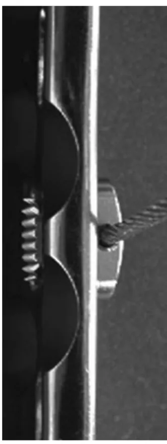
Considering all these limitations,“cerclage only” fixation of the proximal fragment might be favourable. However, the conventional cerclage positioning devices either have no bone contact or require pins through monocortical drill holes at the risk of further weakening the thin bone. Moreover, sufficient depth of the drill holes for adequate fixation seems unobtainable in the presence of a canal filling implant. As a result, they do not biomechanically operate as a screw as they hold the cerclage with minimal impact on the bone and rotational stability cannot be guaranteed. To potentially re-solve these limitations, new plate inserts without the need for any drill holes have been developed with a ridged surface protruding from the plate on the bony side to hook into the bone and to provide rotational stability as an extracortical device (Fig.2). We postulate, that the new device in combi-nation with cerclage wire fixation provides rotational stability comparable to a cerclage fixation using the conventional positioning pins. The aim of this report was to test the hypoth-esis in a simplified biomechanical setup and to report prelim-inary results on the clinical implementation of the new device.
Methods
Biomechanical testing
A synthetic polyurethane (PUR) bone model (PR0014, Synbone, Switzerland) was used. The cylinder, with an outer diameter of 40 mm and length of 200 mm, consisted of a cortical wall thickness of three millimetres dense PUR and an
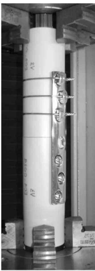
inner porous part simulating trabecular bone. A fracture was simulated by performing transverse osteotomies at half of the length of the cylinders. A reinforcement of the model was established by insertion of a distal part of an uncemented revision hip stem (Revitan® stem, distal straight, Zimmer, Switzerland). Reaming of the model was performed prior to insertion of the stem using a conical reamer with a diameter of 16 mm up to a depth of 60 mm. The hip stem was inserted so that the distal tip of the stem was 20 mm distal to the fracture line. The fractures were then fixed using seven-hole plates (DCP® 4.5 mm broad, stainless steel, Synthes, Zuchwil, Switzerland) with a monocortical and two bicortical screws (4.5 mm cortex screws; Synthes) in the distal three holes. Proximally, the first three holes were instrumented with a wire cerclage (diameter: 1.5 mm; Synthes) using the conventional positioning pins (Ref. 298.839; Synthes) in the control group (Fig. 3a) whereas the new inserts (custom-made device; Synthes) were used in the treatment group (Fig. 3b). To ensure comparable pre-tension and sym-metrical twisting of the cerclage wires, a standardised wire tightening technique was used for all tests as published previ-ously [15]. Thereafter, each of the reconstructions was distorted 20 times ±2° with 15°/s. After this dynamic loading a quasistatic deformation of up to a maximal angle of 5° was performed while recording the torsional moment (Nm) in both groups. Rotational stability was measured using a torsion testing machine (TL500; Zwick; for example, see Fig. 4).
Fig. 1 Conventional radiographs AP view (a ) and lateral views (b ) showing a periprosthetic femoral shaft fracture in an 85-year-old female patient with thin cortical bone due to osteoporosis and a bulky revision stem in the femoral shaft. Conventional radiographs AP views (c, d) after wire-cerclage plating of a periprosthetic femoral shaft fracture using the new plate inserts
Fig. 2 Photograph showing the new insert with a ridged surface protruding from the plate on the bony side with the potential to hook into the bone and to ensure rotational stability
Patient evaluation
Between July 2003 and July 2010, a consecutive series of 18 patients (18 female; mean age 81 years, range 55–92) with periprosthetic hip fractures at/around or distal to the prosthesis tip with a well-fixed stem were treated by the senior author using “cerclage only” fixation, supported by the new plate inserts in the proximal fragment and with bicortical screws in the distal fragment. The patients’ charts were assessed for the patients’ demographics, body mass index (kg/m2
), amount of medical comorbidities, the American Society of Anaesthesiologists Physical Status Classification (ASA) level, the time interval from the index procedure with total hip replacement up until the episode of trauma leading to periprosthetic hip fracture, the postoperative aftercare protocol, the patients’ mobility prior to trauma and after fracture healing at the latest follow-up, intra- and postoperative complications, duration of surgery and blood loss. The radiographic data was analysed to describe the fracture types according to the Van-couver classification [2] and the time until fracture union occurred. The retrospective evaluation focused on the evalua-tion of the time interval until the fractures were radiographically
consolidated, full weight-bearing was allowed and any postop-erative complications needing revision surgery.
Statistical analysis
In the biomechanical setup, the three replicated series “tor-sional load per displacement” of each group were averaged, and the averaged series analysed. Mixed-effects models with the torsional moment (Nm) as the dependent variable and polynomial trends in the rotational displacement (in degree) as a predictor were used to estimate the average curves for the treatment group“new” and for the control group “standard”. The differences in parameters of the two curves were analysed using z-tests. Bonferroni adjustment was applied to have an overall significance level of 5 % for all comparisons. In the clinical setup, p-values are not provided as the statistical analysis used was descriptive. Statistical analysis was performed with SAS 9.2 (SAS Institute Inc., Cary, NC, USA).
Results
Biomechanical testing
The averaged curves for the treatment group“new” and for the control group“standard” presenting the results for the
mixed-Fig. 3 Drawing of the test setup in the experimental group with the standard inserts (intracortical anchorage) (a) for wire cerclage fixation proximally or the new inserts (extracortical anchorage) (b) in a transverse fracture model in synthetic PUR bone with a revision hip stem in situ. The fracture was fixed using screws distally
Fig. 4 Photograph of the test setup in the group with the new device in the transverse fracture model in synthetic PUR bone with a revision hip stem in situ. The fracture was fixed using screws distally and the new plate inserts for wire cerclage fixation proximally
effects model with the torsional moment (Nm) as the depen-dent variable and polynomial trends of grade two in the rotational displacement (in degree) as a predictor are displayed in Fig.5. The value (mean ± standard error) for the slope of the curves was 1.49±0.04 in the group“new” and 1.24±0.04 in the group“standard”. The observed differences were statisti-cally significant (p <0.001), so that an increased torsional load was required to achieve the same rotational displacement in the treatment group using the new device compared to the control group using the standard positioning pins.
Patients
Fixation of the periprosthetic fracture was undertaken using either conventional plates in 15 of 18 cases (LC-DCP® 4.5, broad, stainless steel, Ref. 226.600; Synthes) or 95° condylar plates in three of 18 cases. In the proximal fragment, wire cerclage using the new inserts was applied, whereas in the distal fragment bicortical screws were feasible (e.g. Fig.1c, d). Patient demographics, number of comorbidities, Vancouver classification of the fractures, time from the index procedure with total hip replacement until trauma, postoperative aftercare (partial weight bearing [PWB] = 15–20 kg), time until union, mobility before trauma and osteosynthesis, postoperative mo-bility after fracture consolidation, complications in the postop-erative course are listed in detail in Table1. In addition, the median body mass index (in kg/m2) was 24.8 (range 18.4– 44.5) and the median ASA level was 3 (range 2–4). The time needed for surgery (mean ± SD) was 149±27 min and the estimated blood loss (mean ± SD) was 580±280 ml. Revision surgery was necessary in four of the 18 patients. In one patient, early surgical evacuation of an haematoma one day after
osteosynthesis was necessary due to anticoagulant bleeding. In two patients, revision surgery was necessary due to a re-fracture caused by repeated falls 12 or 97 days after osteosynthesis. Aseptic loosening of the prosthesis stem oc-curred in another patient 18 months after fixation of the periprosthetic hip fracture using the new inserts. The need for revision surgery was unrelated to the new plate insert device.
Discussion
The prevalence of periprosthetic femoral fractures ranges from 1 % in primary total hip arthroplasty to 4 % in revision total hip replacements [16]. However, due to the increasing num-bers of total hip arthroplasties in combination with increasing age and growing daily activities of the elderly at risk for falls, the orthopaedic surgeon will be confronted with an increasing number of periprosthetic fractures in the elderly in the future [16,17]. In general, treatment of these fractures is challenging as experience in the field of orthopaedic revision surgery as well as in trauma surgery is needed [18]. In particular, fixation of fractures occurring at or near the distal tip of the prosthesis with a stable femoral stem (Vancouver B1 or C) [2] is difficult as these fracture types are most frequently associated with complications, due to the inherently unstable facture pattern [1,19]. In cases with a poor bone stock and especially those with thin cortical bone and/or with bulky implants in situ, conventional devices may not be ideal for the stable fixation of the proximal fragment. Thus, current concepts for fracture fixation have to be reconsidered. The purpose of this study was to assess the rotational stability provided by a new fixa-tion reconstrucfixa-tion using specific plate inserts with extracortical anchorage compared to a standard technique.
The stability of cerclage fixation devices depends on the “bone-to-plate” interface and the “wire-to-plate” connection. We therefore indicate that the inserts of the new device hook into the bone and have an extracortical impact on the bone without the disadvantages of conventional inserts requiring monocortical drill holes for anchorage. The “bone-to-plate” interface of these new inserts showed a footprint of the toothed surface in one patient on the occasion of a re-operation due to aseptic loosening of the stem more than one year after the index procedure. Additionally, the inserts have an oval shape ensuring stability on the part of the hardware as the rotation of the oval inserts in oval plate holes is not possible. The conventional plate inserts do not provide such a stable“wire-to-plate” connection. Differences in torsional loads per displacement using cerclage fixation with the new inserts were statistically signif-icant compared to conventional inserts in the biomechanical testing. In addition, in the presence of a bulky implant the necessary insertion depth of the pin may not be achievable, making the implant less stable than in the model setting. In the clinical evaluation, the use of the new device in these specific
Fig. 5 Graph presenting the torsional loads per displacement (mean ± SD; in Nm) measured for the groups using the new device (“new”) or the standard positioning pins (“standard”). Differences in the torsional mo-ments required for rotational displacement were statistically significant (p <0.001)
periprosthetic fracture patterns obtained promising results since fractures healed in all patients. However, it is questionable whether these differences—obtained in a simplified biome-chanical setup—are of clinical relevance. They may even achieve less stability in the clinical situation since the thin cortical bone does not provide the same stability as the material in the model. The use of human femoral specimens, in contrast, would also be limited by the inherent variability of the speci-men, requiring large sample sizes in order to obtain statistically significant results. In addition, the availability of such speci-mens is limited. Further limitations of the study are that (1) the relationship between the length of the plate and the length of the synthetic bone was not evaluated (a shorter fixation could prove to be less rigid), (2) isometric loading with pure torsional force application rather than physiological loading reflecting the in vivo conditions was applied and (3) the impact of soft-tissues (e.g. muscles forces) was not assessed. However, due to the reduced complexity of the model, the statistically signif-icant differences found in fixation strength of the devices used might be related to the stiffness of the reconstruction device rather than to other influences. Additionally, the tension of the wire cerclage was not measured, so that it remains unclear whether the stiffness in terms of tension of the wires is compa-rable. Despite this, a standardised wire tightening technique has been used [15]. A further limitation might be that only a transverse osteotomy was used. A transverse osteotomy does, however, simulate the worst case scenario due to the high rotational instability fracture pattern.
The clinical use of the new device resulted in fracture healing in all cases. Generally, revision surgery was unrelated to the new device; the re-fracture seen in two cases was related to further trauma. The assessment of the mobility status of the patients showed that seven of the 18 patients regained the mobility status that they had prior to trauma. Eleven of the 18 patients did not completely recover from the trauma and required more support or walking aids than before injury. However, fractures were caused by simple falls and, in gen-eral, patients were still at risk for further falls even after the fractures healed—as observed in two of the 18 patients. In retrospect, the use of walking aids might have been modified so that more suitable aids were used in order to prevent further falls from occurring.
In summary, the new inserts required a higher torsional moment for displacement than the standard plate insert in the biomechanical setup. As a result, the achievable rotational stability seems to be sufficient in the clinical setting with thin cortical bone, where screws and conventional inserts cannot be applied in an adequate manner.
Conclusion
The use of the new plate inserts resulted in a higher rotational fixation strength according to a simplified biomechanical model. The extracortical plate fixation in combination with cerclage wires showed fracture healing in all patients in
Table 1 Patient demographics Patient number Age/ sex Comorbidities Fracture THR before
Rehabilitation protocol Union Pre-OP mobility
Post-OP mobility after fracture consolidation
Complications
1 92/F 1 B1 2 years PWB—8 weeks 20 weeks Unaided Walking frame Re-Fracture 2 92/F 2 C 8 years Bed to chair—8 weeks 23 weeks Walking frame Walking frame –
3 55/F >3 B1 13 years Bed to chair—8 weeks 20 weeks Single stick Single stick Aseptic loosening 4 84/F 0 B1 17 years PWB—8 weeks 8 weeks Unaided Two crutches –
5 85/M >3 C 6 months Bed to chair—8 weeks 18 weeks Unaided Transfer to wheelchair – 6 89/F 1 B1 17 years Bed to chair—8 weeks 20 weeks Unaided Unaided – 7 76/F 0 C 12 years Bed to chair—8 weeks 13 weeks Unaided Walking frame –
8 73/F 2 C 2 years PWB—8 weeks 25 weeks Unaided Unaided –
9 77/F 0 B1 1 year PWB—8 weeks 12 weeks Unaided Single stick – 10 84/F 2 B1 12 years Bed to chair—8 weeks 13 weeks Single stick Single stick – 11 87/F >3 C 2 years Bed to chair—8 weeks 13 weeks Walking frame Walking frame – 12 89/F >3 C 17 years Bed to chair—8 weeks 15 weeks Walking frame Transfer to wheelchair – 13 85/F >3 C 4 months PWB—8 weeks 13 weeks Single stick Two crutches Re-fracture 14 74/F >3 B1 19 years Bed to chair—8 weeks 10 weeks Single stick Walking frame – 15 86/F 3 B1 8 years Bed to chair—8 weeks 11 weeks Walking frame Walking frame – 16 70/F 0 B1 8 years Bed to chair—8 weeks 7 weeks Unaided Walking frame –
17 72/F >3 C 3 years Bed to chair—8 weeks 12 weeks Unaided Two crutches Haematoma 18 83/F >3 B1 2 years Bed to chair—8 weeks 6 weeks Two crutches Walking frame –
difficult to treat fractures by conventional means. Therefore, the new device may be a valuable addition to the existing range of implants especially in cases with bulky revision stems in situ and/or poor bone stock. Based on the presented data, the new plate inserts may be a solution for an increasing number of periprosthetic fractures in the elderly presenting with the reported fracture and implant characteristics.
Acknowledgments This work was supported by the RMS Foundation, Bettlach, Switzerland. We thank S. Honold, Mechanics Group, RMS Foundation, Bettlach, Switzerland for her assistance. The implants and instruments were provided by Synthes, Zuchwil, Switzerland. We thank Dr Daniel Dietrich, Institute of Mathematical Statistics and Actuarial Science, University of Bern, Switzerland for performing the statistical analysis and his assistance in the interpretation of the results. We also thank Dr Debra Bickes-Kelleher for linguistic help in the preparation of this manuscript.
Conflict of interest The authors declare that they have no conflict of interest.
References
1. Brady OH, Garbuz DS, Masri BA, Duncan CP (1999) Classification of the hip. Orthop Clin N Am 30(2):215–220
2. Masri BA, Meek RM, Duncan CP (2004) Periprosthetic fractures evaluation and treatment. Clin Orthop Relat Res 420:80–95 3. Rayan F, Haddad F (2010) Periprosthetic femoral fractures in total
hip arthroplasty—a review. Hip Int 20(4):418–426
4. Dennis MG, Simon JA, Kummer FJ, Koval KJ, DiCesare PE (2000) Fixation of periprosthetic femoral shaft fractures occurring at the tip of the stem: a biomechanical study of 5 techniques. J Arthroplasty 15(4):523–528
5. Zdero R, Walker R, Waddell JP, Schemitsch EH (2008) Biomechanical evaluation of periprosthetic femoral fracture fixation. J Bone Joint Surg Am 90(5):1068–1077. doi:10.2106/JBJS.F.01561
6. Buttaro MA, Farfalli G, Paredes NM, Comba F, Piccaluga F (2007) Locking compression plate fixation of Vancouver
type-B1 periprosthetic femoral fractures. J Bone Joint Surg Am 89(9): 1964–1969
7. Erhardt JB, Grob K, Roderer G, Hoffmann A, Forster TN, Kuster MS (2008) Treatment of periprosthetic femur fractures with the non-contact bridging plate: a new angular stable implant. Arch Orthop Trauma Surg 128(4):409–416
8. Konstantinidis L, Hauschild O, Beckmann NA, Hirschmuller A, Sudkamp NP, Helwig P (2010) Treatment of periprosthetic femoral fractures with two different minimal invasive angle-stable plates: biomechanical comparison studies on cadaveric bones. Injury 41(12):1256–1261. doi:10.1016/j.injury.2010.05.007
9. Tsiridis E, Haddad FS, Gie GA (2003) Dall-Miles plates for periprosthetic femoral fractures. A critical review of 16 cases. Injury 34(2):107–110
10. Fulkerson E, Koval K, Preston CF, Iesaka K, Kummer FJ, Egol KA (2006) Fixation of periprosthetic femoral shaft fractures associated with cemented femoral stems: a biomechanical comparison of locked plating and conventional cable plates. J Orthop Trauma 20(2):89–93 11. Schmotzer H, Tchejeyan GH, Dall DM (1996) Surgical management of intra- and postoperative fractures of the femur about the tip of the stem in total hip arthroplasty. J Arthroplasty 11(6):709–717 12. Stevens SS, Irish AJ, Vachtsevanos JG, Csongradi J, Beaupre GS
(1995) A biomechanical study of three wiring techniques for cerclage-plating. J Orthop Trauma 9(5):381–387
13. Noorda RJ, Wuisman PI (2002) Mennen plate fixation for the treat-ment of periprosthetic femoral fractures: a multicenter study of thirty-six fractures. J Bone Joint Surg Am 84-A(12):2211–2215
14. Zhao X, Zhu ZA, Sun YH, Wang Y, Zhao J, Zhang YJ, Dai KR (2012) Nickel-titanium shape-memory sawtooth-arm embracing fixator for periprosthetic femoral fractures. Int Orthop 36(3):619– 626. doi:10.1007/s00264-011-1325-4
15. Meyer DC, Ramseier LE, Lajtai G, Notzli H (2003) A new method for cerclage wire fixation to maximal pre-tension with minimal elongation to failure. Clin Biomech (Bristol, Avon) 18(10):975–980 16. Berry DJ (1999) Epidemiology: hip and knee. Orthop Clin N Am
30(2):183–190
17. Lewallen DG, Berry DJ (1998) Periprosthetic fracture of the femur after total hip arthroplasty: treatment and results to date. Instr Course Lect 47:243–249
18. Tsiridis E, Krikler S, Giannoudis PV (2007) Periprosthetic femoral fractures: current aspects of management. Injury 38(6):649–650 19. Beals RK, Tower SS (1996) Periprosthetic fractures of the femur. An