648 Article Vol. 16, No. 9 Eur. J. Clin. Microbiol. Infect. Dis., 1997, 16:648-652
Evaluation of Commercial Slides for Detection of
Immunoglobulin G against
Bartonella henselae
by
Indirect Immunofluorescence
R. Zbinden 1., N. Michael 1, M. Sekulovski 1, A. von Graevenitz 1, D. Nadal 2
Four commercial slides were compared with in-house slides for the detection of immu- noglobulin G (IgG) against Bartonella henselae in 58 healthy persons from a rural region by an indirect immunofluorescence assay. MRL-BA slides (MRL Diagnostics, USA) and Virion slides (Virion, Switzerland) with agar-derived Bartonella henselae showed IgG titers of >_ 1:256 in 44.8% and 51.7%, respectively, whereas Bion slides (Bios, Germany), MRL-Vero slides (MRL Diagnostics), and in-house slides with cell-associated Bar- tonella henselae showed such titers in 3.4%, 5.1% and 3.4%, respectively. The MRL- Vero slides (Bartonella IgG substrate slides, MRL Diagnostics) were further evaluated with 26 patients with cat scratch disease, 20 patients with lymphadenopathy not due to cat scratch disease, 100 blood donors from an urban area, and 120 blood donors from a mixed urban/rural area. In our mixed urban/rural population the IgG titer of 1:256 had a sensitivity of 84.6% and a specificity of 93.4% for the serodiagnosis of cat scratch disease. Seroprevalence was higher in blood donors from the mixed area (50.8%) than from the urban area (37%). MRL-Vero slides were considered useful for the serodiagnosis of cat scratch disease by indirect immunofluorescence and have replaced our in-house system. However, patients with low IgG titers should be retested three to four weeks after initial sampling to demonstrate a possible rise of IgG titers in paired sera.
Cat scratch disease (CSD) has traditionally been diagnosed in patients with regional lymphadeno- pathy if three of the following criteria are met: con- tact with a cat and the presence of a scratch or a primary lesion; negative studies for other possible causes of lymphadenopathy; a positive skin test; and characteristic histopathological findings (1). By means of culture and the polymerase chain re- action (PCR), Bartonella henselae has been recent- ly recognized as the major causative agent of CSD (2, 3). The CSD skin test has been shown to have a lower sensitivity than P C R for Bartonella henselae and may, for this and for safety reasons, have lost its usefulness (4). Histopathological analyses are only possible following surgical biop- sy. Serology has made important contributions to recognition of the causative agent (5), epidemio-
1 Department of Medical Microbiology, University of Zurich, Gloriastrasse 32, CH-8028 Zurich, Switzerland.
2 Infectious Diseases Unit, University Children's Hospital, CH-8032 Zurich, Switzerland.
logical studies (6), and a better delineation of the clinical spectrum of CSD (7). Thus, serology may become the simplest and most feasible way to di- agnose CSD, provided the tests are reliable (8). We recently demonstrated the intracellular loca- tion of Bartonella henselae after cocultivation with Vero cells. Fixed slides with monolayers of Vero cells with intracellular Bartonella henselae were used in an indirect immunofluorescence assay (IFA) for detecting immunoglobulin G (IgG) to Bartonella henselae (9). The workload associated with cocultivation of Bartonella hense- lae with Vero cells on chamber slides for the in- house IFA was considered reasonable as long as no alternatives were available. In the meantime, several commercial slides for IFA have been de- veloped for the detection of IgG to Bartonella henselae. In the present study we compared our in- house IFA with four commercial IFAs. Further- more, we determined the diagnostic value of one commercial IFA with Vero cell-associated Barto- nella henselae.
Table 1: Immunoglobulin G titers to Bartonella henselae of 58 persons from the rural region determined by indirect immuno-
fluorescence with four commercial slides and an in-house slide.
Agar-derived Cetl-associated
Titer MRL-BA Virion Bion MRL-Vero In-house
< 1:64 6 7 20 25 24 1:64 12 7 27 20 29 1:128 14 14 9 10 3 1:256 7 9 1 2 2 1:512 14 16 1 1 1:1024 3 4 1:2048 2 1 Material and M e t h o d s
Serum Specimens'. Fifty-eight sera (supplied by J. Zihler in
1994, stored at -20~ from individuals living in a rural region in central Switzerland were tested for IgG antibodies to Bar- tonella henselae with four commercial IFAs and the results
compared with those obtained with our in-house system. The individuals had consulted their physicians because of symp- toms not suggestive of CSD. Thirty-three of them were cat owners. Eight sera from four patients with lymphadenopathy who, after cat contact, presented a clear rise of IgG against
Bartonella henselae with the in-house IFA, were included in
this comparison.
Sera collected from 1994 to 1996 from 22 patients with CSD (age range, 3-35 years; mean, 13 years), 20 immunocompetent childrer~ with tymphadenopathy without CSD, 100 blood donors from Zurich (urban area; supplied by R Schenker in 1994), and 120 blood donors from Aarau (mixed urban/rural area; supplied by K. Giger in 1994) were tested with a commercial cell-associated IFA. Sera were stored at -20~ Sera from 15 of the 22 patients with CSD and from the blood donors had been tested previously with the inhouse system (10). The remaining seven patients with CSD had either subacute lymphadenitis with typical histopathological changes or a positive PCR for Bartonella henselae.
Indirect Immunofluorescence Assay. Immunoglobulin G to BartoneUa henselae was detected by an IFA with in-house
slides and the following four commercial slides: MRL-BA slides (prototype #4, lot #R017194; MRL Diagnostics, USA); Virion slides (experimental lot #B:381718.09; Institut Virion, Switzerland) containing blood agar-derived Bartonel- la henselae; MRL-Vero slides (lot #R049395, lot #R067095,
and lot #R028196, Bartonella IgG substrate slides, MRL Diag-
nostics); and Bion slides (Bios, Germany) containing cell- associated Bartonella henselae. Each of the MRL-BA and
MRL-Vero slides with eight wells contained one test field with Bartonella henselae and one test field with Bartonella quintana. Results of the IFA for Bartonella quintana were not
evaluated. The in-house slides were based on intracellular
Bartonella henselae cocultivated with Vero cells as described
previously (9). In brief, Bartonella henselae (strain G-5436;
kindly provided by R. Weyant, Centers for Disease Control and Prevention, Atlanta, USA) was cocultivated with Veto ceils in tissue chamber slides. After two days the sIides were fixed with acetone-ethanol (1:1) and used for the IFA. Sera were overlaid onto the slides at dilutions of 1:64 (if nec- essary, up to 1:2048) and allowed to rest for 30 min at 37~ After washing, IgG was detected by a fluorescein-isothio- cyanate (FITC)-anti-human IgG conjugate for 30 min at
37~ For Virion slides the conjugate (lot 396318.02; Virion) was diluted 1:10; the conjugates for MRL-BA and MRL-Vero slides (goat anti-human IgG FITC; Tago Immunotogicals, Biosource International, USA) and for Bion and in-house slides (Fluoline G; bioM6rieux, Switzerland) were diluted 1:100. After incubation for 30 rain the slides were washed and examined with a fluorescence microscope. The endpoint titer was defined as the dilution that still presented a specific fluorescence.
Statistical Calculations. The optimal cut-off for the commer-
cial Vero cell-associated IFA evaluated was chosen according to the balance desired between sensitivity and specificity (11).
Results
T h e c o m p a r i s o n o f c o m m e r c i a l w i t h i n - h o u s e slides f o r t h e d e t e r m i n a t i o n o f I g G titers to Bar- tonella henselae b y I F A is s h o w n in T a b l e 1. A m o n g t h e 58 i n d i v i d u a l s f r o m t h e r u r a l r e g i o n , titers o f _> 1:256 w e r e d e t e c t e d w i t h t h e agar- d e r i v e d M R L - B A a n d V i r i o n slides in 26 ( 4 4 . 8 % ) a n d 30 ( 5 1 . 7 % ) s e r a a n d titers o f < 1:64 in six ( 1 0 . 3 % ) a n d s e v e n ( 1 2 % ) sera, r e s p e c t i v e l y . I n c o n t r a s t , w h e n e m p l o y i n g B i o n , M R L - V e r o , a n d i n - h o u s e slides w i t h c e l l - a s s o c i a t e d Bartonella henselae, titers o f _> 1:256 w e r e f o u n d in t w o (3.4%), t h r e e ( 5 . 1 % ) , a n d t w o ( 3 . 4 % ) s e r a a n d tit- ors o f < 1:64 in 20 ( 3 4 . 5 % ) , 25 ( 4 3 . 1 % ) , a n d 24 ( 4 1 . 5 % ) sera, r e s p e c t i v e l y . W i t h B i o n slides 9 7 % o f t h e titers w e r e w i t h i n o n e log 2 d i l u t i o n o f t h e c o r r e s p o n d i n g titers o b t a i n e d w i t h t h e i n - h o u s e s y s t e m ; w i t h M R L - V e r o slides all titers w e r e w i t h i n o n e log 2 d i l u t i o n a n d 7 2 . 4 % w e r e i d e n t i c a l to t h o s e o f t h e i n - h o u s e system. H o w e v e r , with t h e t w o c o m m e r c i a l slides w i t h a g a r - d e r i v e d Bartonel- la henselae, 60.3% o f all s e r a h a d t i t e r d i f f e r e n c e s o f t w o o r m o r e l o g 2 d i l u t i o n s c o m p a r e d t o titers o b t a i n e d w i t h t h e i n - h o u s e I F A . T h i r t y - t h r e e o f t h e 58 i n d i v i d u a l s w e r e cat o w n - ers. U s i n g a n y slides w i t h c e l l - a s s o c i a t e d Bartonel- la henselae, cat o w n e r s m o r e f r e q u e n t l y h a d titers
650 Eur. J. Clin. Microbiol. Infect. Dis.
Table 2: Comparison of immunoglobulin G titers in paired sera from four patients with cat scratch disease. Intervals between sera a and b were 42, 21, 24, and 14 days in patients 1, 2, 3, and 4, respectively.
IgG titer
MRL-BA Virion Bion MRL-Vero In-house
Patient 1 a 1:128 1:256 1:512 1:512 1:256 b 1:256 1:256 1:2048 1:1024 1:1024 Patient 2 a 1:64 nd 1:128 1:512 1:128 b 1:128 nd 1:512 1:512 1:512 Patient 3 a 1:64 1:128 1: < 64 1:128 1:64 b 1:128 1:512 1:256 1:1024 1:1024 Patient 4 a 1:128 t:128 1:64 1:128 1:128 b 1:256 1:256 1:256 1:1024 1:512
Fisher's exact test; data not shown). With MRL- B A and Virion slides, the median titers were 1:256 in cat owners and 1:128 in persons without cats; titers of 1:512 were more frequent in cat own- ers (p < 0.05, Fisher's exact test). Since a rise of IgG is a good m a r k e r for an ongoing infection, we included four pairs of sera in this comparison (Ta- ble 2). The slides with blood agar-derived Barto- nella henselae showed in only one patient a four- fold titer rise in paired sera and a higher titer than the corresponding median titer of cat owners. The Bion and MRL-Vero slides showed higher tit- ers than the median titers of cat owners in all four patients.
The diagnostic value of the commercial Vero cell- associated M R L slides was further evaluated. Table 3 shows the IgG titers of 26 patients (including the first sera of the 4 patients from Table 2) with CSD and controls. All control pa- tients with l y m p h a d e n o p a t h y without suspected CSD had titers of < 1:64, except one who had a titer of 1:128. F o u r t e e n of 220 blood donors had titers of 1:256 and one a titer of 1:512. Blood do- nors from the mixed urban/rural area more fre- quently had IgG titers (50.8%) than blood donors
from the urban area (37%) (p < 0.05, Fisher's ex- act test). For the serodiagnosis of CSD in our mixed urban/rural population, the IgG titer of 1:256 had a sensitivity of 84.6% and a specificity of 93.4%.
Discussion
In the present comparison of commercial IFA slides, those containing cell-associated Bartonella henselae revealed lower IgG titers than slides containing blood agar-derived bacilli. Four patients included in this comparison had IgG titers to Bartonella henselae similar to those of the normal rural population when the agar-derived slides were used. We therefore excluded these slides from consideration for replacement of our in- house system. The Vero cell-associated M R L slides showed in 72% the same titer as the in- house slides and were further evaluated for detec- tion of IgG to Bartonella henselae.
Sera from patients with and without CSD and from blood donors from two different geograph-
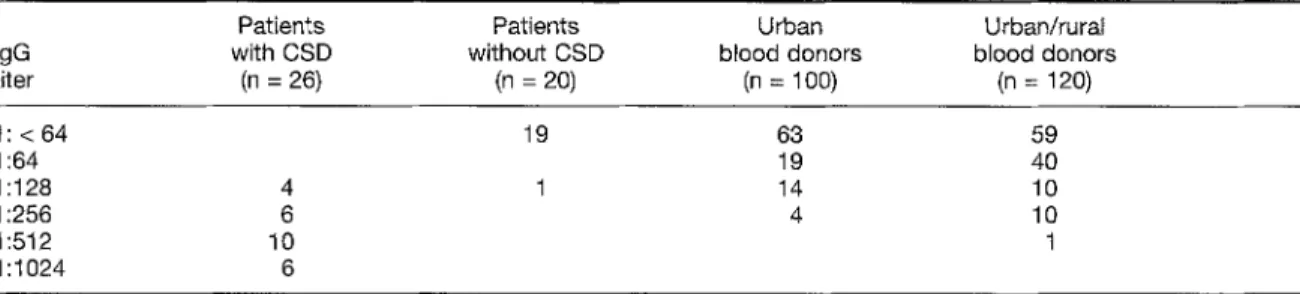
Table 3" Immunoglobulin G titers to Bartonella henselae of patients with cat scratch disease (CSD), control patients, urban blood donors, and mixed urban/rural blood donors using the MRL-Vero slides. When a titer of 1:512 is used as the cut-off, sensitivity is 61.5% and specificity 99.6%; with 1:256 as the cut-off, sensitivity is 84.6% and specificity 93.4%; with 1:128 as the cut-off, sensitivity is 100% and specificity 83.4%.
Patients Patients Urban Urban/rural
IgG with CSD without CSD blood donors blood donors titer (n = 26) (n = 20) (n = 100) (n = 120) 1 : < 64 19 63 59 1:64 19 40 1:128 4 1 14 10 1:256 6 4 10 1:512 10 1 1:1024 6
ical regions were included in this evaluation. To achieve a specificity of over 90%, the titer of 1:256 was chosen as the cut-off. The titer of 1:128 would have had a specificity below 85 % but a sensitivi- ty of 100%. Others have f o u n d that a cut-off titer of 1:100 with a cell-associated system is not sensi- tive enough (40.6%) to confirm a CSD diagnosis; the low sensitivity was attributed to blood sam- pling before an antibody increase had occurred (12). However, the positive predictive value of a titer of 1:100 with the same IFA was only 68.2% (13). A cell-associated IFA with the new Bartonel- la henselae serovariant "Marseille" revealed in three of 100 healthy blood donors a titer of 1:200, which was considered the cut-off titer (14). In a previous study, 11 patients with a Bartonella henselae infection (proven by P C R or character- istic histopathological findings) had titers of 1:256 or higher with the in-house IFA (15). In a further study with the in-house system, titers of 1:512 or higher were f o u n d in 20 of 20 children presenting with subacute l y m p h a d e n o p a t h y and a history of recent cat scratch (10). In three of these CSD patients, the initial titers were only 1:128 and 1:256, but within three weeks a rise to 1:1024 was noted. By contrast, in only one of the 332 controls (including 43 persons from house- holds of the patients) was a titer of 1:512 found (10).
Our most important observation was the differ- ence in IgG titers obtained with the various com- mercial slides, depending on the antigen prepara- tion. It was recently shown in one patient that tit- ers to Bartonella henselae differed greatly, depending on the antigen preparation for the IFA: the patient had a titer of 1:12,800 against a sheep blood agar strain and a titer of 1:200 against the same strain cultivated on endothelial cells (14). A similar discrepancy was described for IgG antibodies against Bartonella quintana grown on sheep blood agar and h u m a n epithelial cells (16).The first IFA to detect IgG to Bartonel- la henselae was based on cocultivation with Vero cells to inhibit autoagglutination of Bartonella henselae (5). The present study showed that cocul- tivation improves the specific detection of IgG to Bartonella henselae. The reasons for this are un- known, but antigenic proteins could perhaps change during cocultivation. Such changes are suggested during phase variation (17). The intra- cellular location of Bartonella henselae in the two commercial cell-associated slides (MRL-Vero and Bion) can be assumed because the specific fluorescence had the same granular aspect as shown previously with our in-house slides (9).
Besides the antigen preparation of Bartonella henselae, epidemiological parameters affect the se- rodiagnosis of Bartonella henselae infection (18). In all IFA systems serum samples of people report- ing cat contact had higher titers than sera of per- sons without cat contact. In line with our previous study of the in-house system (10), we also found by means of the M R L - V e r o slides a higher seroprevalence in blood donors from the mixed urban/rural population than in those from the city. This could be explained by close cat contact in rural regions. L o w IgG titers could relate to un- apparent or past infection with Bartonella hense- lae. Finally, serodiagnosis can be influenced by cross-reacting antibodies against o t h e r bacterial antigens, e.g., Bartonella with Coxiella (19) and other ~-2 protobacteria (20).
We conclude that, using the M R L - V e r o slides, a titer of 1:256 is compatible with CSD in our mixed urban/rural population. For routine pur- poses, the M R L - V e r o slides can replace our in- house system. Until tests to detect IgM to Barto- nella henselae have b e e n thoroughly evaluated, patients with clinical signs and symptoms of CSD plus low specific IgG titers should have paired sera drawn in intervals of three to four weeks in o r d e r to detect a possibly significant an- tibody rise. We believe that serology may replace traditional diagnostic criteria for CSD (8,10), but histology and P C R may still be necessary in atypical clinical situations.
Acknowledgements
]]ae authors thank V. Kaspar for technical assistance; J. Zihler, general practitioner, Unteriberg, Switzerland, for supplying sera from the rural population; K. Giger, Kantonsspital Aarau, Switzerland, for supplying sera of blood donors from the mixed rural/urban area; and, R Schenker, Blutspendezen- trum SRK Zurich, Switzerland, for supplying the sera of blood donors from the urban population.
References
1. Adal KA, Cockerell C J, Petri WA: Cat scratch disease, bacillary angiomatosis, and other infections due to
Rochalimaea. New England Journal of Medicine 1994, 330: 1509-1515.
2. Dolan M J, Wong MT, Regnery RL, Jorgensen JH, Gar- cia M, Peters J, Drehner D: Syndrome of Rochalimaea henselae adenitis suggesting cat scratch disease. An- nals of Internal Medicine 1993, 118: 331-336. 3. Anderson B, Sims K, Regnery R, Robinson L, Schmidt
652 Eur. J. Clin. Microbiol. Infect. Dis.
Rochalimaea henselae DNA in specimens from cat
scratch disease patients by PCR. Journal of Clinical Mi- crobiology 1994, 32: 942-948.
4. Bergmans AMC, Groothedde JW, Schellekens JFP, van Embden JDA, Ossewaarde JM, Schouls LM: Etiology of cat scratch disease: comparison of polymerase chain reaction detection of Bartonella (formerly Rochalimaea)
and Afipia fells DNA with serology and skin tests. Jour-
nal of Infectious Diseases 1995, 171 : 916-923. 5. Regnery RL, Olson JG, Perkins BA, Bibb W: Serologi-
cal response to "Rochalimaea henselae" antigen in sus-
pected cat-scratch disease. Lancet 1992, 339: 1443-1445.
6. Zangwill KM, Hamilton DH, Perkins BA, Regnery RL, Plikaytis BD, Hadler JL, CaRter ML, Wenger JD: Cat scratch disease in Connecticut. Epidemiology, risk fac- tors, and evaluation of a new diagnostic test. New Eng- land Journal of Medicine 1993, 329: 8-13.
7. Itin PH, FIL~ckiger R, Zbinden R, Frei R: Recurrent pyo- genic granuloma with satellitosis - a localized variant of bacillary angiomatosis? Dermatology 1994, 189: 409-412.
8. Dalton M J, Robinson LE, Cooper J, Regnery RL, Olson JG, Childs JE: Use of Bartonella antigens for serologic
diagnosis of cat-scratch disease at a national referral center. Archives of Internal Medicine 1995, 155: 1670-1676.
9. Zbinden R, HSchli M, Nadal D: Intracellular location of
Bartonella henselae cocultivated with Vero cells and used
for an indirect fluorescent-antibody test. Clinical and Di- agnostic Laboratory Immunology 1995, 2: 693-695. 10. Nadal D, Zbinden R: Serology to Bartonella (Rochali-
maea) henselae may replace traditional diagnostic cri-
teria for cat-scratch disease. European Journal of Pedi- atrics 1995, 154: 906-908.
11. Rossing RG, Hatcher WE: A graphic method for the eval- uation of diagnostic tests. Methods of Information in Medicine 1980, 19: 149-156.
12. Dupon M, Savin de Larclause A-M, Brouqui P, Drancourt M, Raoult D, de Mascarel A, Lacut J-Y: Evaluation of set-
ological response to Bartonella henselae, Bartonella quintana and Afipia fells antigens in 64 patients with sus-
pected cat-scratch disease. Scandinavian Journal of In- fectious Diseases 1996, 28: 361-366.
13. Raoult D, Dupont HT, Enea-Mutillod M: Positive predic- tive value of Rochalimaea henselae antibodies in the di-
agnosis of cat-scratch disease. Clinical Infectious Dis- eases 1994, 19: 355.
14. Drancourt M, Birtles R, Chaumentin G, Vandenesch F, Etienne J, Raoult D: New serotype of Bartonella hense- lae in endocarditis and cat-scratch disease. Lancet
1996, 347: 441-443.
15. Goldenberger D, Zbinden R, Perschil I, AItwegg M: Nachweis von Bartonella (Rochalimaea) henselae/B. quintana mittels Polymerase-Kettenreaktion (PCR).
Schweizerische Medizinische Wochenschrift 1996, 126: 207-213.
16. Drancourt M, Mainardi JL, Brouqui P, Vandenesch F, Carta A, Lehnert F, Etienne J, Goldstein F, Acar J, Raoult D: Bartonella (Rochalimaea) quintana endocardi-
tis in three homeless men. New England Journal of Med- icine 1995, 332: 419-423.
17. Anderson BE, Neuman MA: Bartonella spp. as emerg-
ing human pathogens. Clinical Microbiology Reviews 1997, 10: 203-219.
18. Amerein MP, de Briel D, Jauihac B, Meyer P, Monteil H, Piemont Y: Diagnostic value of the indirect immuno- fluorescence assay in cat scratch disease with Bartonel- la henselae and Afipia fells antigens. Clinical and Diag-
nostic Laboratory Immunology 1996, 3: 200-204. 19. La Scola B, Raoult D: Serological cross-reactions be-
tween Bartonella quintana, Bartonella henselae, and Coxiella bumetii. Journal of Clinical Microbiology 1996,
34: 2270-2274.
20. Engbaek K, Koch C: Immunoelectrophoretic character- ization and cross-reactivity of Rochalimaea henselae, Rochalimaea quintana and Afipia fells. Acta Pathologi-
ca, Microbiologica, et Immunologica Scandinavica 1994, 102: 931-942.