Endometriosis
Steroids and protein markers in the follicular fluid
as indicators of oocyte quality in patients
with and without endometriosis
Dorothea M. Wunder,1,2Michael D. Mueller,1Martin H. Birkh ¨auser,1and Nick A. Bersinger1
Submitted October 14, 2004; accepted April 5, 2005
Purpose : To investigate the concentrations of steroid hormones (estradiol, progesterone),
pregnancy–associated protein-A, IGF-binding protein-4 and leptin in the follicular fluid of infertile patients with and without endometriosis.
Methods : Follicular fluid of IVF patients with and without endometriosis was aspirated,
cen-trifuged and stored to analyze the above mentioned hormones and to compare their concen-trations between women with and without endometriosis.
Results : Follicular fluid estradiol levels were significantly higher in controls than in affected
women. The concentrations of the other markers did not differ between the two groups.
Conclusions : Since not only the follicular fluid concentration of estradiol, but also the oocyte
quality is decreased in women with endometriosis, we suggest that estradiol can be considered as a marker not only of oocyte maturity but also of oocyte quality.
KEY WORDS: Endometriosis; follicular fluid; leptin; PAPP-A; steroid hormones.
INTRODUCTION
Endometriosis, which is characterized by the pres-ence of endometrial tissue outside the uterus, is an enigmatic, chronic, progressive and inflammatory disease, associated with local and systemic abnor-malities in the immune response and leading to in-fertility and pain. Leptin, the hormone encoded by the obesity (ob) gene, was firstly thought to have only anti-obesity properties, but many studies (1,2) have shown that leptin also had proinflammatory, im-munoregulatory and angiogenic effects, all of which play a role in the pathogenesis of endometriosis. Leptin influences reproduction and fertility (3) and may be involved in the pathogenesis of endometrio-sis (4). Pregnancy–associated protein-A (PAPP-A)
1Department of Obstetrics and Gynaecology, University of
Berne, Switzerland.
2To whom correspondence should be addressed at
Univer-sit ¨ats-Frauenklinik Inselspital Effingerstrasse 102 3010 Berne, Switzerland; e-mail: dorothea.wunder@insel.ch.
is a high molecular weight, highly glycosylated 2 × 2-heterotetrameric polypeptide linked to the pro-form of the eosinophilic major basic protein (5). Its biological role has been unclear, but it has been recently found (6) to have a protease activity to-wards insulin-like growth factor-binding protein 4 (IGF-bp4) and has been suggested to play a role in follicle selection and granulosa luteinization (7). Re-cently, increased concentrations of PAPP-A in the peritoneal fluid of women with endometriosis were reported (8). IGFs are stimulating steroidogenesis and IGF-bps are effective antigonadotropins; they both play a key role in ovarian follicular develop-ment (9). Insulin-like growth factor binding protein-4 (IGF-bpprotein-4) is an inhibitor of IGF action and with that a potent inhibitor of FSH stimulated granu-losa steroidogenesis (10). By regulating FSH action, both IGF and IGF-bps play a key role in ovar-ian follicular development (9). By cleaving IGF-bp4, PAPP-A is providing higher IGF-II activity in the follicles, which is important for follicle growth and survival.
The outcome of in vitro fertilization (IVF) treat-ment is poorer for patients with endometriosis than for patients with other causes of infertility (11,12,13,14,15), but whether the reduced pregnancy rate in endometriosis is due to embryo quality, the endometrial environment, or both is controversial. Consequently, it is most critical to elucidate the pathophysiological mechanism of endometriosis and to find out why this disease has a negative influence on fertility. It was shown that oocytes of women with endometriosis are of inferior quality and have lower implantation rates in IVF than oocytes from women with other origins of infertility (12). There is increas-ing evidence that not only the gonadotropins, but also cytokines, growth factors as well as the locally produced steroid hormones themselves are modu-lating the folliculogenesis. It is to be expected that the follicular fluid in patients with endometriosis is different from the one from other women (fertile or infertile) and this may be responsible for the in-ferior quality and implantation potential of these oocytes.
Controlled ovarian stimulation in assisted repro-duction gives us the unique opportunity to study the dynamic changes occurring in follicular fluid (FF) composition under the influence of gonadotropins. FF contains a variety of growth factors, cytokines, prostaglandins and steroid hormones secreted by the cells of the ovarian follicle. The objective of the present study was to investigate the mechanisms as-sociated with infertility in patients with endometrio-sis through the analyendometrio-sis of the steroid hormones estradiol (E2) and progesterone (Prog), the hormone
leptin and the protein markers pregnancy-associated plasma protein-A (PAPP-A) and insulin-like growth factor-binding protein-4 (IGF-bp4) in the follicu-lar fluid of infertile patients with and without the disease.
MATERIALS AND METHODS Patient Characteristics
The present study contained 326 analyses of follic-ular fluid of patients undergoing assisted reproduc-tion between September 1999 and December 2003. The fluids were collected at oocyte pick-up from patients undergoing IVF and embryo transfer. For being included in the study, the patient in reproduc-tive age (maximum 42 years) had to provide written consent for the treatment by intracytoplasmic sperm injection (ICSI). Only ICSI-cycles were included to
have a more homogenous population. Hormonal values, gynecological ultrasound results and cervical smears had to be normal, and no infectious diseases (Rubella, HIV, Hepatitis B, Hepatitis C, Syphilis) were allowed to be present. Criteria for exclusion from an IVF/ICSI-treatment were acute or chronic infectious diseases, severe psychiatric problems, or being a carrier of severe genetic diseases. Diagnosis of endometriosis was based on histological analyses of laparoscopically taken biopsies. In our study group there were 47 patients with diagnosed endometriosis, of which 33 were mild (rAFS stages I or II) and 14 severe (rAFS stages III or IV) cases and 279 controls.
Protocol for Controlled Ovarian Stimulation
Ovarian stimulation and oocyte retrieval were usually performed using a “long” protocol, with downregulation starting in the luteal phase of the previous cycle with the agonist Triptorelin, 0.1 mg per day s.c. (Decapeptyl, Ferring pharmaceuticals, Wallisellen, Switzerland) until complete pituitary desensitization was documented. Stimulation was initiated, once sonographic evidence indicated no ovarian follicular activity and serum levels of human chorionic gonadotropin (hCG) and estradiol were below 2 mIU/mL and 130 pmol/L, respectively, in most cases with recombinant FSH (Gonal-F, Serono Pharma, Geneva, Switzerland or Puregon, Organon Pharmaceuticals, Pf ¨affikon, Switzerland) or with menopausal gonadotropin (hMG, Pergonal, Serono or Humegon, Organon). Follicular matura-tion was assessed by ultrasound and serum estradiol measurements. hCG (5,000–10,000 IU, Profasi, Serono or Pregnyl, Organon) was administered when at least three follicles exceeded 17 mm in diameter and the estradiol concentration per mature follicle exceeded 1000 pmol/L.
For low responders the short protocol was used. Downregulation was performed on the first day of the cycle with Triptorelin, 0.05 mg per day. Gonadotrophin stimulation (see above) was initi-ated on the second day of the cycle after sono-graphic evidence indicated no ovarian follicular activity and serum hCG was below 2 mIU/mL. hCG (10,000 IU) was given as above to induce ovulation.
Short and long protocols were equally distributed in both groups, the short protocol accounting for less than 5% in both groups. Similar distribution was
also the case with the different gonadotrophins used (rFSH or HMG).
In the case of hyper-responders with E2
lev-els greater than 10,000 pmol/L and consequently an increased risk of developing a hyperstimulation syndrome, only 5,000 IU hCG were administered. In the endometriosis group, there were nine hy-perresponders (of totally 47 patients), in the con-trol group there were 65 (of 279), p= 0.7066, (n.s. by Fishers Exact test). Concerning the low re-sponse, there were nine (in a total of 47 patients) in the endometriosis group, and 38 (of 279) in the control group, p= 0.1544 (n.s. by Fishers Exact test).
Thirty-five to 36 h after the administration of hCG, the fluid containing the oocytes was retrieved from all follicles by needle aspiration, with transvaginal ultrasound guidance and under routine intravenous sedation.
Assessment for Oocyte Maturity, ICSI and Embryo Culture
Hyaluronidase denuded oocytes were assessed for maturity. Only metaphase-II oocytes, identified by the presence of the first polar body, were chosen for fertilization. ICSI was performed 3–6 h after oocyte recovery by using previously described (16) tech-niques and instrumentation.
Embryo Grading, Selection and Transfer
Fertilization was assessed 17 h after IVF or ICSI. Only normally fertilized oocytes (two pronuclei (PN) and two polar bodies) were considered for em-bryo transfer, and cryopreservation was performed at this stage when more than two normally fertilized oocytes were available. The cells with the highest PN score and with the best morphological grade were selected for transfer and cultured for another 20– 30 h at 37◦C in fresh CO2 equilibrated IVF medium
(Vitrolife, G ¨oteborg, Sweden). The remaining, nor-mally fertilized oocytes were cryopreserved in the PN stage according to the Swiss law. Embryo de-velopment was evaluated 2 to 3 days after ICSI or insemination by determining the number of blas-tomeres and the relative proportion of anucleate cell fragments. Embryos with less than 10% frag-ments, with 10–20% fragfrag-ments, and with 20–30% fragments were referred to as grade 1, 2 and 3, respectively.
Measurement of Hormones and Cytokines in Follicular Fluid
FF samples from individual follicles were pooled and centrifuged for 10 min at 500× g and the super-natants were stored at−30◦C until analyzed further. Fractions of FF with massive blood contamination were excluded.
Steroid Hormones. Estradiol-17β and proges-terone were determined by competitive radio-immunoassay using coated tube methodology. The kits (”Coat-a-Count”) were manufactured by DPC, Los Angeles, USA and obtained from Buhlmann Laboratories, Basle, Switzerland. Samples were diluted 1:250 (estradiol) and 1:500 (progesterone) in the diluent provided. The 3-h incubation protocol was used. Intra- and interassay coefficients of variance at halfmaximal binding were 4.3 and 6.8% for estradiol, and 3.9 and 5.5% for progesterone, respectively.
Pregnancy-Associated Plasma Protein A (PAPP-A). This protein was determined with a double antibody microplate enzyme immunometric assay (ELISA), marketed by Buhlmann Laboratories, Switzerland and involving a capture antibody puri-fied by negative affinity chromatography (17). A di-lution of 1:11 was used; the method and assay param-eters are described elsewhere (17).
IGF-Binding Protein 4 (IGF-bp4). This analyte was quantified similarly by microplate ELISA. The monoclonal capture antibody, the biotinylated
polyclonal (goat) detection antibody and the
recombinant human IGF-bp4 (Asp22-Glu258)
standard were obtained from R&D systems. Briefly, microplate wells (Maxisorp, Nunc, Denmark) were coated with 100 µL of the capture antibody (2 µg/mL) in phosphate buffered saline (PBS) overnight at 4◦C, and excess sites were then blocked, without previous washing, with bovine serum albu-min (BSA, 0.5 mg/mL, Fluka, Buchs, Switzerland) in PBS for 1 h at 30◦C. Then the plates were washed in two aspiration cycles using PBS with Tween-20 (PBST, 0.1% v/v; Sigma, Fluka, Buchs, Switzerland) and follicular fluid samples (1:50) and standard (16 to 0.25 ng/mL in serial 1:2 steps) were diluted in PBS containing 5% (v/v) Tween-20 without carrier protein, and incubated for 2 h at 30◦C under slow shaking (300 rpm) in a Thermostar dry incuba-tor/shaker. After three washing steps with PBST, the detection antibody (400 ng/mL in PBS containing 5% (v/v) Tween-20 and 2% (v/v) heat-inactivated normal goat serum obtained from the Scottish Antibody
Production Unit, Carluke, Scotland) was added and the plates incubated as above. After three more washing cycles, streptavidin-conjugated horseradish peroxidase compound (Southern Biotech, USA; Bioreba, Reinach, Switzerland), diluted 1:4000 in Blotto (Pierce, USA; Socochim, Lausanne, Switzerland), was added and the plates incubated for 30 min under the same conditions. After extensive washing (five aspirations) the colour was developed using the same protocol as for leptin (18).
Leptin. This hormone was quantified with an ELISA developed in our laboratory using mono-clonal “matched-pair” (capture and detection) an-tibodies obtained from R&D Systems, Abingdon, England. The recently described (18) method was followed with the exception that the samples (1:50 di-lution) and standards (1000–15.6 pg/mL in serial 1:2 steps) were incubated in RD5P medium (R&D Systems, England). Subsequent incubations were in Blotto (Pierce, USA). The chromogenic detection method and the coefficients of variance have been reported (18).
Statistics
Except for the normally distributed clinical vari-ables (Table I) non-parametric, two-tailed Mann– Whitney U test and 2 × 2 contingency tables were used as indicated. A p value of 0.05 or less was ac-cepted as significant.
RESULTS
Clinical Data and IVF Treatment Outcome
In our total sample of 326 women undergoing IVF treatment and fulfilling the inclusion criteria, 47 were diagnosed with endometriosis. Table I, top section, shows the pretreatment and clinical data. There were no statistically significant differences, including ovar-ian reserve, between the groups. The results for the outcome parameters of the IVF treatment are given in the second part of Table I.
Steroid Hormones, PAPP-A, IGFbp-4 and Leptin Concentrations in Follicular Fluids
Follicular fluid concentrations for estradiol were significantly reduced in endometriosis patients in comparison to the non-endometriosis controls. On the other hand, no significantly different follicular fluid levels between the two groups were observed
Table I. Pre-IVF Treatment and Clinical Parameters and IVF
Outcome Data for the Study Patients of the Two Groups, Controls and Cases of Endometriosis
Control group Endometriosis p
Clinical parameter (n= 279) group (n= 47) value Maternal age (years) 33.2± 4.1 33.0± 3.7 0.7254a
(Mean± SD)
Body mass index (kg/m2) 23.9± 3.8 22.8± 3.7 0.0575a (Mean± SD) Basal FSH (mIU/mL) 7.7 (2.4–24.8) 7.6 (2.8–19.8) 0.7044b (Median, Range) Oocytes retrieved (N) 8.6± 4.5 7.8± 4.4 0.3767a Embryos transferred/ET 1.72± 0.67 1.51± 0.69 0.0498a Cancelled transfers (N)c 24 of 279 5 of 47 0.5869d No Pregnancy 202 of 279 37 of 47 0.4760d (all cycles) Biochemical 15 of 279 3 of 47 0.7276d pregnancies Miscarriages 7 of 279 1 of 47 1.00 Ongoing 55 of 279 6 of 47 0.3157d pregnancies Pregnancy rate 30.2 23.8 per transfer (%) aBy Student’s t-test. bBy Mann–Whitney U-test.
cFertilization and cleavage failures or threatening hyperstimula-tion syndrome.
dBy Fisher’s Exact test (2× 2 contingency table).
for progesterone, leptin, PAPP-A and IGF-bp4. The results are presented in Table II.
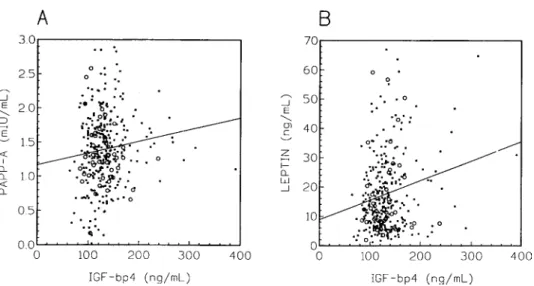
Leptin concentrations in the follicular fluid correlated strongly and significantly (p < 0.0001) with the body mass index, irrespective of the absence or presence of endometriosis but in an even more pronounced way in the latter group. The results are presented in Fig. 1. PAPP-A levels were strongly (p < 0.0001) and positively correlated to progesterone in both groups, and negatively to estradiol in the controls (p= 0.0201). Moreover, PAPP-A was positively correlated to IGF-bp4 in the control group (p= 0.0293, Fig. 2A), with leptin presenting the same pattern to an even stronger extent (p= 0.0005, Fig. 2B). No associations, however, were found between steroid hormones and leptin.
DISCUSSION
We have found a significantly decreased follicular fluid concentration of estradiol in endometriosis patients compared to controls, but no difference in progesterone levels which is in contrast to Pellicer et al. (19) who found that these levels were increasing
Table II. Ovarian Steroid Hormones, Leptin, PAPP-A and IGF-Binding
Protein-4 (Median, Range) in the Pooled Follicular Fluid of Women with and Without Endometriosis
Hormone or Control group Endometriosis
growth factor (n= 279) group (n= 47) p value
Estradiol (nmol/L) 823 (79–5832) 641 (134–3545) 0.0279 Progesterone (µmol/L) 28.92 (1.59–119.2) 28.31 (7.29–45.09) 0.9328 Leptin (ng/mL) 14.15 (1.23–66.95) 11.38 (2.01–56.73) 0.1655 PAPP-A (mIU/L) 1.37 (0.02–2.89) 1.31 (0.66–2.58) 0.6143
IGF-bp4 (ng/mL) 123 (64–184) 122 (84–188) 0.7722
with the severity of the disease. This discrepancy is difficult to explain, but it may be related to lower numbers in the mentioned study (particularly the controls) and to different distribution of the patients between the various types of infertility (tubal vs. male) in the control group. Other studies of natural and stimulated cycles did not indicate any differences of steroid levels in the serum and FF in endometriosis patients compared with controls. We have, however, also observed slightly lower estradiol levels in the serum (on the day of oocyte retrieval) in our endometriosis patients (median, 2.88 nmol/L) than in the controls (median, 4.34 nmol/L), and serum and follicular fluid estradiol levels correlated
Fig. 1. Follicular fluid leptin concentrations as a function of the
body mass index. The individual values and the regression lines are represented as dots and a solid line for the controls and as open circles and a dashed line for the cases of endometrio-sis. Regression parameters for slope, r and p are 2.136, 0.4192, and < 0.0001 for controls and 3.002, 0.5334, and <0.0001 for en-dometriosis, respectively.
strongly with each other in both groups (p < 0.01 by Spearman Rank correlation analysis). In our IVF centre, more than 90% of the couples were referred for reasons of male infertility; our control population could thus be closer to the normal, fertile one which is reacting better to the gonadotrophin stimulation, resulting in higher FF estradiol levels. Our signif-icantly increased estradiol levels together with the better pregnancy rates of the control group in our study correspond to the finding that fertilized oocytes came from follicles whose FF contained higher estra-diol levels, and the best cleavage rate is obtained from follicles containing more estradiol (20).
Data on leptin in the context of fertility are con-troversial. An earlier report suggested low serum lev-els to be related to infertility (3), a finding which has not been confirmed. One study reported an increased leptin concentration in peritoneal fluid in women with peritoneal endometriosis (21). Information on leptin levels in the follicular fluid of endometriosis patients is scarce, but we are confirming the paral-lelism between serum and follicular fluid via correla-tion of the latter with the BMI. The effect of leptin on ovarian steroidogenesis is altogether controver-sial, and only limited data exist regarding the ovarian function and leptin. A positive relationship between the increase in estrogen and leptin was reported (22), and another study suggested a direct stimulation by leptin of estradiol, but not of progesterone produc-tion in cultured granulosa cells (23). Another study (24), however, indicated no effect of leptin on estra-diol or progesterone production in granulosa cell culture and Ghizzoni et al. (25) even suggested an inhibition of steroid biosynthesis by leptin in such cultures. In this study we did not observe any as-sociations amongst follicular fluid leptin and steroid hormone levels.
The results of studies trying to establish a cor-relation between leptin levels and the outcome of IVF treatment are controversial, too. Better preg-nancy rates in the case of lower serum leptin levels at
Fig. 2. Follicular fluid PAPP-A (A, left panel) and leptin (B, right panel) concentrations as a function of
the concentration of IGF-bp4. The individual values and the regression lines are represented as dots and a solid line for the controls and as open circles for the cases of endometriosis. Regression parameters for slope, r and p values in the control group are 1.72× 10⁻³, 0.125, and 0.0365 for PAPP-A, and 0.067, 0.206, and 0.0005 for leptin, respectively. No significant association was observed for these parameters in the endometriosis group.
baseline or at the day of the administration of hCG were reported (26), and the same was suggested to be the case for follicular fluid in a conference abstract at a recent Annual Meeting of ESHRE (2003). These observations would be in theoretical agreement with the reported decreased pregnancy rates and con-comitantly elevated levels of leptin in endometriosis patients (11). Moreover, the reduced estradiol levels in the follicular fluid observed here may strengthen the hypothesis of the inhibitory role of leptin at the level of the granulosa cell, although the causal rela-tionship of this interaction can only be elucidated by in vitro studies at the cellular level. Another report (27), however, did not find any association of FF lep-tin levels and outcome, which is in agreement with our own observation from this study (p = 0.5386).
Data on the body mass index (BMI) in women with endometriosis are scarce and controversial. One study (28) reported endometriosis patients to be un-derweight more frequently, which is in contrast to McCann et al. (29) who did not observe a significant difference of body fat distribution or BMI between women with endometriosis and controls. In the pop-ulation of our own study presented here, the BMI in the endometriosis group was indeed lower than in the controls (Table I), but statistical significance was not reached.
The knowledge on the possible function and clin-ical usefulness of PAPP-A outside pregnancy is still
very thin. Increased PAPP-A levels have been found in the peritoneal fluid of women with endometrio-sis in one study (8), but results on PAPP-A in the FF of such patients are not available. Concerning the outcome, a positive relationship between ele-vated follicular fluid PAPP-A levels and the chance of achieving a pregnancy in that cycle is supported by an observation (30) that PAPP-A gene expres-sion in human ovaries is restricted to healthy follicles. We have observed a significant, positive correlation between PAPP-A and IGF-bp4 levels in the FF of our control group (p= 0.0365) which is difficult to explain: The reported role of PAPP-A as a specific protease to IGF-bp4 would rather suggest a negative association. The situation in the ovary, however, has not been specifically explored and may differ from the one in fibroblasts, trophoblast, and endometrium. An autoregulatory “short” loop may thus be func-tioning for PAPP-A at the level of the follicle. On the other hand, we have observed a pronounced positive correlation between follicular fluid levels of PAPP-A and progesterone, while the association with estra-diol is negative. The further investigation of this ex-tremely complicated pattern for the role of ovarian PAPP-A will be very challenging; one of the major problems is certainly the difficulty to select a proper control population with absolutely fertile women undergoing ICSI treatment only because of pure male factor. It will thus always remain difficult to
compare the observations from different projects and to reconcile results which seem inconsistent or even contradictory.
A recent report has demonstrated that pre-IVF surgery did not improve the subsequent embryo and outcome parameters (31). The gained knowledge on the basics of folliculogenesis, however, will improve long-term treatment of infertility.
CONCLUSIONS
In conclusion, while the follicular fluid levels of progesterone, PAPP-A, IGF-bp4 and leptin showed no significant differences between IVF patients with and without endometriosis, the results of our study indicate that FF estradiol concentrations contrast sig-nificantly between the two groups. As the oocyte quality is also decreased in women with endometrio-sis, estradiol can be considered as a marker not only of oocyte maturity but also of oocyte quality, but this without the reduced estradiol necessarily being the consequence of poor quality, i.e. the causal relation-ship cannot be determined in this type of study. In vitro investigations would be required to address this point, but they are illegal in Switzerland. Moreover, the assessment of the role of the other markers and their clinical potential in fertility related investiga-tions requires a larger number of endometriosis cases as well as in vitro studies.
ACKNOWLEDGMENTS
We are indepted to Ms. von Wyl in the IVF lab-oratory and to Ms. Vaucher in the research unit for skillful technical assistance.
REFERENCES
1. Lord GM, Matarese G, Howard JK, Baker RJ, Bloom SR, Lechler RI: Leptin modulates the T-cell immune response and reverses starvation-induced immunosuppression. Nature 1998;394:897–901
2. Sierra-Honigmann MR, Nath AK, Murakami C: Biological ac-tion of leptin as an angiogenic factor. Science 1998;281:1683– 1686
3. Barash IA, Cheung CC, Weigle DS, Ren H, Kabigting EB, Kuijper JL, Clifton DK, Steiner RA: Leptin is a metabolic sig-nal to the reproductive system. Endocrinology 1996;137:3144– 3147
4. Matarese G, Alviggi C, Sanna V, Howard JK, Lord GM, Carravetta C, Fontana S, Lechler RI, Bloom SR, De Placido G: Increased leptin levels in serum and peritoneal fluid of
patients with pelvic endometriosis. J Clin Endocrinol Metab 2000;85:2483–2487
5. Oxvig C, Sand O, Kristensen T, Kristensen L, Sottrup-Jensen L: Isolation and characterisation of circulating complex be-tween human pregnancy-associated plasma protein-A and proform of eosinophil major basic protein. Biochim Biophys Acta. 1994;1201:415–423
6. Lawrence JB, Oxvig C, Overgaard MT, Sottrup-Jensen L, Gleich GJ, Hays LG, et al.: The insulin-like growth factor (IGF)-dependent IGF binding protein-4 protease secreted by human fibroblasts is pregnancy-associated plasma protein-A. Proc Nat Acad Sci USA 1999;96:3149–3153
7. Conover CA, Faessen GF, Ilg KE, Chandrasekher YA, Christiansen M, Overgaard MT, Oxvig C, Giudice LC: Pregnancy-associated plasma protein-A is the insulin-like growth factor binding protein-4 protease secreted by human ovarian granulosa cells and is a marker of dominant follicle se-lection and the corpus luteum. Endocrinology 2001;142:2155– 2158
8. Arici A, Matalliotakis I, Goumenou A, Koumantakis G, Fragouli Y, Mahutte NG: Increased pregnancy-associated plasma protein-4 (PAPP-A) concentrations in peritoneal fluid of women with endometriosis. Am J Reprod Immunol 2003;49:70–74
9. Adashi EY: Insulin-like growth factors as determinants of fol-licular fate. J Soc Gynecol Invest 1995;2:721–726
10. Mason HD, Cwyfan-Hughes S, Holly JM, Franks S: Potent inhibition of human ovarian steroidogenesis by insulin-like growth factor binding protein-4 (IGFBP-4). J Clin Endocrinol Metab 1998;83:284–287
11. Cahill DJ, Wardle PG, Harlow ChR, Hull MG: Effect of pro-gesterone therapy on follicular development, related hormone concentrations and fertilization in vitro in unstimulated cy-cles and unexplained and endometriosis-associated infertility. Hum Reprod 1996;11:647–650
12. Simon C, Gutierrez A, Vidal A, De los Santos M, Tar`ın JJ, Remoh`ı J Pellicer A: Outcome of patients with endometriosis in assisted reproduction: Results from in vitro fertilization and oocyte donation. Hum Reprod 1994;9:725–729
13. Pellicer A, Oliveira N, Ruiz A, Remoh`ı J, Sim `on C: Explor-ing the mechanism(s) of endometriosis-related infertility: An analysis of embryo development and implantation in assisted reproduction. Hum Reprod 1995;10 (Suppl.2):91–97
14. Arici A, Oral E, Bukulmez O, Duleba A, Olive DL, Jones EE. The effects of endometriosis on implantation: Results from the Yale University in vitro fertilization and embryo transfer pro-gram. Fertil Steril 1996;65:603–607
15. Harlow ChR, Cahill DJ, Maile LA, Talbot WM, Mears J, Wardle PG, et al.: Reduced preovulatory granulosa cell steroidogenesis in women with endometriosis. J Clin En-docrinol Metab 1996;81:426–429
16. Tesarik J, Sousa M: Key elements of highly efficient in-tracytoplasmatic sperm injection technique: Ca+ fluxes and oocyte cytoplasmic dislocation. Fertil Steril 1995;64:770– 776
17. Bersinger NA, Zakher A, Huber U, Pescia G, Schneider H: A sensitive immunoassay for pregnancy-associated plasma pro-tein A (PAPP-A): A possible first trimester method of screen-ing for Down syndrome and other trisomies. Arch Gynecol Obstet 1995;256:185–192
18. Malek A, Willi A, Muller J, Sager R, Bersinger NA: Capacity for hormone production of cultured trophoblast cells obtained
from placentae at term and in early pregnancy. J Assist Re-prod Genet 2001;18:299–303
19. Pellicer A, Valbuena D, Bauset C, Albert C, Bonilla-Musoles F, Remoh`ı J, Sim `on C: The follicular endocrine environment in stimulated cycles of women with endometriosis: Steroid lev-els and embryo quality. Fertil Steril 1998;69:1135–1141 20. Botero-Ruiz W, Laufer N, De Cherney AH, Polan ML,
Haseltine FP, Behrman HR: The relationship between follic-ular fluid steroid concentrations and successful fertilization of human oocytes. Fertil Steril 1984;41:820–826
21. De Placido G, Alviggi C, Carravetta C, Pisaturo ML, Sanna V, Wilding M, Lord GM, Matarese G: The peritoneal fluid con-centration of leptin is increased in women with peritoneal but not ovarian endometriosis. Hum Reprod. 200;16:1251–1254 22. Hardie L, Trayhurn P, Abramovich D, Fowler P: Circulating
leptin in women: A longitudinal study in the menstrual cycle and during pregnancy. Clin Endocrinol (Oxf) 1997;47:101–106 23. Kitawaki J, Kusuki I, Koshiba H, Tsukamoto K, Honjo H: Leptin directly stimulates aromatase activity in human luteinized granulosa cells. Mol Hum Reprod 1999;5:708–713 24. Agarwal SK, Vogel K, Weitsman SR, Magoffin DA: Leptin
antagonizes the insulin-like growth factor I augmentation of steroidogenesis in granulosa cells of the human ovary. J Clin Endocrinol Metab 1999;84:1072–1076
25. Ghizzoni L, Barreca A, Mastorakos G, Furlini M, Vottero A, Ferrari B, Chrousos GP, Bernasconi S: Leptin inhibits steroid
biosynthesis by human granulosa-lutein cells. Horm Metab Res 2001;33:323–328
26. Tsai EM, Yang CH, Chen SC, Lin YH, Chen HS, Hsu SC, Lee JN: Leptin affects pregnancy outcome of in vitro fertilization and steroidogenensis of human granulosa cells. J Ass Reprod Genet 2002;19:169–176
27. Dorn C, Reinsberg J, Kupka M, van der Ven H, Schild RL: Leptin, VEGF, IGF-1, and IGFBP-3 concentrations in serum and follicular fluid of women undergoing in vitro fertilisation. Arch Gynecol Obstet 2003;268:187–193
28. Dmowski WP, Radwanska E: Current concepts on pathology, histogenesis and etiology of endometriosis. Acta Obstet Gy-necol Scand (Suppl) 1984;123:29–33
29. McCann SE, Freudenheim JL, Darrow SL, Batt RE, Zielezny MA: Endometriosis and body fat distribution. Obstet Gynecol 1993;82:545–549
30. Hourvitz A, Widger AE, Filho FL, Chang RJ, Adashi EY, Erickson GF: Pregnancy-associated plasma protein-A gene expression in human ovaries is restricted to healthy follicles and corpora lutea. J Clin Endocrinol Metab 2000;85:4916– 4919
31. Garcia-Velasco JA, Mahutte NG, Corona J, Zuniga V, Giles J, Arici A, Pellicer A: Removal of endometriomas before in vitro fertilization does not improve fertility outcomes: a matched, case-control study. Fertil Steril 2004;81:1194– 1197