HAL Id: hal-00330298
https://hal.archives-ouvertes.fr/hal-00330298
Submitted on 14 Oct 2008HAL is a multi-disciplinary open access
archive for the deposit and dissemination of sci-entific research documents, whether they are pub-lished or not. The documents may come from teaching and research institutions in France or abroad, or from public or private research centers.
L’archive ouverte pluridisciplinaire HAL, est destinée au dépôt et à la diffusion de documents scientifiques de niveau recherche, publiés ou non, émanant des établissements d’enseignement et de recherche français ou étrangers, des laboratoires publics ou privés.
Iron oxide deposits associated with the ectosymbiotic
bacteria in the hydrothermal vent shrimp Rimicaris
exoculata
L. Corbari, M.-A. Cambon-Bonavita, G. J. Long, F. Grandjean, M. Zbinden,
Françoise Gaill, P. Compère
To cite this version:
L. Corbari, M.-A. Cambon-Bonavita, G. J. Long, F. Grandjean, M. Zbinden, et al.. Iron oxide deposits associated with the ectosymbiotic bacteria in the hydrothermal vent shrimp Rimicaris exoculata. Biogeosciences Discussions, European Geosciences Union, 2008, 5 (2), pp.1825-1865. �hal-00330298�
BGD
5, 1825–1865, 2008 Bacteriogenic iron oxides L. Corbari et al. Title Page Abstract Introduction Conclusions References Tables Figures ◭ ◮ ◭ ◮ Back CloseFull Screen / Esc
Printer-friendly Version Interactive Discussion Biogeosciences Discuss., 5, 1825–1865, 2008
www.biogeosciences-discuss.net/5/1825/2008/ © Author(s) 2008. This work is distributed under the Creative Commons Attribution 3.0 License.
Biogeosciences Discussions
Biogeosciences Discussions is the access reviewed discussion forum of Biogeosciences
Iron oxide deposits associated with the
ectosymbiotic bacteria in the
hydrothermal vent shrimp Rimicaris
exoculata
L. Corbari1, M.-A. Cambon-Bonavita2, G. J. Long3, F. Grandjean4, M. Zbinden5, F. Gaill5, and P. Comp `ere1
1
Universit ´e de Li `ege, Laboratoire de Morphologie fonctionnelle et ´evolutive, Unit ´e de
Morphologie ultrastructurale et Cellule d’Appui Technologique en Microscopie (Catµ), all ´ee de la chimie, 3, 4000 Li `ege, Belgium
2
Laboratoire de Microbiologie et Biotechnologie des Extr ˆemophiles, Ifremer, centre de Brest, BP 70, 29280 Plouzan ´e, France
3
Department of Chemistry, Missouri University of Science and Technology, University of Missouri-Rolla, Rolla, Missouri 65409-0010, USA
BGD
5, 1825–1865, 2008 Bacteriogenic iron oxides L. Corbari et al. Title Page Abstract Introduction Conclusions References Tables Figures ◭ ◮ ◭ ◮ Back CloseFull Screen / Esc
Printer-friendly Version Interactive Discussion 4
Department of Physics, B5, University of Li `ege, 4000 Sart-Tilman, Belgium
5
UMR CNRS 7138 “Syst ´ematique, Adaptation et Evolution”, Universit ´e Pierre et Marie Curie, 7 Quai St Bernard, B ˆatiment A, 75252 Paris Cedex 05, France
Received: 12 February 2008 – Accepted: 28 February 2008 – Published: 24 April 2008 Correspondence to: L. Corbari (lcorbari@ulg.ac.be)
BGD
5, 1825–1865, 2008 Bacteriogenic iron oxides L. Corbari et al. Title Page Abstract Introduction Conclusions References Tables Figures ◭ ◮ ◭ ◮ Back CloseFull Screen / Esc
Printer-friendly Version Interactive Discussion
Abstract
The Rimicaris exoculata shrimp is considered a primary consumer that dominates the fauna of most Mid-Atlantic Ridge (MAR) hydrothermal ecosystems. These shrimps harbour in their gill chambers an important ectosymbiotic community of chemoau-totrophic bacteria associated with iron oxide deposits. The structure and elemental
5
composition of the minerals associated with these bacteria have been investigated by using X-ray microanalyses, light microscopy, and transmission, environmental scan-ning and scanscan-ning transmission electron microscopy. The nature of the iron oxides in shrimps obtained from the Rainbow vent field at 36◦14.0′N, has also been deter-mined by M ¨ossbauer spectroscopy. This multidisciplinary approach has revealed that
10
the three step-levels of mineral crust found in the Rimicaris exoculata shrimps consist of heavy concretions formed by nanoparticles of two-line ferrihydrite intermixed with minor inorganic SiO2, (Ca,Mg)SO4, and (Ca,Mg)3(PO4)2 minerals that may stabilise the ferrihydrite form of iron oxides. Morphological observations on the bacteria have revealed their close interactions with these minerals and, thus, indicate the biogenic
15
origin of the iron oxide deposits. The evolution of the bacterial density in the three min-eral crust levels is related to the amount of the iron deposits and it is proposed that the lower crust level is the most likely region for the location of the iron-oxidizing bacteria.
1 Introduction
Rimicaris exoculata (Williams and Rona, 1986) is one of the most dominant species
20
found at the Mid-Atlantic Ridge (MAR) hydrothermal vents. This endemic shrimp swarms on the chimney walls, exhibiting a patch-like distribution of up to several thou-sand per square meter (Segonzac et al., 1993). In extreme deep-sea environments, such high population density levels require some specific adaptations. Many hydrother-mal organisms derive their nutrition from chemoautotrophic bacteria through
sym-25
BGD
5, 1825–1865, 2008 Bacteriogenic iron oxides L. Corbari et al. Title Page Abstract Introduction Conclusions References Tables Figures ◭ ◮ ◭ ◮ Back CloseFull Screen / Esc
Printer-friendly Version Interactive Discussion 2006). R. exoculata possess an original ectosymbiotic bacterial community, housed
in its expanded gill chambers and mouth parts (Van Dover et al., 1988; Casanova et al., 1993; Zbinden et al., 2004; Corbari et al., 2008). Even though numerous authors have suggested that, if really ectosymbiotic, the bacteria could be a direct or indirect food source for the shrimp (Segonzac et al., 1993; Rieley et al., 1999; Gebruk et al.,
5
2000; Zbinden et al., 2004). Still undetermined, however, is the origin of the nutritional carbon of R. exoculata and the role of the bacterial ectosymbiosis play as a trophic resource (Pond et al., 1997; Polz et al., 1998; Zbinden and Cambon-Bonavita, 2003). The bacterial community housed in the gill chamber of R. exoculata has been identified as chemoautotrophic bacteria (Wirsen et al., 1993) and phylogenetic analysis revealed
10
that the bacteria could correspond to a single epsilon-proteobacteria phylotype (Polz and Cavanaugh, 1995). Some authors have also hypothesized that the bacteria could acquire their energy from sulphide oxidation (Gebruk et al., 1993; Polz and Cavanaugh, 1995), but this hypothesis has never been confirmed by any culture experiment.
In the absence of cultivated bacteria, recent studies have focussed on the
descrip-15
tion of both the bacteria and the mineral deposits in R. exoculata. Zbinden et al. (2004) described three bacterial morphotypes, individual rods with an approximate size of 0.5×1.5 µm, and two types of multicellular filaments, i.e., thick filaments with 2 to 3 µm diameters and thin filaments with 0.5 to 1 µm diameters, found within the entire gill chamber. They mapped the location of these bacteria and divided their associated
min-20
erals into three functional compartments, that which were considered to represent dis-tinct microenvironments. One of these compartments, the upper pre-branchial cham-ber, houses the highest density of both bacteria and minerals. A recent study (Corbari et al., 2008) focussed on this compartment and delineated the shrimp-bacteria-mineral association throughout the shrimp moult cycle. This study performed on about 300
25
specimens from two vent sites, TAG and Rainbow, indicated that the bacterial commu-nity restarts after each exuviation and gradually colonises the gill chamber in five moult stage-correlated steps. Moreover, the presence of red-brown mineral deposits in the gill chamber, including the mouth parts and branchiostegites, of the R. exoculata has
BGD
5, 1825–1865, 2008 Bacteriogenic iron oxides L. Corbari et al. Title Page Abstract Introduction Conclusions References Tables Figures ◭ ◮ ◭ ◮ Back CloseFull Screen / Esc
Printer-friendly Version Interactive Discussion already been described (Gloter et al., 2004; Zbinden et al., 2004). These deposits have
been identified as hydrous iron oxide in the form of ferrihydrite (Gloter et al., 2004). The extent and density of iron oxide deposits within the gill chamber are both responsible for the external colour of the shrimp, a colour that may be macroscopically observed by transparency through the branchiostegites (Zbinden et al., 2004). The shrimp external
5
colour ranges from white, indicative of no mineral deposits, to dark-red, indicative of a heavily mineralised crust; the colour appears to be highly correlated with the moult stages (Corbari et al., 2008). The fully-formed mineral crust is roughly organised in three step-levels that illustrate the time-related formation and growth of the mineral particles (Corbari et al., 2008). The shrimp-bacteria ectosymbiosis is characterised
10
by the presence of iron oxide deposits suggesting that iron oxidation may represent an alternative energy-pathway for the bacterial community, especially in shrimps found at the Rainbow site (Gloter et al., 2004; Zbinden et al., 2004; Schmidt et al., 2008). Moreover, the simultaneous occurrence of the bacteria and the iron oxide deposits in the gill chamber of R. exoculata may be interpreted as bacterially mediated (Zbinden
15
et al., 2004; Gloter et al., 2004, Anderson et al., 2008).
Several studies have documented the formation and occurrence of iron oxides formed as a result of biotic pathways in natural environments (Fortin et al., 1998; Fortin and Ch ˆatellier, 2003; Fortin and Langley, 2005; Banfield et al., 2000; Kennedy et al., 2003, 2004). In this context, the bacteria-hydrous ferric oxide interactions have also
20
been investigated to determine the direct and/or indirect bacterial influence on the hy-drous ferric oxide formation (see reviews in Fortin and Langley, 2005; Klapper and Straub, 2005). Some authors (Mavrocordatos and Fortin, 2002; Rancourt et al., 2005) have indicated that bacteria, either iron-metabolizing or non-metabolizing, could influ-ence the mineral deposition. They found evidinflu-ence of the biogenic origin of the hydrous
25
ferric oxide, identified the presence of poorly crystallized iron oxides, and determined the typical Fe/O ratios and particle size ranges. Natural biogenic iron oxides generally contain impurities, such as adsorbed or structural SiO2, and phosphate, sulphate, and manganese and aluminium ions, impurities that may influence the spatial organization
BGD
5, 1825–1865, 2008 Bacteriogenic iron oxides L. Corbari et al. Title Page Abstract Introduction Conclusions References Tables Figures ◭ ◮ ◭ ◮ Back CloseFull Screen / Esc
Printer-friendly Version Interactive Discussion and the morphology of the mineral particles (Fortin and Ch ˆatellier, 2003; Ch ˆatellier
et al., 2004). The properties of the iron oxide deposits and the influence of any im-purities on these properties have been studied in samples from natural environments (Fortin and Langley, 2005) but have never been investigated in the case of a bacterial ectosymbiosis.
5
Because the iron oxide deposition could be actively or passively promoted by R.
ex-oculata ectosymbiotic bacteria, the main goal of this study is to investigate in detail the
structure and the composition of the bacteria-associated mineral particles by using var-ious imaging techniques, such as back-scattered electron imaging, transmission elec-tron microscopy, energy dispersive X-ray microanalysis, and M ¨ossbauer spectroscopy.
10
These investigations have been performed on the fully formed mineral crust of pre-moult shrimps, a crust that is divided into three levels related with the successive steps of formation and growth of the mineral particles.
2 Materials and methods
2.1 Shrimp selection and samples treatment
15
Specimens of Rimicaris exoculata were collected during the French cruise “EXOMAR” (August 2005) at the MAR hydrothermal vent site Rainbow (36◦14.0′N, 2300 m depth) by using the suction sampler of the ROV “Victor 6000” operating from the RV “Atalante.” Immediately after retrieval, entire living specimens were either frozen at −80◦C or dis-sected into body parts, branchiostegite and tail, and fixed in a 2.5% glutaraldehyde in
20
seawater 7/10 at pH 7.2 medium.
Observations and analyses were performed on preecdysial specimens, in moult stages D′′′
1 and D2, in agreement with the moult-staging method of Drach and Tch-ernigovteff (1967), based on the development of setae matrices along the uropods borders. The six frozen and four glutaraldehyde-fixed specimens all exhibited an
im-25
agree-BGD
5, 1825–1865, 2008 Bacteriogenic iron oxides L. Corbari et al. Title Page Abstract Introduction Conclusions References Tables Figures ◭ ◮ ◭ ◮ Back CloseFull Screen / Esc
Printer-friendly Version Interactive Discussion ment with the colour categorisation by Corbari et al. (2008). All the observations were
performed on the dorsal median zone of the branchiostegite of R. exoculata (Fig. 1a) because it exhibits a regular bacterial and mineral cover that lines the antero-dorsal compartment of the gill chamber (Zbinden et al., 2004). The complete branchiostegite and some portions were photographed with an Olympus SZ40 stereo microscope.
5
In order to determine the structure and elemental composition of the bacteria-associated minerals, samples were prepared for study by transmission and scanning electron microscopy, X-ray microanalysis, and M ¨ossbauer spectroscopy. During the-ses preparations, contact between air and the samples was avoided to prevent al-teration in the oxidation and/or hydration states of the iron oxide minerals. To avoid
10
this air-contact, frozen specimens were used for compositional analyses and com-pared with glutaraldehyde-fixed specimens. For analytical electron microscopy mea-surements, the samples, dissected from frozen specimens, were directly dehydrated in absolute ethanol and embedded in epoxy resin (Epofix, Struers) through propylene oxide. Glutaraldehyde-fixed samples, conserved in seawater with NaN3, were quickly
15
rinsed in distilled water and dehydrated through an ethanol-propylene oxide series of rinses before embedding in the Epofix resin.
2.2 Scanning Electron Microscopy (SEM) and Energy-Dispersive X-ray (EDX) Micro-analysis
Polished thin slices of 20 to 50 µm thickness were obtained for the branchiostegites of
20
two glutaraldehyde-fixed and two frozen specimens. The specimens were cut as verti-cal cross-sections through the mineral crust, i.e., perpendicular to the branchiostegite cuticle. They were polished by abrasion on diamond disks and finally mirror polished with a non-aqueous 1 µm diamond suspension (ESCIL, PS-1MIC). The polished-thin slices were covered with a conductive silver paint, carbon-coated in a Balzers
BAF-25
400 rotary evaporator, and then maintained in desiccators to prevent air-contact before analysis. Structural mineral observations and elemental energy-dispersive X-ray mi-croanalysis were rapidly performed within two days of preparation in an environmental
BGD
5, 1825–1865, 2008 Bacteriogenic iron oxides L. Corbari et al. Title Page Abstract Introduction Conclusions References Tables Figures ◭ ◮ ◭ ◮ Back CloseFull Screen / Esc
Printer-friendly Version Interactive Discussion scanning electron microscope (FEI XL30 ESEM-FEG), operating at 15 to 20 kV and a
working distance of 10 mm. A total of 15 polished-thin slices were imaged by back-scattered electrons (BSE) and analysed for the elemental composition of the minerals present.
Elemental analyses have been carried out on the surface of 1 to 2 µm size mineral
5
particles. X-ray microanalyses with an acquisition time of 60 s have been obtained for both the glutaraldehyde-fixed and the frozen samples in order to determine whether any mineral transformation took place through chemical reactions during sample prepara-tion. The elemental quantitative analysis used an automatic background subtraction and a ZAF correction matrix has been used to calculate the elemental composition in
10
weight percents and atomic percents.
2.3 Transmission Electron Microscopy (TEM)
Four glutaraldehyde-fixed specimens were post-fixed in osmium 1%, dehydrated in an ethanol-propylene oxide series and then embedded in epoxy resin (SPI-PON 812). Ultrathin sections were obtained with a Reichert-Jung Ultramicrotome (Ultracut E) by
15
using a diamond knife; uranium acetate and lead citrate were used as contrast agents. The specimens were studied with a Jeol (JEM 100-SX) transmission electron micro-scope operating at 80 kV. In order to provide a three-dimensional view of the mineral crust organisation, vertical cross-sections were cut perpendicular to the branchioste-gite cuticle and the mineral surface and horizontal sections were cut at the three levels
20
of mineral crust as the previously defined.
2.4 Scanning Transmission Electron Microscopy (STEM) and Energy-Dispersive X-ray (EDX) Microanalysis
Ultrathin sections of samples from two frozen shrimps were cut as previously de-scribed, placed on a formvar-coated titanium grid and carbon-coated in a Balzers
BAF-25
BGD
5, 1825–1865, 2008 Bacteriogenic iron oxides L. Corbari et al. Title Page Abstract Introduction Conclusions References Tables Figures ◭ ◮ ◭ ◮ Back CloseFull Screen / Esc
Printer-friendly Version Interactive Discussion Tecnai G2 Twin scanning-transmission electron microscope operating at 200 kV.
In order to determine the elemental composition of the strata observed in the mineral particles, scanning transmission electron imaging has been carried out in a both direct bright-field and a high-angle annular dark-field (HAADF) imaging modes. The energy-dispersive X-ray nanoanalyses were performed with a nanoprobe spot size of 1nm in
5
diameter. Profile spectra have been determined on 1 to 1.5 µm length of ten various mineral particles.
2.5 M ¨ossbauer spectroscopy
The M ¨ossbauer spectra have been obtained on samples from frozen shrimps. Two different types of M ¨ossbauer spectral absorbers have been used. The first contained
10
boron nitride mixed with 14 mg/cm2 of lyophilized powder of crust minerals obtained by scrapings from three shrimps. The second consisted in the superimposed bran-chiostegites of one shrimp. The spectra were measured between 4.2 and 295 K on a constant-acceleration spectrometer that utilised a room temperature rhodium ma-trix cobalt-57 source and was calibrated at 295 K with α-iron powder. The estimated
15
relative errors are ±0.005 mm/s for the isomer shifts, ±0.01 mm/s for the quadrupole splittings and line widths, and ca. ±0.5 T for the hyperfine field. The absolute errors are estimated to be approximately twice as large.
2.6 Statistical analysis
The mineralogical compositions and quantifications are reported as mean values ±1
20
standard deviation. Comparisons of the mineral composition between glutaraldehyde-fixed and frozen samples have been evaluated by using a Mann–Whitney U-test, a two-tailed Student’s t-test, a Fisher test, and/or analysis of the variance. P <0.05 was taken as the fiducial limit for statistical significance.
BGD
5, 1825–1865, 2008 Bacteriogenic iron oxides L. Corbari et al. Title Page Abstract Introduction Conclusions References Tables Figures ◭ ◮ ◭ ◮ Back CloseFull Screen / Esc
Printer-friendly Version Interactive Discussion
3 Results
3.1 Mineral crust ultrastructure
Previously Corbari et al. (2008) have described the formation of a thick mineral crust overlying the bacterial community in the medium zone of the branchiostegites of dark-red shrimps. Three levels in the crust were arbitrarily defined according to the mineral
5
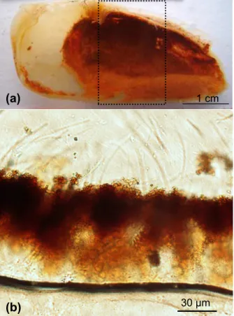
density that gradually increases from the cuticle towards the top surface of the crust. All analysed specimens of Rimicaris exoculata exhibit on the inner side of their bran-chiostegites, a dense, compact, mineral coating with a thickness of up to 100 µm, a mineral coating that corresponds to the mineral crust (Fig. 1a and b). Back scattered electron images of the vertical sections obtained in polished thin slices show the
grad-10
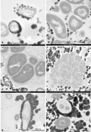
ual increase in mineral density from the cuticle to the surface of the crust (Fig. 2a, c, and e). In contrast, horizontal ultrathin sections obtained at each of the three identified mineral levels reveal different bacterial densities.
The lower level of the mineral crust (Fig. 2a and b) corresponds to the lower side of the mineral crust and is characterised by a heterogeneous distribution of mineral
15
particles, a distribution that is very fine and seems to correspond to clusters of less than 500 nm size (Fig. 2a). Their morphology seems directly related to the bacterial shape. Transmission electron microscopy images (Fig. 2b) reveal that the lower level is characterised by a high density of rod-shaped bacteria; mineral precipitation occurs on the cell walls of these bacteria (Fig. 3a). The mineral precipitates as individual
glob-20
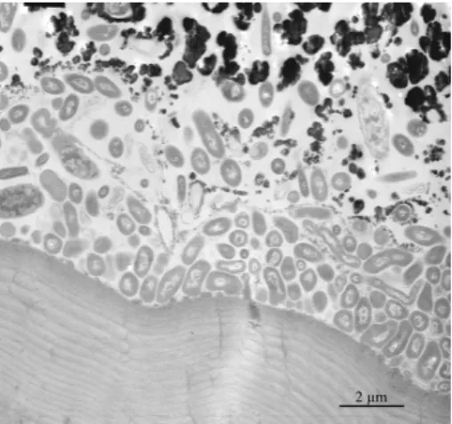
ular particles of 10 to 30 nm diameter, particles that tend to undergo agglomeration into larger particles. High resolution images (Fig. 3b and c) reveal that the minerals are diffuse and mainly precipitate on secretions of the bacteria, i.e., exopolysaccha-rides. TEM observations of vertical sections of all the analysed samples reveal that the bacteria form a dense community close to the cuticle, a location in which mineral
25
particles are almost absent because of the probable presence of bacterial secretion (Fig. 4). Interestingly, the methanotrophic bacteria, characterised by their stacks of in-tracytoplasmic membranes (Fig. 3c), are frequently observed at the lower level of the
BGD
5, 1825–1865, 2008 Bacteriogenic iron oxides L. Corbari et al. Title Page Abstract Introduction Conclusions References Tables Figures ◭ ◮ ◭ ◮ Back CloseFull Screen / Esc
Printer-friendly Version Interactive Discussion crust. Most often they aggregate in isolated groups that remain free of any mineral
precipitates (Fig. 3d).
The median level of the crust exhibits larger mineral particles, globular in shape, (Fig. 2c and d). These globular particles often meet to form larger aggregates that ap-pear multiglobular. In the horizontal cross-sections, the mineral density apap-pears rather
5
heterogeneous and consists of highly mineralised patches interspersed with bacteria rich areas. The bacterial density in the median layer is always smaller than in the lower layer; there are fewer rod-shaped bacteria. Moreover, ghosts of bacteria are also observed (Fig. 3e and f) in TEM images. These ghosts have bacterial shapes that are completely enclosed in a heavy mineral sheath. Sometimes the bacteria are still
10
present but appear either to be damaged or as membrane remain (Fig. 3e and f). In other cases, the mineral sheath appears to be empty or to have been recolonised by other rod-shaped bacteria. These observations suggest that mineral formation may influence the survival rate of the bacteria.
The upper level of the mineral crust contains very large particles with diameters of
15
up to 2 µm (Fig. 2e and f). The “rosette-like” particle shapes with deep indentations suggest that they result from the aggregation of several smaller particles. TEM images reveal that the bacteria become very rare in this upper level. As may be observed in the horizontal sections, almost the only bacteria present are a few large, thin, bacterial filaments that perforate throughout the mineral crust. Moreover, the minerals are not in
20
direct contact with the filament cell walls but form large sheaths at some distance from the cell walls (Fig. 2e). Even though the three step-levels of the mineral crust have been arbitrarily defined, they are representative of the different phases of the mineral formation and are characterised by an inverse correlation, from low to upper levels, between the amount of mineral deposits present and the bacterial density.
25
3.2 Iron oxides identified by M ¨ossbauer spectroscopy
M ¨ossbauer spectroscopy has been used to identify both the nature and oxidation states of the iron oxides found in the native minerals through the measurements of the
iron-BGD
5, 1825–1865, 2008 Bacteriogenic iron oxides L. Corbari et al. Title Page Abstract Introduction Conclusions References Tables Figures ◭ ◮ ◭ ◮ Back CloseFull Screen / Esc
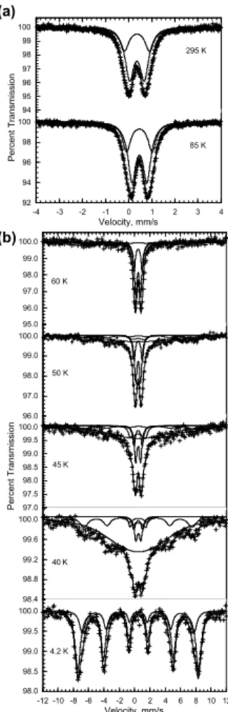
Printer-friendly Version Interactive Discussion 57 isomer shift and quadrupole splitting. The M ¨ossbauer spectra, obtained at 85 and
295 K and between 4.2 and 60 K are shown in Fig. 5a and b, respectively. At 85 and 295 K the spectra consist of broadened quadrupole doublets, whereas below 60 K they consist of a superposition of broadened doublets and sextets. The observed tem-perature dependence of the M ¨ossbauer spectra is typical of small superparamagnetic
5
particles. The spectra have been fit with two symmetric quadruople doublets and one to three magnetic sextets; the average hyperfine parameters are given in Table 1.
The weighted average isomer shift, <δ>, is typical of iron(III) (Shenoy et al., 1978) and spectral analysis indicates that at least 98% of the iron in the mineral crust must be present as iron(III); two percent by spectral area is the approximate detection limit
10
for the presence of any iron(II). The average hyperfine parameters observed at 295 and 4.2 K are typical (Murad et al., 1987) of two-line ferrihydrite. Two-line ferrihydrite, Fe5HO.84H2O, is a poorly crystalline mineral that forms spherical nanoparticles with a diameter of between 2 and 7 nm, a diameter that depends upon both the crystallinity of the material and the presence of impurities. Also for the same reasons, the average
15
hyperfine field observed at 4.2 K may be reduced from 50 to 46.5 T. In the specimens understudy, the average hyperfine field of 47.8 T corresponds to a reasonably well crys-tallised sample. The blocking temperature, i.e., the temperature at which the absorption areas of the doublets and sextets are equal, is 55±2 K. By using the anisotropy con-stant (Murad et al., 1987) of 4×10⁴J/m3 for 5 nm Fe5HO
.
84H2O particles, an average
20
particle diameter of 5.6 nm is obtained. 3.3 Quantitative X-ray microanalyses
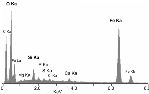
Back-scattered electrons (BSE) images of polished thin slices of frozen specimens pro-vide a detailed map of the mineral particles in the crust. X-ray microanalyses (n=14) performed in ESEM give accurate qualitative and quantitative determinations of the
25
elemental composition of the mineral deposits. These microanalyses reveal the pre-dominance of iron, with a Kαpeak at 6.400 keV and a Kβpeak at 7.059 keV, and oxygen with a Kα peak at 0.5425 keV, in the mineral crust (Fig. 6). Minor amounts of silicon
BGD
5, 1825–1865, 2008 Bacteriogenic iron oxides L. Corbari et al. Title Page Abstract Introduction Conclusions References Tables Figures ◭ ◮ ◭ ◮ Back CloseFull Screen / Esc
Printer-friendly Version Interactive Discussion with a Kα peak at 1.740 keV, calcium with a Kα peak at 3.690 keV and a Kβ peak at
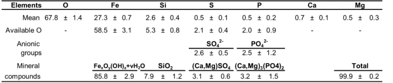
4.012 keV, phosphorus with a Kα peak at 2.013 keV, magnesium with a Kα peak at 1.253 keV, and sulphur with a Kα peak at 2.307 keV, have also been detected. Ele-mental quantitative analyses yield the weight and atomic percentages of the elements present, see Table 2. In order to determine the relative amount of the iron oxides and
5
other minerals in the deposits, we have assumed that the minor elements, such as Si, Ca, Mg, S, and P, are present in the stoichiometric ratio with oxygen as is found in the mineral forms of SiO2, (Ca,Mg)SO4, and (Ca,Mg)3(PO4)2. The elemental analyses also provide the percentages of elements inherent to the sample preparation as carbon from the carbon-coating of the sample and chlorine and oxygen from the embedding
10
resin. Additional spectra and elemental quantitative analyses have been carried out exclusively on the resin in order to quantify the amount of their content inherent to the sample preparation. After subtracting these peripheral elements by use of the refer-ence spectra of the resin and carbon coating on a pure mineral sample, the percent-ages of the remaining available oxygen for each mineral form have been calculated
15
as a function of the atomic percentages; the results of these calculations are given in Table 3. In order to obtain the percentages of the different mineral compounds found in the mineral crust, the two major anions, SO−24 and PO−34 , have been assumed to be in their most probable forms, i.e., (Ca,Mg)SO4and (Ca,Mg)3(PO4)2. Iron(III) oxides correspond to ca. 85% of the minerals present in the crust, as has been confirmed
20
by M ¨ossbauer spectroscopy. In addition the crusts contain ca. 8% of SiO2, ca. 3% (Ca,Mg)SO4and 3% of (Ca,Mg)3(PO4)2.
Comparative elemental X-ray microanalyses carried out on 12 polished thin slices of glutaraldehyde-fixed specimens reveal very similar proportions of the minor SiO2, (Ca,Mg)SO4, and (Ca,Mg)3(PO4)2minerals, a similarity which suggests that these
min-25
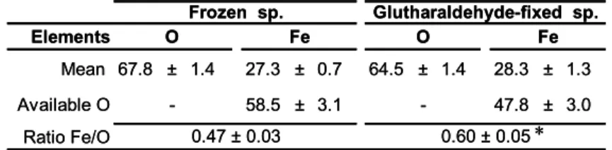
erals are not solubilised or removed by the aqueous preparation procedure. However, these analyses differ from those of the frozen sample by the Fe/O ratio in the iron ox-ide, i.e., after the subtraction of the oxygen from the minor minerals, see Table 4, cor-responding to an Fe/O ratio of 0.47 in the frozen specimens. In contrast, the Fe/O
ap-BGD
5, 1825–1865, 2008 Bacteriogenic iron oxides L. Corbari et al. Title Page Abstract Introduction Conclusions References Tables Figures ◭ ◮ ◭ ◮ Back CloseFull Screen / Esc
Printer-friendly Version Interactive Discussion proaches 0.60 in the glutaraldehyde-fixed specimens. These measured Fe/O ratios are
statistically different as is indicated by a paired t-test which yields t=7.1, d .f =11, and
P =0.00002. Hence, the atomic percentage of oxygen is lower in the
glutaraldehyde-fixed specimens, in which the glutaraldehyde acts as a reducing agent and modifies the iron oxidation state in the mineral particles.
5
3.4 Structure and composition of the crust minerals
TEM images obtained on Ur/Pb contrasted ultra-thin vertical sections indicate that most of the mineral particles, which have diameters ranging from 200 to 600 nm, exhibit with layered features in the lower level of the crust (Fig. 7a and b). Most of the specimens exhibit a multilayered pattern with a periodicity of a few nanometers, a pattern that
sug-10
gests that these particles are composed of ca. 5 to 10 successive strata. All of these strata appear as concentric growth layers originating from a unique nucleation centre, as multiglobular rosette-like particles, particles that change their shape and, for the outer particles, follow the outer particle border (Fig. 7b). Neighbouring particles also exhibit layered patterns that may correspond to similar mineral deposition sequences
15
(Fig. 7a). Mineral nucleation and deposition occurs either close to the rod-shaped bacteria walls or in their near-neighbour environment. Several nucleation centres are located close to the same bacteria and the accumulation of strata leads to the aggrega-tion of mineral particles that, as a consequence, exhibit a rosette-like shape (Fig. 7b). STEM-HAADF images of particles from frozen specimens give an inverted mass
con-20
trast of the strata. These images reveal the reality of the strata in terms of the changing mineral density and/or the composition within the particles (Fig. 7c and d).
In order to determine the exact nature of these strata, X-ray nanoanalyses have been carried out by STEM along a direction perpendicular to the strata of some particles observed in the ultrathin sections. The experimental procedure for obtaining these line
25
profiles of 1 to 1.5 µm length is based on the acquisition of a sequence of ca. 700 X-ray spectra per line profile. Subsequent data analysis can differentiate different elements through the number of counts that correspond to a given element found along the
BGD
5, 1825–1865, 2008 Bacteriogenic iron oxides L. Corbari et al. Title Page Abstract Introduction Conclusions References Tables Figures ◭ ◮ ◭ ◮ Back CloseFull Screen / Esc
Printer-friendly Version Interactive Discussion line. Figure 8 illustrates a typical elemental profile for iron, oxygen, and silicon along
a 1.2 µm line through a stratified mineral particle. The clear lighter coloured strata in Fig. 8a correspond to a higher iron and oxygen content than is found in the dark strata, see Fig. 8b. In spite of a low number of counts, the silicon profile seems to correlate well with those of iron and oxygen. In order to more accurately characterise the nature
5
of the mineral strata, quantitative elemental analyses have been performed at specific points, i.e., points 1, 2, and 3 in Fig. 8a, at well separated strata along the line profile. The results (Table 5) confirm that the iron and oxygen atomic percentages are high at the lighter strata, points 1 and 3, and very low in the dark strata, position 2.
4 Discussion
10
A multifacitated analysis carried out on the mineral crust of R. exoculata reveals a mineral content of 85% iron(III) oxide and 15% of other minerals such as, SiO2, (Ca,Mg)SO4, and (Ca,Mg)3(PO4)2. The M ¨ossbauer spectral results indicate that the iron(III) oxide as two-line ferrihydrite, Fe5HO.84H2O, which is present in small particles of less than 5 nm diameter. A transmission and scanning electron microscopic study
15
of the mineral crust reveals the particles in present in stratified layers suggesting a sequential deposition with alternating layers of differing mineral density and composi-tion. The description of the bacteria-mineral interactions suggests that both biotic and abiotic mineral particles coexist in the R. exoculata ectosymbiosis.
4.1 Iron oxides
20
M ¨ossbauer spectral results have confirmed that the mineral layer which coats the bran-chiostegite of Rimicaris exoculata is mainly composed of a hydrous iron(III) oxide iden-tified as two-line ferrihydrite, Fe5HO
.
84H2O. These results are in agreement with the conclusions based on transmission electron microscopy-electron energy loss (TEM-EELS) analyses of mineral particles of R. exoculata mouth parts (Gloter et al., 2004).
BGD
5, 1825–1865, 2008 Bacteriogenic iron oxides L. Corbari et al. Title Page Abstract Introduction Conclusions References Tables Figures ◭ ◮ ◭ ◮ Back CloseFull Screen / Esc
Printer-friendly Version Interactive Discussion Further, the particle diameter of 2 to 7 nm obtained from the M ¨ossbauer spectral results
is in complete agreement with that obtained by high-resolution electron microscopic im-ages (Gloter et al., 2004). However, although an earlier TEM-EELS analysis (Gloter et al., 2004) indicated a mixture of 55 to 66% iron(III) and 45 to 34% iron(II), only iron(III) has been detected in the M ¨ossbauer spectra of R. exoculata. Hence, if it is assumed
5
that limit of detection for iron(II) is 2%, iron(III) represents more than 98% of the total iron present. This difference in the oxidation state of iron may result from the experi-mental procedure used herein and the glutaraldehyde-fixation used for the TEM-EELS analysis as is discussed below.
M ¨ossbauer spectroscopy utilises bulk samples of the mineral crust and hence, the
10
results are averaged over a rather large number of small particles, in contrast with the results obtained from electron microscopic techniques that are representative of small portions of the samples. Further, there is neither radiation damage nor preparative damage of the absorbers in the M ¨ossbauer spectral experiments. In contrast, exposure to an electron beam can result in atomic displacement, electronic reduction,
electron-15
beam sputtering and/or heating, electrostatic charging, and radiolysis (Egerton et al., 2004). In a recent study using EELS to evaluate the effects of electron beam damage to ferrihydrite, Pan et al. (2006) observed the reduction of iron(III) to iron(II). These results highlight how investigations carried out under high vacuum in a transmission electron microscope may cause substantial and perhaps unsuspected changes to a
20
mineral sample (Michel et al., 2007).
The presence of ferrihydrite as an essential mineral component of the R.
exocu-lata crust is a sign of a biogenic origin of the iron oxide deposits. Indeed, the most
common extracellular biogenic iron oxides include oxyhydroxides, e.g., goethite, lepi-docrocite, akaganeite, and poorly ordered phases, e.g., two-line and six-line ferrihydrite
25
(Cornell and Schwertmann, 2003; Fortin and Langley, 2005). Furthermore, it is com-monly accepted that the product of microbial micro-aerobic iron(II) oxidation is often identified as a poorly crystalline ferrihydrite. The observation of two-line ferrihydrite thus supports the hypothesis of the presence of iron-oxidisers among the
ectosymbi-BGD
5, 1825–1865, 2008 Bacteriogenic iron oxides L. Corbari et al. Title Page Abstract Introduction Conclusions References Tables Figures ◭ ◮ ◭ ◮ Back CloseFull Screen / Esc
Printer-friendly Version Interactive Discussion otic bacterial community of R. exoculata (Zbinden et al., 2004) and validates the first
observations on bacterial cultures (Cambon-Bonavita, pers. com.). In hydrothermal environments, ferrihydrite has previously been identified because it is commonly inter-mixed with lithoautotrophic iron(II) oxidizing bacteria, bacteria that act as a causative agent in the formation of the ferrihydrite (Emerson and Moyer, 2002; Kennedy et al.,
5
2003; Kennedy et al., 2004; Little et al., 2004). Very few vent animals have been discov-ered to live in close association with iron oxide deposits. To the best of our knowledge, only the scaly-foot gastropod found in the hydrothermal vents at the Indian Ridge has been shown to exhibit scale-shaped structures, mineralised with iron sulphides on its foot (Goffredi et al., 2003; Waren et al., 2003). These structures are associated with
10
bacteria but an iron isotopic analysis indicates that sulphur and iron in the sclerites orig-inate from hydrothermal fluids rather than from bacteria (Suzuki et al., 2006). Herein, the presence of ferrihydrite in close interaction with hydrothermal metazoan has been discovered for the first time, in the specific case of the vent shrimp R. exoculata.
Elemental quantitative analyses have been performed to determine the Fe/O ratio,
15
a ratio that is commonly used as a signature in biogenic iron oxides. Because of the influence of the sample preparation for ultrastructural and elemental analyses, an al-ternative experimental procedure has been adopted in this study to reduce damage to the samples containing both bacteria and minerals. The samples for mineralogical analyses were frozen until used and were directly dehydrated by ethanol to avoid both
20
air-contact and any aqueous chemical fixation. This procedure is based on the work of Mavrocordatos and Fortin (2002), who investigated the influence of sample preparation on poorly ordered biotic hydrous iron oxide. To assess the impact of sample fixation on the composition of the R. exoculata minerals, Fe/O ratios determined through identical procedures have been compared between frozen and glutaraldehyde-fixed samples.
25
The Fe/O ratio of the glutaraldehyde-fixed minerals is significantly different from that of the frozen minerals. Thus sample preparation may modify the oxidation state of iron. The use of glutaraldehyde, a reducing agent, in addition to electron beam dam-age in TEM-EELS studies can explain the higher percentdam-age of iron(II) reported by
BGD
5, 1825–1865, 2008 Bacteriogenic iron oxides L. Corbari et al. Title Page Abstract Introduction Conclusions References Tables Figures ◭ ◮ ◭ ◮ Back CloseFull Screen / Esc
Printer-friendly Version Interactive Discussion Gloter et al. (2004) in their mineral samples. The Fe/O ratio in frozen minerals has
been measured to be 0.47, after subtraction of the oxygen associated with minor min-erals. This value cannot be compared with the Fe/O ratio of 0.33 obtained by Gloter et al. (2004) because both their analytical approach and mineralogical interpretations are quite different from those used herein. In contrast, the calculated Fe/O ratio of 0.47
5
may be compared with the 0.417 ratio obtained for abiotic ferrihydrite (Mavrocordatos and Fortin, 2002, and references therein). This higher value can be assigned to the bacterial influence and may be considered as evidence for the biogenic origin of iron oxide deposits.
In conclusion, for future research on R. exoculata minerals, sample preparation and,
10
more specifically, the drying process and fixation of the biological samples must be accurately delineated because they both influence the surface properties of ferrihydrite and hence, the iron oxidation state and the Fe/O ratio.
4.2 Intrinsic inorganic constituents
Even though ferrihydrite represents the main component of the mineral crust in R.
ex-15
oculata, elemental and quantitative analyses of the mineral particles have revealed the
presence of minor elements, such as Si, P, Ca, S, and Mg. These elements are not con-sidered as impurities (Gloter et al., 2004) but rather as intrinsic inorganic constituents or ligands as suggested by Ch ˆatellier et al. (2004) because these authors found that their presence during the oxidation process can affect the mineralogy as well as the size
20
and structure of the iron oxide particles. Because of their large surface areas, small particles of natural biogenic iron oxides generally contain adsorbed elements and ions, such as silicate, sulphate, phosphate, and manganese and aluminium cations (Fortin and Ch ˆatellier, 2003; Fortin and Langley, 2005). For example, nanoparticles of iron oxyhydroxide formed during the mineralisation process, can adsorb phosphate and
sil-25
icate ions (Gilbert and Banfield, 2005). The adsorption of elements on ferrihydrite from the surrounding aqueous milieu can affect its subsequent mineral ordering processes. Specifically, adsorption of silicates has been found to inhibit the conversion of
ferrihy-BGD
5, 1825–1865, 2008 Bacteriogenic iron oxides L. Corbari et al. Title Page Abstract Introduction Conclusions References Tables Figures ◭ ◮ ◭ ◮ Back CloseFull Screen / Esc
Printer-friendly Version Interactive Discussion drite to more crystalline iron oxides, such as hematite and goethite (Kennedy et al.,
2003; Ch ˆatellier et al., 2004). X-ray nanoanalyses and elemental profiles performed on stratified R. exoculata mineral particles have revealed that silicates are already present in the early stages of mineral development. Thus, in R. exoculata, it is evident that inorganic ligands co-precipitate with iron oxide and are closely associated with it in the
5
nanoparticles because the silicate portion cannot be located separately from the iron oxide even on the nanometric scale reached in STEM. In the global characterisation of the mineral particles, we have assumed that they correspond to separated minerals such as silica, (Ca,Mg)3(PO4)2, and (Ca,Mg)SO4, even though it is very probable that silicate, sulphate, phosphate, and magnesium and calcium cations are complexed with
10
ferrihydrite and influence or stabilise this poorly crystalline form of iron oxide. The high silica content of ca. 8% found in the R. exoculata mineral crusts presumably result from the silicon concentration in the vent fluid, specifically the 6.9 mM concentration at the Rainbow site (Schmidt et al., 2007) a conclusion that as is also supported by com-parison with the minerals found in the TAG shrimps (Corbari and Comp `ere, personal
15
communication).
In order to further assess the distribution of the intrinsic inorganic constituents, X-ray spectra acquired at line profiles have been performed on mineral particles exhibiting stratified features. At the atomic level, the results indicate that both iron and the other inorganic ligands are already associated within nanosized mineral particles. These
ob-20
servations reveal that iron, oxygen, and silicon proportions are constant but that the stratification is mainly the consequence of parallel variations of the concentration of all the constituting elements. We can therefore consider the iron oxide globular par-ticles rather like mineral concretions. Bacteria-associated mineral concretions appear to be identical and stratification seems to follow the same sequence within
neighbour-25
ing mineral particles. These observations suggest that micro-environmental variations can occur in the nearby environment during the formation of the mineral particles. Homogenous patterns of stratification on neighbouring particles can be explained by either environmental variations found at the shrimp growth level or by variations in the
BGD
5, 1825–1865, 2008 Bacteriogenic iron oxides L. Corbari et al. Title Page Abstract Introduction Conclusions References Tables Figures ◭ ◮ ◭ ◮ Back CloseFull Screen / Esc
Printer-friendly Version Interactive Discussion bacterial metabolism or activities. Similar patterns of stratification have been previously
described on sediment microfacies and delineated as microstromatolite (Boulvain et al., 2001). The “microstromatolite” aspect of the mineral concretions in R. exoculata provides one more argument in favour of the biogenic origin of the iron oxides.
4.3 Bacteria-mineral interactions
5
In R. exoculata, iron oxide is complexed with intrinsic inorganic ligands in the presence of an important bacterial community. Both bacteria and the intrinsic inorganic ligands may play a role in mineral deposition. The term of biogenic iron oxides is commonly used to refer to iron oxide formed in the presence of bacteria. It also includes iron oxides formed as a direct result of microbial metabolism, i.e., through enzyme activities,
10
or by passive mechanisms through which bacterial secretions trigger the formation and precipitation of iron oxides minerals (Fortin and Ch ˆatellier, 2003).
In R. exoculata, mineral deposition only occurs when the bacterial community is well-developed on the inner side of the branchiostegite (Corbari et al., 2008). This observa-tion both contradicts the idea that iron oxide deposiobserva-tion could only result from a passive
15
chemical-induced precipitation and supports the idea that bacteria must participate in mineral formation. Ferrihydrite formed in the absence of bacteria may be metastable and, typically after few days, must transform into a more structurally ordered iron ox-ide, such as hematite or goethite (Cornell and Schwertmann, 2003). Experiments performed on bacteriogenic ferrihydrite minerals obtained from the hydrothermal vents
20
in the Axial Volcano, in the Pacific Ridge, demonstrated that even if they were sub-jected to heating of up to 80◦C, these minerals did not undergo a phase transition, and therefore suggesting that the presence of bacteria inhibited the ferrihydrite transforma-tion (Kennedy et al., 2004). Hence, the presence of ferrihydrite deposits inside the gill chamber of R. exoculata appears to result from the presence of an abundant bacterial
25
community. If the moult cycle is used as a time-scale, the first ferrihydrite deposits are only observed when the bacterial density reaches a maximum in the early preecdysial individuals, i.e., the light-red or medium-red individuals, in stages D0to D1(Corbari et
BGD
5, 1825–1865, 2008 Bacteriogenic iron oxides L. Corbari et al. Title Page Abstract Introduction Conclusions References Tables Figures ◭ ◮ ◭ ◮ Back CloseFull Screen / Esc
Printer-friendly Version Interactive Discussion al., 2008). During the ten day moult cycle of the vent shrimp, the first mineral
parti-cles appear as ferrihydrite (Corbari and Comp `ere, unpublished data) as early as the second post-moult day and continue to deposit until the tenth day, just before exuvia-tion. Hence, we conclude that the bacterial community contributes of the stabilisation of the iron oxide in the form of ferrihydrite. The bacteria organic moieties can hinder its
5
transformation into a more crystallized iron oxide (Kennedy et al., 2004) or favour the incorporation of the minor ligands.
TEM observations of the bacterial morphotypes and their mineral interactions help to elucidate the biogenic origin of the ferrihydrite deposits inside the gill chamber of
R. exoculata. Different ways of mineral deposition have been identified, based on the
10
recurrent observations of both bacterial morphotypes and mineral morphologies. Two association modes between minerals and rod shaped bacteria have been observed. In the first, iron oxide deposition occurs in close contact to the rod cell walls and, in the second, the iron oxide precipitates on polysaccharide or proteinaceous extracellular secretions at a significant distance from the bacterial cells. Such bacterial-mineral
15
relationships have previously been described, suggesting that rods are mainly involved in iron oxide formation (Anderson et al., 2008). The precipitation of iron oxides on or near the bacterial cell walls raises the question: Is it possible to determine whether the ectosymbiotic bacteria found in the gill chamber of R. exoculata are actively or passively depositing iron oxide minerals?
20
Passive reactions linked to biogenic iron oxide production are related to the reactivity of the bacterial cell walls. Mineral formation on the bacteria is generally not controlled by the organism but, rather, it results from the chemistry of the cell environment and the physicochemistry of the bacterial surface, (Fortin and Langley, 2005) a result that implies the adsorption and/or nucleation of iron oxide particles on bacterial cell walls
25
(Fortin and Ch ˆatellier, 2003). Whatever types of cell surface structure the cell may have, the main charged chemical constituents found in these structures at neutral pH are carboxyl, phosphoryl, and amino groups (Douglas and Beveridge, 1998). We thus suspect that similar mineral-bacteria associations take place in R. exoculata and that
BGD
5, 1825–1865, 2008 Bacteriogenic iron oxides L. Corbari et al. Title Page Abstract Introduction Conclusions References Tables Figures ◭ ◮ ◭ ◮ Back CloseFull Screen / Esc
Printer-friendly Version Interactive Discussion there is passive deposition of iron oxides. In addition, it is unclear whether precipitation
of iron(III) minerals in close vicinity of the cells, or even at the cell surface, is harmful for the cells as a result of limiting substrate diffusion and uptake, as is commonly assumed (Hallberg and Ferris, 2004). The observed ghosts of bacteria surrounded by a dense mineral coating suggest that these bacteria are probably not iron-oxidising bacteria
5
because deposition of heavy minerals on their cell walls should improve their survival rate.
The active metabolic processes of iron oxidation in iron metabolizing bacteria have to be distinguished from the indirect biologically induced iron oxide deposition in which the bacterial cell surface simply acts as a passive deposition template (Konhauser,
10
1997; Klapper and Straub, 2005). The most studied iron-oxidising bacteria are the neutrophilic aerobic iron(II) oxidisers from Gallionella and Leptothrix genus, oxidis-ers that produce extracellular organic polymoxidis-ers that nucleate iron(III) precipitates. It has been suggested that these microbes use these strategies to avoid encrustation of the metabolising cells that would lead to impaired substrate uptake and metabolite
15
release, and might even cause cell death (Hallberg and Ferris, 2004; Kappler et al., 2005). Hence, if iron-oxidizing bacteria are present among the ectosymbiotic commu-nity in R. exoculata, they could use this strategy to keep their metabolism active and only those with extracellular secretion would be involved in iron oxide formation. The presence of a mineral boundary between the dense rod population and the mineralised
20
area suggests a strategy involving the production of an extracellular organic material in order to prevent mineral deposition directly on the bacterial cell walls. Finally, the weak mineral deposition, maximal rod-shaped bacterial density, and presence of extracellu-lar secretions suggest that iron-oxidising bacteria may be located in this layer, a layer that may act as a potential reserve for active ectosymbiotic bacteria.
25
Interestingly, the appearance of methanotrophic bacteria clusters also support the above described mechanism in which bacteria exude organic substances to prevent any mineral deposition directly on their cell walls. Further, the presence of methan-otrophic bacteria suggests a more diversified bacterial community than previously
men-BGD
5, 1825–1865, 2008 Bacteriogenic iron oxides L. Corbari et al. Title Page Abstract Introduction Conclusions References Tables Figures ◭ ◮ ◭ ◮ Back CloseFull Screen / Esc
Printer-friendly Version Interactive Discussion tioned (Segonzac et al., 1993; Zbinden et al., 2004; Corbari et al., 2008). Herein,
methanotrophic bacteria show intact internal structures, i.e., stalks, and are distributed in clusters that indicate an active metabolism.
The three step-levels in the mineral crust formation previously described (Corbari et al., 2008) indicates that iron oxide particle growths are continuously initiated from
5
the lower level, in close association with growing bacteria and subsequently grow into the median and upper levels. The mineralisation within the gill chamber could be de-scribed as a dynamic process in which particles increase in size and are simultane-ously pushed upward by the formation of new particles. As has been illustrated herein, the lower level exhibits the highest bacterial density and is mainly composed of rod
10
bacteria. This level may be considered as a bacterially active layer and its evolution in time, based on the moult cycle, shows continuous growth (Corbari et al., 2008).
5 Conclusions
The multidisciplinary approach used in the present study provides new details about the iron oxide deposits associated with ectosymbiotic bacteria in Rimicaris exoculata.
15
The mineral crust has been identified as a dense layer of two-line ferrihydrite nanopar-ticles intermixed with other intrinsic inorganic constituents, mainly SiO2. Various in-vestigations on these nanoparticles have revealed that these minerals all contribute to the mineral particles deposited in the gill chamber. Their subtraction from the con-cretion composition has led to an of Fe/O ratio of 0.47 in the remaining ferrihydrite,
20
a ratio that suggests a bacteriogenic origin for the ferrihydrite. More morphological evidence has been obtained that determines the role of bacterial morphotypes in the “active” deposition of iron oxides. The process of mineralisation in the gill chambers of
R. exoculata remains complex because the combined effects of the intrinsic inorganic
constituents and the bacterial cells is difficult to disentangle. But the evolution of the
25
bacterial density in the three levels of the mineral crust is closely related to the amount of iron deposited and it is proposed that the lower level is the likely region where the
BGD
5, 1825–1865, 2008 Bacteriogenic iron oxides L. Corbari et al. Title Page Abstract Introduction Conclusions References Tables Figures ◭ ◮ ◭ ◮ Back CloseFull Screen / Esc
Printer-friendly Version Interactive Discussion iron-oxidising bacteria are located. But the presence of a more diversified bacterial
community raises the question on the metabolic or genetic diversity of these bacteria. Because the main studies on R. exoculata ectosymbiosis have been performed on shrimps from the vent site Rainbow, the influence of the chemical vent environment should be studied in the future by comparing ectosymbiosis and its associated minerals
5
in R. exoculata specimens collected at different vent sites, for instance TAG, Logatchev, and Snake Pit. This indirect approach could be used to evaluate how representative is the R. exoculata ectosymbiosis and, thus to determine whether iron oxidation rep-resents the most favourable energetic-pathways for ectosymbiotic bacteria (Schmidt et al., 2008).
10
Acknowledgements. The authors thank A. Godfroy, the chief scientist of the EXOMAR cruise,
as well as the captain and crew of the RV “Atalante” and the ROV “Victor” team. The au-thors also wish to express their appreciation to N. Decloux for her excellent technical assis-tance with transmission and scanning electron microscopy. This work was partly funded with the help of the MOMARNET program. The fellowship of L. Corbari and a part of this work
15
were supported by the Belgian Fund for Joint Basic Research FNRS (F.R.F.C Belgium, con-ventions no. 2.4594.07.F). The authors also thank the centre of Microscopy of Li `ege (CATµ; dir. R. Cloots) for giving access to high performance equipment EM, funded by F.R.F.C and FEDER. F. Grandjean acknowledges, with thanks, the financial support of the FNRS, Belgium, through grants 9.456595 and 1.5.064.05.
20
References
Anderson, L., Halary, S., Lechaire, J.-P., Boudier, T., Frebourg, G., Marco, S., Zbinden, M., and Gaill, F.: Tomography of bacteria-mineral associations within the deep-sea hydrothermal vent shrimp rimicaris exoculata, Comptes Rendus Chimie, 11, 268–280, 2008.
Banfield, J. F., Welch, S. A., Zhang, H., Ebert, T. T., and Penn, L. P.: Aggregation-based
25
crystal growth and microstructure development in natural iron oxyhydroxide biomineralization products, Science, 289, 751–754, 2000.
Boulvain, F., De Ridder, C., Mamet, B., Pr ´eat, A., and Gillan, D.: Iron microbial communities in belgian frasnian carbonate mounds, Facies, 44, 47–60, 2001.
BGD
5, 1825–1865, 2008 Bacteriogenic iron oxides L. Corbari et al. Title Page Abstract Introduction Conclusions References Tables Figures ◭ ◮ ◭ ◮ Back CloseFull Screen / Esc
Printer-friendly Version Interactive Discussion
Casanova, B., Brunet, M., and Segonzac, M.: L’impact d’une ´epibiose bact ´erienne sur la mor-phologie fonctionnelle de crevettes associ ´ees `a l’hydrothermalisme m ´edio-atlantique, Cah. Biol. Mar., 34, 573–588, 1993.
Cavanaugh, C., McKiness, Z., Newton, I., and Stewart, F.: Marine chemosynthetic symbioses, in: The prokaryotes, edited by: Dworkin, M., Falkow, S., Rosenberg, E., Schleifer, K., and
5
Stackebrandt, E., Springer, New York, 475–507, 2006.
Ch ˆatellier, X., Fortin, D., West, M., Leppard, G., and Ferris, F.: Effect of the presence of bacterial surfaces during the synthesis of fe-oxides by oxidation of ferrous ions, Eur. J. Miner., 13, 705–714, 2001.
Ch ˆatellier, X., West, M. M., Rose, J., Fortin, D., Leppard, G. G., and Ferris, F. G.:
Characteri-10
zation of iron-oxides formed by oxidation of ferrous ions in the presence of various bacterial species and inorganic ligands, Geomicrobiol. J., 21, 99–112, 2004.
Corbari, L., Zbinden, M., Cambon-Bonavita, M.-A., Gaill, F., and Comp `ere, P.: Bacterial sym-bionts and mineral deposits in the branchial chamber of the hydrothermal vent shrimp
Rimi-caris exoculata: Relationship to moult cycle, Aquatic Biology, 1, 225–238, 2008.
15
Cornell, R. and Schwertmann, U.: The iron oxides – structure, properties, occurrences and uses, 2nd edition, Weinheim ed., Wiley-VCH Verlag, 664 pp., 2003.
Desbruyeres, D., Biscoito, M., Caprais, J.-C., Colaco, A., Comtet, T., Crassous, P., Fouquet, Y., Khripounoff, A., Le Bris, N., and Olu, K.: Variations in deep-sea hydrothermal vent communi-ties on the Mid-Atlantic Ridge near the Azores plateau, Deep-sea Res. PtI, 48, 1325–1346,
20
2001.
Douglas, S. and Beveridge, T. J.: Mineral formation by bacteria in natural microbial communi-ties, FEMS Microbiol. Ecol., 26, 79–88, 1998.
Drach, P. and Tchernigovtzeff, C.: Sur la m ´ethode de d ´etermination des stades d’intermue et son application g ´en ´erale aux crustac ´es, Vie et Milieu, 18, 595–609, 1967.
25
Edwards, K. J., Bach, W., McCollom, T. M., and Rogers, D. R.: Neutrophilic iron-oxidizing bac-teria in the ocean: Their habitats, diversity, and roles in mineral deposition, rock alteration, and biomass production in the deep-sea, Geomicrobiol. J., 21, 393–404, 2004.
Egerton, R. F., Li, P., and Malac, M.: Radiation damage in the TEM and SEM, Micron, 35, 399–409, 2004.
30
Emerson, D. and Moyer, C. L.: Neutrophilic Fe-oxidizing bacteria are abundant at the Loihi seamount hydrothermal vents and play a major role in Fe oxide deposition, Appl. Environ. Microb., 68, 3085–3093, 2002.
BGD
5, 1825–1865, 2008 Bacteriogenic iron oxides L. Corbari et al. Title Page Abstract Introduction Conclusions References Tables Figures ◭ ◮ ◭ ◮ Back CloseFull Screen / Esc
Printer-friendly Version Interactive Discussion
Fortin, D., Ferris, G., and Scott, S.: Formation of Fe-silicates and Fe-oxides on bacterial sur-faces in samples collected near hydrothermal vents on the southern explorer ridge in the northeast Pacific ocean, American Mineralogist, 83, 1399–1408, 1998.
Fortin, D. and Ch ˆatellier, X.: Biogenic iron-oxides, Recent Research Developments in Mineral-ogy, 3, 47–63, 2003.
5
Fortin, D. and Langley, S.: Formation and occurrence of biogenic iron-rich minerals, Earth-Sci. Rev., 72, 1–19, 2005.
Gebruk, A., Pimenov, N., and Savvichev, A.: Feeding specialization of bresiliid shrimps in the tag site hydrothermal community, Mar. Ecol.-Prog. Ser., 98, 247–253, 1993.
Gebruk, A. V., Southward, E. C., Kennedy, H., and Southward, A. J.: Food sources, behaviour,
10
and distribution of hydrothermal vent shrimps at the mid-atlantic ridge, J. Mar. Biol. Ass. UK, 80, 485–499, 2000.
Gilbert, B. and Banfield, J. F.: Molecular-scale processes involving nanoparticulate minerals in biogeochemical systems, Rev. Mineral. Geochem., 59, 109–155, 2005.
Gloter, A., Zbinden, M., Guyot, F., Gaill, F., and Colliex, C.: TEM-EELS study of natural
ferrihy-15
drite from geological-biological interactions in hydrothermal systems, Earth Planet. Sci. Lett., 222, 947–957, 2004.
Goffredi, S., Hurtado, L., Hallam, S., and Vrijenhoek, R.: Evolutionary relationships of deep-sea vent and cold seep clams (mollusca: Vesicomyidae) of the “Pacifica/lepta” Species complex, Marine Biology, 142, 311–320, 2003.
20
Hallberg, R. and Ferris, F. G.: Biomineralization by Gallionella, Geomicrobiol. J., 21, 325–330, 2004.
Kappler, A., Schink, B., and Newman, D. K.: Fe(III) mineral formation and cell encrustation by the nitrate-dependent Fe(II)-oxidizer strain bofen1, Geobiology, 3, 235–245, 2005.
Kappler, A. and Straub, K. L.: Geomicrobiological cycling of iron, Rev. Mineral. Geochem., 59,
25
85–108, 2005.
Kennedy, C. B., Martinez, R. E., Scott, S., and Ferris, F. G.: Surface chemistry and reactivity of bacteriogenic iron oxidesfrom axial volcano, juan de fuca ridge, north-east pacific ocean., Geobiology, 1, 59–69, 2003.
Kennedy, C. B., Scott, S. D., and Ferris, F. G.: Hydrothermal phase stabilization of 2-line
ferri-30
hydrite by bacteria, Chem. Geol., 212, 269–277, 2004.
Konhauser, K. O.: Bacterial iron biomineralisation in nature, FEMS Microbiol. Rev., 20, 315– 326, 1997.
BGD
5, 1825–1865, 2008 Bacteriogenic iron oxides L. Corbari et al. Title Page Abstract Introduction Conclusions References Tables Figures ◭ ◮ ◭ ◮ Back CloseFull Screen / Esc
Printer-friendly Version Interactive Discussion
Little, C., Glynn, S., and Mills, R. A.: Four-hundred-and-ninety-million-year record of bacterio-genic iron oxide precipitation at sea-floor hydrothermal vents, Geomicrobiol. J., 21, 415–429, 2004.
Mavrocordatos, D. and Fortin, D.: Quantitative characterization of iron oxides formed on bacte-rial walls by tem-eels, Am. Miner., 87, 940–946, 2002.
5
Michel, F. M., Ehm, L., Antao, S. M., Lee, P. L., Chupas, P. J., Liu, G., Strongin, D. R., Schoonen, M. A. A., Phillips, B. L., and Parise, J. B.: The structure of ferrihydrite, a nanocrystalline material, Science, 316, 1726–1729, 2007.
Murad, E. and Johnston, J. H.: M ¨ossbauer spectroscopy applied to inorganic chemistry, edited by: Long, G. J., Plenum Press New York, 507 pp., 1987.
10
Murad, E. and Schwertmann, U.: Iron oxide mineralogy of some deep-sea ferromanganese crusts, Am. Miner., 73, 1395–1400, 1988.
Page, H. M., Fisher, C. R., and Childress, J.: The role of suspension-feeding in the nutritional biology of a deep-sea mussel with methanotrophic symbionts, Mar. Biol., 104, 251–257, 1990.
15
Pan, Y., Brown, A., Brydson, R., Warley, A., Li, A., and Powell, J.: Electron beam damage studies of synthetic 6-line ferrihydrite and ferritin molecule cores within a human liver biopsy, Micron, 37, 403–411, 2006.
Polz, M. F. and Cavanaugh, C. M.: Dominance of one bacterial phylotype at a Mid-Atlantic Ridge hydrothermal vent site, PNAS, 92, 7232–7236, 1995.
20
Polz, M. F., Robinson, J. J., Cavanaugh, C. M., and Van Dover, C. L.: Trophic ecology of massive shrimp aggregations at a mid-atlantic ridge hydrothermal vent site, Limnol. Oceanogr., 43, 1631–1638, 1998.
Pond, D., Bell, M., Dixon, D., and Sargent, J.: Occurrence of 16:2(n-4) and 18:2(n-4) fatty acids in the lipids of the hydrothermal vent shrimp Rimicaris exoculata: Nutritional and trophic
25
implications, Mar. Ecol.-Prog. Ser., 156, 167–174, 1997.
Rieley, G., Dover, C. L. V., Hedrick, D. B., and Eglinton, G.: Trophic ecology of Rimicaris
exoculata: A combined lipid abundance/stable isotope approach, Mar. Biol., 133, 495–499,
1999.
Schmidt, C., Vuillemin, R., Le Gall, C., Gaill, F., and Le Bris, N.: Geochemical energy sources
30
for microbial primary production in the environment of hydrothermal vent shrimps, Mar. Chem., 108, 18–31, 2008.
Geo-BGD
5, 1825–1865, 2008 Bacteriogenic iron oxides L. Corbari et al. Title Page Abstract Introduction Conclusions References Tables Figures ◭ ◮ ◭ ◮ Back CloseFull Screen / Esc
Printer-friendly Version Interactive Discussion
chemistry of hydrothermal fluids from the ultramafic-hosted logatchev hydrothermal field, 15◦N on the Mid-Atlantic Ridge: Temporal and spatial investigation, Chem. Geol., 242, 1–
21, 2007.
Segonzac, M., de Saint-Laurent, M., and Casanova, B.: L’ ´enigme du comportement trophique des crevettes Alvinocarididae des sites hydrothermaux de la dorsale M ´edio-Atlantique, Cah.
5
Biol. Mar., 34, 535–571, 1993.
Shenoy, G. K., Wagner, F. E., and Kalvius, G. M.: M ¨ossbauer isomer shifts, edited by: Shenoy, G. K. and Wagner, F. E., North-Holland Amsterdam, 49 pp., 1978.
Suzuki, Y., Kopp, R. E., Kogure, T., Suga, A., Takai, K., Tsuchida, S., Ozaki, N., Endo, K., Hashimoto, J., Kato, Y., Mizota, C., Hirata, T., Chiba, H., Nealson, K. H., Horikoshi, K., and
10
Kirschvink, J. L.: Sclerite formation in the hydrothermal-vent “Scaly-foot” Gastropod-possible control of iron sulfide biomineralization by the animal, Earth Planet. Sci. Lett., 242, 39–50, 2006.
Van Dover, C., Fry, B., Grassle, F., Humphris, S., and Rona, P. A.: Feeding biology of the shrimp
Rimicaris exoculata at hydrothermal vents on the mid-atlantic ridge., Mar. Biol., 98, 209–216,
15
1988.
War ´en, A. S., Bengtson, S. K., Goffredi, S., and Van Dover, C. L.: A hydrothermal-vent gastro-pod with iron sulfide biomineralized dermal sclerites, Science, 302, 1007, 2003.
Williams, A. and Rona, P. A.: Two new caridean shrimps (bresiliidae) from a hydrothermal field on the mid-Atlantic Ridge., J. Crustac. Biol., 6, 446–462, 1986.
20
Wirsen, C. O., Jannasch, H. W., and Molyneaux, S.: Chemosynthetic microbial activity at Mid-Atlantic Ridge hydrothermal vent sites, J. Geophys. Res., 98, 9693–9703, 1993.
Zbinden, M. and Cambon-Bonavita, M.-A.: Occurrence of deferribacterales and entomoplas-matales in the deep-sea alvinocarid shrimp Rimicaris exoculata gut, FEMS Microbiol. Ecol., 46, 23–30, 2003.
25
Zbinden, M., Le Bris, N., Gaill, F., and Comp `ere, P.: Distribution of bacteria and associated min-erals in the gill chamber of the vent shrimp Rimicaris exoculata and related biogeochemical processes, Mar. Ecol.-Prog. Ser., 284, 237–251, 2004.
BGD
5, 1825–1865, 2008 Bacteriogenic iron oxides L. Corbari et al. Title Page Abstract Introduction Conclusions References Tables Figures ◭ ◮ ◭ ◮ Back CloseFull Screen / Esc
Printer-friendly Version Interactive Discussion
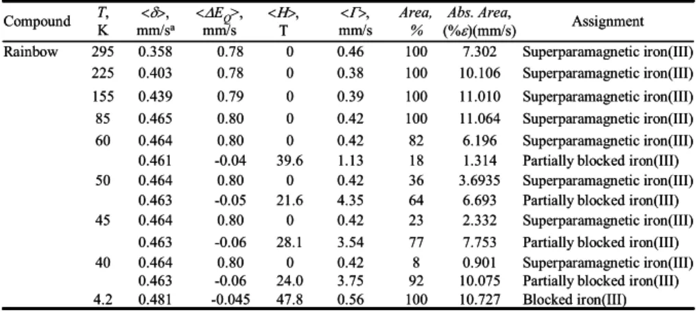
Table 1. M ¨ossbauer spectral parameters obtained for the Rimicaris exoculata hydrothermal shrimp.aThe isomer shifts are given relative to room temperature α-iron powder.
678 679 680 681 682 683 684 685 686 Blocked iron(III) 10.727 100 0.56 47.8 -0.045 0.481 4.2
Partially blocked iron(III) 10.075 92 3.75 24.0 -0.06 0.463 Superparamagnetic iron(III) 0.901 8 0.42 0 0.80 0.464 40
Partially blocked iron(III) 7.753 77 3.54 28.1 -0.06 0.463 Superparamagnetic iron(III) 2.332 23 0.42 0 0.80 0.464 45
Partially blocked iron(III) 6.693 64 4.35 21.6 -0.05 0.463 Superparamagnetic iron(III) 3.6935 36 0.42 0 0.80 0.464 50
Partially blocked iron(III) 1.314 18 1.13 39.6 -0.04 0.461 Superparamagnetic iron(III) 6.196 82 0.42 0 0.80 0.464 60 Superparamagnetic iron(III) 11.064 100 0.42 0 0.80 0.465 85 Superparamagnetic iron(III) 11.010 100 0.39 0 0.79 0.439 155 Superparamagnetic iron(III) 10.106 100 0.38 0 0.78 0.403 225 Superparamagnetic iron(III) 7.302 100 0.46 0 0.78 0.358 295 Rainbow Assignment Abs. Area, (%ε)(mm/s) Area, % <Γ>, mm/s <Η>, Τ <ΔEQ>, mm/s <δ>, mm/sa T, K Compound Blocked iron(III) 10.727 100 0.56 47.8 -0.045 0.481 4.2
Partially blocked iron(III) 10.075 92 3.75 24.0 -0.06 0.463 Superparamagnetic iron(III) 0.901 8 0.42 0 0.80 0.464 40
Partially blocked iron(III) 7.753 77 3.54 28.1 -0.06 0.463 Superparamagnetic iron(III) 2.332 23 0.42 0 0.80 0.464 45
Partially blocked iron(III) 6.693 64 4.35 21.6 -0.05 0.463 Superparamagnetic iron(III) 3.6935 36 0.42 0 0.80 0.464 50
Partially blocked iron(III) 1.314 18 1.13 39.6 -0.04 0.461 Superparamagnetic iron(III) 6.196 82 0.42 0 0.80 0.464 60 Superparamagnetic iron(III) 11.064 100 0.42 0 0.80 0.465 85 Superparamagnetic iron(III) 11.010 100 0.39 0 0.79 0.439 155 Superparamagnetic iron(III) 10.106 100 0.38 0 0.78 0.403 225 Superparamagnetic iron(III) 7.302 100 0.46 0 0.78 0.358 295 Rainbow Assignment Abs. Area, (%ε)(mm/s) Area, % <Γ>, mm/s <Η>, Τ <ΔEQ>, mm/s <δ>, mm/sa T, K Compound α