ORIGINAL ARTICLE
Incidental prostate cancer prevalence at radical
cystoprostatectomy
—importance
of the histopathological work-up
C. Wetterauer&M. Weibel&J. R. Gsponer&T. Vlajnic&T. Zellweger&S. Bütikofer&G. Müller&H. Püschel&
A. Bachmann&T. C. Gasser&L. Bubendorf&C. A. Rentsch
Received: 12 May 2014 / Revised: 25 August 2014 / Accepted: 12 September 2014 / Published online: 1 October 2014 # Springer-Verlag Berlin Heidelberg 2014
Abstract The reported incidental prostate cancer prevalence rates at radical cystoprostatectomy cover a range from 4 to 60 %. We investigated the influence of the histopathological work-up on prostate cancer prevalence rates. We identified 114 patients who had undergone cystoprostatectomy for blad-der cancer between 2000 and 2012. Complete histopatholog-ical assessment was defined as follows: (i) complete embed-ding of the prostate gland, (ii) sectioning of 15 or more prostate sections, and (iii) processing as whole mount slides. Prostate cancer prevalence rates derived from complete and incomplete histopathological assessments were compared. The overall prostate cancer prevalence rate was 59.6 %. A mean of 14.4 macroscopic tissue sections (thickness 3–5 mm) were sectioned. Sectioning≥15 sections resulted in a prostate cancer detection rate of 75 %, compared to 42.6 % when sectioning <15 sections (p<0.001). Complete embedding yielded a prostate cancer detection rate of 72.3 and of 23.1 % for partly embedded prostates (p<0.0001). Prostate cancer was detected in 68.8 % of the whole mounted samples and in 38.2 % of the samples sectioned as standard slides (p<0.01); according to the criteria described by Epstein and Ohori, 44.1 % of the detected prostate cancers were clinically significant. The quality of the histopathological work-up sig-nificantly influences prostate cancer detection rates and might
at least partially explain the highly variable reported incidental prostate cancer prevalence rates at cystoprostatectomy (CP). The high proportion of significant prostate cancer found in our series calls for a careful surgical approach to the prostate during CP.
Keywords Incidental prostate cancer . Bladder cancer . Radical cystoprostatectomy . Histology
Introduction
With a proportion of 24.4 % of male malignancies, pros-tate cancer represents the most common solid neoplasm among men [28]. Bladder cancer makes up 6.3 % of all cancers among men in Europe and ranks as the 4th most common cancer for men, with approximately 95,000 new-ly diagnosed cases each year [13, 28]. The prevalence of both prostate and bladder cancers increases in the aging population. As radical cystoprostatectomy (CP) is per-formed mostly in elderly patients, the coincidental detec-tion of both tumors is not uncommon. Highly variable prevalence rates of incidental prostate cancer at radical CP have been reported, ranging from 4 [30] to 60 % [29]. Importantly, the histopathological work-up of the prostate, more specifically, the macroscopic section thickness [3, 11] and the embedding technique [3, 14], is known to influence prostate cancer detection rates. Here, we assessed the prevalence rate of incidental prostate cancer in CP specimens in a highly industrialized town area and compared our findings to the reported rates worldwide. Furthermore, we analyzed the histopathological character-istics, their potential clinical relevance, and the influence of the histopathological technique on the detection of incidental prostate cancer.
C. Wetterauer and M. Weibel contributed equally to this work. C. Wetterauer
:
S. Bütikofer:
G. Müller:
H. Püschel:
A. Bachmann:
T. C. Gasser:
C. A. Rentsch (*)Department of Urology, University Hospital Basel, Spitalstrasse 21, 4031 Basel, Switzerland
e-mail: crentsch@uhbs.ch
M. Weibel
:
J. R. Gsponer:
T. Vlajnic:
H. Püschel:
L. Bubendorf Institute of Pathology, University Hospital Basel, Basel, Switzerland T. ZellwegerMaterials and methods
This retrospective study was approved by the local ethics committee (EKBB). One hundred thirty-nine patients having undergone radical CP for bladder cancer (n=133) and other indications between 2000 and 2012 were identified by searches in the archives of the Departments of Urology and Pathology at University Hospital Basel and by searches in the accounting systems. Twenty-five of these patients did not qualify for the analysis because either prostate cancer had been diagnosed prior to surgery or they had been treated for other reasons (n=6) than bladder cancer like low-capacity bladder or contracted, nonfunctional bladder after radiation therapy. Finally, 114 men who had undergone radical CP for bladder cancer were included in the analysis. The medical reports were reviewed for individual patient characteristics. The histopathology reports were screened for the TNM clas-sification of bladder and prostate cancers and for tumor his-tology, including Gleason score, surgical margins, maximal tumor diameter, complete or partial sampling, and number of macroscopic sections. All histological samples were reviewed according to the current classifications (i.e., TNM 2009, WHO 2004, ISUP 2005).
To determine the maximal tumor diameter, the edges of each tumor were outlined and the resulting distances were measured in millimeters; if a tumor was present in consecutive sections, the greatest measurement in all three dimensions (length, width, or depth) was considered the maximum diam-eter. In patients with complete transurethral resection of blad-der (TUR-B) tumors and no evidence of tumors in the bladblad-der at cystectomy, the final histology at TUR-B before the cystectomy was noted. Radical CP samples were processed in accordance with the descriptions in the guidelines of the Swiss Society of Pathology [12]. For histopathological grad-ing, the 2004 WHO classification was used.
For macroscopic tissue sectioning, a mean of 14.4 (median 15, range 2–37) sections with a thickness between 3 and 5 mm were used. Of the total 114 processed samples, 26 (23.1 %) had been partially and 83 (76.1 %) had been completely embedded. For five samples, no information on the complete-ness of the embedding was available. Eighty (70.2 %) samples were prepared as whole mount sections, and 34 (29.8 %) samples were processed as standard specimen slides.
We applied the following most commonly used criteria for nonsignificant prostate cancer [22]: (i) Gleason score≤6 with-out Gleason pattern 4 or 5, (ii) organ-confined disease, and (iii) tumor volume <0.5 cm3.
To determine the maximal tumor diameter, the edges of each tumor were outlined and the resulting distances were measured in millimeters; if a tumor was present in consecutive sections, the greatest measurement in all three dimensions (length, width, or depth) was considered the maximum diam-eter. Pathological reports did not encompass tumor volume
but did indicate tumor diameter. Based on the mathematical formula of spherical volume (4/3 πr3), we calculated the diameter for a tumor with a volume of 0.5 cm3and concluded that a tumor with a diameter larger than 1 cm would therefore be defined as significant.
All statistical inference testing and data visualization were performed using R 3.0.1. [24]. For continuous data, Wilcoxon rank sum tests were used, and for categorical data, Fisher’s exact tests or Pearson’s chi-squared tests were applied as indicated. The clinical follow-up information regarding PSA course, survival, progression, or recurrence was incomplete, and the data quality did not allow for further statistical anal-ysis. P values lower than 0.05 were considered statistically significant.
Results
Detailed characteristics for all 114 bladder cancer patients are given in Table1. Most of the detected bladder cancers were diagnosed as urothelial carcinomas (n=108; 94.7 %), while the remaining diagnoses were small cell carcinoma, sarcomatoid carcinoma, squamous cell carcinoma, mixed car-cinoma, and urothelial dysplasia.
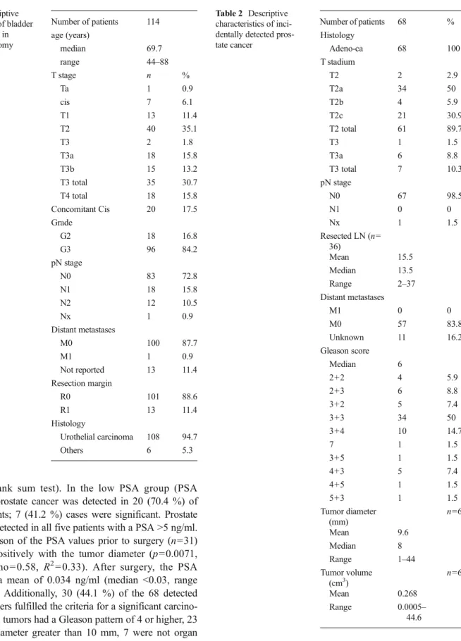
Incidental prostate cancer was identified in 68 (59.6 %) of the 114 CP specimens. The patient characteristics for the patients with incidental prostate cancer are given in Table2. All prostate cancers were histologically classified as adeno-carcinomas. In three patients, high-grade prostatic intraepithelial neoplasia (PIN) was diagnosed. Median age of patients with prostate cancer was comparable to that of the whole investigated population, and no significant differ-ence in age between patients with (69.4 years) and without (69.7 years) prostate cancer was observed. Four (5.9 %) had a Gleason score of 4. The majority of patients (n=45) exhibited a Gleason score of 5–6 (66.2 %), and 19 patients (27.9 %) had Gleason scores ≥7. Sixty-one (89.7 %) of the 68 tumors displayed organ-confined growth, while extra-capsular exten-sions were observed in 7 (10.3 %). Of note, none of the 67 prostate cancer patients for whom the lymph node status had been determined demonstrated lymph node metastases. The mean number of resected lymph nodes was 15.5 (median 13.5; range 2–37). Overall, 62 (91.2 %) of the specimens showed tumor-free surgical margins (R0), and 6 (8.8 %) displayed positive margins (R1). Positive margins were iden-tified dorsally (n=1), laterally (n=1), in the apex (n=2), and the basis (n=2).
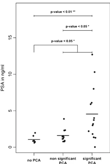
PSA had been tested prior to surgery for 32 patients and upon follow-up for 20 patients. The mean PSA prior to sur-gery was 2.78 ng/ml (median 1.55 ng/ml, range 0.03– 12 ng/ml). The mean preoperative PSA value of the groups with prostate cancer (3.3 ng/ml) and without detected prostate cancer (1.1 ng/ml) was significantly different (Fig.1,p<0.05,
Wilcoxon rank sum test). In the low PSA group (PSA <4 ng/ml), prostate cancer was detected in 20 (70.4 %) of the 27 patients; 7 (41.2 %) cases were significant. Prostate cancer was detected in all five patients with a PSA >5 ng/ml. The comparison of the PSA values prior to surgery (n=31) correlated positively with the tumor diameter (p=0.0071, Spearman rho = 0.58, R2= 0.33). After surgery, the PSA dropped to a mean of 0.034 ng/ml (median <0.03, range <0.03–0.09). Additionally, 30 (44.1 %) of the 68 detected prostate cancers fulfilled the criteria for a significant carcino-ma. Eighteen tumors had a Gleason pattern of 4 or higher, 23 featured a diameter greater than 10 mm, 7 were not organ confined, and 13 met several of the criteria for clinical signif-icance (Table3). The mean preoperative PSA in the subgroup of insignificant prostate cancer (1.4 ng/ml) was significantly lower than the mean PSA in the subgroup of significant prostate cancer (5.4 ng/ml,p=<0.01) (Fig.1).
In the group with incidental prostate cancer, a mean of 16.4 (median 17, range 5–34) tissue sections were sectioned. Fewer than 15 sections were used for 54 (47.4 %) of the prostates, and 15 or more sections were used for 60 (52.6 %) prostates. Table 2 Descriptive
characteristics of inci-dentally detected pros-tate cancer Number of patients 68 % Histology Adeno-ca 68 100 T stadium T2 2 2.9 T2a 34 50 T2b 4 5.9 T2c 21 30.9 T2 total 61 89.7 T3 1 1.5 T3a 6 8.8 T3 total 7 10.3 pN stage N0 67 98.5 N1 0 0 Nx 1 1.5 Resected LN (n= 36) Mean 15.5 Median 13.5 Range 2–37 Distant metastases M1 0 0 M0 57 83.8 Unknown 11 16.2 Gleason score Median 6 2+2 4 5.9 2+3 6 8.8 3+2 5 7.4 3+3 34 50 3+4 10 14.7 7 1 1.5 3+5 1 1.5 4+3 5 7.4 4+5 1 1.5 5+3 1 1.5 Tumor diameter (mm) n=68 Mean 9.6 Median 8 Range 1–44 Tumor volume (cm3) n=68 Mean 0.268 Range 0.0005– 44.6 Table 1 Descriptive characteristics of bladder cancer detected in cystoprostatectomy specimens Number of patients 114 age (years) median 69.7 range 44–88 T stage n % Ta 1 0.9 cis 7 6.1 T1 13 11.4 T2 40 35.1 T3 2 1.8 T3a 18 15.8 T3b 15 13.2 T3 total 35 30.7 T4 total 18 15.8 Concomitant Cis 20 17.5 Grade G2 18 16.8 G3 96 84.2 pN stage N0 83 72.8 N1 18 15.8 N2 12 10.5 Nx 1 0.9 Distant metastases M0 100 87.7 M1 1 0.9 Not reported 13 11.4 Resection margin R0 101 88.6 R1 13 11.4 Histology Urothelial carcinoma 108 94.7 Others 6 5.3
This resulted in the detection of 23 (42.6 %) and 45 (75 %) incidental prostate cancers (p<0.001; Fisher’s exact test), respectively, as presented in Fig.2. Eighty-three (76.1 %) of the total 114 prostates were embedded completely, and 26 (23.9 %) were embedded partially. For five prostates, the embedding technique was not reported. Prostate cancer was detected in 60 (72.3 %) of the 83 completely embedded samples and in 6 (23.1 %) of the 26 partially embedded
samples (p<0.0001; Fisher’s exact test; Fig. 2). Significant prostate cancer was detected in 28 (33.7 %) of the 83 completely embedded samples and in 1 (3.8 %) of the 26 partially embedded samples (p<0.01; Fisher’s exact test).
We compared the rates of complete embedding with the detection rate of prostate cancer in the periods between 2000 and 2006 and 2007 and 2012. The rates of completely embed-ded samples in the two periods were 55.3 and 91.9 %, respec-tively. The first period yielded an overall prostate cancer prev-alence rate of 50 %, and the second period yielded 67.7 %.
Whole mount sections were used for 80 (70.2 %) samples, and standard slides were used for 34 (29.8 %) samples. In 55 (68.8 %) of the whole mount sections and in 13 (38.2 %) of the standard slides, prostate cancer could be detected (p<0.01; Fisher’s exact test; Fig.2).
Discussion
Here, we explored the prevalence of incidental prostate cancers in CP specimens in a highly industrialized town area, where most of the CP specimens are processed centrally at a single pathology institute. Comparison of complete versus incomplete specimen processing indicated prostate cancer prevalence rates reaching up to 75 % for completely processed specimens.
Based on previous publications, including a total of 11,553 patients, the mean prevalence rates of prostate cancer at rad-ical CP in Europe and globally were 25.2 and 26.5 %, respec-tively (Table 4). Detection rates are influenced by technical variables [3,11], study populations [3], and geographic [23, 28] factors; e.g., studies from Asia generally reported lower rates than those from the USA and Europe (Table4). Com-pared to these previously reported rates, the 59.6 % in our study represent one of the highest published prevalence rates of incidental prostate cancer in CP specimens (Table4). Gen-erating a best-case scenario and analyzing the subgroup of 53 patients with complete embedding, the use of 15 or more sections and whole mount sections results in a prevalence rate of 75.5 %. This extraordinary high proportion is unlikely to be greatly biased by the retrospective nature of the study because our study was based on a consecutive series of CP specimens. Our high prevalence of incidental prostate cancer also clearly exceeded the rates of most previous autopsy series [7]. Only Sakr et al. discovered unexpected prostate cancer at a similarly high rate of 55 and 64 % of autopsied patients who had died in their 6th and 7th decades, respectively, after a histological examination of their whole prostates [25].
To the best of our knowledge, this is the first report on the prevalence of incidental prostate cancer in CP specimens in Switzerland. It could be speculated that the high prevalence in an industrialized town area may be due to an increased expo-sure to potential carcinogenic factors that drive prostate can-cer. However, to date, there has been no published evidence Fig. 1 Comparison of preoperative PSA values in the groups without,
with nonsignificant, and with significant prostate cancer
Table 3 Further criteria for the clinical relevance of prostate cancer (n = patients with prostate cancer fulfilling the indicated criteria)
Number Percent Gleason score including a pattern≥4 18 26.5
pT stage≥T3 7 10.3
Max tumor diameter >10 mm 23 33.8 More than one criterion for relevant cancer 13 19.1
<60 years 13 19.1
≥60 <75 years 35 51.5
≥75 years 20 29.4
that a high prevalence of incidental prostate cancer could be driven by exposure to carcinogens in the context of the sub-stantial presence of chemical and pharmaceutical industries or by other, as yet undefined, exposures in our region.
Instead, the extent of histological processing appears to be the most influential factor to explain the high prostate cancer prevalence (Fig.2). Several published results have shown that lower section thickness increases prostate cancer detection rates [11,30]. In our study, the prostates were processed into 3–5 mm tissue slices and paraffin blocks, and sections from 15 or more tissue blocks significantly raised the prostate cancer detection rates. Although the section number can depend on the size of the prostate gland, our results suggest that prostate cancer detection rates increased proportionally with the num-ber of tissue blocks and sections. The same was true for complete versus incomplete embedding of the prostate, which is in line with other published series [3, 14]. Furthermore, complete embedding significantly improved (p<0.01) the detection of significant prostate cancer compared to partial embedding in our series. The use of the whole mount tech-nique [19] resulted in a significantly (p<0.01) higher prostate cancer detection rate of 68.8 %, compared to the use of regular slides (38.2 %). As a caveat, 89.3 % of the prostates processed
as whole mount sections, but only 26.5 % of those processed as regular slides were completely embedded. Finally, the overall prostate cancer prevalence rate of 67.7 % between 2007 and 2012, when most of the samples were embedded completely, indicates that a consequent use of complete em-bedding, the use of whole mount sections, and/or a high section number can significantly influence the prostate cancer prevalence rates in CP series.
The clinical relevance of incidental prostate cancers detect-ed at radical CP remains questionable. Two studies demon-strated significantly worse survival after radical CP for pa-tients with concurrent tumors [8,27], while Pritchett found no difference in the mortality rates [11]. In a recent series of 1476 patients, 22 % of the detected prostate cancers were classified as significant. However, the most influential factor in this series was not the presence of relevant prostate cancer but prostatic infiltration of a urothelial carcinoma [6]. In our series, almost half of the prostate cancers met criteria for significant prostate cancer (Table3) but none had detectable lymph node metastases (Table2). Our rate of clinically sig-nificant prostate cancer is comparable to the other reported rates (Table 4), even though the reported rates are diverse, based on the different criteria being applied [22]. Elevated Fig. 2 Detection rates of incidental prostate cancer, according to the pathological work-up. a <15 versus >15 prostate sections. b Complete versus partial embedding. c Regular slides versus whole mount slides. The numbers inbrackets indicate the percentage of specimens in each indicated group
Table 4 Worldwide reported rates of incidental prostate cancer in cystoprostatectomy specimens
Author Region Year N Mean
age Section thickness (mm) Sampling PCa total PCa % Significant PCa (%) North America
Winfield [3] Iowa, USA 1987 80 63.7 Complete 22 27.5 11 (50 %) Pritchett [11] South Carolina, USA 1988 165 65 Partial 45 27.3 8 (17.8 %) Montie [11] Cleveland, USA 1989 84 62 4–5 Complete 39 46.4 6 (15 %) Kabalin [3] Standford, USA 1989 66 64 3 Complete 25 37.9 3 (12 %)
Abbas [11] Miami, USA 1996 40 64.3 2–3 Partial 18 45
Ward [3] Rochester, USA 2004 129 69 30 23.3 18 (60 %)
Revelo [11] Nashville, USA 2004 121 67.4 5 Complete 50 41.3 24 (48 %) Abdelhady [3] Ontario, Canada 2007 204 67 Complete 58 28.4 18 (31 %)
Weizer [3] Michigan, USA 2007 35 65 16 45.7 4 (25 %)
Bruins [6] Los Angeles, USA 2013 1476 69 3–5 559 37.9 123 (8.3 %) Europe
Moutzouris [11] Athens, Greece 1999 59 66.5 5 Complete 16 27.1
Conrad [11] Hamburg, Germany 2001 133 60 3 Complete 58 43.6 11 (19 %) Prange [3] Hamburg, Germany 2001 85 64 4 Complete 41 49.0 4 (10 %) Cindolo [3] Naples, Italy 2001 165 69 3 Partial/complete 17 10.3
Kouriefs [3] Gillinghan, England 2005 128 23 18
Delongchamp [11] Paris, France 2005 141 62 4 Complete 20 14.2 14 (70 %) Montironi [3] Ancona, Italy 2005 132 61 Complete 55 42 %
Ruffion [11] Lyon, France 2005 100 62 2.5 Complete 51 51 6 (12 %) Rocco [11] Milano, Italy 2006 63 67 3 Complete 34 54 12 (35 %) Winkler [29] London, England 2007 97 2 Partial 58 60 31 (53 %)
Barbisan [5] Marche, Italy 2008 248 68 3 123 49.6 23 (18.7 %)
Mazzuchelli [18] Ancona, Italy 2009 248 68 3 Complete 123 49.6 23 (18.7 %)
Gakis [15] Tübingen, Germany 2010 95 68 4–5 26 27.4 7 (27 %)
Buse [8] Germany 2012 1122 65.6 2–5 200 17.8
Fritsche [14] Regensburg, Germany 2012 295 68 4 Partial/complete 91 30.8 41 (45 %) Alsinnawi [2] Dublin, Ireland 2012 108 64 4 Complete 35 32.4 10 (28.5 %) Sruogis [27] Vilnius, Lithuania 2012 81 61.3 27 33.3 13 (48.1 %)
Pignot [21] France 2013 4251 70.2 905 21.7
Wetterauer Basel, Switzerland 2013 115 68.9 3–5 Partial/complete 68 59.6 40 (58.8 %) Asia
Lee [30] Kweishan, Taiwan 2006 248 63 5 Complete 10 4
Yang [30] Thaijung, Taiwan 1999 49 67.8 3 Complete 16 32.6
Nakagawa [20] Tokyo, Japan 2009 349 65 5 91 26.1 68 (74.7 %)
Jin [16] Hangzhou, China 2008 264 70.9 5 Complete 37 14 12 (32.4 %) Zhu [30] Shanghai, China 2009 92 67.1 5 Complete 3 3.3 1 (33.3 %) Australia
Ahmadi [1] Sydney, Australia 2012 129 50 38.8 35 (70 %)
Middle east
Aydin [3] Turkey 1999 121 67.1 17 14
Hosseini [3] Theran, Iran 2007 50 62.5 Partial 7 14 4 (57 %)
Aytac [4] Bursa, Turkey 2011 300 62 3–5 Complete 60 20 40 (66.7 %)
North America 2400 862 35.9 Europe 7551 1903 25.2 Asia 1002 157 15.7 Australia 129 50 38.8 Middle East 471 84 17.8 Total 11,553 3056 26.5
PSA may be predictive of the presence of significant cancer and a large tumor diameter, as suggested by our comparison; however, the patient numbers are low, and the clinical data are insufficient for making final conclusions about these findings. Srougi et al. proposed a cystectomy in combination with the enucleation of adenomas [3], and TUR-P with preservation of the capsule prior to cystectomy was reported as an option by Colombo et al. [10]. These techniques might be applicable for selected patients for whom the preservation of potency is paramount, but in light of our findings, they may stand against the principles of cancer surgery. If attempted though, it seems reasonable to perform prostate biopsy before such surgery as even low PSA values cannot exclude the presence of prostate cancer (Fig. 1). Given the high risk of prostate cancer in patients undergoing CP, special attention to the removal of the prostate seems advisable. Once the presence of prostate cancer has been confirmed after CP, specific oncologic follow-up may be indicated. At least in high-risk prostate cancer, the oncologic follow-up might include regular PSA monitoring in order to detect residual, recurrent, or metastatic disease.
It has been suggested that bladder cancer patients have an increased risk of being diagnosed with prostate cancer [17, 26]. In fact, a higher coincidence rate of bladder and prostate tumors compared to their presence in the general population has been reported [9]. Such a correlation may be explained partly by the relatively high age of patients and the fact that exposure of patients with either tumor type to urologists favors the detection of other urogenital cancers, if present. Finally, there is currently no compelling evidence for a com-mon link between prostate and bladder cancers.
Several studies have reported the influence of specific pathologic sampling techniques on prostate cancer detection rates in CP specimen. To the best of our knowledge, we are the first to assess the influence of specific aspects of pathologic sampling within one series at a single Pathology Institute and to report the incidental prevalence rate of PCa at CP in Switzerland. Compared to the extremely variable reported rates of incidentally discovered prostate cancers, our reported rate of 59.6 % ranks among the highest published rates. Subgroup analysis of samples with complete embedding, the use of 15 or more sections, and whole mount sections resulted in prevalence rates of more than 75 % in our series.
Conclusions
We report a high prevalence of incidental prostate cancer at CP of 59.6 %. This is more than twice as high as the mean reported European and worldwide prevalence of 26.5 %. We demonstrate the paramount importance of a complete histo-pathological work-up for estimating the true prevalence of prostate cancer in a given population and for the detection of
significant prostate cancer. Our results call for a careful surgi-cal approach to the prostate at radisurgi-cal cystoprostatectomy.
Conflict of interest The authors declare that they have no conflict of interest.
References
1. Ahmadi N, Delprado WJ, Brooks AJ, Brenner PC, Coombes GM, Grant A, Patel MI (2012) Cancer identified incidentally in the pros-tate following radical cystoprospros-tatectomy: an Australian study. ANZ J Surg. doi:10.1111/ans.12015
2. Alsinnawi M, Loftus B, Flynn R, McDermott T, Grainger R, Thornhill JA (2012) The incidence and relevance of prostate cancer in radical cystoprostatectomy specimens. Int Urol Nephrol 44:1705– 1710. doi:10.1007/s11255-012-0224-y
3. Autorino R, Di Lorenzo G, Damiano R, Giannarini G, De Sio M, Cheng L, Montironi R (2009) Pathology of the prostate in radical cystectomy specimens: a critical review. Surg Oncol 18:73–84. doi:
10.1016/j.suronc.2008.07.006 S0960-7404(08)00063-7
4. Aytac B, Vuruskan H (2011) Clinicopathologic features of incidental prostatic adenocarcinoma in radical cystoprostatectomy specimens. World J Surg Oncol 9:81. doi: 10.1186/1477-7819-9-811477-7819-9-81
5. Barbisan F, Mazzucchelli R, Scarpelli M, Lopez-Beltran A, Cheng L, Kirkali Z, Montironi R (2009) Urothelial and incidental prostate carcinoma in prostates from cystoprostatectomies for bladder cancer: is there a relationship between urothelial and prostate cancer? BJU Int 103:1058–1063. doi:10.1111/j.1464-410X.2008.08207.xBJU8207
6. Bruins HM, Djaladat H, Ahmadi H, Sherrod A, Cai J, Miranda G, Skinner EC, Daneshmand S (2013) Incidental cancer of the prostate in patients with bladder urothelial carcinoma: comprehensive analy-sis of 1476 radical cystoprostatectomy specimens. J Urol. doi:10. 1016/j.juro.2013.05.034
7. Bubendorf L, Schopfer A, Wagner U, Sauter G, Moch H, Willi N, Gasser TC, Mihatsch MJ (2000) Metastatic patterns of prostate cancer: an autopsy study of 1,589 patients. Hum Pathol 31:578–583 8. Buse S, Hofner T, Muller SC, Hermann E, Wieland WF, May M, Stief CG, Bastian PJ, Hohenfellner M, Haferkamp A (2013) Characterization and risk stratification of prostate cancer in patients undergoing radical cystoprostatectomy. Int J Urol. doi:10.1111/iju. 12073
9. Chun TY (1997) Coincidence of bladder and prostate cancer J Urol 157:65–67
10. Colombo R, Bertini R, Salonia A, Naspro R, Ghezzi M, Mazzoccoli B, Deho F, Montorsi F, Rigatti P (2004) Overall clinical outcomes after nerve and seminal sparing radical cystectomy for the treatment of organ confined bladder cancer. J Urol 171:1819–1822. doi:10. 1097/01.ju.0000123781.49896.fe
11. Damiano R, Di Lorenzo G, Cantiello F, De Sio M, Perdona S, D’Armiento M, Autorino R (2007) Clinicopathologic features of prostate adenocarcinoma incidentally discovered at the time of radi-cal cystectomy: an evidence-based analysis. Eur Urol 52:648–657. doi:10.1016/j.eururo.2007.06.016
12. Dirnhofer St. BL, Lehr HA, Landau B, Zenklusen HR (2011) Qualitätsrichtlinien SGpath
13. Ferlay J, Steliarova-Foucher E, Lortet-Tieulent J, Rosso S, Coebergh JW, Comber H, Forman D, Bray F (2013) Cancer incidence and mortality patterns in Europe: estimates for 40 countries in 2012. Eur J Cancer 49:1374–1403. doi: 10.1016/j.ejca.2012.12.027S0959-8049(13)00007-5
14. Fritsche HM, Aziz A, Eder F, Otto W, Denzinger S, Wieland WF, May M, Hofstadter F, Hartmann A, Burger M (2012) Potentially clinically relevant prostate cancer is found more frequently after complete than after partial histopathological processing of radical cystoprostatectomy specimens. Virchows Arch 461:655–661. doi:
10.1007/s00428-012-1328-6
15. Gakis G, Schilling D, Bedke J, Sievert KD, Stenzl A (2010) Incidental prostate cancer at radical cystoprostatectomy: implications for apex-sparing surgery. BJU Int 105:468–471. doi: 10.1111/j.1464-410X.2009.08739.xBJU8739
16. Jin XD, Chen ZD, Wang B, Cai SL, Yao XL, Jin BY (2008) Incidental prostate cancer in radical cystoprostatectomy speci-mens. Asian J Androl 10:809–814. doi:10.1111/j.1745-7262. 2008.00420.x
17. Kurokawa K, Ito K, Yamamoto T, Takechi H, Miyamoto S, Suzuki K, Yamanaka H (2004) Comparative study on the prevalence of clinically detectable prostate cancer in patients with and without bladder cancer. Urology 63:268–272. doi:10.1016/j.urology.2003. 09.027S009042950301015X
18. Mazzucchelli R, Barbisan F, Scarpelli M, Lopez-Beltran A, van der Kwast TH, Cheng L, Montironi R (2009) Is incidentally detected prostate cancer in patients undergoing radical cystoprostatectomy clinically significant? Am J Clin Pathol 131:279–283. doi:10.1309/ AJCP4OCYZBAN9TJUP06J2Q5065544X5J
19. Montironi R, Cheng L, Mazzucchelli R, Scarpelli M, Kirkali Z, Montorsi F, Lopez-Beltran A (2009) Critical evaluation of the pros-tate from cystoprospros-tatectomies for bladder cancer: insights from a complete sampling with the whole mount technique. Eur Urol 55: 1305–1309. doi:10.1016/j.eururo.2008.10.032S0302-2838(08) 01251-7
20. Nakagawa T, Kanai Y, Komiyama M, Fujimoto H, Kakizoe T (2009) Characteristics of prostate cancers found in specimens removed by radical cystoprostatectomy for bladder cancer and their relationship with serum prostate-specific antigen level. Cancer Sci 100:1880– 1884. doi:10.1111/j.1349-7006.2009.01267.x
21. Pignot G, Salomon L, Neuzillet Y, Masson-Lecomte A, Lebacle C, Patard JJ, Lunardi P, Rischmann P, Pasticier G, Bernhard JC, Cohen J, Timsit MO, Verkarre V, Peyronnet B, Verhoest G, Le Goux C, Zerbib M, Brecheteau F, Bigot P, Larre S, Murez T, Thuret R, Lacarriere E, Champy C, Roupret M, Comperat E, Berger J, Descazeaud A, Toledano H, Bastide C, Lavilledieu S, Avances C, Delage F, Valeri A, Molimard B, Houlgatte A, Gres P, Donnaint A,
Kleinclauss F, Legal S, Doerfler A, Koutlidis N, Cormier L, Hetet JF, Colls P, Arvin-Berod A, Rambeaud JJ, Quintens H, Soulie M, Pfister C, The members of The Oncologic Committee of the French Association of U (2013) Clinicopathological characteristics of inci-dental prostate cancer discovered from radical cystoprostatectomy specimen: a multicenter French study. Ann Surg Oncol. doi:10.1245/ s10434-013-3340-8
22. Ploussard G, Epstein JI, Montironi R, Carroll PR, Wirth M, Grimm MO, Bjartell AS, Montorsi F, Freedland SJ, Erbersdobler A, van der Kwast TH (2011) The contemporary concept of significant versus insignificant prostate cancer. Eur Urol 60:291–303. doi:10.1016/j. eururo.2011.05.006S0302-2838(11)00495-7
23. Quinn M, Babb P (2002) Patterns and trends in prostate cancer incidence, survival, prevalence and mortality. Part II: individual countries. BJU Int 90:174–184
24. R CT (2013) A language and environment for statistical computing. R Foundation for Statistical Computing, Vienna, Austria. URLhttp:// www.R-project.org/
25. Sakr WA, Grignon DJ, Crissman JD, Heilbrun LK, Cassin BJ, Pontes JJ, Haas GP (1994) High grade prostatic intraepithelial neoplasia (HGPIN) and prostatic adenocarcinoma between the ages of 20–69: an autopsy study of 249 cases. In Vivo 8:439–443
26. Singh A, Kinoshita Y, Rovito PM Jr, Landas S, Silberstein J, Nsouli I, Wang CY, Haas GP (2008) Higher than expected association of clinical prostate and bladder cancers. J Urol 179:S2–S5. doi:10. 1016/j.juro.2008.03.130S0022-5347(08)00731-3
27. Sruogis A, Ulys A, Smailyte G, Kardelis Z, Kulboka A, Anglickiene G, Samalavicius N, Anglickis M (2012) Incidentally found prostate cancer and influence on overall survival after radical cystoprostatectomy. Prostate Cancer 2012:690210. doi:10.1155/ 2012/690210
28. WHO (2008) Estimated cancer incidence, mortality, prevalence and disability-adjusted life years (DALYs) worldwide in 2008
29. Winkler MH, Livni N, Mannion EM, Hrouda D, Christmas T (2007) Characteristics of incidental prostatic adenocarcinoma in contempo-rary radical cystoprostatectomy specimens. BJU Int 99:554–558. doi:
10.1111/j.1464-410X.2006.06660.x
30. Zhu YP, Ye DW, Yao XD, Zhang SL, Dai B, Zhang HL, Shen YJ, Zhu Y, Shi GH (2009) Prevalence of incidental prostate cancer in patients undergoing radical cystoprostatectomy: data from China and other Asian countries. Asian J Androl 11:104–108. doi:10.1038/aja. 2009.15aja200815