HAL Id: hal-03034414
https://hal-normandie-univ.archives-ouvertes.fr/hal-03034414
Submitted on 1 Dec 2020
HAL is a multi-disciplinary open access
archive for the deposit and dissemination of
sci-entific research documents, whether they are
pub-lished or not. The documents may come from
teaching and research institutions in France or
abroad, or from public or private research centers.
L’archive ouverte pluridisciplinaire HAL, est
destinée au dépôt et à la diffusion de documents
scientifiques de niveau recherche, publiés ou non,
émanant des établissements d’enseignement et de
recherche français ou étrangers, des laboratoires
publics ou privés.
Fluorophore-assisted click chemistry through copper(I)
complexation
Victor Flon, Magalie Bénard, Damien Schapman, Ludovic Galas, Pierre-Yves
Renard, Cyrille Sabot
To cite this version:
Victor Flon, Magalie Bénard, Damien Schapman, Ludovic Galas, Pierre-Yves Renard, et al..
Fluorophore-assisted click chemistry through copper(I) complexation. Biomolecules, MDPI, 2020,
10 (4), pp.619. �10.3390/biom10040619�. �hal-03034414�
Article
1
Fluorophore-assisted click chemistry through
2
copper(I) complexation
3
Victor Flon, 1 Magalie Bénard, 2 Damien Schapman, 2 Ludovic Galas, 2 Pierre-Yves Renard, 1
4
Cyrille Sabot 1,*
5
1 Normandie Univ, CNRS, UNIROUEN, INSA Rouen, COBRA (UMR 6014), 76000 Rouen, France
e-mail@e-6
mail.com
7
2 Normandie Univ, UNIROUEN, INSERM, PRIMACEN, 76000 Rouen (France)
8
* Correspondence: [email protected]
9
Received: date; Accepted: date; Published: date
10
Abstract: The copper-catalyzed alkyne-azide cycloaddition (CuAAC) is one of the most powerful
11
chemical strategies for selective fluorescent labeling of biomolecules in vitro or in biological systems.
12
In order to accelerate the ligation process and ensure efficient formation of conjugates under diluted
13
conditions, external copper(I) ligands or sophisticated copper(I) chelating azides are used. This
14
latter strategy, however, increases the bulkiness of the triazole linkage, thus perturbing the
15
biological function or dynamic behavior of the conjugates. In a-proof-of-concept study, we
16
investigated the use of an extremely compact fluorophore-based copper(I) chelating azide in order
17
to accelerate the CuAAC with concomitant fluorescence labeling; In our strategy, the fluorophore is
18
able to complex copper(I) species while retaining its photophysical properties. It is believed that this
19
unprecedented approach which was applied for the labeling of a short peptide molecule and the
20
fluorescent labeling of live cells, could be extended to other families of nitrogen-based fluorophores
21
in order to tune both the reaction rate and photophysical characteristics.
22
Keywords: click chemistry, CuAAC, chelating fluorophore, chelating azide, fluorescent labeling,
23
azaphthalimide, Kondrat’eva ligation
24
1. Introduction
25
The copper-catalyzed alkyne-azide cycloaddition (CuAAC) also known as click chemistry,[1]
26
has attracted tremendous interest in recent years for the site-specific in vitro modification of
27
biomolecules such as proteins, glycans, lipids or nucleic acids, as well as for the bioorthogonal
28
fluorescent labeling of cell extracts or living systems.[2-4] Prominent applications in this latter area
29
include among others the understanding of diverse biological processes,[5-6] the development of
30
detection tools,[7] and the real-time live-cell imaging and therapy[8].[9-12] In contrast to other
31
successful bioorthogonal chemistries such as strain promoted alkyne azide cycloaddition
32
(SPAAC)[13] and tetrazine-based inverse electron demand DielsAlder ligation (IEDDA),[14,15]
33
CuAAC ligation has been widely adopted due to the small size and inertness of the alkyne and azide
34
handles that can be incorporated into biomolecules by using the genetic code expansion, or the
35
cellular metabolic machinery,[16] and to the fact that small triazole adducts also impose a minimal
36
perturbation of resulting conjugates.
37
Reaction of alkynes with azides generally involves the in situ formation of copper(I) catalyst
38
from a copper(II) source (e.g. CuSO4) in the presence of reducing agent, sodium ascorbate. Although
39
coordinating ligands are not strictly required for CuAAC,[5] their combine use has shown to
40
significantly accelerate the alkyne-azide cycloaddition which thus ensures efficient bioconjugation
41
under diluted conditions imposed by biological systems, while preventing both the deactivation of
42
the copper(I) catalyst by biomolecules, and copper-mediated oxidative damages.
43
Tris(triazolylmethyl)amine derivatives such as TBTA or its water-soluble analogues THPTA, and
44
BTTAA constitute an important family of such accelerating ligands (Figure 1a).[17-19]
46
Figure 1. Strategies of copper ligands
47
48
However, ligands, copper sources, and fluorescent bioorthogonal handle are generally required
49
in excess amounts relatively to the chemical reporter group for the bioconjugate reaction to proceed
50
accordingly with high yields.[20] These ligands are mostly used for cell-surface labeling, or require
51
covalent attachment to cell penetrating peptides to facilitate their cellular uptake.[21] To circumvent
52
these limitations, another strategy consists in using azides equipped with a chelating moiety which
53
are capable of complexing the copper(I) species and thus accelerate the reaction in the absence of
54
external ligands by both facilitating the formation of the metallacycle intermediate and increasing the
55
electrophilicity of the azide function.[22-24] Highly performant azide chelating systems were
56
designed in this context (Figure 1b).[25,26] Nevertheless, these chelating azides are constituted of
57
polycyclic ligands with aromatic characters, and when linked to bulky and rigid fluorophores,
water-58
solubilizing groups are required to counterbalance their overall hydrophobicity. As a consequence,
59
subsequent biological, physico-chemical properties of the corresponding conjugates, in particular for
60
short labeled biomolecules or pharmacophores, may be dramatically altered, which questions the
61
benefit of using small alkyne, azide handles in CuAAC, with respect to other biorthogonal
62
reactions.[27]
63
Following these considerations, in order to perform bioconjugate reaction with alkyne modified
64
biomolecules, we want to report the unprecedented use of an extremely compact fluorophore-based
65
ligand capable of complexing copper(I) species while retaining its photophysical characteristics
66
useful for bioimaging applications. We previously reported the Kondrat’eva ligation based on a
one-67
pot Diels-Alder/aromatization of 5-alkoxyoazole with maleimide to furnish the corresponding
68
azaphthalimide fluorophore, which has found different applications in chemical biology.[28-29]
69
Interestingly, this small bicyclic azaphthalimide dye displays relatively high excitation and emission
70
wavelengths (ex. 420 nm, em. 520 nm) suitable for live-cell imaging. Herein, we wish to combine both
71
the fluorescent properties of the azaphthalimide dye and its underestimated copper-binding ability
72
to design a readily available and extremely compact fluorescent copper-chelating azide (Figure 2).
73
Figure 2. Fluorophore-based cooper(I) ligand for CuAAC-mediated fluorescent labeling.
74
2. Materials and Methods
75
2.1. General information
76
All chemicals were used as received from commercial sources without further purification.
77
Solvents, unless otherwise stated, were purchased in reagent grade or HPLC grade and used as
78
received, except tetrahydrofuran which was freshly distilled over sodium prior to use. PBS (pH 7.4,
0.1 M) and aq. mobile phases for HPLC were prepared with water purified by means of a MilliQ
80
system (purified to 18.2 MΩ cm). All reactions were monitored by thin layer chromatography (TLC)
81
and/or RP-HPLC. TLC were carried out on Merck DC Kieselgel 60 F-254 aluminum sheets.
82
Visualization of spots was performed under a UV lamp at λ = 254 or 365 nm, and/or staining with a
83
KMnO4 solution/K2CO3 + 5% NaOH, and developed with heat. Flash column chromatography
84
purifications were performed manually on silica gel (40–63 µM) under pressurized air flow.
85
Instruments and Methods. HPLC system A: RP-HPLC analyses were performed with a Thermo
86
Fischer Ultimate 3000 RS instrument, equipped with a diode array detector (DAD-3000RS). The
87
temperature of the column compartment was fixed at 25 °C. A Thermo Hypersyl GOLD® C18 column
88
(1.9 µm, 2.1 × 50 mm) was used with a binary solvent system composed of MeCN and 0.1% aq. formic
89
acid (aq. FA, pH 2) as eluents (linear gradient from 5 to 100% MeCN over 10 min) at a flow rate of
90
0.600 mL/min. System B: Semi-preparative RP-HPLC were performed with an Interchim puriFlash®
91
4250 instrument equipped with a diode array detector, and a Thermo Hypersyl GOLD® C18 column
92
(5 µm, 30.0 × 250 mm) with MeCN and water as eluents (linear gradient 10–65% MeCN for 50 min)
93
at a flow rate of 40 mL/min. System C: Semi-preparative RP-HPLC were performed with an Interchim
94
puriFlash® 4250 instrument equipped with a diode array detector, and a Thermo Syncronis® C18
95
column (5 µm, 21.2 × 250 mm) with MeCN and 0.1% aq. FA as eluents (linear gradient 5–95% MeCN
96
for 35 min) at a flow rate of 15 mL/min. System D: Semi-preparative RP-HPLC were performed with
97
an Interchim puriFlash® 4250 instrument equipped with a diode array detector, and a Thermo
98
Hypersyl GOLD® C18 column (5 µm, 20.0 × 250 mm) with MeCN and water as eluents (linear gradient
99
25–70% MeCN for 25 min) at a flow rate of 40 mL/min. System E: Semi-preparative RP-HPLC were
100
performed with a Thermo Scientific SPECTRASYSTEM liquid chromatography system (P4000)
101
equipped with a UV-visible 2000 detector, and a Hypersyl GOLD® C18 column (5 µm, 20.0 × 250 mm)
102
with MeCN and 0.1% aq. FA as eluents (linear gradient 10–90% MeCN for 30 min) at a flow rate of
103
10 mL/min.
104
High Resolution Mass spectrometry (HRMS) were obtained by using a Waters Micromass LCT
105
Premier XE® equipped with an orthogonal acceleration time-of-flight (oa-TOF) and an electrospray
106
source in positive mode.
107
1H, 13C and NMR spectra were recorded on Bruker 300 machine operating at ambient
108
temperature. The solvent resonance was used as the internal standard for 1H-NMR (chloroform-d3 at
109
7.26 ppm; DMSO-d6 at 2.50 ppm ; MeOD-d4 at 3.31 ppm) and 13C-NMR (chloroform-d3 at 77.0 ppm ;
110
DMSO-d6 at 29.8 ppm ; MeOD-d4 at 49.0 ppm). Chemical shift (δ) were quoted in parts per million
111
(ppm). Coupling constants (J) were quoted in Hertz (Hz). The following abbreviations were used to
112
give the multiplicity of the NMR signals: s: singlet, bs: broad singlet, d: doublet, t: triplet, dd: doublet
113
of doublet…
114
Fluorescence spectroscopic studies (emission/excitation spectra and time course for kinetics
115
monitoring) were performed on a Varian Cary Eclipse® spectrophotometer using a quartz
116
fluorescence cell (Hellma, 104F-QS, 10 × 4 mm, lightpath: 10 mm, chamber volume 1.4 mL) and
117
excitation/emission spectra were recorded at 20 °C. UV-visible absorption spectra were obtained on
118
a Varian Cary 60 UV-Vis® (Varian, standard cell, 10 × 10 mm, chamber volume 3.5 mL) at 20 °C.
119
Fluorescence quantum yields were measured on a ORIBA Fluorolog 3 spectrophotometer with
120
a quartz fluorescence cell (Hellma, 104F-QS, lightpath:10×10 mm, chamber volume : 3.5 mL, excitation
121
and emission slit: 2 nm) at 25 °C by a relative method using Lucifer Yellow (ΦF = 21% in Water, 430
122
nm)[30] as a standard. The following equation was used to determine the relative fluorescence
123
quantum yield:124
125
F (X) = (As/Ax) (Fx/Fs) (nx/ns)² F (S)126
127
where A is the absorbance (in the range of 0.01-0.1 a.u.), F is the area under the emission curve,
128
n is the refractive index of the solvents (at 25 °C) used in measurements, and the subscripts s and x
129
represent standard and unknown, respectively. The following refractive index values were used:
130
1.337 for PBS 0.1 M pH 7.4.
2.2. Chemical synthesis
132
Compounds 6,[25] 7,[31], 10[25] and 14[32] were prepared as described previously.
133
134
Ethyl 2-(5-ethoxy-2-methyloxazol-4-yl)acetate 2. To a suspension of P2O5 (1.85 g, 13 mmol, 3 eq),
135
CaO (1.25 g, 22 mmol, 5 eq) and Celite® (400 mg) in dry chloroform (40 mL) stirred vigorously at room
136
temperature, was added diethyl N-acetylaspartate (1 g, 4.32 mmol) dissolved in dry CHCl3 (5 mL).
137
The mixture was brought to reflux for 3 h, then P2O5, CaO and Celite® were further added in the
138
aforementioned amounts, and the reaction mixture was refluxed for further 3 h. The mixture was
139
cooled to 0 ° C and a solution of saturated aq NaHCO3 (100 ml) was added slowly with stirring for
140
30 min. Water (20 ml) was added and the solution was extracted with DCM (3 x 100 ml). The organic
141
phases were combined and then washed with a solution of saturated aq NaCl (150 mL), dried over
142
MgSO4 and filtered through Celite®. The solvents were evaporated to dryness and the crude product
143
was purified by chromatography on silica gel in an AcOEt / Cyclohexane elution system (1:3 v/v) to
144
(1:1 v/v), the desired product was obtained as a yellow oil (620 mg, 2.90 mmol, 67%). The 1H RMN
145
analysis is in agreement with the one described in the literature[33] : 1H RMN (300MHz, CDCl3) δ =
146
4.17 (q, J = 7.2 Hz, 2 H), 4.14 (q, J = 7.2 Hz, 2 H), 3.41 (s, 2 H), 2.34 (s, 3 H), 1.36 (t, J = 7.2 Hz, 3 H), 1.28147
(t, J = 7.2 Hz, 3 H).148
149
2-(5-Ethoxy-2-methyloxazol-4-yl)ethan-1-ol 3. To a suspension of LiAlH4 (810 mg, 21.30 mmol, 2.5
150
eq) in anhydrous THF (120 mL) cooled at 0 °C, the oxazole ethyl
2-(5-ethoxy-2-methyloxazol-4-151
yl)acetate 2 (1.8 g, 8.53 mmol) in dry THF (20 mL) was added dropwise. After 15 min at 0 °C, the
152
solution was allowed to warm at room temperature and further stirred for 3 h. Then, the mixture was
153
cooled to 0 °C and water (5 mL), a solution of aq NaOH (5 M, 5 mL), and water (15 mL) were added
154
successively. The mixture was stirred for 15 min, then MgSO4 was added. The resulting cake was
155
filtered through Celite®, washed with ethyl acetate and the filtrate was evaporated to dryness. The
156
crude reaction mixture was then purified quickly by chromatography on a short plug of silica gel
157
(AcOEt) and the desired product was obtained as yellow oil (650 mg, 3.80 mmol, 46%). 1H RMN
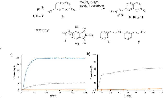
158
(300MHz, CDCl3) δ = 4.09 (q, J = 7.2 Hz, 2 H), 3.77 (t, J = 6 Hz, 2 H), 2.56 (t, J = 6 Hz, 2 H), 2.26 (s, 3 H),
159
1.30 (t, J = 7.2 Hz, 3 H). 13C RMN (75MHz, CDCl3) δ = 154.1, 152.6, 114.2, 70.5, 61.5, 27.6, 15.0, 14.2. IR
160
(neat, cm-1) : 3347, 2930, 1671, 1583, 1379, 1264, 1237, 1018, 1049, 603, 473. HRMS (ESI+) : Calc. for
161
C8H14NO3 [M+H]+ : 172.0974, found : 172.0972.
162
163
2-(5-Ethoxy-2-methyloxazol-4-yl)ethyl methanesulfonate 4. To the alcohol 2-
(5-ethoxy-2-164
methyloxazol-4-yl) ethan-1-ol 3 (300 mg, 1.75 mmol) in dry THF (30 mL) at 0 °C, triethylamine (500
165
L, 3.68 mmol, 2.1 eq) and mesyl chloride (270 L, 3.50 mmol, 2 eq) were successively added dropwise
166
and the resulting mixture was stirred at room temperature for 3 h. Water (30 mL) was added to the
167
solution, and the mixture was further stirred for 15 min. The aqueous phase was extracted with
168
AcOEt (3 × 20 mL), the organic phases were combined and then washed with a solution of saturated
169
aq NaCl (30 mL), dried over MgSO4 and filtered through Celite®. The solvent was evaporated to
170
dryness and the crude product was purified by chromatography on silica gel (AcOEt), the desired
171
product was obtained as a yellow oil (430 mg, 1.72 mmol, 98%). 1H RMN (300MHz, CDCl3) δ = 4.40
172
(t, J = 6.9 Hz, 2 H), 4.13 (q, J = 7.2 Hz, 2 H), 2.96 (s, 3 H), 2.80 (t, J = 6.9 Hz, 2 H), 2.30 (s, 3 H), 1.35 (t, J
173
= 7.2 Hz, 3 H). RMN 13C (75MHz, CDCl3) δ = 154.9, 152.8, 111.0, 70.6, 68.1, 37.4, 25.1, 15.0, 14.2. IR
174
(neat, cm-1) : 2983, 2936, 1673, 1444, 1349, 1267, 1169, 1018, 951, 902, 800, 526. HRMS (ESI+) : calc. for
175
C9H15NO5S [M+H]+ : 250.0749 ; found : 250.0753.
176
177
4-(2-Azidoethyl)-5-ethoxy-2-methyloxazole 5. To a solution of oxazole 2-
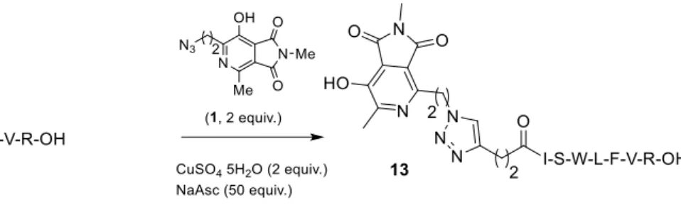
(5-ethoxy-2-methyloxazol-178
4-yl) ethyl methanesulfonate 4 (400 mg, 1.60 mmol) in dry DMF (8 mL), sodium azide (620 mg, 9.60
179
mmol, 6 eq) was added. After 15 min stirring at room temperature, the solution was heated at 50 ° C
180
for 8 h. Then, the mixture was cooled to RT and water (10 mL) was added. The aqueous phase was
181
extracted with AcOEt (2 × 30 mL) and the combined organic phases were washed with a solution of
182
saturated aq NaCl (15 mL), dried over MgSO4, filtered through Celite® and then the solvent was
183
evaporated to dryness. The crude reaction mixture was then purified by chromatography on silica
184
gel in a AcOEt / Cyclohexane elution system (1:5 v/v) to (1:1 v/v), the desired product was obtained
185
as yellow oil (240 mg, 1.22 mmol, 76%). 1H RMN (300MHz, CDCl3) δ = 4.15 (q, J = 7.2 Hz, 2 H), 3.5 (t,
186
J = 6.9 Hz, 2 H), 2.63 (t, J = 6.9 Hz, 2 H), 2.32 (s, 3H), 1.37 (t, J = 7.2 Hz, 3 H). 13C RMN (75MHz, CDCl3)
187
δ = 154.56, 152.60, 112.74, 70.39, 49.83, 24.75, 15.01, 14.28. IR (neat, cm-1) : 2979, 2930, 2094 (N3), 1671,
188
1587, 1379, 1265, 1229, 1171, 1020, 952, 644. HRMS (ESI+) : Calc. for C8H12N4O2 [M+H]+ : 197.1039 ;
189
found : 197.1042.190
191
6-(2-Azidoethyl)-7-hydroxy-2,4-dimethyl-1H-pyrrolo[3,4-c]pyridine-1,3(2H)-dione 1.N-192
methylmaleimide (47 mg, 0.43 mmol, 1.05 eq) was added to a solution of 4- (2-azidoethyl)
-5-ethoxy-193
2-methyloxazole 5 (80 mg, 0.41 mmol) in toluene (1.5 mL), was added. After stirring at room
194
temperature for 15 min and then at 70 ° C for 6 h, the mixture was concentrated under reduced
195
pressure. The residue was dissolved in CDCl3 (1 mL) and then formic acid (10 L, 0.265 mmol, 0.65
196
eq) was added. The mixture was allowed to stir at room temperature overnight. The reaction mixture
197
was concentrated under reduced pressure and the resulting residue was purified by semi-preparative
198
RP-HPLC according to system B to afford the desired product (43 mg, 0.16 mmol, 40%) as a yellow
199
solid. Melting point: 107.3 – 108.5 °C. 1H RMN (300 MHz, MeOD) δ = 3.69 (t, J = 6.9 Hz, 2 H), 3.16 (t, J
200
= 6.9 Hz, 2 H), 3.06 (s, 3 H), 2,67 (s, 3 H) ppm. 13C RMN (75MHz, MeOD) δ = 169.51, 168.58, 157.58,
201
147.41, 146.43, 122.67, 122.15, 50.07, 32.95, 23.88, 19.77 ppm. IR (neat, cm-1) : 3304, 2944, 2100 (N3),
202
1698, 1433, 1382, 1233, 1004, 756, 519. HRMS (API+) : Calc. for C11H12N5O3 [M+H]+ : 262.0940 ; found :
203
262.0946.204
205
7-Hydroxy-2,4-dimethyl-6-(2-(4-(2-oxo-2H-chromen-7-yl)-1H-1,2,3-triazol-1-yl)ethyl)-1H-pyrrolo[3,4-206
c]pyridine-1,3(2H)-dione 9. To a solution of 6- (2-azidoethyl) -7-hydroxy-2,4-dimethyl-1H-pyrrolo
[3,4-207
c] pyridine-1,3 (2H) –dione 1 (13.55 mg, 0.052 mmol) in a solution of H2O/t-BuOH (200 L, 1:1, v/v),
208
were successively added the 7-ethynylcoumarin (8.65 mg, 0.052 mmol, 1 eq)[1] in DCM (100 L),
209
sodium ascorbate (10.3 mg, 0.052 mmol, 1 eq), and copper sulfate pentahydrate CuSO4.5H2O (1.3 mg,
210
0.052 mmol, 1 eq). The reaction mixture was stirred at room temperature in the darkness for 4 h
211
(analytical RP-HPLC control, system A) and then the crude reaction mixture was concentrated under
212
reduced pressure. The resulting residue was triturated in Et2O, then EtOH. The pellet recovered after
213
centrifugation was purified by preparative RP-HPLC according to system C, the desired product (5
214
mg, 0.12 mmol, 23%) was obtained as a yellow solid. Melting point: 179.9 – 181.2. °C. 1H RMN (300
215
MHz, DMSO-d6) δ = 8.72 (s, 1 H), 8.04 (d, J = 9.6 Hz, 1 H), 7.84 - 7.75 (m, 3 H), 6.46 (d, J = 9.6 Hz, 1 H),216
4.85 (t, J = 6.9 Hz, 2 H), 3.49 (t, 2 H), 2.97 (s, 3 H), 2.59 (s, 3 H) ppm. 13C RMN (75MHz, DMSO-d6) δ =217
168.00, 166.46, 159.94, 155.49, 154.08, 145.72, 144.73, 143.92, 134.31, 129.12, 123.17, 121.17, 118.15,218
115.76, 112.13, 47.49, 32.35, 23.56, 19.50 ppm. IR (neat, cm-1) : 2924, 1706, 1681, 1621, 1430, 1223, 1000,219
845, 754, 616, 590, 524, 456. HRMS (ESI+) : Calc. for C22H18N5O5 [M+H]+ : 432.1308 ; found : 432.1302.
220
221
7-(1-Phenethyl-1H-1,2,3-triazol-4-yl)-2H-chromen-2-one 11. To a solution of 2-phenylethyl azide 7
222
(89 mg, 0.52 mmol) in a solution of H2O/t-BuOH (1.5 mL, 1:1, v/v) were successively added 7-ethynyl
223
coumarin (97 mg, 0.52 mmol, 1 eq) in DCM (300 L), sodium ascorbate (104 mg, 0.52 mmol, 1 eq) and
224
copper sulfate pentahydrate CuSO4.5H2O (13 mg, 0.05 mmol, 0.1 eq). The reaction mixture was
225
allowed to stir at room temperature in the darkness for 12 h, then concentrated under reduced. The
226
obtained residue was dissolved in AcOEt (1 mL) and then purified by chromatography on silica gel
227
in a AcOEt / Cyclohexane elution system (3:1, v/v) to (3:0, v/v). The desired product (110 mg, 3.47
228
mmol, 66%) was obtained as a yellow solid. Melting point: 180 – 181.9 °C. 1H RMN (300MHz,
DMSO-229
d6) δ = 8.71 (s, 1 H), 8.04 (d, J = 9.3 Hz, 1 H), 7.79 (t, J = 7.8 Hz, 3 H), 7.25 (t, J = 6.5 Hz, 5 H), 6.46 (d, J =230
9.3 Hz, 1 H), 4.68 (t, J = 6.9 Hz, 2 H), 3.23 (t, J = 6.9 Hz, 2 H) ppm. 13C RMN (75MHz, DMSO-d6) δ =231
159.9, 154.1, 144.8, 143.9, 137.5, 134.2, 129.1, 128.7, 128.4, 126.6, 122.8, 121.1, 118.1, 115.8, 112.0, 50.8,232
35.5 ppm. IR (neat, cm-1) : 3121, 2924, 1702, 1620, 1455, 1222, 1151, 939, 838, 754, 696, 614, 494. HRMS233
(ESI+) : Calc. for C19H16N3O2 [M+H]+ : 318.1243 ; found : 318.1244.
234
Alkyne-ISWLFVR-OH 12. The 2-chlorotrityl chloride resin (1.0 g, 1.60 mmoles/g, 1.60 mmoles)
236
was swollen for 20 min in DCM (15 mL) under nitrogen bubbling. A solution of the amino acid
Fmoc-237
Arg(pbf)-OH (1.04 g, 1.60 mmoles, 2 eq) and DIPEA (1.64 mL, 9.60 mmoles, 6 eq) in DCM (10 mL)
238
was added on the resin, and agitated with nitrogen bubbling for 2.5 h at room temperature. The
239
resulting resin was then treated with MeOH (~ 5 mL) and agitated under nitrogen bubbling for 15
240
min. Then, the resin was washed successively by DCM/MeOH/DIPEA (15 mL, 17:2:1, v/v/v, repeated
241
3 times), DCM (3 × 15 mL), DMF (3 × 15 mL), DCM (3 × 15 mL) and then MeOH (3 × 15 mL). The resin
242
was dried under vacuum and the attachment level of the first residue was determined by UV/VIS
243
(55%, 0.88 mmoles).[34] Fmoc-Arg (pbf) -2-chlorotrityl resin (0.88 mmoles, 1 eq) was swollen for 20
244
min in DCM (15 mL) under nitrogen bubbling and then washed with DMF (3 × 15 mL). Deprotection
245
of the Fmoc group was performed with piperidine 20% in DMF (10 mL) for 1 min with nitrogen
246
bubbling. The resin was filtered and then deprotection was again performed for 10 min. After
247
washing with DMF (3 × 15 mL), subsequent Fmoc-amino acids were coupled in excess (2.64 mmol, 3
248
eq) on the deprotected amino-acid bearing resin with HBTU (2.95 eq) and DIPEA (6 eq) in DMF (~ 5
249
mL) for 1 h with nitrogen bubbling at room temperature. The coupling reactions were performed
250
twice with a filtration of the resin between them. The resin was washed with DMF (3 × 15 mL), and a
251
new cycle of deprotection / coupling was performed. The final H-peptide-resin was successively
252
washed with DMF (3 × 15mL), DCM (3 × 15 mL), MeOH (3 × 15 mL) and then was dried under
253
vacuum. The coupling of the 4-pentynoic acid (87 mg, 0.88 mmoles, 2 eq) was performed on half of
254
the N-term primary amine free ISWLFVR-resin (0.44 mmoles, 1 eq) with HBTU (326 mg, 0.86 mmol,
255
1.95 eq) and DIPEA (300 L, 1.76 mmoles, 4 eq) in DMF (~ 5 mL) for 2 h at room temperature, under
256
nitrogen bubbling. The resulting peptide was cleaved from the resin with a solution of TFA/TIS/H2O
257
(~ 10 mL, 95: 2.5: 2.5, v/v/v) for 2 h at room temperature. The cleavage solution was evaporated and
258
the peptide was precipitated with cold diethyl ether followed by washing by centrifugation 3 times
259
with diethyl ether (9000 rpm, 10 min). The resulting residue was solubilized in water and lyophilized
260
before a purification by semi-preparative RP-HPLC according to system D. The desired product was
261
obtained as a white solid (62 mg, 0.06 mmol, 14%). HRMS (ESI+) : Calc. for C51H74N11O10 [M+H]+ :
262
1000.5620 ; found : 1000.5628. HPLC System A (λ = 254 nm) : tR = 3.66 min.
263
264
Azapthalimide-ISWLFVR-OH 13. To a solution of the peptide-alkyne 12 in DMF (2 mg, 1.25 mL,
265
1.6 mM, 1 eq) was added a solution of azaphthalimide-azide 1 in DMF (2.04 mg, 1.25 mL, 3.2 mM, 2
266
eq). The mixture was then diluted with PBS buffer solution (2.50 mL, 0.1 M, pH = 7.4) and a solution
267
of copper sulfate pentahydrate in MilliQ water (1 mg, 2.50 mL, 1.6 mM, 2 eq) was added. The reaction
268
mixture was stirred at room temperature for 10 min, then a solution of sodium ascorbate in water
269
(19.81 mg, 2.50 mL, 40 mM, 50 eq) was added. The reaction was monitored by analytical RP-HPLC,
270
system A. The crude reaction mixture was then directly purified (without prior treatment) by
271
preparative RP-HPLC according to system E. The desired product (1.2 mg, 0.95 mmol, 38%) was
272
obtained as a yellow solid. HRMS (ESI+) : Calc. for C62H85N16O13 [M+H]+ : 1261.6482 ; found : 1261.6497.
273
HPLC System A (λ = 254 nm) : tR = 3,63 min.
274
275
2.2. Live cells labeling
276
PC12 cells line, derived from a pheochromocytoma of the rat adrenal medulla, are cultured in a
277
humidified incubator at 37 °C in an atmosphere of 5 % CO2. Cells were grown in Dulbecco's modified
278
Eagle's medium (DMEM) (Thermo Fisher Scientific, Illkirch, France) supplemented with 7 % heat‐
279
inactivated fetal bovine serum (Sigma–Aldrich), 7 % horse serum (Lonza Bioscience, Walkersville,
280
MD, USA), 2.5 % HEPES (4‐(2‐hydroxyethyl)‐1‐piperazine ethanesulfonic acid) (Thermo Fisher
281
Scientific), 1 % glutamine (Thermo Fisher Scientific), 100 units/mL penicillin and 100 μg mL−1
282
streptomycin (Thermo Fisher Scientific).
283
284
PC12 cells were plated on glass bottom dishes (MatTek Corporation, MA, USA) for staining and
285
confocal microscopy observation (SP8). After medium removal and PBS rince, cells were incubated
or not (“control” and “cocktail alone” conditions) for 30 min at 37°C in an atmosphere of 5 % CO2
287
with a 50 µM Alkyne 1-(4-pentyn-1-yl)-1H-pyrrole-2,5-dione 14 solution in PBS 0.1 M pH 7.4 (10 mL,
288
containing 0.05% acetonitrile) prepared from a 5 mM alkyne 14 in acetonitrile/milliQ water 5:95 (1
289
mL stock solution). “Cocktail” solution with azide 1 (50 mM), copper sulfate pentahydrate (50 mM),
290
sodium ascorbate (1.25 mM), prepared 45 min before incubation (azide 1 stock solution 10 mM in
291
DMSO/water 10:90 (500 mL) was added the copper sulfate pentahydrate stock solution 20 mM in
292
water (250 mL). After 30 min of incubation at 20 °C, the sodium ascorbate stock solution 0.5 M in
293
water (250 mL) was added, and the mixture was further incubated for 15 min at 20 °C). After 2 washes
294
with PBS, cells were incubated during 30 min at 37 °C in an atmosphere of 5 % CO2 with the “cocktail”
295
solution implemented with 1 µM of the non-ionic surfactant polyol Pluronic F-127 (Thermo Fisher
296
Scientific). Cells were washed one time with PBS before to be visualized under microscope.
297
298
Images (1024x1024 pixels) were acquired by using an upright fixed-stage Leica TCS SP8 confocal
299
microscope (Leica Microsystems, Nanterre, France) equipped with diode laser at 405 nm to excite
300
fluorophore and a conventional scanner at 400Hz. Using a 25× objective (NA 0.95, water immersion)
301
fluorescence emission was detected through a hybrid detector (HyD) in photon counting mode with
302
a specific band from 500 to 550 nm. Mosaic image (square of 5 by 5 images at 1024x1024 pixels) was
303
performed to obtain a large view and a merging was realized after acquisition through module LAS
304
X Navigator. For image acquisitions, focuses on cells were performed in bright-field mode before
305
fluorescence acquisition. For the fluorescence quantification, all values are expressed as fluorescence
306
intensity (A.U.) means of at least 100 cells +/- sem . Statistical analysis was performed using the
307
GraphPad Prism 4 software (GraphPad Software Inc., San Diego, CA) and a one-way Analysis of
308
Variance (ANOVA) with a Tukey-Kramer multiple comparisons tests
309
3. Results and Discussion
310
3.1. Synthesis of the fluorophore-based chelating azide
311
The fluorescent chelating agent 1 was prepared from commercial L-aspartic acid diethyl ester
312
hydrochloride (Scheme 1). First, the reported oxazole ester 2 obtained in two steps,[33] was reduced
313
into the corresponding alcohol derivative 3 in 46% isolated yield. The hydroxyl group was then
314
converted into mesylate intermediate 4, which was subsequently displaced with azide ion to afford
315
compound 5 in 74% over two steps. Finally, the oxazole 5 underwent a [4+2]
316
cycloaddition/aromatization process with N-methylmaleimide to furnish the corresponding
317
azaphthalimide 1 in 40% isolated yield. Importantly, this compound proved to be relatively soluble
318
in PBS pH 7.4 as the only solvent, up to 4 mM (Scheme 1a and b). Interestingly, LogS value of
319
azaphthalimide 1 was found to be -1.58, which is in the range of those calculated for water-soluble
320
tris(triazolylmethyl)amine THPTA and BTTAA (Table S8).
321
322
323
Scheme 1. a) Synthetic access to the azaphthalimide 1. b) Photographs of 1 (1 mg/mL PBS 0.1 M pH
324
7.4) taken under visible light (1) or under 365 nm light (2).
3.1. Comparative study with chelating and non-chelating azide
326
The azaphthalimide 1 in hand, its ability to accelerate 1,3-dipolar cycloadditions was compared
327
to a reported effective but not fluorescent pyridine-based chelating azide, 2-(2-azidoethyl)pyridine 6,
328
[25] as well as a non-chelating derivative, 2-phenylethyl azide 7. We chose these three azide-based
329
reagents, since in all of them, the azide function is separated from the aromatic ring by a two
carbon-330
atom alkyl chain length in order to form a 6-membered metallocycle system (for chelating azides 1
331
and 7 only). The fluorogenic 7-ethynyl coumarin 8 whose fluorescence increases upon reaction with
332
azides,[25] was chosen as the click partner in order to monitor the click reactions progress by
333
fluorescence spectroscopy. For compound 6 and 7, an excitation and emission wavelength of 320 nm
334
and 400 nm was selected, corresponding to the absorption and emission of the triazole-substituted
335
coumarin (Figures S2-S3). With regards to azaphthalimide azide 1, the coumarin and azaphthalimide
336
scaffold display complementary photophysical properties to constitute a suitable FRET pair with an
337
excitation at the coumarin wavelength (λex = 320 nm), and an emission at the azaphthalimide emission
338
wavelength (λem = 550 nm, Figure S1). For each triazole product, a calibration curve was performed
339
to convert fluorescence intensity signal into product conversion (Figures S4-S6). Of note, the presence
340
of copper(I) did not significantly modify the fluorescence emission of either triazole products 9-11.
341
First, azide 1, 6 and 11 were reacted with coumarin 8, and CuSO4.5 H2O (17.5 M each), followed
342
by the addition of sodium ascorbate(0.437 mM, 25 equiv.) in aqueous medium (Figure 3a).
343
344
345
Figure 3. Comparison of kinetics of CuAAC reactions of: a) 7-ethynyl coumarin (17.5 M), with azide
346
(1 in grey, 6 in blue, and 7 in orange) in the presence of CuSO4.5H20 (17.5 M) and sodium ascorbate
347
(437 M) in DMF:PBS pH 7.4:H2O (1:1:2); b) 7-ethynyl coumarin (175 M), with azide (1 in grey, and
348
7 in orange) in the presence of CuSO4.5H20 (175 M) and sodium ascorbate (4.375 M) in DMF:PBS
349
pH 7.4:H2O (1:1:2).
350
First, the 2-(2-azidoethyl)-pyridine 6 led to the rapid formation of the triazole product (Figure
351
3a, curve in blue), while on the other hand, the non-chelating azide 7 gave no detectable product,
352
even after 100 min of reaction (curve in orange). Meanwhile, the azaphthalimide enables the
353
formation of 20% of triazole. Although this conversion is significantly lower than that of 6, this result
354
showed that the azapthalimide was able to accelerate the reaction presumably by acting as a bidentate
355
ligand for CuAAC reactions. This chelating ability has been further validated with reactions carried
out at 175 M, and monitored by HPLC (Figure 2b, grey curve). In fact, a plateau at c.a. 60%
357
conversion yield was quickly reached while almost no product was obtained with the non-chelating
358
compound (orange curve). The reaction performed with the azaphthalimide was approximatively
359
100 times faster than the one achieved with the phenyl scaffold. Importantly, the presence of the
360
copper complex did not impact the absorption and emission wavelengths of either fluorophores
361
(Figure S1).
362
3.1. Applications
363
The potency of a small fluorophore-assisted click chemistry for minimal conjugate
364
perturbation was illustrated through the fluorescent labeling of a heptapeptide-based short
365
biomolecule (Scheme 2). The starting peptide was prepared according to standard solid-phase
366
peptide synthesis protocols with the incorporation of the pentynoic acid at the N-terminus (see
367
Materials and Methods).
368
369
Scheme 2. Labeling of alkyne-oligopeptide 12 with azide 1.
370
The reaction was monitored by HPLC and showed a clean and complete reaction within 30 min
371
(Figure 3a). The isolated conjugate 13 obtained in 38% isolated yield after RP-HPLC and
372
lyophilization was subject to photophysical measurements. Absorption and emission spectra
373
recorded either in the presence or absence of copper(I) (generated in situ from CuSO4.5H2O and
374
sodium ascorbate), showed no change in position of peak maxima, which were found at λabs ~ 416 nm
375
and λem ~ 525 nm, respectively (Figure 3b). Only a widening of the absorption peak was observed.
376
Besides, quantum yield for the green fluorescence-labeled peptide was found to be around 8% in the
377
presence and absence of in situ generated copper(I) determined in PBS pH 7.4 at 25 °C. This value is
378
in line with those reported for similar azaphthalimide derivatives.[29]
379
380
381
382
Figure 3. Green-fluororescence labeling of peptide 12. a) RP-HPLC chromatogram of peptide-alkyne
383
12 (top) and labeled peptide 13 after 30 min reaction (bottom). b) Normalized absorption (solid line)
384
and emission (dashed line) spectra of azaphthalimide-containing peptide 13 (in black) in the presence
385
of in situ generated copper(I) (grey line) in PBS pH 7.4 at 20 °C.
386
After establishing that small conjugates could be readily labeled in vitro using an heptapeptide
387
as model, then, the labeling of alkyne-modified live cells with azaphthalimide azide 1 was
388
investigated. As a proof-of-concept study, the alkyne reporter group was first chemically
389
incorporated into live cells by using a maleimide derivative 14 (50 M in PBS for 30 min at 37 °C), a
390
functional group known to react rapidly and covalently with biological thiols through a Michael
391
addition process.[35] Cells were washed twice with PBS to remove unreacted alkyne-maleimide 14
392
in the supernatant, then a 50 M solution of azaphthalimide-based chelating azide 1 in PBS pretreated
393
with a 50 M solution of in situ generated copper(I) (cocktail solution), was incubated for 15 min with
394
cells at 37 °C, then rinsed with PBS. Subsequent confocal analysis microscopy revealed a strong
395
fluorescence labeling of cells at 500-550 nm (Figure 4), highly statistically significant compared to
396
control conditions (fluorescence poorly detectable in cells with only PBS incubation (ctrl) and
397
“cocktail alone” when cells that were not incubated with the alkyne reporter group, thus
398
demonstrating successful CuAAC mediated ligation of azaphthalimide azide and alkyne
399
incorporated into cells, data not shown). Images revealed some discernible vesicles that could be
400
identify as lipid droplets, in which the lipophilic alkyne reporter presumably accumulates (Figures
401
4A,B, S7).[36]
402
403
Figure 4. A, B. PC12 cells in « Cocktail+Alkyne » conditions, acquired under mozaic confocal image
404
for cell population observation (A) and single image for cellular observation (B). C. Fluorescence
405
intensity graph showing high significance (***p<0.001) of the fluorescence intensity level after
406
« Cocktail+Alkyne » incubation versus « cocktail alone » and PBS incubation (ctrl). D. Chemical
407
structure of alkynyl maleimide 14.
4. Conclusions
409
Herein, we disclosed an unprecedented “all-in-one” fluorophore-based chelating azide with an
410
exceptionally low molecular weight (260 Da), which proved useful for the fluorescent labeling of
411
small biomolecules, and biological systems. In this approach, the azaphthalimide played the role of
412
both the copper ligand and fluorophore in order to accelerate the CuAAC reaction with concomitant
413
installation of a fluorescent tag. Importantly, the fluorescence properties of the native fluorophore
414
were conserved upon its complexation to cooper(I). This process advantageously does not require
415
neither an external ligand, a fluorophore, nor water solubilizing group which complicate the
416
synthesis and increase the risk of impacting the mobility of azides and properties of conjugated
417
molecules. This strategy could be extended to other families of nitrogen-containing fluorophores in
418
order to further improve the ligation rates or to red-shift the fluorescence emission.[37,38]
419
Supplementary Materials: The following are available online at www.mdpi.com/xxx/s1, 1H and 13C NMR
420
spectra for all new compounds; absorption, emission, and excitation spectra for compounds 9-11; additional
421
information regarding live cells labeling.
422
Acknowledgments: This work was partially supported by the Centre National de la Recherche Scientifique
423
(CNRS), INSA Rouen, Normandie Rouen University, and the Labex SynOrg (ANR-11-LABX-0029). We also
424
thank Albert Marcual (CNRS) for HRMS analyses.
425
Conflicts of Interest: The authors declare no conflict of interest.
426
References
427
428
1. Rostovtsev, V. V.; Green, L. G.; Fokin, V. V.; Sharpless, K. B. A Stepwise Huisgen Cycloaddition Process:
429
Copper(I)-Catalyzed Regioselective “Ligation” of Azides and Terminal Alkynes. Angew. Chem., Int. Ed.
430
2002, 41, 2596.
431
2. Tornøe, C. W.; Christensen, C.; Meldal, M. Peptidotriazoles on Solid Phase: [1,2,3]-Triazoles by
432
Regiospecific Copper(I)-Catalyzed 1,3-Dipolar Cycloadditions of Terminal Alkynes to Azides. J . Org.
433
Chem. 2002, 6, 3057.
434
3. Bruyat, P.; Gautier, A.; Jean, L.; Renard, P.-Y. Use of an Air-Stable Cu(I)-NHC Catalyst for the Synthesis of
435
Peptidotriazoles. J. Org. Chem. 2018, 83, 13515.
436
4. Farzan, V. M.; Ulashchik, E. A.; Martynenko-Makaev, Y. V.; Kvach, M. V.; Aparin, I. O.; Brylev, V. A.;
437
Prikazchikova, T. A.; Maklakova, S. Y.; Majouga, A. G.; Ustinov, A. V.et al. Automated Solid-Phase Click
438
Synthesis of Oligonucleotide Conjugates: From Small Molecules to Diverse N-Acetylgalactosamine
439
Clusters. Bioconjugate Chem. 2017, 28, 2599.
440
5. Simon, C.; Lion, C.; Spriet, C.; Baldacci-Cresp, F.; Hawkins, S.; Biot, C. One, Two, Three: A Bioorthogonal
441
Triple Labelling Strategy for Studying the Dynamics of Plant Cell Wall Formation In Vivo. Angew. Chem.,
442
Int. Ed. 2018, 57, 16665.
443
6. Jiang, H.; English, B. P.; Hazan, R. B.; Wu, P.; Ovryn, B. Tracking Surface Glycans on Live Cancer Cells with
444
Single-Molecule Sensitivity. Angew. Chem., Int. Ed. 2015, 54 (6), 1765.
445
7. Mas Pons, J.; Dumont, A.; Sautejeau, G.; Fugier, E.; Baron, A.; Dukan, S.; Vauzeilles, B. Identification of
446
Living Legionella pneumophila Using Species-Specific Metabolic Lipopolysaccharide Labeling. Angew.
447
Chem., Int. Ed.2014, 53, 1275.
448
8. Yuan, Y.; Xu, S.; Cheng, X.; Cai, X.; Liu, B. Bioorthogonal Turn-On Probe Based on Aggregation-Induced
449
Emission Characteristics for Cancer Cell Imaging and Ablation. Angew. Chem., Int. Ed. 2016, 55, 6457.
450
9. Haldón, E.; Nicasio, M. C.; Pérez, P. J. Copper-catalysed azide–alkyne cycloadditions (CuAAC): an update.
451
Org. Biomol. Chem. 2015, 13, 9528.
452
10. Li, L.; Zhang, Z. Development and Applications of the Copper-Catalyzed Azide-Alkyne Cycloaddition
453
(CuAAC) as a Bioorthogonal Reaction. Molecules 2016, 21, 1393.
454
11. Kenry; Liu, B. Bio-orthogonal Click Chemistry for in vivo Bioimaging. Trends Chem. 2019, 1, 763.
455
12. Yang, M.; Li, J.; Chen, P. R. Transition metal-mediated bioorthogonal protein chemistry in living cells.
456
Chem. Soc. Rev. 2014, 43, 6511.
457
13. Agard, N. J.; Prescher, J. A.; Bertozzi, C. R. A Strain-Promoted [3 + 2] Azide−Alkyne Cycloaddition for
458
Covalent Modification of Biomolecules in Living Systems. J. Am. Chem. Soc. 2004, 126, 15046.
14. Blackman, M. L.; Royzen, M.; Fox, J. M. Tetrazine Ligation: Fast Bioconjugation Based on
Inverse-Electron-460
Demand Diels−Alder Reactivity. J. Am. Chem. Soc. 2008, 130, 13518.
461
15. Devaraj, N. K.; Weissleder, R.; Hilderbrand, S. A. Tetrazine-Based Cycloadditions: Application to
462
Pretargeted Live Cell Imaging. Bioconjugate Chem. 2008, 19, 2297.
463
16. Kyung Jin, L.; lt; sup; gt; lt; sup; gt; Deokhee, K.; lt; supet al. Site-Specific Labeling of Proteins Using
464
Unnatural Amino Acids. Mol. Cells 2019, 42 (5), 386.
465
17. Hong, V.; Presolski, S. I.; Ma, C.; Finn, M. G. Analysis and Optimization of Copper-Catalyzed Azide–
466
Alkyne Cycloaddition for Bioconjugation. Angew. Chem., Int. Ed. 2009, 48, 9879.
467
18. Besanceney-Webler, C.; Jiang, H.; Zheng, T.; Feng, L.; Soriano del Amo, D.; Wang, W.; Klivansky, L. M.;
468
Marlow, F. L.; Liu, Y.; Wu, P. Increasing the Efficacy of Bioorthogonal Click Reactions for Bioconjugation:
469
A Comparative Study. Angew. Chem., Int. Ed. 2011, 50, 8051.
470
19. Rodionov, V. O.; Presolski, S. I.; Gardinier, S.; Lim, Y.-H.; Finn, M. G. Benzimidazole and Related Ligands
471
for Cu-Catalyzed Azide−Alkyne Cycloaddition. J. Am. Chem. Soc. 2007, 129, 12696.
472
20. Jones, L. H.; Beal, D.; Selby, M. D.; Everson, O.; Burslem, G. M.; Dodd, P.; Millbank, J.; Tran, T.-D.;
473
Wakenhut, F.; Graham, E. J. S.et al. In-cell click labelling of small molecules to determine subcellular
474
localisation. J. Chem. Biol. 2011, 4, 49.
475
21. Li, S.; Wang, L.; Yu, F.; Zhu, Z.; Shobaki, D.; Chen, H.; Wang, M.; Wang, J.; Qin, G.; Erasquin, U. J.et al.
476
Copper-catalyzed click reaction on/in live cells. Chem. Sci. 2017, 8, 2107.
477
22. Kuang, G.-C.; Guha, P. M.; Brotherton, W. S.; Simmons, J. T.; Stankee, L. A.; Nguyen, B. T.; Clark, R. J.; Zhu,
478
L. Experimental Investigation on the Mechanism of Chelation-Assisted, Copper(II) Acetate-Accelerated
479
Azide–Alkyne Cycloaddition. J. Am. Chem. Soc. 2011, 133, 13984.
480
23. Brotherton, W. S.; Michaels, H. A.; Simmons, J. T.; Clark, R. J.; Dalal, N. S.; Zhu, L. Apparent
Copper(II)-481
Accelerated Azide−Alkyne Cycloaddition. Org. Lett. 2009, 11, 4954.
482
24. Uttamapinant, C.; Tangpeerachaikul, A.; Grecian, S.; Clarke, S.; Singh, U.; Slade, P.; Gee, K. R.; Ting, A. Y.
483
Fast, Cell-Compatible Click Chemistry with Copper-Chelating Azides for Biomolecular Labeling. Angew.
484
Chem., Int. Ed. 2012, 51, 5852.
485
25. Bevilacqua, V.; King, M.; Chaumontet, M.; Nothisen, M.; Gabillet, S.; Buisson, D.; Puente, C.; Wagner, A.;
486
Taran, F. Copper-Chelating Azides for Efficient Click Conjugation Reactions in Complex Media. Angew.
487
Chem., Int. Ed. 2014, 53, 5872.
488
26. Sallustrau, A.; Bregant, S.; Chollet, C.; Audisio, D.; Taran, F. Scalable and practical synthesis of clickable
489
Cu-chelating azides. Chem. Commun. 2017, 53, 7890.
490
27. Birch, D.; Christensen, M. V.; Staerk, D.; Franzyk, H.; Nielsen, H. M. Fluorophore labeling of a
cell-491
penetrating peptide induces differential effects on its cellular distribution and affects cell viability. BBA -
492
Biomembranes 2017, 1859, 2483.
493
28. Jouanno, L. A.; Chevalier, A.; Sekkat, N.; Perzo, N.; Castel, H.; Romieu, A.; Lange, N.; Sabot, C.; Renard, P.
494
Y. Kondrat'eva ligation: Diels-Alder-based irreversible reaction for bioconjugation. J. Org. Chem. 2014, 79,
495
10353.
496
29. Renault, K.; Jouanno, L.-A.; Lizzul-Jurse, A.; Renard, P.-Y.; Sabot, C. Fluorogenic Behaviour of the
Hetero-497
Diels–Alder Ligation of 5-Alkoxyoxazoles with Maleimides and their Applications. Chem. Eur. J. 2016, 22,
498
18522.
499
30. Stewart, W. W. Synthesis of 3,6-disulfonated 4-aminonaphthalimides. J. Am. Chem. Soc. 1981, 103, 7615.
500
31. Li, M.; Dong, K.; Zheng, Y.; Song, W. Copper-catalyzed cascade click/nucleophilic substitution reaction to
501
access fully substituted triazolyl-organosulfurs. Org. Biomol. Chem. 2019, 17, 9933.
502
32. Nunes, J. P. M.; Morais, M.; Vassileva, V.; Robinson, E.; Rajkumar, V. S.; Smith, M. E. B.; Pedley, R. B.;
503
Caddick, S.; Baker, J. R.; Chudasama, V. Functional native disulfide bridging enables delivery of a potent,
504
stable and targeted antibody–drug conjugate (ADC). Chem. Commun. 2015, 51, 10624.
505
33. Jouanno, L. A.; Sabot, C.; Renard, P. Y. Expeditious microwave-assisted synthesis of 5-alkoxyoxazoles from
506
alpha-triflyloxy esters and nitriles. J. Org. Chem. 2012, 77, 8549.
507
34. Gude, M.; Ryf, J.; White, P. D. An accurate method for the quantitation of Fmoc-derivatized solid phase
508
supports. J. Pept. Sci. 2002, 9, 203.
509
35. Renault, K.; Fredy, J. W.; Renard, P.-Y.; Sabot, C. Covalent Modification of Biomolecules through
510
Maleimide-Based Labeling Strategies. Bioconjugate Chem. 2018, 29, 2497.
511
36. Cohen, S. Lipid Droplets as Organelles. Int Rev Cell Mol Biol. 2018 ; 337, 83.
37. Atkins, R. L.; Bliss, D. E. Substituted coumarins and azacoumarins. Synthesis and fluorescent properties. J.
513
Org. Chem. 1978, 43, 1975.
514
38. Takano, H.; Narumi, T.; Nomura, W.; Tamamura, H. Microwave-Assisted Synthesis of Azacoumarin
515
Fluorophores and the Fluorescence Characterization. J. Org. Chem. 2017, 82, 2739.
516
517