HAL Id: hal-01285650
https://hal.sorbonne-universite.fr/hal-01285650
Submitted on 9 Mar 2016
HAL is a multi-disciplinary open access
archive for the deposit and dissemination of
sci-entific research documents, whether they are
pub-lished or not. The documents may come from
teaching and research institutions in France or
abroad, or from public or private research centers.
L’archive ouverte pluridisciplinaire HAL, est
destinée au dépôt et à la diffusion de documents
scientifiques de niveau recherche, publiés ou non,
émanant des établissements d’enseignement et de
recherche français ou étrangers, des laboratoires
publics ou privés.
Distributed under a Creative Commons Attribution - NonCommercial| 4.0 International
License
Tuning Eye-Gaze Perception by Transitory STS
Inhibition
Ana Saitovitch, Traian Popa, Hervé Lemaitre, Elza Rechtman, Jean-Charles
Lamy, David Grévent, Raphael Calmon, Sabine Meunier, Francis Brunelle,
Yves Samson, et al.
To cite this version:
Ana Saitovitch, Traian Popa, Hervé Lemaitre, Elza Rechtman, Jean-Charles Lamy, et al.. Tuning
Eye-Gaze Perception by Transitory STS Inhibition. Cerebral Cortex, Oxford University Press (OUP),
2016, �10.1093/cercor/bhw045�. �hal-01285650�
O R I G I N A L A R T I C L E
Tuning Eye-Gaze Perception by Transitory STS
Inhibition
Ana Saitovitch
1
, Traian Popa
3
, Hervé Lemaitre
1,2
, Elza Rechtman
1
,
Jean-Charles Lamy
3
, David Grévent
1
, Raphael Calmon
1
, Sabine Meunier
3
,
Francis Brunelle
1
, Yves Samson
4
, Nathalie Boddaert
1
and Monica Zilbovicius
1
1
INSERM U1000, Department of Pediatric Radiology, Hôpital Necker Enfants Malades, AP-HP, University René
Descartes, PRES Sorbonne Paris Cité, UMR 1163, Institut Imagine, Paris, France,
2Faculté de Médecine, Université
Paris-Sud, Paris, France,
3Inserm U1127, CNRS UMR 7225, Sorbonne Universités, UPMC Univ. Paris 06, UMR S 1127,
Institut du Cerveau et de la Moelle épinière, ICM, Centre de Neuro-imagerie de Recherche, CENIR, Paris, France and
4
Stroke Center, Groupe Hospitalier Pitié-Salpêtrière, AP-HP, Université Pierre et Marie Curie, Paris, France
Address correspondence to Ana Saitovitch, Service de Radiologie Pédiatrique, Hôpital Necker, 149 rue de Sèvres 75015 Paris, France. Email: a.saitovitch@gmail.com
Abstract
Processing eye-gaze information is a key step to human social interaction. Neuroimaging studies have shown that superior temporal sulcus (STS) is highly implicated in eye-gaze perception. In autism, a lack of preference for the eyes, as well as anatomo-functional abnormalities within the STS, has been described. To date, there are no experimental data in humans showing whether it is possible to interfere with gaze processing by modulating STS neural activity. Here, we measured eye-gaze perception before and after inhibitory transcranial magnetic stimulation (TMS) applied over the posterior STS ( pSTS) in young healthy volunteers. Eye-gaze processing, namely overt orienting toward the eyes, was measured using eye tracking during passive visualization of social movies. Inhibition of the right pSTS led participants to look less to the eyes of characters during visualization of social movies. Such effect was specific for the eyes and was not observed after inhibition of the left pSTS nor after placebo TMS. These results indicate for thefirst time that interfering with the right pSTS neural activity transitorily disrupts the behavior of orienting toward the eyes and thus indirectly gaze perception, a fundamental process for human social cognition. These results could open up new perspectives in therapeutic interventions in autism.
Key words: eye-gaze perception, social cognition, STS, TMS
Introduction
The processing of eye-gaze information is a key step to engage in social interactions. This ability is characteristic of humans and nonhuman primates living in complex social environments (Adolphs 2003). Eye contact helps infer the intentions and
feelings of the conspecifics, which is crucial for survival and so-cial integration (Klein et al. 2009). Spontaneous perception and acute analysis of the eye movement is essential for maintaining optimal social relationships throughout the primate lifespan. Moreover, the preference for the eyes as a privileged attention target is evident extremely early in the normal development,
© The Author 2016. Published by Oxford University Press.
This is an Open Access article distributed under the terms of the Creative Commons Attribution Non-Commercial License (http://creativecommons.org/ licenses/by-nc/4.0/), which permits non-commercial re-use, distribution, and reproduction in any medium, provided the original work is properly cited. For commercial re-use, please contact journals.permissions@oup.com
Cerebral Cortex, 2016, 1–9 doi: 10.1093/cercor/bhw045 Original Article 1 at UPMC on March 9, 2016 http://cercor.oxfordjournals.org/ Downloaded from
suggesting that this preference is a core mechanism for the subsequent development of a larger expertise of human social cognition (Frith and Frith 2012). As the most reliable cue to un-derstanding what another person is thinking, feeling or in-tending, the eyes become, in fact, a“window to the soul.” Interestingly, in developmental disorders, such as autism spectrum disorders (ASD), this behavior seems to be disrupted and deficits in eye contact are a hallmark of autism (Jones and Klin 2013). Indeed, in both adults and children with ASD, a lack of preference for the eyes has been demonstrated (Klin et al. 2002;Jones et al. 2008), which may account for their difficulties in social interactions.
A substantial body of evidence has emphasized the import-ance of the superior temporal sulcus (STS) in gaze perception. In monkeys, single unit recording studies have consistently indi-cated the involvement of the posterior STS ( pSTS;Perrett et al. 1985). In humans, neuroimaging studies have described STS acti-vation, mainly within the right STS, in the processing of eye gaze (Pelphrey, Morris, Michelich, et al. 2005;Hadjikhani et al. 2008;
Sato et al. 2008;Nummenmaa et al. 2010;Carlin and Calder 2013). Indeed, several functional MRI studies have revealed a well-defined anatomical region within the pSTS consistently plicated in gaze perception. Furthermore, a number of brain im-aging studies have reported the presence of anatomical and functional abnormalities within the pSTS in children and in adults with ASD (Boddaert et al. 2004;Pelphrey, Morris, McCarthy 2005;Zilbovicius et al. 2006;Duchesnay et al. 2011;Philip et al. 2012).
To date, no experimental data exist for humans that demon-strate whether gaze processing can be altered by an artificial modulation of the STS neural network. We hypothesize that a transitory inhibition of the right pSTS would selectively inter-fere with eye-gaze processing. To test this hypothesis, we exam-ined eye-tracking recordings from young healthy volunteers during passive visualization of naturalistic social movies, with-out any specific task performance. To provide an ecological and naturalistic setting, we have used short movie fragments
ex-tracted from commercial films. We aimed to measure the
changes in gaze perception, namely overt orienting toward the eyes, induced by an inhibitory theta-burst transcranial magnetic stimulation (TMS) applied to the right pSTS.
Materials and Methods
Participants
Thirty healthy volunteers, divided into 2 groups, participated in thefirst phase of the study. Group 1 was composed of 16 partici-pants who underwent the Sham–Actual Inhibition protocol applied to the right pSTS (5 women; 22.1 ± 2.5 years); Group 2 was composed of 14 participants who underwent the Sham– Sham protocol applied to the right pSTS (3 women; 23.1 ± 3.1 years). After analysis comparing these 2 groups, additional data were collected to form a third group (Group 3). Group 3 was com-posed of 14 participants who underwent the Sham–Actual Inhib-ition protocol applied to the left pSTS (3 women; 24.1 ± 3.4 years). There was no significant age difference between the 3 groups (F2,41
= 1.67; P = 0.20). All participants were right-handed, had normal or corrected-to-normal sight, were free of psychiatric, neurological, and general health problems, and presented no contraindication for the TMS. All participants provided written informed consent in accordance with the Ethical Committee at the Saint-Louis Hos-pital, Paris, France, and the participants were monetarily compen-sated for their participation in this study.
Experimental Design
The study was initially based on 2 different experimental sets: 1) Sham–Actual Inhibition protocol applied to right pSTS (Group 1; Fig. 1A); 2) Sham–Sham protocol applied to the right pSTS (Group 2; Fig.1B). In Group 1, actual inhibition was delivered to
the right pSTS with a continuous theta-burst stimulation (cTBS). This type of TMS has effects that largely outlast (by at least 30 min) the stimulation duration (40 s), which allows the experimental measures to be conducted separately from the stimulation itself (Huang et al. 2005). However, this long-lasting effect did not allow for a classical crossover randomized ex-perimental design. Therefore, a Sham–Sham protocol, in which actual STS inhibition was replaced by a second sham, was de-signed and applied to Group 2. Finally, to verify the specificity of thefindings for the target area in the right pSTS, subsequently to analysis comparing Groups 1 and 2, additional data were col-lected in a new experimental set: Sham–Actual Inhibition proto-col applied to left pSTS (Group 3; Fig. 1C) in which actual inhibition was delivered to the left pSTS. All subjects were blinded to the protocol type and to the intervention. In addition, after the experiment subjects were debriefed and did not mani-fest knowledge regarding sham or active condition.
Before the TMS session, an anatomical 3DT1 MRI scan was acquired for all participants. Based on 4 fMRI studies on gaze per-ception (Pelphrey, Morris, Michelich, et al. 2005;Hadjikhani et al. 2008;Sato et al. 2008;Nummenmaa et al. 2010), the precise target location within the pSTS was identified on the individual scans. A baseline eye-tracking measure was performed prior to the TMS procedures. The participants underwent thefirst intervention (sham) and eye-tracking measures were recorded at 1, 6, and 12 min after the intervention. The participants receiving the Sham–Actual Inhibition protocol underwent the second inter-vention in the form of an actual inhibition (Fig.1A,C).
Eye-track-ing measures were recorded at 1, 6, and 12 min after the inhibition. The participants receiving the Sham–Sham protocol underwent the second intervention in the form of a sham (Fig. 1B). Eye-tracking measures were recorded at 1, 6, and 12 min after the sham intervention. The entire session lasted approximately 90 min.
Structural MRI
All participants underwent a 3D T1-weighted FSPGR sequence
(TR/TE/TI/NEX: 10.5/2.2/600/1,flip angle 10°, and matrix size 256 × 192, yielding 124 axial slices at a thickness of 1.2 mm and a 22 cmfield of view) acquired with a 1.5 T (Signa General Electric) scanner at the Necker Hospital, Paris, France.
Targeting the pSTS
The cortical 3D representation of each individual was recon-structed from the previously acquired structural MRI using the eXimia software (Nextim Ltd, Helsinki, Finland). The target was visually identified on the 3D reconstruction by an experi-mented neuro-radiologist based on the mean Talairach coordi-nates (x = 50, y =−53, z = 15) provided by 4 fMRI studies on the gaze perception, mainly on passive viewing of eye movements (Pelphrey, Morris, Michelich, et al. 2005;Hadjikhani et al. 2008;
Sato et al. 2008;Nummenmaa et al. 2010). The frameless stereo-taxic neuronavigation system localizes the coil placement and the orientation with an accuracy below 2 mm, which allows a maximum mismatch of 4 mm between the TMS hotspot and the MRI target (Neggers et al. 2004). The spatial resolution of 2 | Cerebral Cortex
at UPMC on March 9, 2016
http://cercor.oxfordjournals.org/
Figure 1. Experimental design: (A) Group 1: Sham–Actual Inhibition protocol applied to the right pSTS. The white square on the beginning of the time line indicates the baseline eye-tracking measure. The yellow squares over time indicate each eye-tracking measure performed after the sham (1, 6, and 12 min); the green squares over time indicate each eye-tracking measure performed after the inhibitory TMS administered over the right pSTS (1, 6, and 12 min). (B) Group 2: Sham–Sham protocol applied to the right pSTS. The white square on the beginning of the time line indicates the baseline eye-tracking measure. The yellow squares over time indicate each eye-tracking measure performed after thefirst sham (1, 6, and 12 min) and after the second sham (1, 6, and 12 min) administered over the right pSTS. (C) Group 3: Sham–Actual Inhibition protocol applied to the left pSTS. The white square on the beginning of the time line indicates the baseline eye-tracking measure. The yellow squares over time indicate each eye-tracking measure performed after the sham (1, 6, and 12 min); the green squares over time indicate each eye-tracking measure performed after the inhibitory TMS administered over the left pSTS (1, 6, and 12 min).
at UPMC on March 9, 2016
http://cercor.oxfordjournals.org/
the magnetic pulse cone being of approximately 1–2 cm assures that the induced electricalfield encompasses the target region. Actual Inhibition
The repetitive TMS was delivered with a continuous theta-burst pattern (cTBS) to inhibit the right or left pSTS. Continuous TBS is assumed to activate preferentially an inhibitory cascade of events, as demonstrated on the primary motor cortex. Such a stimulation paradigm would induce a depression in the excitabil-ity of pyramidal neurons within the target area, reversibly redu-cing the cortical excitability for at least 30 min and up to 1 h (Huang et al. 2005). The cTBS inhibition was delivered with a SuperRapid2 (Magstim Co., Whitland, UK) via a figure-eight cooled coil with a wing diameter of 70 mm. The coil was held in such a way as the handle (and the inducedfield) would be per-pendicular to the targeted. The cTBS consisted of trains of 3 mag-netic pulse separated by 20 ms (50 Hz); each train was repeated every 200 ms (5 Hz) until the total number of pulses (n = 600) was delivered (total stimulation time: 40 s). In this protocol, the cTBS was delivered with an intensity of 90% of the active motor threshold (AMT) of thefirst interosseous right hand muscle. The AMT was defined as the lowest intensity of a single magnetic pulse delivered over the primary motor cortex (M1), which pro-duced a motor evoked potential >0.2 mV in at least 5 of the 10 trials when the subject exerted a 10% maximum voluntary target muscle contraction using visual feedback (Rothwell 1997). The subjects were required to wear MRI-grade earplugs during the procedure.
Sham TMS
The sham TMS consisted of 600 pulses delivered in the same cTBS pattern with a special sham coil (double 70 mm air-cooled sham coil; Eng Spc. SP15878, Magstim Co., Whitland, UK), de-signed to replicate the appearance and operation of the standard double 70 mm coil used in the active condition. This coil provides discharge noise and a slight sensory stimulation without stimu-lating cortical tissue. The subjects were blinded to the type of intervention performed. The subjects were required to wear MRI-grade earplugs during the procedure.
Eye Tracking
The study was performed using the Tobii™ T120 eye tracker equipment, based on infra-red technology, consisting of a 17-in. TFT monitor with a resolution of 1280 × 1024 pixels, from which the stimuli were presented in full screen, and the gaze be-havior was simultaneously recorded. The eye-tracking system was completely noninvasive with little indication that the eye movements were being tracked and with no artificial constraints of the head or body movements. The system tracked both eyes to a rated accuracy of 0.5° with a sampling rate of 60 Hz. The Tobii™ equipment was connected to a HP Pavilion dv6 laptop computer (Windows 7 Professional).
The participants were individually tested and were seated fa-cing the eye-tracker monitor at a distance of approximately 60 cm; the experimenter sat next to the participant to control the computer without interfering with the viewing behavior. A calibration test consisting of 5 registration points was performed before each set of stimuli. The calibration test was repeated if the examiner considered one of the 5 points not valid according to the eye-tracker criteria (recorded gaze extrapolating the limits of the calibration-designed area or the absence of recording for
one of the 5 points). All participants matched general recording quality criteria, based on the amount of valid and missing data, as indicated by Tobii Studio™ software. A repeated-measure ANOVA with recoding quality as the dependent variable showed no significant interaction between group and TMS type (F2,41= 0.62; P = 0.55). The participants were instructed that they
would see a sequence of movie fragments and all they had to do was watch them. The stimuli creation, the calibration proce-dures, and the data acquisition and visualization were performed using the Tobii Studio™ software.
Stimuli
To have the most ecological and naturalistic stimuli set, we have deliberately used movies fragments extracted from French com-mercialfilms (25 fps). A total of 8 movie fragments, 10 s each, were selected and assembled together (seeSupplementary Ma-terial). Six fragments displayed social scenes with 2 characters engaged in peer to peer social interactions (Le Petit Nicolas®),
and 2 fragments displayed a simple nonsocial scene with a red balloonflying against a blue sky (Le ballon rouge®), to control
for changes linked to the perception of nonbiological movement (Fig.2). Sounds in the movies were dialogs in the social scenes and soft music in the nonsocial scenes. Factors as scene back-ground, characters’ position, balloon size, or speed were not con-trolled for. Sevenfinalized movies, 80 s each, were created. Each finalized movie presented all 8 fragments in a randomized order and a different movie was presented in each measure (baseline, 3 time points after thefirst intervention and 3 time points after the second intervention).
A pilot study was conducted prior to the current experiment to investigate putative habituation effects due to the repetitive visualization of the movie fragments. Fourteen participants successively watched the 7finalized movies presented in this study. Results showed that repetitive visualization of the stimuli set has no effect on gaze pattern. Indeed, number offixations to the eyes did not differ over the 7 visualizations (F1,13= 0.85;
P = 0.37) (unpublished data).
Data Analyses
In each movie fragment, the following dynamic areas of interest (AOIs) were selected for analysis: the eyes and mouth of the char-acters in the social movie fragments and the balloon in the non-social movie fragments. Eye-tracking software interpolates the shape and position of the AOI, so that it moves smoothly from one frame to the next. More importantly, AOIs sizes and shapes remained stable across measures. The number offixations in each AOI was recorded using the Tobii Studio™ software. A fix-ation event was defined as such by the Tobii fixfix-ation filter based on 0.42 pixels/ms threshold. Number offixations was se-lected since it is an absolute variable that informs on exploratory behavior toward a defined region: higher number of fixations in-dicates that people further explore the region. The number of fixations to the eyes was pooled together from all social frag-ments for each one of the 7 visualizations. The same procedure was applied for thefixations made to the mouth and to the bal-loon. To render the behaviors comparable across all subjects, we used adjusted values (i.e., number offixations to each AOI di-vided by the baseline data). Finally, for each AOI data from the 3 time points after each intervention were averaged (since mea-surements remained stable after each intervention), providing 1 final single data after each intervention.
4 | Cerebral Cortex
at UPMC on March 9, 2016
http://cercor.oxfordjournals.org/
We initially compared Groups 1 and 2, by performing a re-peated-measures ANOVA, using Bonferroni corrections for mul-tiple comparisons, for each AOI. The average number of fixations after each intervention served as the repeated factor, and the TMS type (sham or actual inhibition) and group (Group 1 and Group 2) served as the independent factor. Following re-sults from thisfirst analysis, additional data were collected (Group 3) to confirm that the reduction in number of fixations to the eyes was a specific consequence of inhibition of the right pSTS, and further analysis was performed comparing Groups 1, 2, and 3. The average number offixations after each intervention served as the repeated factor, and the TMS type (sham or actual inhibition) and group (Group 1; Group 2 and Group 3) served as the independent factor. In all analyses, TMS type by group inter-action was investigated.
Results
Transitory inhibition of the right pSTS by TMS induced modifica-tions in spontaneous perception of the eyes. Repeated-measures ANOVA comparing Groups 1 and 2 showed an interaction be-tween TMS type (sham or actual inhibition) and group (Group 1 and Group 2) on the number offixations to the eyes (F1,28= 4.74;
P = 0.04). Indeed, a significant reduction in the number of fixa-tions to the eyes during the visualization of social movies was ob-served only in the group receiving TMS actual inhibition applied to the right pSTS (F1,15= 13.84, P = 0.002) and not in the group
re-ceiving sham TMS applied to the right pSTS (F1,13= 1.53, P = 0.24).
In addition, we did notfind any significant interaction between TMS type (sham or actual inhibition) and group (Group 1 and
Group 2) regarding the number offixations made to other AOIs, that is, the mouth in the social scenes (F1,28= 0.07, P = 0.80) and
the balloon in the nonsocial control scenes (F1,28= 0.05, P = 0.81).
Following results from analysis comparing Groups 1 and 2, additional data on inhibition of the left pSTS were collected (Group 3) to confirm that the reduction in number of fixations to the eyes was a specific consequence of inhibition of the right pSTS. Further analysis comparing the TMS effects on gaze pat-tern among all 3 groups was performed. Results showed an inter-action between TMS type (sham or actual inhibition) and group (Group 1; Group 2 and Group 3) on the number offixations to the eyes (F2,41= 3.76; P = 0.03). Indeed, a significant reduction in
the number offixations to the eyes during the visualization of so-cial movies was observed only in the group receiving TMS actual inhibition applied to the right pSTS (F1,15= 13.84, P = 0.002) and
not in the group receiving sham TMS applied to the right pSTS (F1,13= 1.53, P = 0.24) nor in the group receiving TMS actual
inhib-ition applied to the left pSTS (F1,13= 0.14, P = 0.71) (Fig.3). In
add-ition, we did notfind any significant interaction between TMS type (sham or actual inhibition) and group (Group 1; Group 2 and Group 3) regarding the number offixations made to other AOIs, that is, the mouth in the social scenes (F2,41= 0.48, P = 0.96)
and the balloon in the nonsocial control scenes (F2,41= 0.12,
P = 0.89).
Discussion
This TMS study demonstrates for thefirst time that the artificial disruption of the right pSTS neural network interferes with the spontaneous act of looking to the eyes. As predicted, inhibition
Figure 2. Example of stimuli set: for each eye-tracking measure, afinalized stimuli set assembling 8 movie fragments of 10 s each (6 displaying social scenes and 2 displaying nonsocial scenes) was presented in a randomized order.
at UPMC on March 9, 2016
http://cercor.oxfordjournals.org/
of the right pSTS induced a selective change in the gaze pattern of healthy volunteers, that is, fewerfixations to the eyes during the visualization of naturalistic social movies (Fig.3). This result does not seem to be associated with a disruption of global visual
perception, since the effect was observed specifically for percep-tion of eyes of characters, while no significant changes in gaze pattern were observed for the perception of the mouth of charac-ters in the social movies nor for the perception of the moving
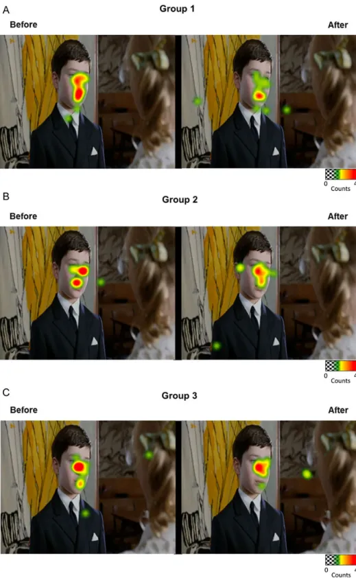
Figure 3. Reduction in the number offixations to the eyes only after inhibitory TMS applied to the right pSTS. (A) Examples of close-up heatmap from group data before and after each intervention (Sham or inhibitory TMS) in the 3 groups (warm colors denote a greater number offixations and cold colors denote fewer fixations). Scenes were selected for illustrative purposes. Heatmaps illustrate the reduction in number offixations to the eye at the group level only in Group 1, while Groups 2 and 3 show no significant changes. (B) The plots illustrate that a significant reduction in the normalized values of number of fixations to the eyes during the visualization of naturalistic social movies was only observed after inhibitory TMS applied over the right pSTS (Group 1) and not after Sham applied to the right pSTS (Group 2) nor after inhibitory TMS applied to the left pSTS (Group 3). The error bars represent the SEM. *P = 0.002.
6 | Cerebral Cortex
at UPMC on March 9, 2016
http://cercor.oxfordjournals.org/
object (balloon) in the nonsocial movies, even though total visu-alization time was shorter for the nonsocial scenes. Importantly, no significant changes in gaze pattern were observed in partici-pants receiving the Sham–Sham protocol, strongly suggesting that the observed changes were indeed due to the inhibition of the right pSTS neural activity and were not related to a placebo ef-fect or to the repetitive visualization of the movies. Furthermore,
the absence of changes in gaze pattern following all placebo stimu-lations points to an extreme intraindividual stability of spontan-eous gaze behavior, which could be considered an individual signature in social behavior (Fig.4).
Interestingly, the inhibition of the left pSTS had no impact on the spontaneous perception of the eyes. This result suggests an asymmetry in the cortical processing of eye-gaze information,
Figure 4. Whole-frame heatmaps representing group gaze behaviors before and after each intervention in (A) Group 1 (inhibitory TMS applied over the right pSTS), (B) Group 2 (Sham applied over the right pSTS), and (C) Group 3 (inhibitory TMS applied over the left pSTS) (selected for illustrative purposes).
at UPMC on March 9, 2016
http://cercor.oxfordjournals.org/
occurring predominantly within the right pSTS, which has been proposed by previous brain imaging studies (Pelphrey and Carter 2008;Greene and Zaidel 2011). For instance, an fMRI study inves-tigating gaze perception showed strongly right lateralized activa-tion within the STS (Pelphrey et al. 2004). In addition, another study investigating the brain network involved in the event-related potential response to direct gaze reported significant clusters in the right STS region (Conty et al. 2007). Moreover, a re-cent fMRI study showed that, although stimuli presenting gaze shifts elicited significant bold responses in both right and left pSTS, only the right pSTS was sensitive to the social meaning of different gaze directions, with enhanced responses when the eyes shifted toward rather than away from the viewer (Ethofer et al. 2011). Finally, it has been suggested that the left STS would be further implicated in the processing of multimodal in-formation, such as integration of speech and gesture (Willems et al. 2009;Holle et al. 2010).
Few previous studies have used TMS to better characterize the role of the STS in the perception of biological motion. Indeed, it has been shown that inhibitory rTMS applied over the pSTS tem-porarily impairs the ability to detect and discriminate point-light animations depicting human movement (Grossman et al. 2005). It has also been shown that judgment of unfamiliar faces as being trustworthy or untrustworthy was disrupted when rTMS was delivered over the STS (Dzhelyova et al. 2011). In addition, single-pulse TMS applied over the right temporal cortex has been shown to impair subject’s perception of gaze shifts over presentation of 2 consecutive face stimuli (Pourtois et al. 2004). However, the implication of the right pSTS in the very particular and subtle behavior of spontaneous looking to the eyes during passive visualization of ecological social stimuli, verified by an objective non-self-reported measure such as eye tracking, had not yet been demonstrated.
The present results provide then 2 majorfindings. First, these results help to elucidate some of the basic mechanisms of social cognition, by establishing a direct link between a very specific be-havior (looking to the eyes) and a localized anatomical region (the right pSTS). Indeed, recent fMRI studies have indicated that the right pSTS is highly implicated in gaze perception (Hadjikhani et al. 2008;Sato et al. 2008;Nummenmaa et al. 2010;Carlin and Calder 2013). However, due to the lack of lesion models circum-scribed to this region, no direct evidence of this association ex-ists. Here, the right pSTS virtual lesion caused a significant and selective decrease in perception of the eyes, providing direct evi-dence implicating the right pSTS in this precise behavior.
Secondly, these results show that it is possible to interfere with a key behavior of social cognition, opening up interesting perspectives on interventions in psychiatric disorders. By dis-rupting the right pSTS neural network in healthy volunteers, we artificially induced a gaze pattern that is similar to the gaze pattern observed in persons with autism, who present anatomic-al and functionanatomic-al abnormanatomic-alities of the pSTS (Klin et al. 2002; Zil-bovicius et al. 2006). Indeed, a core symptom of autism is deficits in social perception, mainly a lack of preference for the eyes, which has been demonstrated in several eye-tracking studies (Klin et al. 2002;Pelphrey et al. 2002;Jones et al. 2008). If stimula-tion of the STS by excitatory TMS is able to change this pattern and induce an increase in looking to the eyes, new perspectives on therapeutic interventions for ASD could emerge.
Supplementary Material
Supplementary material can be found at: http://www.cercor. oxfordjournals.org/.
Funding
The study was supported by AP-HP PHRC and Fondation de France grants. A.S. received funding from Fondation Orange. T.P. and J-C. L. received funding from the program“Investissements d’avenir” ANR-10-IAIHU-06, Paris Institute of Translational Neuroscience. Eye-Tracking device wasfinanced by “Les Amis d’Arthur” association. Funding to pay the Open Access publication charges for this article was provided by INSERM U1000.
Notes
We thank Michel Siksik for assistance with video work. Conflict of Interest: None declared.
References
Adolphs R. 2003. Cognitive neuroscience of human social behav-iour. Nat Rev Neurosci. 4:165–178.
Boddaert N, Chabane N, Gervais H, Good CD, Bourgeois M, Plumet MH, Barthelemy C, Mouren MC, Artiges E, Samson Y, et al. 2004. Superior temporal sulcus anatomical abnormal-ities in childhood autism: a voxel-based morphometry MRI study. Neuroimage. 23:364–369.
Carlin JD, Calder AJ. 2013. The neural basis of eye gaze processing. Curr Opin Neurobiol. 23:450–455.
Conty L, N’Diaye K, Tijus C, George N. 2007. When eye creates the contact! ERP evidence for early dissociation between direct and averted gaze motion processing. Neuropsychologia. 45:3024–3037.
Duchesnay E, Cachia A, Boddaert N, Chabane N, Mangin JF, Martinot JL, Brunelle F, Zilbovicius M. 2011. Feature selection and classification of imbalanced datasets: application to PET images of children with autistic spectrum disorders. Neuroimage. 57:1003–1014.
Dzhelyova MP, Ellison A, Atkinson AP. 2011. Event-related repeti-tive TMS reveals distinct, critical roles for right OFA and bilat-eral posterior STS in judging the sex and trustworthiness of faces. J Cogn Neurosci. 23:2782–2796.
Ethofer T, Gschwind M, Vuilleumier P. 2011. Processing social aspects of human gaze: a combined fMRI-DTI study. Neuroimage. 55:411–419.
Frith CD, Frith U. 2012. Mechanisms of social cognition. Annu Rev Psychol. 63:287–313.
Greene DJ, Zaidel E. 2011. Hemispheric differences in atten-tional orienting by social cues. Neuropsychologia. 49: 61–68.
Grossman ED, Battelli L, Pascual-Leone A. 2005. Repetitive TMS over posterior STS disrupts perception of biological motion. Vision Res. 45:2847–2853.
Hadjikhani N, Hoge R, Snyder J, de Gelder B. 2008. Pointing with the eyes: the role of gaze in communicating danger. Brain Cogn. 68:1–8.
Holle H, Obleser J, Rueschemeyer SA, Gunter TC. 2010. Integration of iconic gestures and speech in left superior temporal areas boosts speech comprehension under adverse listening condi-tions. Neuroimage. 49:875–884.
Huang YZ, Edwards MJ, Rounis E, Bhatia KP, Rothwell JC. 2005. Theta burst stimulation of the human motor cortex. Neuron. 45:201–206.
Jones W, Carr K, Klin A. 2008. Absence of preferential looking to the eyes of approaching adults predicts level of social disabil-ity in 2-year-old toddlers with autism spectrum disorder. Arch Gen Psychiatry. 65:946–954.
8 | Cerebral Cortex
at UPMC on March 9, 2016
http://cercor.oxfordjournals.org/
Jones W, Klin A. 2013. Attention to eyes is present but in decline in 2–6-month-old infants later diagnosed with autism. Nature. 504:427–431.
Klein JT, Shepherd SV, Platt ML. 2009. Social attention and the brain. Curr Biol. 19:R958–R962.
Klin A, Jones W, Schultz R, Volkmar F, Cohen D. 2002. Visual fix-ation patterns during viewing of naturalistic social situfix-ations as predictors of social competence in individuals with autism. Arch Gen Psychiatry. 59:809–816.
Neggers SF, Langerak TR, Schutter DJ, Mandl RC, Ramsey NF, Lemmens PJ, Postma A. 2004. A stereotactic method for image-guided transcranial magnetic stimulation validated with fMRI and motor-evoked potentials. Neuroimage. 21:1805–1817. Nummenmaa L, Passamonti L, Rowe J, Engell AD, Calder AJ. 2010.
Connectivity analysis reveals a cortical network for eye gaze perception. Cereb Cortex. 20:1780–1787.
Pelphrey KA, Carter EJ. 2008. Brain mechanisms for social percep-tion: lessons from autism and typical development. Ann N Y Acad Sci. 1145:283–299.
Pelphrey KA, Morris JP, McCarthy G. 2005. Neural basis of eye gaze processing deficits in autism. Brain. 128:1038–1048.
Pelphrey KA, Morris JP, Michelich CR, Allison T, McCarthy G. 2005. Functional anatomy of biological motion perception in pos-terior temporal cortex: an FMRI study of eye, mouth and hand movements. Cereb Cortex. 15:1866–1876.
Pelphrey KA, Sasson NJ, Reznick JS, Paul G, Goldman BD, Piven J. 2002. Visual scanning of faces in autism. J Autism Dev Disord. 32:249–261.
Pelphrey KA, Viola RJ, McCarthy G. 2004. When strangers pass: processing of mutual and averted social gaze in the superior temporal sulcus. Psychol Sci. 15:598–603.
Perrett DI, Smith PA, Potter DD, Mistlin AJ, Head AS, Milner AD, Jeeves MA. 1985. Visual cells in the temporal cortex sensitive to face view and gaze direction. Proc R Soc Lond B Biol Sci. 223:293–317.
Philip RC, Dauvermann MR, Whalley HC, Baynham K, Lawrie SM, Stanfield AC. 2012. A systematic review and meta-analysis of the fMRI investigation of autism spectrum disorders. Neurosci Biobehav Rev. 36:901–942.
Pourtois G, Sander D, Andres M, Grandjean D, Reveret L, Olivier E, Vuilleumier P. 2004. Dissociable roles of the human somato-sensory and superior temporal cortices for processing social face signals. Eur J Neurosci. 20:3507–3515.
Rothwell JC. 1997. Techniques and mechanisms of action of tran-scranial stimulation of the human motor cortex. J Neurosci Methods. 74:113–122.
Sato W, Kochiyama T, Uono S, Yoshikawa S. 2008. Time course of superior temporal sulcus activity in response to eye gaze: a combined fMRI and MEG study. Soc Cogn Affect Neurosci. 3:224–232.
Willems RM, Ozyurek A, Hagoort P. 2009. Differential roles for left inferior frontal and superior temporal cortex in multimodal in-tegration of action and language. Neuroimage. 47:1992–2004. Zilbovicius M, Meresse I, Chabane N, Brunelle F, Samson Y,
Boddaert N. 2006. Autism, the superior temporal sulcus and social perception. Trends Neurosci. 29:359–366.
at UPMC on March 9, 2016
http://cercor.oxfordjournals.org/