HAL Id: inserm-00668443
https://www.hal.inserm.fr/inserm-00668443
Submitted on 9 Feb 2012
HAL is a multi-disciplinary open access
archive for the deposit and dissemination of
sci-entific research documents, whether they are
pub-lished or not. The documents may come from
teaching and research institutions in France or
abroad, or from public or private research centers.
L’archive ouverte pluridisciplinaire HAL, est
destinée au dépôt et à la diffusion de documents
scientifiques de niveau recherche, publiés ou non,
émanant des établissements d’enseignement et de
recherche français ou étrangers, des laboratoires
publics ou privés.
Prevalence and novelty of PRPF31 mutations in French
autosomal dominant rod-cone dystrophy patients and a
review of published reports.
Isabelle Audo, Kinga Bujakowska, Saddek Mohand-Saïd, Marie-Elise
Lancelot, Veselina Moskova-Doumanova, Naushin Waseem, Aline Antonio,
José-Alain Sahel, Shomi Bhattacharya, Christina Zeitz
To cite this version:
Isabelle Audo, Kinga Bujakowska, Saddek Mohand-Saïd, Marie-Elise Lancelot, Veselina
Moskova-Doumanova, et al.. Prevalence and novelty of PRPF31 mutations in French autosomal dominant
rod-cone dystrophy patients and a review of published reports.. BMC Medical Genetics, BioMed
Central, 2010, 11 (1), pp.145. �10.1186/1471-2350-11-145�. �inserm-00668443�
R E S E A R C H A R T I C L E
Open Access
Prevalence and novelty of PRPF31 mutations in
French autosomal dominant rod-cone dystrophy
patients and a review of published reports
Isabelle Audo
1,2,3,4,5, Kinga Bujakowska
1,2,3, Saddek Mohand-Saïd
1,2,3,4, Marie-Elise Lancelot
1,2,3,
Veselina Moskova-Doumanova
1,2,3, Naushin H Waseem
5, Aline Antonio
1,2,3,4, José-Alain Sahel
1,2,3,4,5,
Shomi S Bhattacharya
1,2,3,5, Christina Zeitz
1,2,3*Abstract
Background: Rod-cone dystrophies are heterogeneous group of inherited retinal disorders both clinically and
genetically characterized by photoreceptor degeneration. The mode of inheritance can be autosomal dominant,
autosomal recessive or X-linked. The purpose of this study was to identify mutations in one of the genes, PRPF31,
in French patients with autosomal dominant RP, to perform genotype-phenotype correlations of those patients, to
determine the prevalence of PRPF31 mutations in this cohort and to review previously identified PRPF31 mutations
from other cohorts.
Methods: Detailed phenotypic characterization was performed including precise family history, best corrected
visual acuity using the ETDRS chart, slit lamp examination, kinetic and static perimetry, full field and multifocal ERG,
fundus autofluorescence imaging and optic coherence tomography. For genetic diagnosis, genomic DNA of ninety
families was isolated by standard methods. The coding exons and flanking intronic regions of PRPF31 were PCR
amplified, purified and sequenced in the index patient.
Results: We showed for the first time that 6.7% cases of a French adRP cohort have a PRPF31 mutation. We
identified in total six mutations, which were all novel and not detected in ethnically matched controls. The
mutation spectrum from our cohort comprises frameshift and splice site mutations. Co-segregation analysis in
available family members revealed that each index patient and all affected family members showed a
heterozygous mutation. In five families incomplete penetrance was observed. Most patients showed classical signs
of RP with relatively preserved central vision and visual field.
Conclusion: Our studies extended the mutation spectrum of PRPF31 and as previously reported in other
populations, it is a major cause of adRP in France.
Background
Rod-cone dystrophies, also called retinitis pigmentosa
(RP), are a clinically and genetically heterogeneous
group of inherited retinal disorders usually primarily
affecting rods with secondary cone degeneration [1-4]. It
represents a progressive disorder which often starts with
night blindness and leads to visual field constriction,
abnormal color vision and can eventually lead to loss of
central vision and complete blindness. It is the most
common inherited form of severe retinal degeneration,
with a frequency of about 1 in 4000 births and more
than 1 million individuals affected worldwide. The mode
of inheritance can be X-linked (5-15%), autosomal
dominant (30-40%) or autosomal recessive (50-60%).
The remaining patients represent isolated cases for
which the inheritance trait cannot be established [5].
To date, mutations in 20 different genes are associated
with autosomal dominant RP (adRP) http://www.sph.
uth.tmc.edu/Retnet/. The majority of prevalence studies
reveal rhodopsin (RHO) being the most frequently
mutated gene in adRP [6].
PRPF31 was also proposed to
* Correspondence: christina.zeitz@inserm.fr
1INSERM, UMRS968, Paris, F-75012, France
Full list of author information is available at the end of the article
© 2010 Audo et al; licensee BioMed Central Ltd. This is an Open Access article distributed under the terms of the Creative Commons Attribution License (http://creativecommons.org/licenses/by/2.0), which permits unrestricted use, distribution, and reproduction in any medium, provided the original work is properly cited.
represent a major gene underlying this disorder. It is
located on chromosome 19q13.42, encompasses 14
exons and codes for a ubiquitously expressed
pre-mRNA splicing factor [7]. According to published
reports,
PRPF31 mutation prevalence ranges from 1 to
8% in adRP cohorts from various geographical origins,
with higher frequencies reported in the United States
[8-15].
To date over 40 mutations have been located in
differ-ent parts of the gene. The mutation spectrum comprises
missense, splicing, regulatory and nonsense mutations.
In addition small or gross insertions, small
insertion-deletions, and small or gross deletions were identified
(http://www.sph.uth.tmc.edu/Retnet/,
http://www.retina-international.org/sci-news/prp31mut.htm) (Table 1).
The phenotype, age of onset and the severity of the
disease in adRP patients varied with different
PRPF31
mutations. In addition, in some families the same
muta-tion was even associated with a range of phenotypic
var-iations [15]. Furthermore, several studies revealed that
incomplete penetrance is a common feature in families
showing
PRPF31 mutations with an asymptomatic
muta-tion carrier having a carrier child, who fully manifests
the disease [16-18].
Our comprehensive study reported here aims to
per-form for the first time genotype-phenotype correlations
in a French adRP cohort with
PRPF31 mutations. All
patients were recruited from the same clinical center,
namely the Quinze-Vingts hospital in Paris. We will
pre-sent the prevalence of
PRPF31 mutations in this cohort
and compare our findings with other studies.
Methods
Clinical assessment
Ninety families with a provisional diagnosis of
autoso-mal dominant rod-cone dystrophy, (adRP) were
ascer-tained in the Clinical Investigating Centre of
Quinze-Vingts Hospital. Informed consent was obtained from
each patient and normal controls after explanation of
the study and its potential outcome. The study protocol
adhered to the tenets of the Declaration of Helsinki and
was approved by the local ethics committee. Each
patient underwent full ophthalmic examination with
clinical assessment as described earlier [6]. For
addi-tional family members who could not come to our
cen-tre for examination, ophthalmic records were obtained
from local ophthalmologists.
Mutation detection
Total genomic DNA was extracted from peripheral
blood leucocytes according to manufacturer
recommen-dation (Puregen Kit, Qiagen, Courtaboeuf, France).
Sub-sequently, direct genomic sequencing of
PRPF31 was
performed. All 14 exons of which exons 2-14 are coding,
and flanking intronic regions of
PRPF31 were PCR
amplified in 10 fragments (PRPF31 RefSeq NM_015629)
using oligonucleotides previously described [7] and a
polymerase (HotFire, Solis Biodyne, Estonia) in the
pre-sence of 2.5 mM MgCl
2and at an annealing
tempera-ture of 60°C. The PCR products were enzymatically
purified (ExoSAP-IT, USB Corporation, Cleveland, Ohio,
USA purchased from GE Healthcare, Orsay, France) and
sequenced with a commercially available sequencing
mix (BigDyeTerm v1.1 CycleSeq kit, Applied Biosystems,
Courtaboeuf, France). The sequenced products were
purified on a presoaked Sephadex G-50 (GE Healthcare)
96-well multiscreen filter plate (Millipore, Molsheim,
France), the purified product analyzed on an automated
48-capillary sequencer (ABI 3730 Genetic analyzer,
Applied Biosystems) and the results interpreted by
applying a software (SeqScape, Applied Biosystems). At
least 192 commercially available control samples were
used to validate the pathogenicity of the novel sequence
variants (Human random control panel 1-3, Health
Pro-tection Agency Culture Collections, Salisbury, United
Kingdom).
Multiplex Ligation dependent Probe Amplification
(MLPA) was performed using a commercially available
kit (SALSA MLPA kit P235-B1 Retinitis, MRC
Hol-land). The MLPA reactions were carried out according
to the manufacturer’s instructions and analyzed on an
automated 48-capillary sequencer (ABI 3730 Genetic
analyzer, Applied Biosystems). MLPA data analysis was
performed using GeneMarker (Softgenetics) and
addi-tionally Coffalyser (MRC Holland) software. Five
con-trol DNAs were included in each MLPA run and the
data was interpreted in terms of the ratio of each
probe signal between the control and patient DNA
samples. Samples with probe ratio values below 0.6
were considered as deletions and values above 1.4 as
duplications.
Results and Discussion
Samples included in this study are part of a French
cohort of adRP patients that were previously screened
for
RHO mutations and we noted 16.5% of cases with
known or novel
RHO mutations [6].
In the current study we report the identification of
novel
PRPF31 mutations in six of the 90 adRP index
patients. In total two deletions, three duplications and a
splice site mutation all over the gene were identified
(Table 2). The respective deletions and duplications
were predicted to lead to premature stop codons. The
novel splice site mutation c.527 + 2T>C resides in the
highly conserved donor site of exon 6, which is
pre-dicted to lead to skipping of exon 6. Co-segregation
analysis revealed in all but one family incomplete
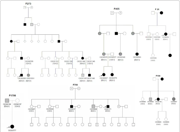
pene-trance (Table 2, Figure 1).
Audo et al. BMC Medical Genetics 2010, 11:145 http://www.biomedcentral.com/1471-2350/11/145
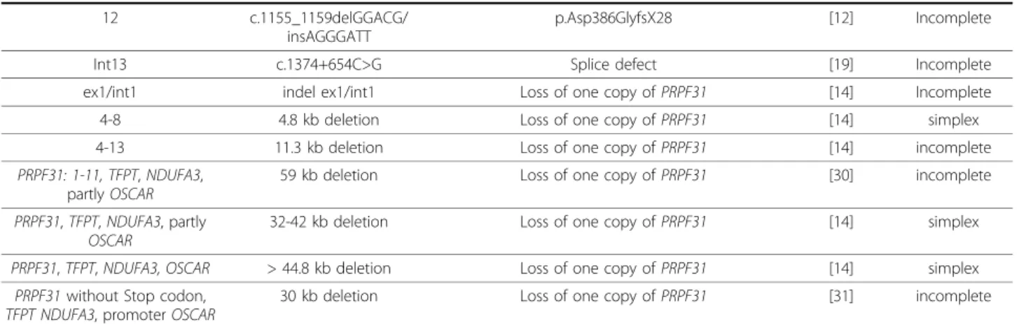
Table 1 Previously described
PRPF31 mutations in adRP patients
Exon/Intron Nucleotide Exchange Protein Effect Publication Information about penetrance Int1 c.1-2481G>T (formerly: IVS1
+1G>T)
splice defect [27] incomplete 2 c.79G>T p.Glu27X [15] Incomplete Int2 c.177+1G>A splice defect [13]
[23]
Simplex Simplex 3 c.220C>T p.Gln74X2 [13] Simplex 4 c.319C>G p.Leu107Val (interferes with splice site leading to
frameshift)
[23] Simplex Int4 c.323-2A>G Splice defect [23] Simplex 5 c.331_342del p.His111_Ile114del [22] High 5 c.358_359delAA p.Lys120GlufsX122 [9] Simplex 5 c.390delC p.Asn131MetfsX67 (formerly p.Asn131fs7ter197) [13] segregates in 3
affected 5 c.413C>A (formerly
c.412C>A)
p.Thr138Lys [15] Incomplete Int5 c.421-1G>A splice defect [28] Incomplete 6 c.421G>T p.Glu141X [13] Simplex Int6 c.527+1G>T splice defect [29] Incomplete Int6 c.527+1G>A splice defect [9] Incomplete Int6 c.527+3A>G splice defect [7,16]
[15]
Incomplete incomplete Int6 c.528-1G>A splice defect [15] not tested Int6 c.528-3_45del (previous
description: IVS6-3 to -45 del)
splice defect [7,12] Incomplete 7 c.581C>A p.Ala194Glu [7] Simplex 7 Formerly: 580-581dup33bp formerly: in frame insertion of 11 amino acids [7] Simplex 7 c.636delG p.Met212IlefsX27 (formerly: Met212fs/ter238) [13] segregates in 2
affected 7 c.646G>C p.Ala216Pro [7] Incomplete 8 c.732_737delins20bp p.Met244fsX248 [11] segregated in 2
affected 8 c.758_767del p.Gly253AlafsX65 (formerly: p.Gly253fs/ter317) [13] Simplex 8 c.769_770insA p.Thr258AspfsX21 (formerly: frameshift, 20 novel
amino acids then STOP) (formerly:Lys257fsX277)
[7] [11]
Simplex incomplete 8 c.785delT p.Phe262SerfsX59 [10]
8 c.828_829delCA p.His276GlnfsX2 (formerly p.His276fsX237) [11] Incomplete Int8 c.856-2A>G splice defect [23] segregated in 2
affected 9 c.871G>C p.Ala291Pro [13] Simplex 9 c.877_910del p.Arg293_Arg304>ValfsX17 [23] Incomplete 9 c.895T>C p.Cys299Arg [13] Incomplete 10 c.973G>T p.Glu325X [13] Simplex 10/int10 1049_IVS10+20del/insCCCCT splice defect [13] Simplex
Int10 c.1073+1G>A (Formerly: IVS10+1G>A)
splice defect [13] segregates in 5 affected 11 c.1115_1125del p.Arg372GlnfsX99 (formerly: frameshift, 98 novel
amino acids then STOP)
[7] Incomplete 11 c.1142delG p.Gly381GlufsX32 [12] Incomplete Int11 c.1146+2T>C Splice defect [15] Incomplete
To detect large deletions in those patients (60
patients) where no mutations was detected by direct
sequencing approaches, MLPA studies were performed.
However, no additional deletion was found by this
method.
Phenotypic characteristics of the 6 index patients are
summarized in table 3 and 4. Four of them were females
and two males with age ranging from 23 to 44 years
with an average age of 34.5 years. Age at time of
diagno-sis ranged from 5 to 43 with an average of 19.5 years.
Symptoms that led to the diagnosis were dominated by
night blindness in all patients but one (CIC01171). Two
patients also complained of visual field constrictions
(CIC00034 and CIC01171). Refractive errors were
vari-able. Central visual acuity was relatively preserved
except in one patient (CIC00140), age 31, who had
decreased central vision that just qualified her for legal
blindness. Preserved central vision was well correlated
with preserved responses to central hexagons in
multifo-cal ERG. All patients showed visual field constriction
with some peripheral perception, except for one patient
(CIC00607), age 23, who had a relatively normal
binocu-lar visual field. This patient was the only one with
detectable responses for both scotopic and photopic
condition on ERG. Color vision was normal in 3
patients or showed tritan defect in one eye for one
patient (CIC00607) and both eyes for patients CIC00140
and CIC03777 who also had low visual acuity. Anterior
segment examination showed moderate posterior
sub-capsular cataract only in one patient (CIC00034), age
42. Fundus examination showed typical peripheral signs
of RP with variable posterior pole involvements (Figure
2) including one patient with cystoid macular edema
(CIC00607, Figure 2B), one patient with an area of
par-afoveal well-demarcated atrophy (CIC00034, Figure 2C),
two
patients
with
perifoveal
atrophic
changes
(CIC01171, Figure 2D, CIC03777, Figure 2F) and one
patient with foveal thinning (CIC00140, Figure 2E). This
would suggest that central involvement is not
uncom-mon in the course of the disorders and that central
changes can occur as early as age 31 (patient CIC00140,
Figure 2E). These cone dysfunction and macular
changes can lead to further decrease in central vision
and central cone survival should be the major target of
future therapeutic intervention.
Due to the small number of index patients included in
this study, it is difficult to draw general conclusions on
phenotypic variability and phenotype/genotype
correla-tion. However, our cohort still shows on one hand one
patient with reduced but still detectable rod-cone
Table 1 Previously described
PRPF31 mutations in adRP patients (Continued)
12 c.1155_1159delGGACG/ insAGGGATT
p.Asp386GlyfsX28 [12] Incomplete Int13 c.1374+654C>G Splice defect [19] Incomplete ex1/int1 indel ex1/int1 Loss of one copy of PRPF31 [14] Incomplete 4-8 4.8 kb deletion Loss of one copy of PRPF31 [14] simplex 4-13 11.3 kb deletion Loss of one copy of PRPF31 [14] incomplete PRPF31: 1-11, TFPT, NDUFA3,
partly OSCAR
59 kb deletion Loss of one copy of PRPF31 [30] incomplete PRPF31, TFPT, NDUFA3, partly
OSCAR
32-42 kb deletion Loss of one copy of PRPF31 [14] simplex PRPF31, TFPT, NDUFA3, OSCAR > 44.8 kb deletion Loss of one copy of PRPF31 [14] simplex
PRPF31 without Stop codon, TFPT NDUFA3, promoter OSCAR
30 kb deletion Loss of one copy of PRPF31 [31] incomplete
If possible, mutations are indicated according to NM_015629 by using the recommendations of human genome variation society: http://www.hgvs.org/rec.html and/or the nomenclature of the original publication are given.
Table 2 Novel
PRPF31 mutations in a French adRP cohort.
Index (families) Exon Nucleotide Exchange Protein Effect Information about Penetrance CIC00398 (F273) 4 c.269_273del p.Tyr90CysfsX21 incomplete
CIC00607 (F405) Int6 c.527+2T>C splice defect incomplete
CIC00034 (F28) 7 c.666dup p.Ile223TyrX56 segregates (1 affected show mutation, 3 unaffected no mutation) CIC03777 (F1706) 8 c.709_734dup p.Cys247X incomplete
CIC01171 (F700) 9 c. 873_897dup p.Thr300GlyfsX32 incomplete CIC00140 (F108) 10 c.997delG p.Glu333SerfsX5 incomplete
Mutations are indicated according to NM_015629.3 by using the recommendations of human genome variation society: http://www.hgvs.org/rec.html.
Audo et al. BMC Medical Genetics 2010, 11:145 http://www.biomedcentral.com/1471-2350/11/145
responses and well preserved central vision (CIC00607)
at age 23 and on the other hand one legally blind
patient with undetectable ERG (CIC00140), at age 31,
suggesting variable severity of the disorder. This variable
severity of the disorder is in accordance with previous
reports, which also mentioned the possible role of
unknown modifier genes [15]. Further longitudinal
stu-dies are required to document retinal degeneration
kinetics and especially macular involvement in order to
prepare the patients for future treatment.
With the study presented here we report 6 novel
mutations in a French cohort leading to variable severity
of adRP. To our knowledge, this is the first report on
PRPF31 mutation screening and prevalence in the
French population and it further expands the mutation
spectrum causing adRP. In total two deletions, three
duplications and a splice site mutation were identified
(Table 2). To date only few
PRPF31 variations have
been reported to be recurrent (Table 1). This holds also
true for our study. Consistent with previous reports, we
propose that these mutations also lead to loss of
func-tion of PRFP31 and thus to haploinsufficiency [19-21].
In five of our families incomplete penetrance was
observed. Only in one family (family 28) no
asympto-matic mutation/or obligate carriers were reported. This
was confirmed on three unaffected family members who
did not reveal a mutation. However, due to the small
size of the family, incomplete penetrance cannot be
for-mally excluded for this mutation. To our knowledge, to
date only one large Chinese family was reported with
high penetrance [22], suggesting that most of the
PRPF31 mutations are indeed associated with
incom-plete penetrance. Although the presumed mechanism to
explain this phenomenon is allelic imbalance with
over-Figure 1 Pedigrees of adRP patients withPRPF31 mutations and co-segregation in available family members. Filled symbols represent affected, unfilled unaffected and dotted asymptomatic individuals. Question marks indicate that it is not clear whether the individual is affected or not. Squares depict males, circles females. Arrows mark the index patients. Equation symbols represent unaffected alleles. The identified mutations were abbreviated as followed: A = c.269_273del, B = c.527+2T>C, C = c.666dup, D = c.709_734dup, E = c.873_897dup and F = c.997delG.
Table 3 Clinical data of affected members from families with adRP due to
PRPF31 mutations
Family and PRPF31 mutation Patient Age at time of testing Age at time of diagnosisSex Family history Symptoms
at time of diagnosis
BCVA OD/OS Refraction
Lens Fundus examination OCT FAF
F273 CIC398 23 5 M From North of Brittany
Father affected and few
Night blindness 20/25 20/20 -6.25(-1.75)20° -5.50(-1.25)175°
clear Normal disc color, narrowed retinal vessels, little RPE changes in the
periphery,
Preserved foveal lamination
Loss of AF outside the vascular arcades, perifoveal
ring of increased AF F405 CIC00607 23 20 F One sister affected, cousins
on maternal side affected Mother not affected incomplete penetrance
From French descent
Night blindness late teens 20/32 20/32 +0.75(-2.75)15° Plano(-1.75)175°
clear Bilateral ERM Normal disc color; no narrowing of
blood vessels; little changes in the periphery
with few bone spicules
Bilateral ERM Bilateral
CME
Perifoveal ring of increased AF; foveal changes due to
CME
F28 CIC00034 42 18 F Family from Cameroun,
daughter mother and one brother affected, no notion
of incomplete penetrance Night blindness and visual field constriction 20/32 20/32 -3.25(-0.75)65° -3.25(-0.75)130° Small posterior subcapsular opacities
Disc pallor narrowed, blood vessels, RPE changes in periphery, bilateral atrophic lesion
off the fovea
Preserved foveal lamination
Loss of AF outside the vascular arcades, round eccentric parafoveal area of
loss of AF, no ring of AF
F1706 CIC03777 44 8 F Paternal grand-mother,
great-grand mother and one great-uncle on father side affected, French family from
Jewish Ashkenazi ancestry
Night blindness since age 7 20/125 20/160 +0.5(-1.25)40° +0.25(-1.50)155°
pseudophakic Pale optic disc, narrowed retinal vessels
Preserved foveal lamination
Loss of AF outside the vascular arcades, patchy loss
of AF within the posterior pole with no ring of AF F700 CIC01171 44 43 M One elder brother affected,
one niece affected from one of his unaffected sister, one
uncle on father side Incomplete penetrance family originating from the
Mauritius Island Visual field constriction, no real night blindness 20/32 20/25 -0.50(-3.75)15° -0.25(-3.75)175°
clear Normal disc color, narrowed retinal vessels,
RPE changes in the periphery
Preserved foveal lamination
Loss of AF outside the vascular arcades; small perifoveal ring of increased autofluorescence with some perifoveal areas of loss of
AF
F108 CIC00140 31 23 F Mother, maternal
grand-mother affected; family from Ivory Coast Night blindness since birth 20/500 20/200 +1.50(-1.25)95° +1.50(-1.25)75°
clear No pale optic disc; narrowed retinal vessels, some RPE changes in the
periphery
Foveal thinning
Loss of AF outside the vascular arcades, increased AF within the foveal region associated with some
patchy loss of AF
BCVA Best Corrected Visual Acuity; CME: Cystoid Macular Edema; ERM: Epi Retinal Membrane; AF: autofluorescence; OCT: optic coherence tomography OD: Oculis dextra (right eye); OS: Oculis Sinistra (center eye); RPE: Retinal Pigment Epithelium.
Audo et al . BMC Medical Genetics 2010, 11 :145 http://ww w.biomedcen tral.com/1471 -2350/11/145 Page 6 o f 9
expression of the wild-type allele, compensating for the
non-functional allele in asymptomatic carriers [21,23],
the exact mechanism how this over-expression happens
remains to be solved.
Patient data and mouse
in vivo studies strongly
sug-gest that the disease mechanism is caused by
haploinsufficiency rather than dominant negative effect.
A recent study in mice demonstrated that p.A216P
mutation as well as deletion of
Prpf31 exon 7 in mice
lead to null alleles [24]. Mice heterozygous for these
mutations did not reveal signs of retinal degeneration in
histological, ERG and fundus examination, however in
Table 4 Functional data.
Patient Color vision Binocular Goldman visual field, III4 isopter
Full field ERG Multifocal ERG CIC00398 ODS normal at 28
saturated Farnworth Hue
20° both horizontally and vertically with a large island of perception in temporal and inferior periphery
Only residual cone responses Relatively well preserved central
responses CIC00607 OD tritan defect; OS
normal at Farnsworth 15Desaturated Hue
180° horizontally ×110° vertically Rod-cone dysfunction with 80% of normal for scotopic 3.0cd.s/m2ERG amplitude and 50% of
normal for photopic 3.0cd.s.m2
Relatively well preserved central
responses CIC00034 ODS normal at 28
saturated Farnworth Hue
20° both horizontally and vertically with 2 bitemporal island of perception
in periphery
ND Only preservation of responses to central hexagons CIC03777 ODS tritan defext at 28
saturated Farnworth Hue
20° both horizontally and vertically ND Only residual responses to central hexagons CIC01171 ODS normal at 28
saturated Farnworth Hue
20° both horizontally and vertically ND Only preservation of responses to central hexagons CIC00140 ODS tritan defext at 28
saturated Farnworth Hue
30° both horizontally and vertically with a large island of perception in temporal and inferior periphery
ND Only residual responses to central hexagons
NP: not performed; ND: not detectable, OD: Oculus Dexter; OS: Oculus Sinister.
Figure 2 Color fundus photograph and autofluorescence imaging of the right eye for each index patient: A: CIC0398; B: CIC00607; C: CIC00034; D: CIC01171; E CIC00140; F: CIC03777.
homozygous state they were embryonic lethal,
demon-strating lack of function of the mutant
Prpf31 alleles. In
a number of studies a cytotoxic effect of
PRPF31
muta-tions has been suggested [25,26]. We believe that this
toxicity plays a minor role in the development of the
disease since asymptomatic mutation carriers do not
develop retinal degeneration.
The study presented here reveals a prevalence of 6.7%
in adRP cases due to
PRPF31 mutations. This is higher
than in another study from UK with 5% [15], in a study
from India (4%), from Japan with 3% [12], from Spain
with 1.7% [11] and from China with 1% [10]. A
preva-lence study by Sullivan and co-workers (2006) in 200
US families of presumably UK origin revealed 5.5% of
cases with
PRPF31 mutations. However, these numbers
were corrected to 8% when MLPA studies revealed
lar-ger deletions, which were not detectable by direct
sequencing approaches [14]. In contrast to these
find-ings, our MLPA studies did not reveal any large
dele-tions or duplicadele-tions in this cohort. Therefore, we
conclude that genomic rearrangements in the
PRPF31
gene are not common in the French adRP cohort.
Conclusions
With the study presented here we report six novel
mutations in a French cohort leading to variable severity
of adRP in families with mainly incomplete penetrance.
In 6.7% of this cohort
PRPF31 mutations were detected,
rendering this gene a major gene for adRP in France.
Consistent with previous reports, we propose that
muta-tions in
PRPF31 are mainly not recurrent, lead to loss of
function of PRFP31 and thus to haploinsufficiency.
Acknowledgements
The authors are grateful to patients and family members described in this study, to Thierry Léveillard, Dominique Santiard-Baron, Christine Chaumeil and clinical staff for their help in clinical data and DNA collection. The project was financially supported by the Department of Paris, Foundation Fighting Blindness (I.A. FFB Grant N°: CD-CL-0808-0466-CHNO and the CIC503 recognized as an FFB center, FFB Grant No: C-CMM-0907-0428-INSERM04), ANR NIHR Biomedical Research Centre for Ophthalmology and The Special Trustees of Moorfields Eye Hospital London, Foundation Voir et Entendre (C.Z), EU FP6, Integrated Project‘EVI-GENORET’ (LSHG-CT-2005-512036) and Ville de Paris et Région Ile de France.
Author details
1
INSERM, UMRS968, Paris, F-75012, France.2UPMC Univ Paris 06, UMR_S 968, Institut de la Vision, Paris, F-75012, France.3CNRS, UMR_7210, Paris, F-75012,
France.4Centre Hospitalier National d’Ophtalmologie des Quinze-Vingts, INSERM-DHOS CIC 503, Paris, F-75012, France.5UCL-Institute of
Ophthalmology, Bath Street, London, UK. Authors’ contributions
IA contributed to the design of the study, the acquisition and interpretation of clinical data, and drafted the manuscript. KB contributed to the design and interpretation of the MLPA studies. S MS contributed to the acquisition and interpretation of clinical data. M-E L, V M-D, NH W and A A performed the DNA extraction and sequence analysis. J-A S contributed to the design of the study. SSB contributed to the design of the study, and helped to draft the manuscript. CZ contributed to the design of the study, the
acquisition and interpretation of molecular genetic data, and drafted the manuscript. All authors read and approved the final manuscript. Competing interests
The authors declare that they have no competing interests. Received: 21 May 2010 Accepted: 12 October 2010 Published: 12 October 2010
References
1. Birch DG, Fish GE: Rod ERGs in retinitis pigmentosa and cone-rod degeneration. Invest Ophthalmol Vis Sci 1987, 28:140-150.
2. Carter-Dawson LD, LaVail MM, Sidman RL: Differential effect of the rd mutation on rods and cones in the mouse retina. Invest Ophthalmol Vis Sci 1978, 17:489-498.
3. Cideciyan AV, Hood DC, Huang Y, Banin E, Li ZY, Stone EM, Milam AH, Jacobson SG: Disease sequence from mutant rhodopsin allele to rod and cone photoreceptor degeneration in man. Proc Natl Acad Sci USA 1998, 95:7103-7108.
4. Milam AH, Li ZY, Fariss RN: Histopathology of the human retina in retinitis pigmentosa. Prog Retin Eye Res 1998, 17:175-205.
5. Hartong DT, Berson EL, Dryja TP: Retinitis pigmentosa. Lancet 2006, 368:1795-1809.
6. Audo I, Manes G, Mohand-Said S, Friedrich A, Lancelot ME, Antonio A, Moskova-Doumanova V, Poch O, Zanlonghi X, Hamel CP, et al: Spectrum of rhodopsin mutations in French autosomal dominant rod-cone dystrophy patients. Invest Ophthalmol Vis Sci 2010, 51:3687-3700.
7. Vithana EN, Abu-Safieh L, Allen MJ, Carey A, Papaioannou M, Chakarova C, Al-Maghtheh M, Ebenezer ND, Willis C, Moore AT, et al: A human homolog of yeast pre-mRNA splicing gene, PRP31, underlies autosomal dominant retinitis pigmentosa on chromosome 19q13.4 (RP11). Mol Cell 2001, 8:375-381.
8. Daiger SP, Bowne SJ, Sullivan LS: Perspective on genes and mutations causing retinitis pigmentosa. Arch Ophthalmol 2007, 125:151-158. 9. Gandra M, Anandula V, Authiappan V, Sundaramurthy S, Raman R,
Bhattacharya S, Govindasamy K: Retinitis pigmentosa: mutation analysis of RHO, PRPF31, RP1, and IMPDH1 genes in patients from India. Mol Vis 2008, 14:1105-1113.
10. Lim KP, Yip SP, Cheung SC, Leung KW, Lam ST, To CH: Novel PRPF31 and PRPH2 mutations and co-occurrence of PRPF31 and RHO mutations in Chinese patients with retinitis pigmentosa. Arch Ophthalmol 2009, 127:784-790.
11. Martinez-Gimeno M, Gamundi MJ, Hernan I, Maseras M, Milla E, Ayuso C, Garcia-Sandoval B, Beneyto M, Vilela C, Baiget M, et al: Mutations in the pre-mRNA splicing-factor genes PRPF3, PRPF8, and PRPF31 in Spanish families with autosomal dominant retinitis pigmentosa. Invest Ophthalmol Vis Sci 2003, 44:2171-2177.
12. Sato H, Wada Y, Itabashi T, Nakamura M, Kawamura M, Tamai M: Mutations in the pre-mRNA splicing gene, PRPF31, in Japanese families with autosomal dominant retinitis pigmentosa. Am J Ophthalmol 2005, 140:537-540.
13. Sullivan LS, Bowne SJ, Birch DG, Hughbanks-Wheaton D, Heckenlively JR, Lewis RA, Garcia CA, Ruiz RS, Blanton SH, Northrup H, et al: Prevalence of disease-causing mutations in families with autosomal dominant retinitis pigmentosa: a screen of known genes in 200 families. Invest Ophthalmol Vis Sci 2006, 47:3052-3064.
14. Sullivan LS, Bowne SJ, Seaman CR, Blanton SH, Lewis RA, Heckenlively JR, Birch DG, Hughbanks-Wheaton D, Daiger SP: Genomic rearrangements of the PRPF31 gene account for 2.5% of autosomal dominant retinitis pigmentosa. Invest Ophthalmol Vis Sci 2006, 47:4579-4588.
15. Waseem NH, Vaclavik V, Webster A, Jenkins SA, Bird AC, Bhattacharya SS: Mutations in the gene coding for the pre-mRNA splicing factor, PRPF31, in patients with autosomal dominant retinitis pigmentosa. Invest Ophthalmol Vis Sci 2007, 48:1330-1334.
16. Al-Maghtheh M, Vithana E, Tarttelin E, Jay M, Evans K, Moore T, Bhattacharya S, Inglehearn CF: Evidence for a major retinitis pigmentosa locus on 19q13.4 (RP11) and association with a unique bimodal expressivity phenotype. Am J Hum Genet 1996, 59:864-871. 17. Evans K, al-Maghtheh M, Fitzke FW, Moore AT, Jay M, Inglehearn CF,
Arden GB, Bird AC: Bimodal expressivity in dominant retinitis pigmentosa genetically linked to chromosome 19q. Br J Ophthalmol 1995, 79:841-846. Audo et al. BMC Medical Genetics 2010, 11:145
http://www.biomedcentral.com/1471-2350/11/145
18. McGee TL, Devoto M, Ott J, Berson EL, Dryja TP: Evidence that the penetrance of mutations at the RP11 locus causing dominant retinitis pigmentosa is influenced by a gene linked to the homologous RP11 allele. Am J Hum Genet 1997, 61:1059-1066.
19. Rio Frio T, McGee TL, Wade NM, Iseli C, Beckmann JS, Berson EL, Rivolta C: A single-base substitution within an intronic repetitive element causes dominant retinitis pigmentosa with reduced penetrance. Hum Mutat 2009, 30:1340-1347.
20. Rio Frio T, Wade NM, Ransijn A, Berson EL, Beckmann JS, Rivolta C: Premature termination codons in PRPF31 cause retinitis pigmentosa via haploinsufficiency due to nonsense-mediated mRNA decay. J Clin Invest 2008, 118:1519-1531.
21. Vithana EN, Abu-Safieh L, Pelosini L, Winchester E, Hornan D, Bird AC, Hunt DM, Bustin SA, Bhattacharya SS: Expression of PRPF31 mRNA in patients with autosomal dominant retinitis pigmentosa: a molecular clue for incomplete penetrance? Invest Ophthalmol Vis Sci 2003, 44:4204-4209. 22. Wang L, Ribaudo M, Zhao K, Yu N, Chen Q, Sun Q, Wang L, Wang Q: Novel
deletion in the pre-mRNA splicing gene PRPF31 causes autosomal dominant retinitis pigmentosa in a large Chinese family. Am J Med Genet A 2003, 121A:235-239.
23. Rivolta C, McGee TL, Rio Frio T, Jensen RV, Berson EL, Dryja TP: Variation in retinitis pigmentosa-11 (PRPF31 or RP11) gene expression between symptomatic and asymptomatic patients with dominant RP11 mutations. Hum Mutat 2006, 27:644-653.
24. Bujakowska KM, Maubaret C, Chakarova CF, Tanimoto N, Beck SC, Fahl E, Humphries MM, Kenna P, Makarov E, Makarova O, Paquet-Durand F, Ekström PA, van Veen T, Leveillard T, Humphries P, Seeliger MW, Bhattacharya SS: Study of gene targeted mouse models of splicing factor gene Prpf31 implicated in human autosomal dominant retinitis pigmentosa (RP). Invest Ophthalmol Vis Sci 2009, 50:5927-5933. 25. Huranova M, Hnilicova J, Fleischer B, Cvackova Z, Stanek D: A mutation
linked to retinitis pigmentosa in HPRP31 causes protein instability and impairs its interactions with spliceosomal snRNPs. Hum Mol Genet 2009, 18:2014-2023.
26. Yuan L, Kawada M, Havlioglu N, Tang H, Wu JY: Mutations in PRPF31 inhibit pre-mRNA splicing of rhodopsin gene and cause apoptosis of retinal cells. J Neurosci 2005, 25:748-757.
27. Liu JY, Dai X, Sheng J, et al: Identification and functional characterization of a novel splicing mutation in RP gene PRPF31. Biochem Biophys Res Commun 2008, 367:420-426.
28. Xia K, Zheng D, Pan Q, et al: A novel PRPF31 splice-site mutation in a Chinese family with autosomal dominant retinitis pigmentosa. Mol Vis 2004, 367(10):361-365.
29. Chakarova CF, Cherninkova S, Tournev I, et al: Molecular genetics of retinitis pigmentosa in two Romani (Gypsy) families. Mol Vis 2006, 12:909-914.
30. Kohn L, Bowne SJ, Sullivan LS, et al: Breakpoint characterization of a novel approximately 59 kb genomic deletion on 19q13.42 in autosomal-dominant retinitis pigmentosa with incomplete penetrance. Eur J Hum Genet 2009, 17:651-655.
31. Abu-Safieh L, Vithan EN, Mantel I, et al: A large deletion in the adRP gene PRPF31: evidence that haploinsufficiency is the cause of disease. Mol Vis 2006, 12:384-388.
Pre-publication history
The pre-publication history for this paper can be accessed here: http://www.biomedcentral.com/1471-2350/11/145/prepub
doi:10.1186/1471-2350-11-145
Cite this article as: Audo et al.: Prevalence and novelty of PRPF31 mutations in French autosomal dominant rod-cone dystrophy patients and a review of published reports. BMC Medical Genetics 2010 11:145.
Submit your next manuscript to BioMed Central
and take full advantage of:
• Convenient online submission
• Thorough peer review
• No space constraints or color figure charges
• Immediate publication on acceptance
• Inclusion in PubMed, CAS, Scopus and Google Scholar
• Research which is freely available for redistribution Submit your manuscript at