HAL Id: inserm-02462594
https://www.hal.inserm.fr/inserm-02462594
Submitted on 31 Jan 2020
HAL is a multi-disciplinary open access archive for the deposit and dissemination of sci-entific research documents, whether they are pub-lished or not. The documents may come from teaching and research institutions in France or abroad, or from public or private research centers.
L’archive ouverte pluridisciplinaire HAL, est destinée au dépôt et à la diffusion de documents scientifiques de niveau recherche, publiés ou non, émanant des établissements d’enseignement et de recherche français ou étrangers, des laboratoires publics ou privés.
Xeroderma Pigmentosum Groups C and A in Algerian
Patients with Deregulation of both Transcription and
DNA Repair
Nicolas Le May, N Calmels, Y Abiayad, L Boukli, M Semer, A Serradj,
Jean-Marc Egly, Laugel Laugel
To cite this version:
Nicolas Le May, N Calmels, Y Abiayad, L Boukli, M Semer, et al.. Xeroderma Pigmentosum Groups C and A in Algerian Patients with Deregulation of both Transcription and DNA Repair. Journal of Case Reports and Studies, 2018, 6 (4), �10.15744/2348-9820.6.401�. �inserm-02462594�
Xeroderma Pigmentosum Groups C and A in Algerian Patients with
Deregulation of both Transcription and DNA Repair
Le May N
*1,2,3,4, Calmels N
5,6, Abiayad Y
7, Boukli L
7, Semer M
1,2,3,4, Serradj A
7, Egly JM
*1,2,3,4and Laugel V
*5,6 1IGBMC, Department of Functional Genomics and Cancer, CNRS/INSERM/University of Strasbourg, Illkirch Cedex, CU Strasbourg, France2Centre National de la Recherche Scientifique, Illkirch, France
3Institute National de la Santé et de la Recherche Médicale, Illkirch, France 4Université de Strasbourg, Illkirch, France
5Department of Pediatric Neurology, Strasbourg University Hospital, Avenue Moliere, Strasbourg Cedex, France 6Laboratory of Medical Genetics, University of Strasbourg, Strasbourg, France
7Etablissement Hospitalier Universitaire d’Oran, Service de Dermatologie, Ibn Rochd, Oran, Algeria
*
Corresponding author: Le May N, IGBMC, Department of Functional Genomics and Cancer, CNRS/
INSERM/University of Strasbourg, BP 163, 67404 Illkirch Cedex, CU Strasbourg, France, Tel: 33388653450,
E-mail: nlemay@igbmc.fr
Case Report Open Access
Citation: Le May N, Calmels N, Abiayad Y, Boukli L, Semer M, et al. (2018) Xeroderma Pigmentosum
Groups C and A in Algerian Patients with Deregulation of both Transcription and DNA Repair. J Case Rep
Stud 6(4): 401
Volume 6 | Issue 4 Journal of Case Reports and Studies ISSN: 2348-9820
Xeroderma Pigmentosum (XP) is a rare autosomal recessive disorder characterized by an extreme sensitivity to UV rays from sunlight,
a high incidence of skin cancer and occasional neurological symptoms. XP, primarily defined as a DNA repair syndrome, has been found associated with defects in the Nucleotide Excision Repair (NER) pathway, and more recently by transcriptional deregulation. XP results from mutations in eight genes (XPA to XPG and XPV) coding for proteins involved in NER.
Keywords: Xeroderma pigmentosum; Algerian Patients; DNA Repair; Nucleotide-Excision Repair; Transcription
Abstract
We report here two cases of XP patients from Algeria, describe their clinical features, identify the causative mutations, and molecularly define their etiology. We determined that each XP individual bears XPC and XPA mutations respectively. Both mutations disrupt expression of their corresponding genes: while the XPC p.Val548Alafs*25 variant was not expressed, the truncated XPA p.Arg228* variant was detected in the patient’s cells. Unscheduled DNA synthesis (UDS) and Recovery of RNA synthesis after DNA damage (RRS) assays, as well as immunofluorescence on Ultraviolet-irradiated patient cells showed deficiency in the NER pathway. Moreover, we also found that the patients’ cells were defective in transcription, especially certain Retinoic-acid receptor (RAR)-responsive genes. Altogether our data revealed both DNA repair and transcriptional defaults that defined the molecular etiology for these two XP individuals, and may help to understand some of the patients’ clinical features.
List of abbreviations: XP: Xeroderma pigmentosum; NER: Nucleotide-Excision Repair; UDS: Unscheduled DNA Synthesis; RRS:
Recovery of RNA Synthesis after DNA Damage; RAR: Retinoic-acid Receptor; CPD: Cyclobutane Pyrimidine Dimers; 6-4PP: 6-4 Photoproducts; GGR: Global Genome Repair; TCR: Transcription-Coupled Repair; NR: Nuclear Receptor; RARß2: Retinoic Acid Receptor Isoform ß2; CYP26: Cytochrome P450 Family 26; DAPK1: Death Associated Protein Kinase 1 (DAPK1); GAPDH: Glyceraldehyde-3-Phosphate Dehydrogenase
Introduction
Xeroderma Pigmentosum (XP) is a rare autosomal recessive disorder. Complementation tests by cell fusion have shown the existence of at least seven XP complementation groups: XP-A to XP-G, corresponding to mutations on XPA to XPG genes, and a separate group variant (XP-V) related to mutations on the gene coding for DNA polymerase η [1]. The products of these genes are involved in Nucleotide Excision Repair (NER), the principal DNA repair pathway for removal of a variety of DNA damages induced by genotoxic attack such as Ultraviolet (UV) irradiation or anti-tumor drugs. XP has been found associated with defects in NER pathway.
Journal of Case Reports and Studies 2 This genetic disease is characterized by a wide variety of clinical features. XP patients present an extreme sensitivity to UV rays from sunlight. This condition mostly affects the eyes and areas of skin exposed to the sun and results in a high incidence of skin cancers (more than 1000-fold in comparison with normal individuals) as well as increased cancer susceptibility in many other tissues, including breast and lung. Approximately 30% of affected individuals have neurological symptoms, including acquired microcephaly and progressive cognitive impairment. These neurological disorders are often found in XP-A, XP-B, XP-D and XP-F groups associated with severe phenotypes, but are rare in the XP-C group. XP is rare in the United States and Europe (1/100000), but relatively more frequent in Japan (1/ 22000) and in North Africa (1/10000) due to the high rate of consanguinity (for example 29.8% in Tunisia) [2-5].
UV irradiation of human cells results in DNA damage, consisting primarily of cyclobutane pyrimidine dimers (CPD) and 6-4 photoproducts (6-4PP) that can be eliminated through two sub-pathways of the NER: the global genome repair (GGR) removes DNA damage from the entire genome, whereas the transcription-coupled repair (TCR) corrects DNA lesions located on the actively transcribed strand which block elongating RNA polymerase II [6]. In GGR, the XPC-HR23B and UV-DDB complexes recognize the damage-induced DNA distortion. In TCR, RNA Pol II stalled in front of a lesion on the transcribed DNA strand initiates the recruitment of the TCR-specific sensors CSB and CSA. Both NER sub-pathways then recruit TFIIH, which unwinds the DNA via its ATPase/helicase activities; XPA and RPA facilitate expansion of the unwound DNA bubble around the damage. The endonucleases XPG and XPF-ERCC1 then promote removal of the damaged oligonucleotide, before the re-synthesis machinery fills in the DNA gap.
XP has been primarily defined as a DNA repair syndrome due to the inability of patient cells to eliminate DNA lesions via NER pathways. However, studies in the last decade suggested that some of their phenotypes may also stem from transcriptional deregulation [7]. Indeed, we have demonstrated that the NER factors XPC, CSB, XPA, RPA, XPG and ERCC1-XPF are recruited with the transcription machinery controlled by nuclear receptors (NR) such as retinoic-acid receptor (RAR), influencing local chromatin remodeling around responsive gene promoters [8]. Moreover, primary fibroblasts derived from XP patients bearing mutations on XPC, XPA, XPG, XPF, XPB and XPD also present transcriptional deregulation [9,10].
In this report, we describe the clinical features and identify the mutations of two XP-C and XP-A individuals from Algeria. We also determined how these mutations disrupt NER and deregulate expression of genes such as those responsive to RAR, explaining the molecular etiology for these XP patients.
Methodology
The XP-C patient, hereafter called EHUO1, is a male Algerian born to first-cousin consanguineous parents. He is the second child of five siblings (Figure 1A). He showed bilateral conjunctivitis from 1 week of age and cutaneous photosensitivity from 8 months of age, with marked erythema in sun-exposed skin areas. Cutaneous symptoms later evolved to include lentigines, hypo-pigmented macules and eventually poikilodermia. Multiple actinic keratosis lesions, several basal cell carcinomas of the head and neck area and a botryomycoma of the neck had to be removed before the age of 18 years. Bilateral visual impairment due to corneal opacification was confirmed at age 10 with complete blindness of the right eye and severely impaired vision of the left eye. This patient showed no extra-cutaneous symptoms and no mental retardation at the time of his latest clinical assessment, at age 18.
Patients
Figure 1: Algerian Xeroderma pigmentosum patients (A) Patient EHUO1, at age of 19 years, had cutaneous symptoms later
evolved to include lentigines, hypo-pigmented macules, multiple actinic keratosis lesions, several basal cell carcinomas of the head and neck area. He showed no extra-cutaneous symptom and no mental retardation at the time of his latest clinical assessment at age 18; (B) Patient EHUO3, at age of 10 years, had cutaneous photosensitivity with marked erythema of sun-exposed areas. Cutaneous symptoms progressively included multiple lentigines, sclerodactyly and acrocyanosis together with mild sun-induced conjunctival hyperemia. Patient EHUO3 showed moderate mental retardation. No skin tumor had developed at age 10
3 Journal of Case Reports and Studies The XP-A patient, hereafter called EHUO3, is a male patient born to Algerian parents after an uneventful pregnancy (Figure 1B). No known consanguinity was reported in the family. Cutaneous photosensitivity was reported from the age of 7 months with marked erythema of sun-exposed areas. Hypo- and hyperpigmented macules were reported at age 1. Cutaneous symptoms progressively included multiple lentigines, sclerodactyly and acrocyanosis, together with mild sun-induced conjunctival hyperemia. This patient shows moderate mental retardation No skin tumor had developed at age 10.
Informed consent was obtained for all participants, including the parents of the children.
Informed consent
Primary fibroblast cultures were established from biopsies of unaffected skin obtained from the two XP individuals (EHUO1 and EHUO3) and their related mothers (EHUO2 and EHUO4). Fibroblasts were routinely grown in Dulbecco’s modified Eagle medium (DMEM) (1g/L glucose) w/GLUTAMAX (Life Technologies, Inc., Rockville, MD) supplemented with 10% of Fetal Calf Serum (FCS) and Penicillin (100 UI/mL) + Streptomycin (100 ug/mL).
Cell lines and culture conditions
Genomic DNA was extracted from fibroblast cultures using QIAamp DNA mini kit from QIAGEN. DNA samples from the two XP patients (EHUO1 and EHUO3) were studied by next-generation sequencing (NGS), targeting 16 genes of the NER pathway (XPA to XPG and POLH genes included) [11]. Briefly, libraries obtained by multiplex amplification were sequenced with Ion Personal Genome Machine (PGM, Life Technologies) using Ion 316 chip. Sequencing data were analyzed by the Torrent Suite v4.4 (Life Technologies). Subsequent variant annotation and ranking were performed using VaRank v1.4.0 (Geoffroy V et al, PeerJ. 2015) configured with Alamut Batch (Interactive biosoftware). XPA and XPC mutations identified by NGS were confirmed by Sanger DNA sequencing and familial segregation analyses were performed by the same assay on maternal samples (EHU02 and EHUO4). The sequences of the primers used for amplifying the two XPA and XPC loci are indicated in Table 1. Sequences were obtained on a 3500 Genetic Analyzer (Applied Biosystems) using the Big Dyev1.1 sequencing standard kit (Life Technologies), aligned with the Sequencing Pilot software (JSI) and compared with the corresponding genomic DNA reference sequences (GRCh37; NM_000380.3 for XPA and NM_004628.4 for XPC).
Genomic DNA sequencing
Reverse Forward Primers GCCAGGTGACCTTCACTGAAACTT TGTACATGGCTGAAAGCTTGATGGAG XPA (exon 6) ACCCAACATAGTGCTGGGCATA TGGCCCTCCAAAGCAGAGGAAA XPC (exon 9)
Table 1: List of primers used in genomic DNA sequencing
Total RNA was isolated from the different fibroblasts using a GenElute Mammalian Total RNA Miniprep kit (Sigma) and reverse transcribed with SuperScript IV reverse transcriptase (Invitrogen). The quantitative PCR was done using QuantiTect SYBR green (QIAGEN) and the Lightcycler 480 (Roche). For the analysis of RAR-target genes, twelve hours before ligand treatment, cells were incubated with phenol red-free medium containing charcoal treated FCS and 40mg/ml gentamycin. Cells were then treated with 10μM all-trans retinoic acid (ATRA) (MP) for 6 hours. The primer sequences for the different genes used in qPCR are indicated in Table 2. The mRNA expression of the different analyzed genes was normalized to that of the housekeeping gene glyceraldehyde-3-phosphate dehydrogenase (GAPDH).
XPC, XPA and RAR-target gene mRNA expression analysis
Reverse Forward Primers ACGCCTGCTTCACCACCTTC AGCTCACTGGCATGGCCTTC GAPDH_mRNA
QuantiTect Primer Assay (QIAGEN, QT00029519) Hs_XPA_1_SG
QuantiTect Primer Assay (QIAGEN, QT00080381) Hs_XPC_1_SG TACACGCTCTGCACCTTTAGC CCAGCAAGCCTCCATGTTC RARB2_mRNA CCAAAGAGGAGTTCGGTTGA CGAGCACTCGTGGGAGAG CYP26A1_mRNA CAGGCCTGGGACATTGTCAT TGAGTGTTGCCAGAAGCGAT DAPK1_mRNA
Table 2: List of primers used in mRNA expression analysis
Approximatively 2x10⁶ fibroblasts were plated in 10 cm plates (Falcon) and after 24 hours cells were washed, harvested, lysed in
RIPA buffer (10 mM Tris-HCl [pH 8.0], 140 mM NaCl, 1% Triton X-100, 0.1% Na-deoxycholate, 0.1% SDS) and incubated for 15
Journal of Case Reports and Studies 4
Cells were plated on coverslips in 6-well plates at a confluency of 7x10⁴ cells per well. After 2 days, cells were irradiated with a
range of UV-C doses, and then incubated for 23 hours with DMEM supplemented with Fetal Bovine Serum (FBS). Cells were labelled with 5-ethynyl-uridine (EU; Invitrogen) for 2 hours, and then washed with PBS, followed by fixation, permeabilization and an azide-coupling reaction and DAPI staining (Click-iT RNA HCS Assay, Invitrogen). Finally, coverslips were washed in Phosphate Buffer Solution (PBS), and mounted on glass slides with Ibidi Mounting Medium (Biovalley). Photographs of the cells were taken with a fluorescent microscope (Imager.Z2) equipped with a CCD camera (AxioCam, Zeiss). The images were processed and analyzed with ImageJ. At least 50 cells were randomly selected, and the average nuclear fluorescence intensity was calculated.
Recovery of RNA synthesis after DNA damage (RRS) assay
Cells were plated on coverslips in 6-well plates at a confluency of 7x10⁴ cells per well. After 2 days, cells were irradiated with a
range of UV-C doses, and then incubated for 23 hours with DMEM supplemented with Fetal Bovine Serum (FBS). Cells were labelled with 5-ethynyl-uridine (EU; Invitrogen) for 2 hours, and then washed with PBS, followed by fixation, permeabilization and an azide-coupling reaction and DAPI staining (Click-iT RNA HCS Assay, Invitrogen). Finally, coverslips were washed in Phosphate Buffer Solution (PBS), and mounted on glass slides with Ibidi Mounting Medium (Biovalley). Photographs of the cells were taken with a fluorescent microscope (Imager.Z2) equipped with a CCD camera (AxioCam, Zeiss). The images were processed and analyzed with ImageJ. At least 50 cells were randomly selected, and the average nuclear fluorescence intensity was calculated.
Unscheduled DNA synthesis (UDS) assay
Approximatively 2x10⁵ fibroblasts were plated in Lab-tek chambered coverglass (Thermo scientitific) and 24 hours later cells were
washed, covered with an isopore polycarbonate filter with 5um pores (EMD Millipore) and irradiated with a 254 nm UV-C lamp at 120J/m2. 15 minutes after irradiation cells were fixed with 4% paraformaldehyde (PFA) for 10 minutes, then washed with PBS and
permeabilized with PBS 0.1% tritonX-100 for 10 minutes. DNA was denaturated with 10% HCl at room temperature (RT) for 20 minutes, and then cells were washed with PBS. Blocking and incubation with antibodies were performed in 10% heat inactivated FCS, washes were done with PBS 0.1% triton X-100. Nuclei were counterstained with DAPI and cells were mounted using the ProLong Gold antifade reagent (Molecular Probes). Microscopy pictures were taken at a TCS SP2 microscope (Leica) based on an inverted microscope (DMIRBE; Leica; 63x Plan Apochromat, NA 1.4; LCS software Leica), Z stack width was 0.5µm.
Immunofluorescence
Antibodies against XPC (2076) were produced at the IGBMC. XPB (S-19) and XPA (FL-723) antibodies were purchased from Santa-Cruz Biotechnology. For immunofluorescence, antibodies against CPD (clone TDM-2 Cosmo Bio Co LTD), XPB (anti TFIIH p89 antibody, Santa Cruz Biotechnology, S-19), XPC (A301-122A, Bethyl), XPA (Ab-1, 2F15 from NeoMarkers) and secondary antibodies (Goat anti mouse alexa 488 and goat anti rabbit alexa 546 from Jackson Laboratories) were purchased.
Antibodies
Results
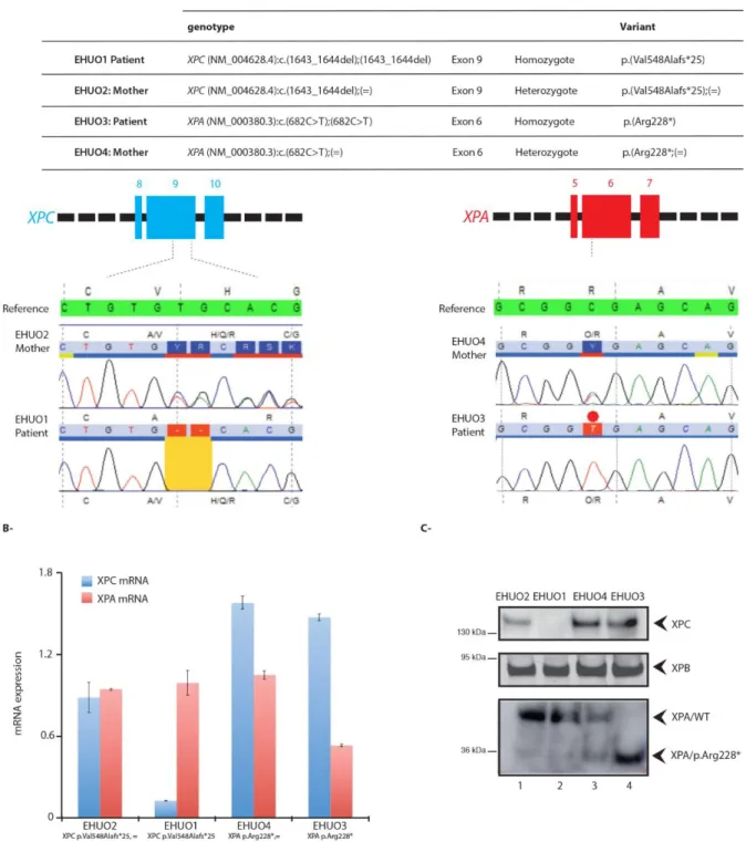
To pinpoint their genetic defects, genomic DNA from fibroblasts derived from the patients and the mothers were isolated. The two patients were homozygous for a single mutation each (Figure 2A). We identified one frameshift mutation (p.Val548Alafs*25) in XPC for the EHUO1 patient, due to a deletion (c.1643_1644delTG) on exon 9 (Figure 2A, left panel). The EHUO3 patient carried the nonsense mutation c.682C>T on exon 6 of XPA, leading to a premature termination at amino acid 228 (p.Arg228*) (Figure 2A, right panel). For both patients, their mother was heterozygous for the same mutation. Although the fathers’ DNA was not available for analysis, it is presumed that they are also heterozygous carriers for these XPC and XPA mutations, which are often found in North Africa [4,12,13].
Identification of the XPC and XPA patient mutations
We next evaluated the consequences of these XPC and XPA mutations on the expression of corresponding transcripts and proteins. The EHUO1 fibroblasts had a strongly reduced level of XPC mRNA compared with the EHUO2 parental fibroblasts, and an absence of the protein (Figure 2B and 2C, lanes 1-2). In EHUO3 fibroblasts, a slight reduction of XPA mRNA was observed (Figure 2B). The expressed XPA protein was truncated with a molecular weight around 30kDa; this isoform was also detected within the heterozygous EHUO4 maternal cells (Figure 2C, lanes 3-4).
min at 4 oC before centrifugation. After denaturation, the protein samples were electrophoresed on polyacrylamide-SDS gels and
transferred onto Hybond-C membrane. The blots were probed with antibodies against XPC, XPA and XPB. Detection was carried out with the chemiluminescence system (Pierce) and the Amersham Imager 600 (GE Healthcare).
5 Journal of Case Reports and Studies
Figure 2: Mutations on XPC and XPA identified for the XP patients and occurrence on the related transcripts and proteins (A) Sequence
of XPC in genomic DNA from fibroblasts derived from EHUO1 patient and EHUO2 mother and of XPA in genomic DNA from fibroblasts derived from EHUO3 patient and EHUO4 mother. The XPC variant for the EHUO1 patient was a frameshift mutation (p.Val548Alafs*25) due to deletion c.1643_1644delTG on XPC exon 9. His mother was heterozygous for the same mutation. The fibroblasts from the EHUO3 patient carried the nonsense mutation c.682C>T on the exon 6 of XPA, leading to a premature termination at amino acid 228 (p.Arg228*). His mother was heterozygous for the same mutation; (B) Relative mRNA expression of XPC and XPA in EHUO1, EHUO2, EHUO3 and EHUO4 fibroblasts;
(C) Relative protein expression of XPC, XPA and XPB analysed by Western Blot from whole cell extract from EHUO1, EHUO2, EHUO3 and
EHUO4 fibroblasts
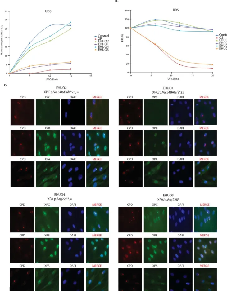
We next performed the unscheduled DNA synthesis (UDS) assay to measure the ability of these different cells to perform GGR, using UV-C irradiation to create 6-4PP and CPD [14]. The fibroblasts from both patients and parents, as well as reference fibroblasts from an XP patient a normal individual, were submitted to increased doses of UV. EHUO1 and EHUO3 fibroblasts exhibited a very low UDS (with UDS levels comparable to the XP positive control; around 10% of the EHUO2 and EHUO4 maternal fibroblasts and control) (Figure 3A).
Journal of Case Reports and Studies 6
Figure 3: Consequences of XPC and XPA mutations on NER pathways; (A) GG-NER pathway analysis on control (dark blue circle), XP (red
circle), EHUO1 (purple circle), EHUO2 (green circle), EHUO3 (blue circle) and EHUO4 (orange circle) fibroblasts measured by UDS assay; (B) TCR-NER pathway analysis on control (dark blue circle), CS (red circle), EHUO1 (purple circle), EHUO2 (green circle), EHUO3 (blue circle) and EHUO4 (orange circle) fibroblasts measured by RRS assay; (C) Recruitment of XPC, XPA and XPA to localized DNA damage (CPD) in EHUO1, EHUO2, EHUO3 and EHUO4 cells following UV irradiation. Cells were irradiated with a 254nm UV-C lamp at 120J/m2 dose and 15
minutes after irradiation were fixed and immunofluorescent staining was carried out. While the CPD photoproducts were detected in both XP and maternal cells, the NER proteins were only co-localized in the parental fibroblasts
7 Journal of Case Reports and Studies We also investigated TCR capacity of the patient fibroblasts by performing the recovery of RNA synthesis (RRS) assay upon UV irradiation. EHUO1 cells exhibited normal recovery of RNA synthesis, with similar levels to fibroblasts from parental EHUO2 and a normal individual (Figure 3B). However, the XPA-mutated EHUO3 cells failed to recover RNA synthesis after UV irradiation, just like the known TCR-deficient CS cells [15] (Figure 3B).
We next studied the dynamics of the NER factors in both XP-C and XP-A cells using localized UV irradiation combined with fluorescent immunostaining [16]. Confocal immunostaining showed that 15 minutes post UV irradiation, XPC co-localized with CPD in parental EHUO2 and EHU04 cells, allowing the subsequent recruitment of XPB, a subunit of TFIIH, and XPA at the damaged sites (Figure 3C, upper panels). Compared to parental fibroblasts, the absence of XPC in cells derived from XP-C patient EHUO1 led to the loss of co-localization between CPD and either XPB or XPA, in line with previous studies showing the sequential recruitment of XPC, TFIIH, XPA, RPA, and finally XPG/XPF NER factors to DNA lesions [16,17]. In EHUO3 patient cells, the truncated XPA failed to recognize the CPD damage sites which had already recruited XPC and XPB/TFIIH, (Figure 3C, lower panels).
Altogether these results indicated that both XPC and XPA mutations cause defective NER in these patients.
Having previously demonstrated the involvement of NER factors in RAR-target gene expression, and that these genes are downregulated in cells derived from XP patients, we tested whether these XPC and XPA mutations also altered expression of
Cells with XPC and XPA mutations have gene expression defects
Figure 4: Consequences of XPC and XPA mutations on RAR-target genes transactivation
Relative mRNA expression of RARβ2, CYP26A1, DAPK1 genes in EHUO1, EHUO2, EHUO3 and EHUO4 fibroblasts, 6 hours after ATRA treatment. Error bars represent the standard deviation of three independent experiments
Journal of Case Reports and Studies 8
Discussion
We described here the phenotype of two Algerian XP-C and XP-A patients originating from sun-exposed villages in the Oran city surroundings. The common XPC p.Val548Alafs variant present in the homozygous state in 87% of XP-C patients has been previously reported in patients mainly originating from North Africa [19,20]. The XPA p.Arg228* variant is present in 12% of XP-A patients worldwide, its frequency increasing to 87.5% in North Africa [13,21]. These patients exhibit extreme sensitivity to sunlight with some skin cancer development including melanocyte and keratinocyte malignancy, and eventually multiple basal cell carcinomas and invasive squamous cell carcinomas and melanomas [22]. The clinical features of these patients are linked to exposure to sunlight, and the complementation group and precise nature of the mutation. Some of the XP patients have neurological problems and intellectual deficiency that could be the due to progressive neuronal degeneration, also resulting in sensorineural deafness, ataxia and microcephaly. Although most of the above clinical features may be explained by deficiencies in DNA repair, and more precisely in NER, as shown above, we also show here that there are defects in gene expression, illustrated by impaired induction of RAR-target genes. Retinoids maintain certain skin functions and have therefore been proposed as a treatment for skin problems, such as in topical anti-acne and anti-wrinkle agents. Beside these functions, retinoids are also involved in the regulation of a large set of genes. As an example, CYP26 enzymes have a role in determining the cellular exposure to RA by metabolizing RA, and DAPK1 acts as an inhibitor of RIG-I, signaling production of type I interferon and as a tumor suppressor, downregulated in multiple cancer types [23,24]. A defect in the expression of these genes might lead to detrimental consequences, which could explain some clinical features in these patients, which warrants further attention.
As well as describing the biological phenotype of these two patients due to their environmental and social situations, our report emphasized for the first time the dual molecular etiology of XP patients, demonstrating both transcriptional and DNA repair defects. The identification of genes and the pathways regulated by NER factors would help to (i) determine relevant markers for an early and specific diagnosis, and to (ii) anticipate/predict the cancer risk among the different symptoms.
Acknowledgements
We are grateful to F. Coin and E. Compe for fruitful discussions. We also are indebted to the IGBMC cell culture facilities. We thank Cathy Obringer and Géraldine Greff for their excellent technical support. This study was supported by l’Agence Nationale de Recherche (ANR-12-BSU8-0017-01), la Fondation ARC pour la Recherche contre le Cancer (ARC n°SL220130607082), the Ligue contre Le cancer (CCIR-GE 2015). Maryssa Semer was supported by le prix d'encouragement à la recherche de la province Sud (Nouvelle Calédonie).
Statements
N.C, M.S, V.L, N.LM conducted experiments and analyzed data. Y.A, L.B, A.S conducted the survey. N.LM, JM.E., A.S., V.L conceived the project, supervised the research and wrote the manuscript with input and editing from all the authors. N.LM submitted the study. Authors declare no conflict of interest and no competing interests.
these genes [18]. Upon ATRA treatment, we observed in the EHUO2 and EHUO4 parental fibroblasts transactivation of several RAR-target genes, including Retinoic acid receptor isoform ß2 (RARß2), cytochrome P450 family 26 (CYP26) (involved in cell proliferation and RA metabolism) and the tumor suppressor gene death associated protein kinase 1 (DAPK1) (Figure 4). Such ATRA-mediated induction was strongly decreased in XP-C EHUO1 and XP-A EHUO3 patient fibroblasts compared to the parental cells. We thus demonstrate a correlation between the identified XPC and XPA mutations and the deregulation of RAR-responsive genes.
References
7. Compe E, Egly JM (2016) Nucleotide Excision Repair and Transcriptional Regulation: TFIIH and Beyond. Annu Rev Biochem 85: 265-90.
8. May NL, Egly JM, Coin F (2010) True lies: the double life of the nucleotide excision repair factors in transcription and DNA repair. J Nucleic Acids 10.4061/2010/616342.
1. Bootsma D, Kraemer KH, Cleaver JE, Hoeijmakers JHJ (2002) Nucleotide excision repair syndromes: xeroderma pigmentosum, Cockayne syndrome, and trichothiodystrophy In: The Genetic Basis of Human Cancer (2nd Edn) McGraw-Hill, USA.
2. Kleijer WJ, Laugel V, Berneburg M, Nardo T, Fawcett H, et al. (2008) Incidence of DNA repair deficiency disorders in western Europe: Xeroderma pigmentosum, Cockayne syndrome and trichothiodystrophy. DNA Repair 7: 744-50.
3. Hirai Y, Kodama Y, Moriwaki S, Noda A, Cullings HM, et al. (2006) Heterozygous individuals bearing a founder mutation in the XPA DNA repair gene comprise nearly 1% of the Japanese population. Mutat Res 601: 171-8.
4. Soufir N, Ged C, Bourillon A, Austerlitz F, Chemin C, et al. (2010) A prevalent mutation with founder effect in xeroderma pigmentosum group C from north Africa. J Invest Dermatol 130: 1537-42.
5. Ben Halim N, Ben Alaya Bouafif N, Romdhane L, Kefi Ben Atig R, Chouchane I, et al. (2013) Consanguinity, endogamy, and genetic disorders in Tunisia. J Community Genet 4: 273-84.
6. Hanawalt PC, Spivak G (2008) Transcription-coupled DNA repair: two decades of progress and surprises. Nat Rev Mol Cell Biol 9: 958-70.
9. Le May N, Fradin D, Iltis I, Bougnères P, Egly JM (2012) XPG and XPF endonucleases trigger chromatin looping and DNA demethylation for accurate expression of activated genes. Mol Cell 47: 622-32.
9 Journal of Case Reports and Studies
17. Riedl T, Hanaoka F, Egly JM (2003) The comings and goings of nucleotide excision repair factors on damaged DNA. Embo J 22: 5293-303.
18. Le May N, Mota-Fernandes D, Vélez-Cruz R, Iltis I, Biard D, et al. (2010) NER factors are recruited to active promoters and facilitate chromatin modification for transcription in the absence of exogenous genotoxic attack. Mol Cell 38: 54-66.
12. Satokata I, Tanaka K, Miura N, Narita M, Mimaki T, et al. (1992) Three nonsense mutations responsible for group A xeroderma pigmentosum. Mutat Res 273: 193-202.
13. Nishigori C, Zghal M, Yagi T, Imamura S, Komoun MR, et al. (1993) High prevalence of the point mutation in exon 6 of the xeroderma pigmentosum group A-complementing (XPAC) gene in xeroderma pigmentosum group A patients in Tunisia. Am J Hum Genet 53: 1001-6.
14. Kelly CM, Latimer JJ (2005) Unscheduled DNA synthesis: a functional assay for global genomic nucleotide excision repair. Methods Mol Biol 291: 303-20. 15. Mayne LV, Lehmann AR (1982) Failure of RNA synthesis to recover after UV radiation: an early defect in cells from individuals with Cockayne's syndrome and xeroderma pigmentosum. Cancer Res 42: 1473-8.
16. Volker M, Moné MJ, Karmakar P, van Hoffen A, Schul W, et al. (2001) Sequential assembly of the nucleotide excision repair factors in vivo. Mol Cell 8: 213-24.
19. Khan SG, Oh KS, Shahlavi T, Ueda T, Busch DB, et al. (2006) Reduced XPC DNA repair gene mRNA levels in clinically normal parents of xeroderma pigmentosum patients. Carcinogenesis 27: 84-9.
20. Li L, Bales ES, Peterson CA, Legerski RJ (1993) Characterization of molecular defects in xeroderma pigmentosum group C. Nat Genet 5: 413-7.
21. Cleaver JE, Thompson LH, Richardson AS, States JC (1999) A summary of mutations in the UV-sensitive disorders: xeroderma pigmentosum, Cockayne syndrome, and trichothiodystrophy. Hum Mutat 14: 9-22.
22. Benhamou S, Sarasin A (2000) Variability in nucleotide excision repair and cancer risk: a review. Mutat Res 462: 149-58.
23. Willemsen J, Wicht O, Wolanski JC, Baur N, Bastian S, et al. (2017) Phosphorylation-Dependent Feedback Inhibition of RIG-I by DAPK1 Identified by Kinome-wide siRNA Screening. Mol Cell 65: 403-15.
24. Kissil JL, Feinstein E, Cohen O, Jones PA, Tsai YC, et al. (1997) DAP-kinase loss of expression in various carcinoma and B-cell lymphoma cell lines: possible implications for role as tumor suppressor gene. Oncogene 15: 403-7.
Submit your next manuscript to Annex Publishers and
benefit from:
Submit your manuscript at
http://www.annexpublishers.com/paper-submission.php
→ Easy online submission process
→ Rapid peer review process
→ Open access: articles available free online
→ Online article availability soon after acceptance for Publication
→ Better discount on subsequent article submission
→ More accessibility of the articles to the readers/researchers within the field
10. Singh A, Compe E, Le May N, Egly JM (2015) TFIIH Subunit Alterations Causing Xeroderma Pigmentosum and Trichothiodystrophy Specifically Disturb Several Steps during Transcription. Am J Hum Genet 96: 194-207.
11. Calmels N, Greff G, Obringer C, Kempf N, Gasnier C, et al. (2016) Uncommon nucleotide excision repair phenotypes revealed by targeted high-throughput sequencing. Orphanet J Rare Dis 11: 26.