HAL Id: hal-02346975
https://hal.archives-ouvertes.fr/hal-02346975
Submitted on 8 May 2020
HAL is a multi-disciplinary open access archive for the deposit and dissemination of sci-entific research documents, whether they are pub-lished or not. The documents may come from teaching and research institutions in France or abroad, or from public or private research centers.
L’archive ouverte pluridisciplinaire HAL, est destinée au dépôt et à la diffusion de documents scientifiques de niveau recherche, publiés ou non, émanant des établissements d’enseignement et de recherche français ou étrangers, des laboratoires publics ou privés.
Bmpr2 Mutant Rats Develop Pulmonary and Cardiac
Characteristics of Pulmonary Arterial Hypertension
Aurélie Hautefort, Pedro Mendes-Ferreira, Jessica Sabourin, Grégoire
Manaud, Thomas Bertero, Catherine Rucker-Martin, Marianne Riou, Rui
Adão, Boris Manoury, Mélanie Lambert, et al.
To cite this version:
Aurélie Hautefort, Pedro Mendes-Ferreira, Jessica Sabourin, Grégoire Manaud, Thomas Bertero, et al.. Bmpr2 Mutant Rats Develop Pulmonary and Cardiac Characteristics of Pulmonary Arterial Hypertension. Circulation, American Heart Association, 2019, 139 (7), pp.932-948. �10.1161/CIRCU-LATIONAHA.118.033744�. �hal-02346975�
BACKGROUND: Monoallelic mutations in the gene encoding bone morphogenetic protein receptor 2 (Bmpr2) are the main genetic risk factor for heritable pulmonary arterial hypertension (PAH) with incomplete penetrance. Several Bmpr2 transgenic mice have been reported to
develop mild spontaneous PAH. In this study, we examined whether rats with the Bmpr2 mutation were susceptible to developing more severe PAH.
METHODS: The zinc finger nuclease method was used to establish rat lines with mutations in the Bmpr2 gene. These rats were then
characterized at the hemodynamic, histological, electrophysiological, and molecular levels.
RESULTS: Rats with a monoallelic deletion of 71 bp in exon 1 (Δ71 rats) showed decreased BMPRII expression and phosphorylated SMAD1/5/9 levels. Δ71 Rats develop age-dependent spontaneous PAH with a low penetrance (16%–27%), similar to that in humans. Δ71 Rats were more susceptible to hypoxia-induced pulmonary hypertension than wild-type rats. Δ71 Rats exhibited progressive pulmonary vascular remodeling associated with a proproliferative phenotype and showed lower pulmonary microvascular density than wild-type rats. Organ bath studies revealed severe alteration of pulmonary artery contraction and relaxation associated with potassium channel subfamily K member 3 (KCNK3) dysfunction. High levels of perivascular fibrillar collagen and pulmonary interleukin-6 overexpression discriminated rats that developed spontaneous PAH and rats that did not develop spontaneous PAH. Finally, detailed assessments of cardiomyocytes
demonstrated alterations in morphology, calcium (Ca2+), and cell contractility
specific to the right ventricle; these changes could explain the lower cardiac output of Δ71 rats. Indeed, adult right ventricular cardiomyocytes from Δ71
rats exhibited a smaller diameter, decreased sensitivity of sarcomeres to Ca2+,
decreased [Ca2+] transient amplitude, reduced sarcoplasmic reticulum Ca2+
content, and short action potential duration compared with right ventricular cardiomyocytes from wild-type rats.
CONCLUSIONS: We characterized the first Bmpr2 mutant rats and showed some of the critical cellular and molecular dysfunctions described in human PAH. We also identified the heart as an unexpected but
potential target organ of Bmpr2 mutations. Thus, this new genetic rat model represents a promising tool to study the pathogenesis of PAH.
© 2018 American Heart Association, Inc.
Aurélie Hautefort, PhD Pedro Mendes-Ferreira, PhD Jessica Sabourin, PhD Grégoire Manaud, MSc Thomas Bertero, PhD Catherine Rucker-Martin, PhD Marianne Riou, MD Rui Adão, PhD Boris Manoury, PhD Mélanie Lambert, MSc Angèle Boet, MD Florence Lecerf, MSc Valérie Domergue, BS Carmen Brás-Silva, PhD Ana Maria Gomez, PhD David Montani, MD, PhD Barbara Girerd, PhD Marc Humbert, MD, PhD Fabrice Antigny, PhD* Frédéric Perros, PhD*
ORIGINAL RESEARCH ARTICLE
Bmpr2 Mutant Rats Develop Pulmonary
and Cardiac Characteristics of Pulmonary
Arterial Hypertension
https://www.ahajournals.org/journal/circ
Circulation
*Drs Antigny and Perros contributed equally.
Key Words: bone morphogenetic
protein receptors, type II ◼ cardiovascular diseases ◼ hypertension, pulmonary
◼ interleukin-6 ◼ models, animal
◼ myocytes, cardiac
Sources of Funding, see page 946
Hautefort et al Characterization of Bmpr2 Mutant Rats
ORIGINAL RESEARCH
AR
TICLE
P
ulmonary arterial (PA) hypertension (PAH) is a se-vere, progressive disease clinically defined by an abnormal increase in pulmonary arterial pressure (PAP) >25 mm Hg and a PA wedge pressure of ≤15 mm Hg at rest, leading to right ventricular (RV) hyper-trophy and ultimately death resulting from right heart failure.1 Increased pulmonary vascular resistancere-sponsible for PAH is the consequence of progressive reduction in the cross-sectional area of the lumen of distal PAs. In 70% of heritable PAH (hPAH) and 15% to 40% of idiopathic PAH (iPAH), the disease develops in the setting of germline autosomal dominant mutations in the bone morphogenetic protein receptor 2 (BMPR2) gene, making BMPR2 mutations the main genetic risk factor for PAH development. However, the low pene-trance (≈20%) of BMPR2 mutations suggests that ad-ditional triggers are required for the development of PAH.2 There is in particular an influence of sex on the
development of PAH, with an approximate female:male ratio of 4:1, depending on the underlying disease pa-thology.3 In the context of BMPR2 mutation, this sexual
dimorphism is also striking because the female trance of the mutations is ≈42% and the male pene-trance is ≈14%.4
Bmpr2 transgenic mice display aberrant pulmonary
vascular cell phenotypes, including excessive prolifera-tion of medial smooth muscle cells (SMCs)5 and
apop-tosis of endothelial cells (ECs). Johnson et al6
demon-strated that Bmpr2 mutations also induce endothelial disruption.
Rat models of pulmonary hypertension (PH) have undoubtedly contributed to a better understanding of the PH process. Rats are more sensitive to PH devel-opment than mice.7 Moreover, recent developments of
novel molecular tools such as transcription activator-like effector nucleases, zinc finger nuclease, and clustered regulatory interspaced short palindromic repeats/Cas9 have enabled researchers to perform genome editing in rats.8 We used these new technologies to establish a
Bmpr2 mutant rat model using the zinc finger nuclease
method.
Accordingly, in this study, using a combination of hemodynamic measurements, molecular biology, elec-tron microscopy, Ca2+ imaging, and electrophysiology
approaches, we report the hemodynamic, histologi-cal, vascular, and molecular characterization of the first
Bmpr2 mutant rat line (harboring a monoallelic
muta-tion) during aging.
METHODS
The Bmpr2 mutant rat lines will be made available to other researchers for purposes of reproducing the results or repli-cating the procedure.
Generation of Bmpr2-Deficient Rats
Bmpr2 mutant rats were established using the zinc
finger nuclease method (for details, see the online-only Data Supplement and Table I in the online-only Data Supplement).
The animal facility is licensed by the French Ministry of Agriculture (agreement C92-019-01). This study was approved by the Committee on the Ethics of Animal Experiments CEEA26 CAP Sud. Animal experiments were approved by the French Ministry of Higher Education. Animal experiments were performed conforming to the guidelines from Directive 2010/63/EU 22 (September 2010) of the European Parliament on the protection of animals used for scientific purposes and complied with French institution’s guidelines for animal care and handling.
Bmpr2 Genotyping by Polymerase Chain
Reaction
See the online-only Data Supplement.
Hemodynamic Measurements, Evaluation
of RV Hypertrophy, and Collection of
Tissues
See the online-only Data Supplement.
Clinical Perspective
What Is New?
• We have created and characterized a new an-imal model of hereditary pulmonary arterial hypertension, which shares not only the same epidemiological characteristics with the human disease but also the same pathophysiological features observed in the pulmonary vascula-ture of patients with heritable pulmonary artery hypertension.
• Noticeably, we were able to show a not-yet-studied effect of bone morphogenetic protein receptor 2 (Bmpr2) mutation specifically on the right ventricle, which compromised intrinsic function, even in non-overloaded states.
What Are the Clinical Implications?
• The clinical implications for our findings are wide and show how BMPR2 mutations compromise not only the pulmonary vasculature but also specifically the heart.
• Our data should result in an improved under-standing of the intrinsic alterations of the right side of the heart in BMPR2 mutation carriers, suggesting a deleterious effect of this mutation in right ventricular adaptation in carriers devel-oping the disease and compromised response of these patients to standard therapy and lung transplantation.
ORIGINAL RESEARCH
AR
TICLE
Histology and Immunohistochemistry
See the online-only Data Supplement.
Real-Time Quantitative Polymerase Chain
Reaction
See the online-only Data Supplement.
Western Blot Analyses
See the online-only Data Supplement.
Isometric Tension Measurement
See the online-only Data Supplement.
Pulmonary Arterial SMC Isolation and
Culture
See the online-only Data Supplement.
Adult Cardiomyocyte Isolation
See the online-only Data Supplement.
SMC Proliferation Assays
See the online-only Data Supplement.
Electrophysiological Recordings
See the online-only Data Supplement.
In Vitro Studies in Isolated Skinned
Cardiomyocytes
See the online-only Data Supplement.
Measurement of [Ca
2+]
i
Transients and
Sarcoplasmic Reticulum Ca
2+Load
See the online-only Data Supplement.
Transmission Electron Microscopy
See the online-only Data Supplement.
Correlative Light and Electron
Microscopy
See the online-only Data Supplement.
Statistical Analysis
Analysis was performed with GraphPad Software version 6 (GraphPad Software Inc, San Diego, CA). All data were verified for normal distributions. Quantitative variables are presented as mean± SEM. Data were analyzed with Student t tests or 1-way ANOVA and Bonferroni multiple-comparisons tests. For the isolated cardiomyocyte experi-ments, 2-way ANOVA with repeated measures was used to analyze most parameters with the Tukey method for post hoc comparisons between groups. The Student t test was
used for calcium half-maximal effective concentration. Values of P<0.05 were considered statistically significant.
RESULTS
Monoallelic Bmpr2 Mutations in Rats
Reduced the BMPRII Canonical Signaling
Pathway in the Lungs
Zinc finger nuclease technology targeting the first exon of the Bmpr2 gene induced a monoallelic deletion of 71 bp (Δ71), which led to loss of an initiation codon (Figure 1A and 1B). In this study, we focused only on males because hemodynamic and histological altera-tions were observed in males but not in females (Figure I in the online-only Data Supplement). Females never-theless had a similar level of pulmonary BMPRII expres-sion (data not shown). We performed Western blot analysis to evaluate the effects of monoallelic Bmpr2 mutations on pulmonary BMPRII protein expression and canonical BMPRII signaling pathways. As expected, we observed a 50% decrease in the expression of BMPRII in the lungs of Bmpr2 mutant rats (Figure 1C), validat-ing our strategy. Because BMPRII signalvalidat-ing involves the SMAD pathway, we also showed a significant decrease in the phosphorylation of SMAD1/5/9 in the lungs of Δ71 rats (Figure 1C). Similar results were obtained in the second Bmpr2 mutant rat line (Δ140 rats; Figure IIA through IIC in the online-only Data Supplement). When we mated rats with monoallelic Bmpr2 mutations, we did not obtain biallelic mutation–bearing rats, and we observed a significant decrease in the number of pups (data not shown). This suggested that biallelic Bmpr2 mutations were lethal in utero, consistent with a previ-ous study demonstrating that homozygosity for a null mutation in Bmpr2 is lethal before gastrulation.9
Bmpr2 Mutant Rats Developed
Spontaneous PH in an Age-Dependent
Manner Concomitant With Alterations in
Distal Microvascular Density
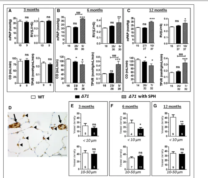
We performed right-sided heart catheterization in wild-type (WT) and Δ71 rats at 3, 6, and 12 months of age. Spontaneous PH (SPH) was defined by an mean PA pressure ≥25 mm Hg. As shown in Figure 2A, no SPH was observed in Δ71 rats at 3 months of age, as observed by RV hypertrophy (Fulton index), cardiac output (CO), total pulmonary vascular resistance (Fig-ure 2A), and carotid artery systemic pressure (data not shown) results similar to those of WT rats. However, 16.7% of animals (5 of 30) had SPH at 6 months of age in association with a significant increase in total pulmonary vascular resistance (Figure 2B). All Δ71 rats exhibited a significant decrease in CO (Figure 2B). At
Hautefort et al Characterization of Bmpr2 Mutant Rats
ORIGINAL RESEARCH
AR
TICLE
1 year, the penetrance of SPH was increased up to 27.8% (10 of 36), with concomitant increases in mean PA pressure, total pulmonary vascular resistance, and RV hypertrophy and a decrease in CO (Figure 2C). CO was also lower in non-SPH rats (Figure 2C). The sec-ond Bmpr2 mutant rat line (Δ140 rats) developed SPH at 12 months of age with a penetrance of 20% (5 of 25; Figure IIF in the online-only Data Supplement). Although Δ71 females did not develop SPH, their CO values tended to be lower at 6 months of age and were significantly lower at 12 months of age (Figure I in the online-only Data Supplement). The lower CO found in mutant rats may nevertheless be related at least in part to lower body weight (Tables II and III in the online-only Data Supplement). However, there is a complex interplay between the bone morphoge-netic protein signaling and body development,10,11 and
Bmpr2 mutations may be responsible for weight and
height abnormalities.
In WT rats, the density of pulmonary small microves-sels (<10 µm), expressed as number of CD31+ vessels
measured in 20 fields, progressively increased over time (23.4±4.7, 30.0±6.1, and 42.1±5.1 at 3, 6, and 12 months, respectively), whereas the density of larger mi-crovessels (between 10 and 50 µm) remained constant (44.0±5.8, 31.0±2.7, and 36.0±3.9, respectively) in WT rats (Figure 2D through 2G). Conversely, the density of
pulmonary small microvessels (<10 µm) did not increase over time in Δ71 rats (18.0±2.8, 17.0±2.7, and 24.0±3 at 3, 6, and 12 months, respectively). Consequently, compared with that in WT rats, the density of pulmo-nary small microvessels became significantly lower at 6 months of age (P<0.05) and even lower at 12 months of age (P<0.01; Figure 2D–2G).
Bmpr2 Mutant Rats With SPH Exhibited
Interleukin-6 Overexpression
In an attempt to identify a molecular marker of disease penetrance, we quantified the expression of the proin-flammatory cytokine interleukin (IL)-6 in lungs from af-fected and unafaf-fected animals compared with controls. Indeed, patients with iPAH exhibit increased IL-6 serum levels, which correlate with their prognoses.12
Consist-ent with these findings, lung-specific IL-6 transgenic mice display SPH in normoxia and develop greatly exag-gerated hypoxia-induced PH,13 whereas IL-6 receptor–
deficient mice show resistance to hypoxia-induced PH.14
These findings suggest that IL-6 has a significant role in the pathogenesis of PH. Consistent with these obser-vations, IL-6 was specifically overexpressed in Bmpr2 mutant rats with SPH, whereas non-SPH rats had levels similar to those of controls (Figure III in the online-only Data Supplement).
Figure 1. Generation and characterization of heterozygous bone morphogenetic protein receptor 2 (Bmpr2) mutant rats.
A, Schematic overview of the Bmpr2 gene with wild-type (WT) and Bmpr2 mutant alleles. We generated Bmpr2 mutant rats with a monoallelic deletion of 71 bp (Δ71) in exon 1 of the Bmpr2 gene, inducing the loss of the start codon (box). B, Validation of deletions by polymerase chain reaction, showing genomic DNA amplicons from rats carrying the WT and deleted alleles. Primers targeting the edges of deleted sequences detecting the WT allele (WT, 400 bp) and the allele with the deletion (ΔEx1, 329 bp). C, Representative Western blots and quantification of BMPR-II expression and phospho-SMAD1/5/9 levels in lungs from WT (n=5; white) and Δ71 (n=4; black) rats. β-Actin was used as loading control. Results are presented as mean±SEM. ns indicates not significant. *P<0.05; ***P<0.001.
ORIGINAL RESEARCH
AR
TICLE
Bmpr2 Mutant Rats Were More Susceptible
to Chronic Hypoxia-Induced PH
To investigate whether Bmpr2 mutations sensitized rats to chronic hypoxia-induced PH, we exposed 3-month-old Δ71 rats to 10% O2 for 3 weeks. Bmpr2 mutations increased the severity of hypoxic PH, as demonstrated by the observation that Δ71 rats had higher mean PA pres-sure values compared with WT rats at the end of the ex-periment (Figure IV in the online-only Data Supplement).
Bmpr2 Mutations Increased the
Muscularization of the Distal Pulmonary
Vascular Bed
Δ71 Rats had higher muscularization of smaller ves-sels (diameter <50 µm) at all analyzed time points (3,
6, and 12 months of age) compared with WT rats (Fig-ure 3A through 3D). Muscularization of larger vessels with diameters of 50 to 100 µm became significantly higher only at the latest time point (12 months of age; Figure 3A through 3D). We obtained similar results in the Δ140 strain (Figure IIG through III in the online-only Data Supplement). Moreover, Western blotting of the proproliferative mitogen-activated protein kinase path-way in lung tissues from Δ71 rats revealed that the phosphorylation of p38 mitogen-activated protein ki-nase was increased compared with that in WT rats at 1 year of age (Figure 3E). In addition, extracellular signal– regulated kinase 1/2 phosphorylation was significantly higher than in WT rats at earlier time points (3 and 6 months of age; Figure 3F). In vitro, PASMCs isolated from 6-month-old Δ71 rats had higher proliferation rates than those isolated from WT rats after treatment
Figure 2. Bone morphogenetic protein receptor 2 (Bmpr2) mutant rats developed spontaneous pulmonary hypertension (SPH) in an age-dependent
manner concomitant with alterations in distal microvascular density.
Assessment of mean pulmonary artery pressure (mPAP), cardiac output (CO), total pulmonary vascular resistance (TPVR), right ventricular hypertrophy (Fulton in-dex; weight ratio of right ventricle [RV] to left ventricle [LV] plus septum [S]) in Δ71 male rats without SPH (black) and wild-type (WT) male rats (white) at 3 (A), 6 (B), and 12 (C) months of age. Bmpr2 mutant rats were thought to have SPH when the mPAP was ≥25 mm Hg. SPH Δ71 rats are indicated with black-striped bars. D through G, Pulmonary vessel density quantification based on CD31 (endothelium marker) labeling of paraffin-embedded lung sections, expressed as number of CD31+ vessels measured in 20 fields (D; vessel diameter >10 µm, arrows; vessel diameter <10 µm, arrowheads) from Δ71 without SPH (black) compared with WT
(white) rats at 3 (E), 6 (F), and 12 (G) months of age. Vessel diameter <10 µm (top) and vessel diameter >10 µm (bottom). Each group at each time point includes different animals. ns indicates not significant. *P<0.05, **P<0.01, ***P<0.001 vs WT. $$$P<0.001 vs rats without SPH.
Hautefort et al Characterization of Bmpr2 Mutant Rats
ORIGINAL RESEARCH
AR
TICLE
with platelet-derived growth factor-BB (10 ng/mL) for 24 hours (Figure 3G).
Bmpr2 Deficiency Increased Perivascular
Fibrillar Collagen Deposition and
Assembly in Distal Pulmonary Arteries
Perivascular fibrillar collagen deposition and assembly were analyzed in Δ71 rats (Figure 4 and Figure V in the online-only Data Supplement). Although increased perivascular collagen deposition was observed in both small (<50 µm; Figure 4) and medium (50–100 µm; Fig-ure V in the online-only Data Supplement) PAs from
Δ71 rats at 3, 6, and 12 months of age compared with WT rats, a significantly higher level of fibrillar collagen assembly was also observed in both small and me-dium PAs in Δ71 rats with SPH at 6 and 12 months of age compared with Δ71 rats without SPH at 6 and 12 months of age.
Bmpr2 Mutations Induced Lung
Inflammation During Aging
We analyzed the effects of Bmpr2 deficiency in pul-monary inflammation by quantifying the expression of pulmonary CD45 protein (a pan leukocyte marker). We
Figure 3. Bone morphogenetic protein receptor 2 (Bmpr2) mutations increased the muscularization of the distal pulmonary vascular bed and enhanced
pulmonary artery smooth muscle cell (PASMC) proliferation.
A through D, Vessel muscularization analysis based on α-actin smooth muscle labeling on paraffin-embedded lung sections (A; top, wild-type [WT]; bottom, Δ71) from Δ71 (black) without spontaneous pulmonary hypertension (SPH) and WT (white) rats at 3 (B), 6 (C), and 12 (D) months of age. Vessel diameter <50 µm (top) and vessel diameter between 50 and 100 µm (bottom). E and F, Western blotting of (E) p38 phosphorylation (p) at 12 months of age and (F) extracellular signal– regulated kinase (ERK) 1/2 phosphorylation at 6 months of age in the lungs of Δ71 (black) rats without SPH and WT (white) rats and quantification of (E) p38 and (F) ERK1/2 phosphorylation levels in the lung of Δ71 (black) without SPH and WT (white) rats at 3, 6, and 12 months of age. β-Actin and α-tubulin were used as loading controls. G, Proliferation was measured with BrdU incorporation. Proliferation of PASMCs from Δ71 without SPH (black) and WT (white) rats after 24 hours of starva-tion, followed by treatment with platelet-derived growth factor-BB (10 ng/mL) for 24 hours (n=7 for each group). ns indicates not significant. *P<0.05; **P<0.01.
ORIGINAL RESEARCH
AR
TICLE
showed that lung CD45 expression was significantly in-creased in Δ71 rats at 1 year of age compared with WT rats (Figure VIA in the online-only Data Supplement). Hematoxylin-eosin staining revealed perivascular leuko-cyte accumulation at this later time point (Figure VIB in the online-only Data Supplement).
Bmpr2 Deficiency Caused Abnormal PA
Contraction, PASMC Depolarization,
Alteration of KCNK3 Function, and
Reduced Endothelium-Dependent PA
Relaxation
We induced contraction of PAs isolated from 6-month-old WT and Δ71 rats by increasing the KCl concentra-tion (from 10–120 mmol/L) to progressively depolarize PASMCs (Figure 5A). The contractile dose-response to KCl was significantly shifted to the left in PAs from Δ71 rats, demonstrating a predisposition to contraction (Fig-ure 5A). We also studied endothelium-dependent PA relaxation from WT and Δ71 rats by evaluating the dose response to acetylcholine after precontraction of PA with the thromboxane A2 agonist U46619 (1 µmol/L). PAs from Δ71 rats exhibited a significant decrease in acetylcholine-induced relaxation compared with WT rats (at 10 µmol/L acetylcholine; Figure 5B). This func-tional alteration occurred with ultrastructural changes in ECs. Indeed, electron microscopy imaging of
remod-eled PAs from Δ71 rats demonstrated disjointed and protrusive ECs containing abnormal vacuoles (Figure VII in the online-only Data Supplement).
Because the membrane potential (Em) of PASMCs partly controls the tone of PAs,15 we used a whole-cell
patch-clamp technique in current-clamp mode to meas-ure resting Em in freshly isolated PASMCs from Δ71 and WT rats. We observed a significant depolarization of the Em in Δ71 rats (Figure 5C). To explain this cellular depo-larization, we analyzed the KCNK3 current (IKNCK3), which strongly contributes to resting Em of PAMSCs.15 Using the
specific KCNK3 inhibitor A293 (200 nmol/L), we isolated the KCNK3 current from all K+ currents (A293-sensitive
current). The A293-sensitive current (IKCNK3) was strongly reduced in freshly isolated PASMCs from Δ71 rats (Fig-ure 5D). KCNK3 channels are known to be insensitive to Tetraethylammonium inhibited by external acidic pH (pH 6.3), and fully activated at basic pH (8.3). In another pro-tocol, we evaluated the KCNK3 current through meas-urement of the Tetraethylammonium-insensitive acidic-sensitive K+ current.15 This current was also reduced in
PASMCs from Δ71 rats, confirming that KCNK3 function was strongly reduced in the context of Bmpr2 muta-tions (Figure 5E). However, KCNK3 expression was not affected at the mRNA level in Δ71 rats (Figure 5F).
To explain the loss of KCNK3 function, we hypoth-esized that KCNK3 was not correctly localized to the plasma membrane. Accordingly, we analyzed the
sub-Figure 4. Bone morphogenetic protein receptor 2 (Bmpr2) mutations increased perivascular fibrillar collagen deposition and assembly in distal
(diameters <50 µm) pulmonary arteries.
Picrosirius Red staining and quantification of orthogonal signals in pulmonary arteries (diameters <50 µm) from Δ71 rats with (black-striped bars) or without
(black) spontaneous pulmonary hypertension (SPH) vs wild-type (WT; white) rats at 3, 6, and 12 months of age (n=5 for each group). Scale bar, 100 µm. *P<0.05, **P<0.01, ***P<0.001 vs WT. $$P<0.01 and $$$P<0.001 vs rats without SPH.
Hautefort et al Characterization of Bmpr2 Mutant Rats
ORIGINAL RESEARCH
AR
TICLE
Figure 5. Bone morphogenetic protein receptor 2 (Bmpr2) deficiency led to abnormal pulmonary artery (PA) contraction, PA smooth muscle cell (SMC)
plasma membrane depolarization, altered potassium channel subfamily K member 3 (KCNK3) function, and endothelium-dependent PA relaxation. A, Left, Dose-response curve established by applying increasing concentrations of KCl (10–120 mmol/L) in PAs isolated from Δ71 (n=4; black curve) and wild-type (WT; n=10; white curve) rats. Right, Half-maximal effective concentration (EC50) of KCl under previous conditions. B, Left, Relaxation dose-response curves established
by applying increasing concentrations of acetylcholine (Ach; 1 nmol/L to 10 µmol/L) on PA segments isolated from Δ71 (n=5; black) and WT (n=7; white) rats. Right, Corresponding bar graph of the percent relaxation at 10–7 mol/L of Ach. C, Resting membrane potential (Em) of PASMCs that were freshly isolated from Δ71 (black)
or WT (white, Em; 3 rats per condition; 25<n<35 cells). D, Current-voltage relationships of A293–K+-sensitive current (I
KCNK3) in PASMCs freshly isolated from Δ71
(n=6–10; black) and WT (n=9–14; white) rats. Bar graph represents KCNK3 current density measured at 60 mV for each condition. E, Left, Representative ramp cur-rents in freshly isolated PASMCs from WT and Δ71 rats, corresponding to the differences between the curcur-rents recorded at pH 8.3 and 6.3, called the acidic-sensitive K+ current. Right, Quantification of KCNK3 current density measured at 0 mV (6 <n <7 cells). F, mRNA expression of KCNK3 in PAs from Δ71 and WT rats (n=7 rats).
G, Transmission electron microscopy of pulmonary arteries from (a) WT and (b) Δ71 without spontaneous pulmonary hypertension (SPH) rats after KCNK3 staining. White squares correspond to ×2 magnification of SMCs 1 and 2. White arrows indicate plasma membrane KCNK3 localization; black arrows, cytoplasmic KCNK3 lo-calization. BL indicates basal lamina; Col, collagen; EC, endothelial cell; E, elastic; F, fibroblast; N, nucleus; and ns, not significant, *P<0.05; **P<0.01; ***P<0.001.
ORIGINAL RESEARCH
AR
TICLE
cellular localization of KCNK3 using correlative light and electron microscopy. As shown in Figure 5G, KCNK3 was expressed mainly at the cell surface of PASMCs from WT rats, whereas KCNK3 was not present at the cell surface of PASMCs from Δ71 rats and was abnor-mally retained in the cytosol and sarcoplasmic reticulum (SR) compartments.
Bmpr2 Mutant Rats Showed Intrinsic RV
Dysfunction
Hemodynamic measurements revealed a decrease in cardiac output in all Δ71 rats at 6 and 12 months of age (Figure 2), suggesting abnormalities in cardiac function in those animals. Using frozen RV and left ventricular (LV) samples from Δ71 rats without SPH at 6 months of age, we next measured the active tension of isolated cardiomyocytes. As shown in Figure 6, isolated skinned cardiomyocytes in RV samples from Δ71 rats showed a significant decrease in active tension development (Fig-ure 6A) and a decreased sensitivity to calcium (P<0.05; Figure 6B and 6C), whereas no changes were observed in passive tension (Figure 6D). Notably, no significant modifications were detected in LV cardiomyocytes (Fig-ure 6E through 6H). The intrinsic RV dysfunction oc-curring in unaffected Δ71 rats was not associated with decreased RV capillarization or RV fibrosis (Figure VIIIA and VIIIB in the online-only Data Supplement).
Bmpr2 Mutant RV Cardiomyocytes
Showed Decreased [Ca
2+]
i
Transients, SR
Ca
2+Load, and Cell Shortening
Decreased CO could also be related to alterations in Ca2+ handling involved in excitation-contraction
cou-pling. We analyzed electrically evoked [Ca2+]
i transients
and cell shortening in isolated RV and LV cardiomyo-cytes from WT and Δ71 rats without SPH (6 months of age). Figure 7A shows representative line-scan images (top) and corresponding fluorescence traces (bottom) of Fluo-4–loaded RV cardiomyocytes from WT and Δ71 rats on field stimulation at 1 Hz. The [Ca2+]
i transient
amplitude was significantly decreased in RV cardiomyo-cytes from Δ71 rats (Figure 7A and 7B), demonstrating compromised Ca2+ handling. The [Ca2+]
i transient
de-cay time was unchanged (Figure 7C), suggesting similar Ca2+ uptake by the SR calcium transport ATPase pump
in WT and Bmpr2-deficient rats. Consistent with this, we found no significant changes in SR Ca2+-ATPase
expression, total phospholamban expression, or phos-pholamban phosphorylation at serine 16/threonine 17 (Figure 7E). The decrease in the amplitude of [Ca2+]
i
transients was associated with a decrease in cell short-ening in RV myocytes from Δ71 rats (Figure 7D). No significant modifications were detected in LV
cardiomy-ocytes (Figure IXA through IXC in the online-only Data Supplement).
SR Ca2+ content is a determinant of the [Ca2+] i
tran-sient peak amplitude; therefore, we estimated SR Ca2+
loading by rapid caffeine application (10 mmol/L), as ex-emplified in Figure 7F for RV cardiomyocytes from WT and Δ71 rats. Caffeine-evoked [Ca2+]
i transients in RV
myocytes were significantly reduced in Bmpr2-deficient cardiomyocytes compared with WT cardiomyocytes (Figure 7F and 7G). Thus, the decrease in the amplitude of [Ca2+]
i transients may be related, at least in part, to
the reduction in SR Ca2+ load. The decay time of
caf-feine-induced [Ca2+]
i transients was similar in myocytes
from WT and Δ71 rats (Figure 7H), suggesting that Na+/Ca2+ exchange activity was maintained. We also
estimated the fractional release in electrically evoked twitches by normalizing the [Ca2+]
i transient amplitude
to the SR Ca2+ load. We showed similar fractional
re-lease in isolated cardiomyocytes from Δ71 rats and WT cardiomyocytes (Figure 7I), suggesting unchanged Ca2+
-induced Ca2+-released efficiency. No significant
modifi-cations were detected in LV cardiomyocytes (Figure IXA through IXE in the online-only Data Supplement).
Bmpr2 Deficiency Was Associated With
Decreased RV Cardiomyocyte Size and
Action Potential Remodeling
Reduction of SR Ca2+ content and [Ca2+]
i transients is
traditionally observed in left-sided heart hypertrophy and failure,16 which is associated with
electrophysiolog-ical remodeling, including action potential prolongation and decreased outward K+ current.16 Using the
whole-cell patch-clamp approach, we evaluated the whole-cell sur-face of isolated RV cardiomyocytes from WT and Δ71 rats (at 6 months of age) by measuring the cell capac-itance. The cell capacitance of cardiomyocytes isolated from Δ71 rats was significantly reduced compared with those from WT rats, indicating a decrease in the cell surface area (Figure 8A). By measuring the diameters of RV cardiomyocytes in histological sections, we con-firmed that the diameter of RV cardiomyocytes was de-creased in Δ71 rats compared with WT rats (Figure 8B), whereas cardiomyocyte size was unchanged in the LV compartment (data not shown).
To determine the effects of Bmpr2 deficiency on RV action potential, we analyzed action potential duration by patch-clamp experiments. We demonstrated that ac-tion potential repolarizaac-tion was significantly shortened at −30 mV in RV cardiomyocytes from Δ71 rats com-pared with WT rats (Figure 8C and 8D). Transient (Ito) and sustained (Isus) outward K+ currents play a critical role in
action potential repolarization. Thus, we analyzed Ito and
Isus in RV cardiomyocytes from Δ71 rats. The results in-dicated that Ito was significantly increased in Δ71 rats,
Hautefort et al Characterization of Bmpr2 Mutant Rats
ORIGINAL RESEARCH
AR
TICLE
Figure 6. Isolated skinned cardiomyocytes specifically from the right ventricle (RV) of bone morphogenetic protein receptor 2 (Bmpr2)–deficient rats
showed decreased active tension development and decreased sensitivity to calcium at 6 months of age.
In the RV compartment (A), the active tension–Ca2+ curve showed a significant decrease in tension development in cardiomyocytes isolated from Δ71 rats without
spontaneous pulmonary hypertension (SPH) compared with those isolated from wild-type (WT) rats. B, The normalized tension–Ca2+ curve showed a rightward
shift in Δ71 rats without SPH compared with WT rats. C, Calcium sensitivity was decreased in Δ71 rats without SPH compared with WT rats. EC50 for Ca2+ is the
Ca2+ concentration at which 50% of tension was developed. D, The passive tension–sarcomere length curve showed no differences between Δ71 rats without SPH
and WT rats. In the left ventricular (LV) compartment (E), the active tension–Ca2+ curve, (F) normalized tension–Ca2+ curve, (G) EC
50, and (H) passive
tension–sarco-mere length curve. ns indicates not significant were not changed between groups. *P<0.05; ***P<0.001.
ORIGINAL RESEARCH
AR
TICLE
whereas Isus was unchanged (Figure 8E and 8F), similar to IK1 (Figure IXF in the online-only Data Supplement), suggesting a key role of Ito in action potential shortening.
DISCUSSION
In this study, we performed hemodynamic, histological, cellular, electrophysiological, and molecular characteri-zation of 2 Bmpr2-deficient rat lines (Δ71 and Δ140).
This study has 5 major findings, as follows. First, we succeeded in generating a model of Bmpr2 haploinsuf-ficiency with a 50% decrease in BMPRII lung expres-sion associated with reduced activation of SMAD1/5/9 downstream signaling. Second, Bmpr2-deficient rats developed SPH in an age-dependent manner with a pen-etrance ranging between 16.7% and 27.8%, perfectly mimicking the PAH penetrance in patients carrying a
BMPR2 mutation, and were more susceptible to chronic
Figure 7. Decreased [Ca2+]
i transients, sarcoplasmic reticulum (SR) Ca2+ load, and cell shortening in right ventricular (RV) cardiomyocytes from bone morphogenetic protein receptor 2 (Bmpr2)–deficient rats at 6 months of age.
A, Top, Line scan of isolated RV cardiomyocytes loaded with Fluo-4/AM from Δ71 and wild-type (WT) rats. Bottom, Corresponding traces of [Ca2+]
i transients.
B, Amplitude of [Ca2+]
i transients (peak F/F0) obtained in RV myocytes field-stimulated at 1 Hz in Δ71 and WT rats. C, Average of [Ca2+]i transients decay time
(milliseconds) in RV myocytes in WT and Δ71 rats. D, Average percent cell shortening in RV myocytes in Δ71 and WT rats. E, Left, Representative Western blots of SR Ca2+-ATPase (SERCA2a), phospho-serine 16 (Ser17) / phospho-threonine 17 phospholamban (PLB), and total PLB levels in RV tissue from Δ71 and WT rats. GAPDH was used as a loading control. Middle and Right, Quantification of SERCA2a expression and the phospho-PLB/PLB ratio (n=12 different rats). F, Evalua-tion of SR Ca2+ load in single RV cardiomyocytes from Δ71 and WT rats. Top, Line scan of isolated RV cardiomyocytes loaded with Fluo-4/AM from WT and Δ71
rats. Bottom, Corresponding traces of caffeine-induced [Ca2+]
i transients. G, Amplitude of caffeine-induced SR Ca2+ load (peak F/F0) recorded in RV myocytes from
Δ71 and WT rats. H, Average of caffeine-evoked SR Ca2+ load decay time in RV myocytes from Δ71 and WT rats. I, Average fractional release in RV myocytes from
Δ71 and WT rats. ns indicates not significant. *P<0.05; ***P<0.001.
Hautefort et al Characterization of Bmpr2 Mutant Rats
ORIGINAL RESEARCH
AR
TICLE
hypoxia-induced PH. Third, this susceptibility to SPH and induced PH was concurrent with relevant pulmonary vascular alterations, including distal muscularization, perivascular matrix remodeling, lower microvascular density, and inflammation. High levels of perivascular fibrillar collagen and pulmonary IL-6 overexpression dis-criminated rats that developed SPH and rats that did not develop SPH. Fourth, Bmpr2 mutations predisposed rats to PA vasoconstriction through functional K+
cur-rent inhibition and subsequent PASMC depolarization. Changes in KCNK3 localization may be one of the main mechanisms responsible for these effects. Bmpr2 muta-tions were also found to be associated with a reduc-tion in endothelium-dependent relaxareduc-tion, which may be consistent with the major ultrastructural changes observed in the pulmonary endothelium of these rats. Finally, Bmpr2 mutant rats showed intrinsic RV dysfunc-tion, highlighted by PH-independent decreases in CO, RV cardiomyocyte size, and action potential duration; decreases in active tension development and sensitiv-ity to Ca2+ in skinned RV cardiomyocytes; and reduced
[Ca2+]
i transients, SR Ca2+ load, and cell shortening. All
these alterations were observed in Bmpr2 mutant rats without SPH. The study of these rats allowed us to de-lineate the primary contribution of the mutation to the phenotype independently of the alterations occurring secondary to PH and subsequent molecular and cellular activations. The former mechanisms are at the heart of PH susceptibility.
The variety of pulmonary vascular alterations that we observed in Bmpr2-mutant rats were consistent with recent findings related to pulmonary hemodynamic disturbances in PAH.17 Indeed, narrowing of resistance
vessels is heterogeneous, and this heterogeneous nar-rowing alone cannot explain the large increase in re-sistance in PAH. Previous studies have suggested that rarefaction could be an important contributor to the hemodynamic changes.17 The abnormally low
micro-vascular density observed in Bmpr2 mutant rats during aging supported this hypothesis. Moreover, patients with PAH harboring BMPR2 mutations are less likely to respond to acute vasodilator testing.18 The reduction
Figure 8. Bone morphogenetic protein receptor 2 (Bmpr2) deficiency is associated with decreased right ventricular (RV) cardiomyocytes size and
action potential (AP) remodeling.
A, RV cardiomyocyte capacitance from Δ71 and wild-type (WT) rats (32–37 cells from 3 rats). B, Quantification of the diameters of RV cardiomyocytes in Δ71 and WT rats (10–15 images per animal from 3–4 rats). C, Representative APs in RV cardiomyocytes from Δ71 and WT rats. D, Analysis of AP duration (APD) at 0-, −30-, and −60-mV membrane repolarization in Δ71 and WT rats (9–14 cells from 3 rats). E, Left, Current-voltage relationship of transient outward-K+ currents (I
to) from
RV cardiomyocytes from Δ71 and WT rats. Right, Average Ito density (10–15 cells from 3 rats). F, Left, Current-voltage relationships of sustained outward-K+
cur-rents (Isus) from RV cardiomyocytes from Δ71 and WT rats. Right, Average Isus density (10–15 cells from 3 rats). ns indicates not significant. *P<0.05; ***P<0.001.
ORIGINAL RESEARCH
AR
TICLE
in endothelium-dependent relaxation found in Bmpr2-deficient animals may partly explain this clinical obser-vation. Increasing evidence also suggests a central role for the biochemical and biomechanical properties of the extracellular matrix in PH.19 However, it is unclear
whether extracellular matrix remodeling is merely an end-stage feature of PH or whether massive fibrosis is necessary to induce pathogenic outcomes. Numerous animal models of PH/PAH exist; however, these accel-erated models of PH development make it difficult to obtain accurate insights into the pathological events. Our Bmpr2 transgenic rat model clarified these points by allowing us to study the early development of peri-arteriolar fibrosis in Bmpr2 mutant rats, even before physiological manifestation of PH (3 months of age). Furthermore, increased vascular fibrosis was markedly pronounced in Bmpr2 mutant rats that develop SPH compared with Bmpr2 mutant rats that did not develop SPH, suggesting a PH-dependent threshold of fibrillar collagen and resulting perivascular stiffness.19 Taken
to-gether, these results reinforce the importance of me-chanical cues in the development of cardiovascular dis-eases such as PH/PAH, even in genetically driven forms. Overall, our results presented a valuable model to study perivascular extracellular matrix properties during PH/ PAH development.
Another valuable finding was the reduced CO ob-served in both Bmpr2-deficient rat lines from 6 months of age. Indeed, data from the Registry to Evaluate Early and Long-Term PAH Disease Management demonstrat-ed >2-fold increases in hazard ratios for mortality a-mong patients with hPAH compared with patients with iPAH.20 Furthermore, RV stroke work index and cardiac
index are both decreased in hPAH,21,22 suggesting the
potential impairment of RV function in this PAH sub-type. Notably, Claessen and colleagues23 have shown
that humans with asymptomatic BMPR2 mutations did not show abnormal RV contractile reserve, as measured by the change in contractility at rest and at peak exer-cise, even when under hypoxic conditions. Despite that, under the same conditions, the increase in RV ejection fraction is significantly lower in BMPR2 carriers while also showing decreased pulmonary arterial compliance and mean PA pressure at rest, and a close-to-signifi-cant decrease in cardiopulmonary fitness, suggesting a small degree of intrinsic RV dysfunction in these pa-tients. Moreover, van der Bruggen et al24 recently
dem-onstrated, by combining in vivo measurements of RV function with molecular and histological analyses of unique RV and LV tissues from patients with PAH and controls, that despite a similar afterload, RV function is more severely compromised in BMPR2 mutation carriers than in noncarriers. These findings suggest that BMPR2 mutations may have a negative effect on RV function in patients with PAH. To date, differences in survival and disease severity have been explained mainly by more
severe pulmonary vascular involvement, leading to a more severe and faster disease trajectory. However, be-cause RV function is the main determinant of prognosis and disease severity and because the BMPR2 gene is also expressed in the RV, RV function may play a role in the different clinical phenotypes of mutation carri-ers and noncarricarri-ers. In animals, impaired hypertrophy attributable to altered cardiac energy metabolism in the RV has been demonstrated in a transgenic mouse model expressing mutant Bmpr2.25 In this study, the
in-duced impairment of cardiomyocyte BMPRII signaling via a receptor cytoplasmic domain mutation limited the ability of the RV to undergo hypertrophy in response to increased pressure. Thus, mutant BMPRII expression in the RV may impair hypertrophic responses resulting in part from fatty acid oxidation defects. Indeed, fatty acid oxidation is the preferred energy source in cardio-myocytes.26 Furthermore, cardiomyocytes derived from
mouse embryonic stem cells mutant for either the cyto-plasmic or kinase domain of BMPRII showed no increase in expression of brain natriuretic peptide in response to phenylephrine-induced hypertrophy,27
demonstrat-ing the incapacity to respond to hypertrophic stimuli. In fact, mice mutant for bmpr2 show decreased exer-cise capacity at baseline and respond worse to pressure overload.28 Of note, induced pluripotent stem
cell–de-rived cardiomyocytes differentiated from patients with hPAH and BMPR2 mutation showed decreased expres-sion of hypertrophy-associated genes (MYH6/7 and
ANP), further corroborating the lack of adaptation of
these cardiomyocytes, which was corrected by BMPR2 mutation correction.29
Cardiomyocytes from the RV of patients with PAH were found to show increased cardiomyocyte stiffness (increased passive tension) and hypercontractility, with increases in both active tension development and im-proved calcium sensitivity.30 Those mechanisms,
sug-gested to cope for the increasing afterload imposed on the RV in PAH, seem to have been lost in our Bmpr2 mutant rats. In contrast, decreased active tension and calcium sensitivity are associated with RV dysfunction in pressure overload31 and could be similar to our
ob-servations in Bmpr2 mutant rats. Indeed, we observed Ca2+ cycling alterations and depressed cell contractility
associated with decreased calcium sensitivity for sarco-meres. Patients with systemic sclerosis–associated PAH show decreased RV functional reserve, demonstrating decreased adaption to exercise and a lack of contrac-tility increase in response to increased load.32 At the
same time, these patients have decreased mean PAP compared with patients with iPAH33,34 and present
with lower cardiomyocyte active force development compared with both patients with iPAH and control subjects,35 similar to our results in Bmpr2 mutant
an-imals. These findings, together with the fact that a-symptomatic patients carrying BMPR2 mutations show
Hautefort et al Characterization of Bmpr2 Mutant Rats
ORIGINAL RESEARCH
AR
TICLE
decreased pulmonary compliance, which is further ag-gravated by hypoxia,36 may explain the susceptibility of
these patients to PAH with a more severe RV dysfunc-tion. Future studies will focus on a more detailed evalu-ation of cardiovascular function (including PV loops and magnetic resonance imaging), which will determine to what extent the RV (and LV) of the Bmpr2 mutant rats is compromised and will aim at determining the adapt-ability of these hearts to increased load.
We functionally evaluated BMPRII deficiency via the decrease in SMAD1/5/8 phosphorylation. BMPRII inter-acts directly with several proteins relevant to regulation of the actin cytoskeleton, including LIMK, TCTEX, and SRC.6 Actin cytoskeleton defect is a common feature
in all classes of BMPR2 mutations found in mouse and human instances of PH/PAH.6 Furthermore, an
alterna-tive spliced variant of BMPR2 lacking exon 12 has been found to be overrepresented in cells from BMPR2 mu-tant patients with PAH. Exon 12 forms the intracyto-plasmic tail domain of BMPRII that interacts with the cytoskeleton regulatory proteins.37 In line with these
results, it is tempting to speculate that the lower PH penetrance in Δ140 rats arising in a paradoxical more altered SMAD signaling background may be related to different cytoskeletal defects. Further investigation is needed to determine the role of actin cytoskeleton on the occurrence and severity of PAH in Δ71 and Δ140 rats.
Δ71 and Δ140 Sprague Dawley rats develop mild SPH. However, it has been demonstrated that variation in strain, even between colonies of the same strain, has a remarkable influence on the nature and severity of the response to an experimental PH trigger such as SU5416, consistent with an important role for genetic modifiers of the PAH phenotype.38 Consistent with this
concept, it was found that chronic hyperglycemia in-duces vascular damage predominantly in PAs, leading to PH in Wistar Kyoto but not in Sprague Dawley rats.39
Further studies are needed to investigate the role of the genetic background on the occurrence and severity of PAH in the context of Bmpr2 mutation.
Our model has 2 main limitations:
1. Bmpr2 mutant female rats did not develop SPH,
whereas the prevalence of iPAH and hPAH is ≈4-fold higher in women than in men.3 Actually, this
is not so surprising because there is a paradox-ical opposite male bias in typparadox-ical rodent mod-els of PH (chronic hypoxia or monocrotaline); in these models, administration of estrogenic com-pounds (eg, estradiol-17 β) is protective (estrogen paradox).40 This paradox is poorly understood.
However, Sehgal et al40 described an estradiol-17
β–sensitive central neuroendocrine mechanism of sex bias that culminated in species-specific male (pulsatile) versus female (more continuous) tem-poral patterns of circulating growth hormone
(GH) levels.40 In adult male rats, GH is released
into the circulation in discrete pulses, with little or no circulating GH detectable during the inter-pulse interval. In female rats, the inter-pulses are more frequent, the pulse heights are lower, and the interpulse levels are higher. In contrast, men show infrequent GH pulses of high magnitude, with very low interpulse levels; however, women show a more continuously high level of GH. On average, women have 80- to 120-fold higher lev-els of circulating GH than men. Thus, a difference between female humans and female rodents is the much higher continuous GH levels in women compared with female rodents. These 80- to 120-fold higher levels and a more continuous pattern of GH, and thus activation of different sets of cell cycle, cell proliferation, and cell migration regula-tory genes, may explain why iPAH is more preva-lent in women than men.40 Moreover, there are
significant physiological differences in hemato-logic and biochemical analytes between male and female Sprague Dawley rats and between rodents and humans.41 For instance, men, premenopausal
women, and postmenopausal women have similar plasma erythropoietin levels,42 whereas
erythro-poietin levels in male rats are lower than in female rats.43 Given that erythropoietin attenuates
pul-monary vascular remodeling in experimental PH through interplay between endothelial progenitor cells and activation of the cytoprotective enzyme heme oxygenase-1,44 this may underlie male
sus-ceptibility to PH in rat. Overall, factors such as ge-netic background, diet, temperature, hormonal cycles, metabolic rate, enzyme activities, and cir-cadian rhythms all can influence the metabolism of an organism.41
2. Bmpr2 mutated rats display variable expression
of the PH phenotype. Actually, the rats used in this study have a Sprague Dawley background, which is an outbred strain. By definition, outbred strains maintain genetic variation to conserve het-erozygosity within its population. Therefore, the likelihood of other mutations contributing as a second hit, promoting the development of PAH, is decreased. The entailed genetic variability as-sociated with these rats is sufficient to change their growth rate,45 highlighting the significant
role of outbred-associated variability, and results in increased susceptibility to certain diseases when polymorphisms/mutations on specific genes are present,46 with low penetrance,47 similar to our
own. Finally, Sprague Dawley rats hyperresponsive to vascular endothelial growth factor inhibition de-velop severe PH, but again with incomplete pen-etrance. Although 72% of male rats developed PH, only 27% of female rats developed PH. These
ORIGINAL RESEARCH
AR
TICLE
findings strengthen the fact that outbred strain (like the Sprague Dawley rat used in our study) can present increased susceptibility to PH as the result of a mutation, but with incomplete penetrance. Moreover, and consistent with our results, these data demonstrated that females are less prone to exhibit a phenotype.48 Together, these findings
argue in favor of the facts that the presence of the same mutation and environment contribute to the development of BMPR2 mutation-associ-ated SPH and that a significantly diverse genetic background, resulting from the use of an outbred species, greatly contributes to a lack of complete penetrance. Moreover, mice with endothelium-specific Bmpr2 deletion49 show PH development
in only 20% of heterozygous mutation–carrying animals, whereas 39% of homozygous animals show increased pulmonary pressures, demonstrat-ing that even in an inbred strain bmpr2 mutation does not result in complete penetrance of PH.
Conclusions
Rat lines with monoallelic mutation in the Bmpr2 gene did not develop severe PAH, but exhibited salient pathobiological features of hPAH, including similar pen-etrance, pulmonary vascular remodeling, and reduced microvascular density, and showed endothelial dysfunc-tion associated with altered vasoreactivity and overac-tivation of mitogenic signaling pathways. Thus, these rats are valuable models for studying PAH susceptibility mechanisms related to BMPR2 mutations. However, the slow onset (6–12 months), low incidence, and mild di-sease may discourage their use as a preclinical model of PAH harboring the late symptomatic clinical presenta-tion of the human disease. Our data also showed that
Bmpr2 mutants exhibited intrinsic cardiomyocyte
dys-function, which could underscore their increased sus-ceptibility to cardiac overload.
ARTICLE INFORMATION
Received March 8, 2018; accepted September 18, 2018.
The online-only Data Supplement is available with this article at https:// www.ahajournals.org/doi/suppl/10.1161/circulationaha.118.033744.
Correspondence
Frédéric Perros, PhD, INSERM U999, Centre Chirurgical Marie Lannelongue, 133 Avenue de la Résistance, F-92350 Le Plessis Robinson, France. Email frederic.perros@inserm.fr
Affiliations
Université Paris-Sud, Faculté de Médecine, Le Kremlin-Bicêtre, France (A.H., G.M., C.R.-M., M.R., M.L., F.L., D.M., B.G., M.H., F.A., F.P.). AP-HP, Centre de Référence de l’Hypertension Pulmonaire Sévère, Département Hospitalo-Univer-sitaire Thorax Innovation, Service de Pneumologie et Réanimation Respiratoire, Hôpital de Bicêtre, Le Kremlin-Bicêtre, France (A.H., G.M., C.R.-M., M.R., M.L., F.L., D.M., B.G., M.H., F.A., F.P.). UMRS 999, INSERM and Université Paris-Sud,
Laboratoire d’Excellence en Recherche sur le Médicament et l’Innovation Théra-peutique, Le Plessis Robinson, France (A.H., G.M., C.R.-M., M.R., M.L., F.L., D.M., B.G., M.H., F.A., F.P.). Centre Chirurgical Marie Lannelongue, Le Plessis Robinson, France (A.H., G.M., C.R.-M., M.R., M.L., A.B., F.L., D.M., B.G., M.H., F.A., F.P.). Department of Surgery and Physiology, Faculty of Medicine, UnIC-Cardiovascular Research and Development Centre, University of Porto, Portu-gal (P.M.-F., R.A., C.B.-S.). Respiratory Division, Department of Department of Chronic Diseases, Metabolism & Aging, KU Leuven–University of Leuven, Bel-gium (P.M.-F.). Signalisation et Physiopathologie Cardiovasculaire, UMR-S 1180, Université Paris-Sud, INSERM, Université Paris-Saclay, 92296, Châtenay-Malabry, France (J.S., B.M., A.M.G.). Université Côte d’Azur, CNRS, IRCAN, Nice, France (T.B.). Animal Facility, Institut Paris Saclay d’Innovation Thérapeutique (UMS IPSIT), Université Paris-Sud, Université Paris-Saclay, Châtenay-Malabry, France (V.D.). Centre de Recherche de l’Institut Universitaire de Cardiologie et de Pneu-mologie de Québec, Laval University, QC, Canada (F.P.).
Acknowledgments
The authors thank Dr Ignacio Anegon, Laurent Tesson, and Séverine Ménoret from Transgenic Rats and Immunophenomics Core Facility (TRIP), platform TRIP–(Immunology–Nantes) for generating Bmpr2 mutant rats. They thank the staff at the Animalerie et Exploration Fonctionnelle (AnimEx) platform for caring for the rat lines. They also thank the Microscopie et Imagerie des Micro-organ-ismes, Animaux et Aliments (MIMA2) imaging facility (Institut national de la re-cherche agronomique [INRA]), Jouy-en-Josas; http://www6.jouy.inra.fr/mima2) for the transmission electron microscopy images and Christine Pechoux and Martine Letheule for excellent technical support. They thank Prof Adelino Leite-Moreira for collaboration with Cardiovascular R&D Unit (UnIC) and Naomi Ka-minsky for quantifying heart capillarization. They acknowledge the support of the European Respiratory Society, Fellowship LTRF 2017 (LTRF-2017 01–00063).
Sources of Funding
Dr Perros received funding from the French National Research Agency (Agence Nationale de la Recherche, Grant ANR-13-JSV1-0011-01) and from the Fonda-tion du Grand défi Pierre Lavoie. Dr Perros also received a Pulmonary Vascular Research Institute BMPR2 Research Grant supported by the Dinosaur Trust. Dr Hautefort is supported by a PhD grant from Région Ile de France (Domaine d’Intérêt Majeur “maladies Cardiovasculaires-Obésité-Rein-Diabète”) and by the Fondation de la Recherche Médicale. G. Manaud and Dr Riou are supported by the Fonds de dotation Recherche en Santé Respiratoire. M. Lambert is sup-ported by Therapeutic Innovation Doctoral School (Ecole Doctorale ED569). Dr Antigny receives funding from the Fondation du Souffle et Fonds de Dotation Recherche en Santé Respiratoire, Fondation Lefoulon-Delalande, and Fondation Legs Poix. Drs Mendes-Ferreira, Adão, and Brás-Silva receive funds from the European Union through the European Regional Development Fund, European Structural and Investment Funds, under Lisbon Portugal Regional Operational Program and Fundação para a Ciência e a Tecnologia (DOCnet: NORTE-01-0145-FEDER-000003; IMPAcT-PTDC/MED-FSL/31719/2017), and Drs Mendes-Ferreira and Adão are supported by Fundação para a Ciência e a Tecnologia (SFRH/BD/87714/2012 and SFRH/BD/96403/2013, respectively).
Disclosures
None.
REFERENCES
1. Hoeper MM, Bogaard HJ, Condliffe R, Frantz R, Khanna D, Kurzyna M, Langleben D, Manes A, Satoh T, Torres F, Wilkins MR, Badesch DB. Definitions and diagnosis of pulmonary hypertension. J Am Coll Cardiol. 2013;62(suppl):D42–D50. doi: 10.1016/j.jacc.2013.10.032
2. Guignabert C, Bailly S, Humbert M. Restoring BMPRII func-tions in pulmonary arterial hypertension: opportunities, challenges and limitations. Expert Opin Ther Targets. 2017;21:181–190. doi: 10.1080/14728222.2017.1275567
3. Mair KM, Johansen AK, Wright AF, Wallace E, MacLean MR. Pulmonary arterial hypertension: basis of sex differences in incidence and treatment response. Br J Pharmacol. 2014;171:567–579. doi: 10.1111/bph.12281 4. Larkin EK, Newman JH, Austin ED, Hemnes AR, Wheeler L, Robbins IM,
West JD, Phillips JA, Hamid R, Loyd JE. Longitudinal analysis casts doubt on the presence of genetic anticipation in heritable pulmonary arterial


![Figure 7. Decreased [Ca 2+ ] i transients, sarcoplasmic reticulum (SR) Ca 2+ load, and cell shortening in right ventricular (RV) cardiomyocytes from bone morphogenetic protein receptor 2 (Bmpr2)–deficient rats at 6 months of age](https://thumb-eu.123doks.com/thumbv2/123doknet/13513705.416316/12.877.98.793.77.759/decreased-transients-sarcoplasmic-reticulum-shortening-ventricular-cardiomyocytes-morphogenetic.webp)

