R E S E A R C H
Open Access
Association between hydroxocobalamin
administration and acute kidney injury after
smoke inhalation: a multicenter
retrospective study
François Dépret
1,2,3,4, Clément Hoffmann
5, Laura Daoud
1, Camille Thieffry
6, Laure Monplaisir
1, Jules Creveaux
5,
Djillali Annane
7, Erika Parmentier
6, Daniel Mathieu
6, Sandrine Wiramus
8, Dominique Demeure DIt Latte
9,
Aubin Kpodji
10, Julien Textoris
11, Florian Robin
12, Kada Klouche
13, Emmanuel Pontis
14, Guillaume Schnell
15,
François Barbier
16, Jean-Michel Constantin
17, Thomas Clavier
18,19, Damien du Cheyron
20, Nicolas Terzi
21,
Bertrand Sauneuf
22, Emmanuel Guerot
23,24, Thomas Lafon
25,26, Alexandre Herbland
27, Bruno Megarbane
28,
Thomas Leclerc
4, Vincent Mallet
29, Romain Pirracchio
30and Matthieu Legrand
1,2,3,5,31*Abstract
Background: The use of hydroxocobalamin has long been advocated for treating suspected cyanide poisoning after smoke inhalation. Intravenous hydroxocobalamin has however been shown to cause oxalate nephropathy in a single-center study. The impact of hydroxocobalamin on the risk of acute kidney injury (AKI) and survival after smoke inhalation in a multicenter setting remains unexplored.
Methods: We conducted a multicenter retrospective study in 21 intensive care units (ICUs) in France. We included patients admitted to an ICU for smoke inhalation between January 2011 and December 2017. We excluded patients discharged at home alive within 24 h of admission. We assessed the risk of AKI (primary endpoint), severe AKI, major adverse kidney (MAKE) events, and survival (secondary endpoints) after administration of hydroxocobalamin using logistic regression models.
Results: Among 854 patients screened, 739 patients were included. Three hundred six and 386 (55.2%) patients received hydroxocobalamin. Mortality in ICU was 32.9% (n = 243). Two hundred eighty-eight (39%) patients developed AKI, including 186 (25.2%) who developed severe AKI during the first week. Patients who received hydroxocobalamin were more severe and had higher mortality (38.1% vs 27.2%, p = 0.0022). The adjusted odds ratio (95% confidence interval) of AKI after intravenous hydroxocobalamin was 1.597 (1.055, 2.419) and 1.772 (1.137, 2.762) for severe AKI; intravenous hydroxocobalamin was not associated with survival or MAKE with an adjusted odds ratio (95% confidence interval) of 1.114 (0.691, 1.797) and 0.784 (0.456, 1.349) respectively.
Conclusion: Hydroxocobalamin was associated with an increased risk of AKI and severe AKI but was not associated with survival after smoke inhalation.
Trial registration:ClinicalTrials.gov, NCT03558646
Keywords: Smoke inhalation, Acute kidney injury, Intensive care unit, Mortality, Burn, Hydroxocobalamin
© The Author(s). 2019 Open Access This article is distributed under the terms of the Creative Commons Attribution 4.0 International License (http://creativecommons.org/licenses/by/4.0/), which permits unrestricted use, distribution, and reproduction in any medium, provided you give appropriate credit to the original author(s) and the source, provide a link to the Creative Commons license, and indicate if changes were made. The Creative Commons Public Domain Dedication waiver (http://creativecommons.org/publicdomain/zero/1.0/) applies to the data made available in this article, unless otherwise stated.
* Correspondence:matthieu.m.legrand@gmail.com
1
Department of Anesthesiology and Critical Care and Burn Unit, AP-HP, GH Saint Louis-Lariboisière, Paris, France
2UMR INSERM 942, Institut National de la Santé et de la Recherche Médicale
(INSERM), Paris, France
Introduction
Patients with smoke inhalation are at a high risk of death or major morbidities. Cyanide is a documented cause for rapid death after intoxication from smoke
[1]. Several cyanide antidotes have been proposed
(i.e., sodium nitrite, amyl nitrite, sodium thiosulfate, 4-dimethylaminophenol, dicobalt edetate, and hydro-xocobalamin) with utilization varying across countries [2]. Several experts suggested using hydroxocobalamin as the first-line treatment after suspected cyanide in-toxication due to its perceived good safety profile [3]. However, other experts raised concerns about liberal-izing the use of hydroxocobalamin after smoke inhal-ation due to a lack of data on both efficacy and tolerance. They have called for additional investiga-tions [4, 5].
Recently, nephrotoxicity of hydroxocobalamin due to oxalate nephropathy was reported in a single-center study among burn patients [4]. The objective of this co-hort study was to assess the association between the use of hydroxocobalamin and the risk of acute kidney injury (AKI) and death in intensive care unit (ICU) patients with smoke inhalation.
Methods
Study population and settings
We conducted a retrospective, multicenter study in 21 ICUs in France. All consecutive adult (≥ 18 years old) patients admitted between January 2011 and De-cember 2017 with the final diagnosis of smoke inhal-ation were included in this study. Patients discharged alive at home within 24 h from admission (reflecting absence of severity) were excluded. Patient files were retrieved using our national coding for smoke inhal-ation injury (code CIM T599 for all patients and as-sociated: X00·0, X 09·0, X09·9, X47·0, X47·08, X47·9, X67·0, X67·9, T58) (Additional file 1: Table S1). This academic investigator-driven study was registered at
ClinicalTrials.gov, NCT03558646. The study protocol was approved by our local research ethical committee (Comité de Protection des Personnes 2013/17NICB). The use of patient’s data was allowed in cases of death and/or when proxies could not be contacted.
Primary outcome
The primary outcome was AKI. AKI was defined accord-ing to Kidney Disease Improvaccord-ing Global Outcome (KDIGO) within 7 days following admission using the serum creatinine criteria [6].
Secondary outcome
Secondary outcomes were severe AKI (severe AKI defined patients by AKI stage 2 or 3), major associated kidney events (MAKE) in the ICU, which includes death and/or
renal replacement therapy (RRT), and/or persistent AKI at ICU discharge. Persistent AKI was defined as an elevated SCreat level from baseline by > 1.5-fold or > 0.3 mg/dL (26.3μmol/L) at ICU discharge or RRT at ICU discharge and survival in the ICU.
Data collections and definitions
Data were collected through a standardized case re-port form. Each form was manually encoded in each participating center using the initials and year of birth of the patient. Admission serum creatinine (SCreat) was used for baseline SCreat. Severe burn was defined
as total body surface area (TBSA) burn ≥ 20% and/or
deep TBSA burn ≥ 10% with organ support on
admis-sion (i.e., mechanical ventilation or need for
vasopres-sors) [7]. When a fiber-optic bronchoscopy was
performed, inhalation injury was classified according to fiber-optic bronchoscopy inhalation injury classifi-cation [8].
Sample size and statistical analysis
Due to the exploratory design of this study, no sam-ple size could be calculated. Categorical variables were presented using percentages and counts, and continuous variables were presented using means and standard deviations or medians with the 25th and 75th percentiles. Categorical variables were compared using the chi-square test or Fisher’s exact test as ap-propriate. Continuous variables were compared using
the Student t test or the Mann–Whitney U test as
appropriate.
Actuarial mortality was plotted using the Kaplan– Meier estimator. Administrative censoring was applied at day 90. The impact of hydroxocobalamin adminis-tration on AKI was estimated using a multivariate lo-gistic regression model adjusting age, comorbidities, aminoglycoside, vancomycin and iodine contrast agent utilization during hospitalization, peak value of cre-atinine phosphokinase, prehospital cardiac arrest, pre-hospital minimal GCS, severe burn, initial sequential organ failure assessment (SOFA) score without the renal item, lactate at admission, need for catechol-amine infusion at admission, the SAPS2 score calcu-lated over the first 24 h, the interaction between hydroxocobalamin administration and prehospital car-diac arrest, and the center. The impact of
hydroxoco-balamin administration on secondary outcomes
(severe AKI, MAKE, survival) was estimated using a multivariate logistic regression model adjusting for the same confounders. An interaction term between prehospital cardiac arrest and hydroxocobalamin was also introduced in the model. Odds ratios were pro-vided together with their 95% confidence intervals.
Table 1 Comparison between patients with or without hydroxocobalamin
Characteristics All patients, N = 739 Hydroxocobalamin, N = 386 No hydroxocobalamin, N = 353 p At admission
- Age in years 50 (36–63) 50 (38–62) 48 (33–64) 0.4858
- Sex female, n (%) 271 (36.7) 140 (36.3) 131 (37.1) 0.9335
- BMI in kg/m2 25 (22–28) 24 (22–28) 25 (22–28) 0.2416
- Prehospital cardiac arrest (%) 46 (6.2) 42 (10.9) 4 (1.1) < 0.0001
- Prehospital GSC /15 15 (9–15) 13 (5–15) 15 (14–15) < 0.0001
Comorbidities
- CKD, n (%) 6 (0.8) 6 (1.6) 0 (0) 0.0315
- CHT, n (%) 141 (19.1) 71 (18.4) 70 (19.9) 0.6872
- Diabetes mellitus, n (%) 54 (7.3) 33 (8.5) 21 (6) 0.2243
- Peripheral artery disease, n (%) 22 (3) 9 (2.3) 13 (3.7) 0.3882
- CHF, n (%) 33 (4.5) 20 (5.2) 13 (3.7) 0.4197
Burn characteristic
- Burn, n (%) 577 (78.1) 286 (74.1) 291 (82.4) 0.0081
- TBSA % 20 (3–47) 15 (0–45) 24 (6–50) 0.0163
- Deep burn TBSA % 9 (0–30) 5 (0–30) 10 (0–32) 0.1985
SOFA at admission 4 (1–7) 5 (2–8) 2 (0–5) < 0.0001
MAP in mmHg 86 (72–101) 86 (68–101) 86 (73–101) 0.4383
Vasopressors, n (%) 226 (30.6) 153 (39.6) 73 (20.7) < 0.0001
HbCO % 3.6 (1.9–9.7) 7 (3–15) 3 (2–5) < 0.0001
Biological data
- Plasma lactate in mmol/L 3.0 (1.8–5.2) 3.5 (2.1–6) 2.6 (1.4–4.1) < 0.0001
- Serum creatinine at admission inμmol/L 76 (59–101) 82 (63–106) 71 (56–93) 0.0031
- Maximal serum creatinine inμmol/L 100 (73–162) 108 (77–182) 90 (71–137) 0.0027
Inhalation fibroscopic status, n (%) 305 (41.3) 105 (27.5) 200 (56.7) < 0.0001
- Grade 0, n 1 (0.1) 0 (0) 1 (0.3) 1
- Grade 1, n 121 (16.4) 31 (8) 90 (25.5) < 0.0001
- Grade 2, n 110 (14.9) 37 (9.6) 73 (20.7) < 0.0001
- Grade 3, n 73 (9.9) 37 (9.6) 36 (10.2) 0.8764
During ICU hospitalization
- In-ICU mortality, n (%) 243 (32.9) 147 (38.1) 96 (27.2) 0.0022
- AKI in the first week, n (%) 288 (39) 166 (43) 122 (34.6) 0.0229
- Stage of AKI - Stage 1, n (%) 102 (13.8) 52 (13.5) 50 (14.2) 0.8682 - Stage 2, n (%) 39 (5.3) 22 (5.7) 17 (4.8) 0.7099 - Stage 3, n (%) 147 (19.9) 92 (23.8) 55 (15.6) 0.0066 - Severe AKI, n (%) 186 (25.2) 114 (29.5) 72 (20.4) 0.0055 RRT at day 7, n (%) 136 (18.8) 86 (22.3) 50 (14.2) 0.006 - RRT in ICU, n (%) 183 (24.8) 107 (27.7) 76 (21.5) 0.0626 - MAKE, n (%) 313 (42.4) 187 (48.4) 126 (35.7) 0.0006 - Shock in ICU, n (%) 402 (54.4) 225 (58.3) 176 (50) 0.0261
- Length of stay in ICU in days 15 (3–44) 11 (2–36) 22 (3–50) 0.0161
- SAPS2 42 (27–60) 49 (31–77) 37 (23–54) < 0.0001
- In-ICU survival, n (%) 496 (67.1) 239 (61.9) 257 (72.8) 0.0022
Nephrotoxic in ICU
- Aminoglycoside during hospitalization 188 (25.4) 81 (21) 107 (30.3) 0.0047
- Glycopeptide during hospitalization 41 (5.5) 16 (4.1) 25 (7.1) 0.1138
- Contrast agent 74 (10) 48 (12.4) 26 (7.4) 0.0299
All data are expressed as median ± 25–75 interquartile or percentage (%)
BMI body mass index, GCS Glasgow coma scale, CKD chronic kidney disease, CHT chronic hypertension, CHF chronic heart failure, TBSA total body surface area, SOFA sequential organ failure assessment,MAP mean arterial pressure, HbCO carboxy hemoglobin, ICU intensive care unit, AKI acute kidney injury, Severe AKI AKI stage 2 and 3, RRT renal replacement therapy, MAKE major associated kidney events, Shock in ICU catecholamine need during ICU stay, SAPS2 simplified acute physiology score 2
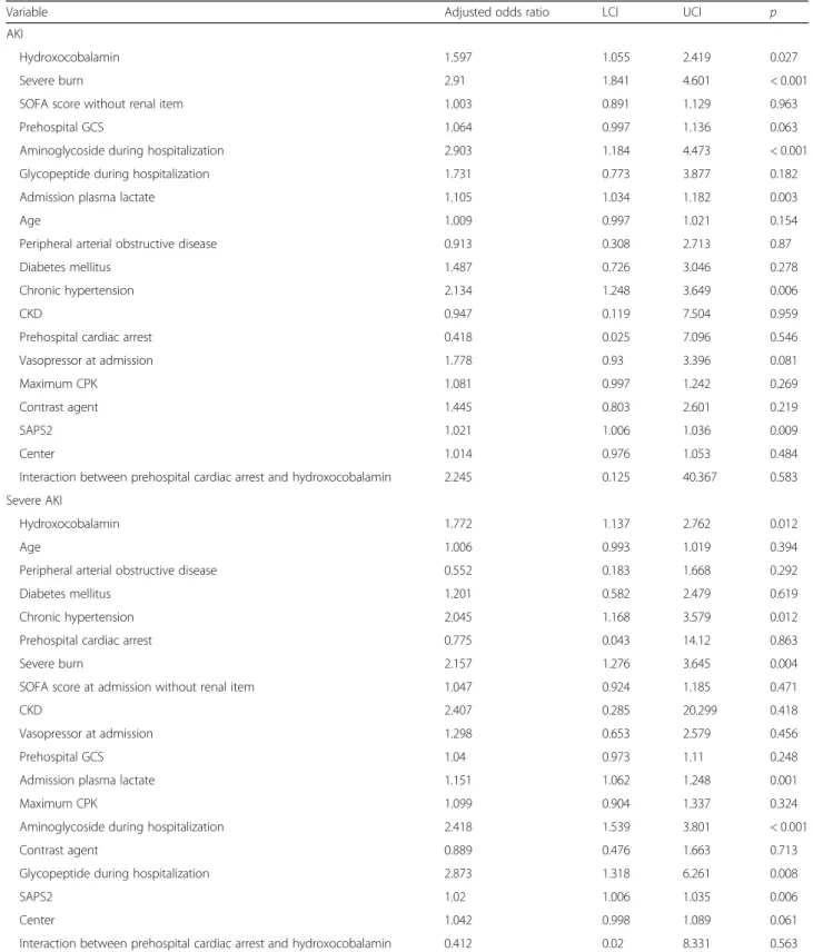
Table 2 Multivariate analyses of factors associated with AKI and severe AKI
Variable Adjusted odds ratio LCI UCI p
AKI
Hydroxocobalamin 1.597 1.055 2.419 0.027
Severe burn 2.91 1.841 4.601 < 0.001
SOFA score without renal item 1.003 0.891 1.129 0.963
Prehospital GCS 1.064 0.997 1.136 0.063
Aminoglycoside during hospitalization 2.903 1.184 4.473 < 0.001
Glycopeptide during hospitalization 1.731 0.773 3.877 0.182
Admission plasma lactate 1.105 1.034 1.182 0.003
Age 1.009 0.997 1.021 0.154
Peripheral arterial obstructive disease 0.913 0.308 2.713 0.87
Diabetes mellitus 1.487 0.726 3.046 0.278
Chronic hypertension 2.134 1.248 3.649 0.006
CKD 0.947 0.119 7.504 0.959
Prehospital cardiac arrest 0.418 0.025 7.096 0.546
Vasopressor at admission 1.778 0.93 3.396 0.081
Maximum CPK 1.081 0.997 1.242 0.269
Contrast agent 1.445 0.803 2.601 0.219
SAPS2 1.021 1.006 1.036 0.009
Center 1.014 0.976 1.053 0.484
Interaction between prehospital cardiac arrest and hydroxocobalamin 2.245 0.125 40.367 0.583 Severe AKI
Hydroxocobalamin 1.772 1.137 2.762 0.012
Age 1.006 0.993 1.019 0.394
Peripheral arterial obstructive disease 0.552 0.183 1.668 0.292
Diabetes mellitus 1.201 0.582 2.479 0.619
Chronic hypertension 2.045 1.168 3.579 0.012
Prehospital cardiac arrest 0.775 0.043 14.12 0.863
Severe burn 2.157 1.276 3.645 0.004
SOFA score at admission without renal item 1.047 0.924 1.185 0.471
CKD 2.407 0.285 20.299 0.418
Vasopressor at admission 1.298 0.653 2.579 0.456
Prehospital GCS 1.04 0.973 1.11 0.248
Admission plasma lactate 1.151 1.062 1.248 0.001
Maximum CPK 1.099 0.904 1.337 0.324
Aminoglycoside during hospitalization 2.418 1.539 3.801 < 0.001
Contrast agent 0.889 0.476 1.663 0.713
Glycopeptide during hospitalization 2.873 1.318 6.261 0.008
SAPS2 1.02 1.006 1.035 0.006
Center 1.042 0.998 1.089 0.061
Interaction between prehospital cardiac arrest and hydroxocobalamin 0.412 0.02 8.331 0.563 AKI acute kidney injury, LCI lower confidence interval, UCI upper confidence interval, p p value, SOFA score sequential organ failure assessment, GCS Glasgow coma scale,CKD chronic kidney disease, CPK creatinine phosphokinase, SAPS2 simplified acute physiology score 2
Missing data were handled using multivariate imput-ation by chained equimput-ations (mice package for R, 50 im-putations) [9].
Because plasma lactate level was shown to correlate with cyanide, we performed subgroup analysis in pa-tients with plasma lactate level at admission above me-dian of the entire cohort and above 8 mmol/L [10, 11]. A subgroup analysis was also performed in patients with severe burns.
Results
Among 854 patients with smoke inhalation, 739 were included in the analysis after exclusion of patients discharged at home within 24 h. Three hundred eighty-six (55.2%) patients received hydroxocobalamin.
Patient’s characteristics and comparison between
pa-tients who received hydroxocobalamin and those who
did not are presented in Table 1. The number of
pa-tients per center is summarized in Additional file 2: Table S2.
Acute kidney injury
Two hundred eighty-eight (39%) patients developed AKI in the first week, including 186 (25.2%) patients with severe AKI. In univariate analysis, the use of hydroxocobalamin was associated with AKI, severe AKI, and RRT (Table 1). After adjusting for potential confounders, hydroxocobalamin remained associated
with AKI and severe AKI and RRT (Table 2, Fig. 1
and Additional file 3: Table S3). Severe burns, amino-glycoside administration, admission plasma lactate
level, chronic hypertension, and SAPS2 were also as-sociated with AKI (Table 2).
Three hundred and thirteen (42.4%) patients met the criteria for MAKE during ICU stay. Hydroxocoba-lamin was not associated with the risk of MAKE in multivariate analysis (OR = 0.784, LCI = 0.456, UCI =
1.349, p = 0.379). Factors associated with MAKE in
ICU are summarized in Additional file 4: Table S4.
ICU mortality
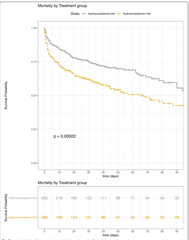
Mortality in ICU was 32.9% (N = 243). Patients who received hydroxocobalamin had higher severity scores and higher mortality rate. Factors associated with mortality in univariate analysis are summarized in Table 3 and Fig. 2. In multivariate analysis, hydroxo-cobalamin was not associated with survival (OR = 1.114, LCI = 0.691, UCI = 1.797, p = 0.657). Age, severe burns, SAPS2, plasma lactate level at admission, and the center were associated with mortality in ICU
(Table 4). Three hundred and ninety-two patients
(53%) had admission plasma lactate level above the median (median = 3.0 [1.8–5.2] mmol/L), and 74
pa-tients had admission plasma lactate level≥ 8 mmol/L
(median = 10.5 [8.9–12.8] mmol/L) (among 681
(92.2%) patients with plasma lactate available at ad-mission, Additional file 5: Table S5). No association between hydroxocobalamin use and survival was ob-served after adjustment for confounding factors in the subgroups of patients with admission plasma lactate level above the median (OR = 0.76; LCI = 0.398, UCI = 1.451, p = 0.403) or in the group of patients with
ad-mission plasma lactate level≥ 8 mmol/L (OR = 2.604,
Fig. 1 Non-adjusted and adjusted odds ratio (lower confidence interval, upper confidence interval) of hydroxocobalamin for AKI (upper panel) and severe AKI (lower panel). Adjusted on the following variables: age, peripheral arterial obstructive disease, diabetes mellitus, chronic hypertension, chronic kidney disease, prehospital cardiac arrest, severe burn, SOFA score without kidney item, vasopressors at admission, prehospital Glasgow coma scale, plasmatic lactate level at admission, maximum creatinine phosphokinase plasmatic level, contrast agent, use of vancomycin and aminoglycosides, and simplified acute physiology score 2
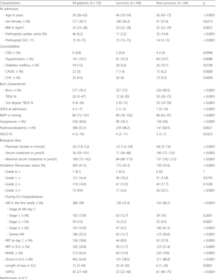
Table 3 Patient characteristics
Characteristics All patients, N = 739 Survivors, N = 496 Non-survivors, N = 243 p At admission
- Age in years 50 (36–63) 46 (33–59) 56 (43–72) < 0.0001
- Sex female, n (%) 271 (36.7) 180 (36.3) 91 (37.4) 0.8214
- BMI in kg/m2 25 (22–28) 24 (22–28) 25 (22–29) 0.0822
- Prehospital cardiac arrest (%) 46 (6.2) 11 (2.2) 35 (14.4) < 0.0001
- Prehospital GSC /15 15 (9–15) 15 (13–15) 14 (3–15) < 0.0001 Comorbidities - CKD, n (%) 6 (0.8) 2 (0.4) 4 (1.6) 0.0948 - Hypertension, n (%) 141 (19.1) 81 (16.3) 60 (24.7) 0.0088 - Diabetes mellitus, n (%) 54 (7.3) 28 (5.6) 26 (10.7) 0.0198 - CAOD, n (%) 22 (3) 7 (1.4) 15 (6.2) 0.0008 - CHF, n (%) 33 (4.5) 20 (4) 13 (5.3) 0.4858 Burn characteristic - Burn, n (%) 577 (78.1) 357 (72) 220 (90.5) < 0.0001 - TBSA % 20 (3–47) 12 (0–30) 50 (20–73) < 0.0001 - 3rd degree TBSA % 9 (0–30) 2 (0–15) 33 (10–58) < 0.0001 SOFA at admission 4 (1–7) 2 (1–5) 7 (3–10) < 0.0001 MAP in mmHg 86 (72–101) 89 (76–102) 80 (62–97) < 0.0001 Vasopressor, n (%) 226 (30.6) 90 (18.1) 136 (56) < 0.0001 Hydroxocobalamin, n (%) 386 (52.2) 239 (48.2) 147 (60.5) 0.0021 HbCO % 4 (2–10) 4 (2–11) 3 (2–7) 0.0323 Biological data
- Plasmatic lactate in mmol/L 3.0 (1.8–5.2) 2.5 (1.4–3.8) 4.8 (3–7.6) < 0.0001 - Serum creatinine inμmol/L 76 (59–101) 71 (56–88) 100 (72–123) < 0.0001 - Maximal serum creatinine inμmol/L 100 (73–162) 84 (68–113) 137 (102–212) < 0.0001 Inhalation fibroscopic status (%) 305 (41.3) 175 (35.3) 130 (53.5) < 0.0001
- Grade 0, n 1 (0.1) 1 (0.2) 0 (0) 1
- Grade 1, n 121 (16.4) 90 (18.2) 31 (12.8) 0.0795
- Grade 2, n 110 (14.9) 67 (13.5) 43 (17.7) 0.1638
- Grade 3, n 73 (9.9) 17 (3.4) 56 (23.1) < 0.0001
- During ICU hospitalization
- AKI in the first week, n (%) 288 (39) 126 (25.4) 162 (66.7) < 0.0001 - Stage of AKI day 7
- Stage 1, n (%) 102 (13.8) 63 (12.7) 39 (16) 0.2601 - Stage 2, n (%) 39 (5.3) 16 (3.2) 23 (9.5) 0.0007 - Stage 3, n (%) 147 (19.9) 47 (9.5) 100 (41.2) < 0.0001 - Severe AKI 186 (25.2) 63 (12.7) 123 (50.6) < 0.0001 - RRT at day 7, n (%) 136 (18.8) 44 (8.9) 92 (37.9) < 0.0001 - RRT in ICU, n (%) 183 (24.8) 58 (11.7) 125 (51.4) < 0.0001 - MAKE, n (%) 313 (42.4) 69 (13.9) 243 (100) < 0.0001 - Shock in ICU, n (%) 402 (54.4) 191 (38.5) 211 (86.8) < 0.0001
- Length of stay in ICU 15 (3–44) 25 (4–50) 6 (1–24) 0.0001
- SAPS2 42 (27–60) 32 (22–46) 61 (46–75) < 0.0001
LCI = 0.361, UCI = 18.79, p = 0.336). In multivariate analysis, hydroxocobalamin was neither associated
with survival (OR = 0.85, LCI = 0.5, UCI = 1.447, p =
0.549) among 405 (54.8%) patients with severe burns (Additional file 6: Table S6).
Discussion
In this retrospective, multicenter study, we observed a higher risk of AKI and severe AKI associated with ad-ministration of hydroxocobalamin after adjustment for potential confounding factors. Hydroxocobalamin was not associated with improved survival.
Cyanide poisoning has been suspected to account for deaths after smoke inhalation [12]. Hydroxocoba-lamin (Cyanokit®, Merck Santé SAS) is approved by the Food and Drug Administration for the treatment
of cyanide poisoning. Hydroxocobalamin chelates
cyanide to form cyanocobalamin, which is excreted by the kidneys. Experts have advocated the use of cyan-ide antidotes after smoke inhalation with suspected cyanide intoxication. While preclinical data have sug-gested improved hemodynamics with hydroxocobala-min in animal models of cyanide intoxication, clinical data are very limited. In two retrospective studies, Borron et al. and Fortin et al. described the outcome of patients receiving hydroxocobalamin after smoke inhalation injury. However, the absence of control groups precluded drawing any conclusion with respect
to the impact of hydroxocobalamin on outcome [13,
14]. Nguyen et al. reported that the use of
hydroxo-cobalamin was associated with fewer episodes of
pneumonia while mortality was unaffected [15].
How-ever, the association between the prevention of pneu-monia lies on very low level of evidence. Other factors, especially with respect to pneumonia prevent-ive strategies, may have accounted for the observation of lower incidence of pneumonia in this before-and-after retrospective study.
In the current study, we observed an association be-tween hydroxocobalamin and the risk of AKI and severe AKI. These results further suggest the potential nephro-toxicity of the drug. We previously showed that hydro-xocobalamin induces oxaluria with a risk of oxalate
nephropathy in burn patients [4]. Kidney biopsies con-firmed renal calcium oxalate crystal deposits. Oxaluria was also reported in healthy volunteers and animals re-ceiving hydroxocobalamin [16]. Nitric oxide chelation could represent an additional mechanism of renal tox-icity of hydroxocobalamin. In the absence of cyanide, hydroxocobalamin binds to nitric oxide. Nitric oxide is a key factor of the renal microcirculatory regulation, and inhibition of the nitric oxide pathway was shown to im-pair renal perfusion and oxygenation and cause kidney damage [17–19].
Cyanide is a rapidly lethal mitochondrial poison. The plasma cyanide level is difficult to measure and not readily available, and none of the patients in-cluded in this study had a plasma cyanide dosage. In-creased serum lactate level > 8 mmol/L was reported to be a fair surrogate biomarker of cyanide poisoning as a biomarker of mitochondrial dysfunction [10]. In our study, hydroxocobalamin had no impact in the subgroup of patients with high plasma lactate level. The lack of evidence of any association between hydroxocobalamin and survival in our study and the risk of AKI question the risk-benefit ratio in these
pa-tients. Of note, hydroxocobalamin costs about €1000/
$1200 per vial.
We acknowledge the limitations of our study. First, the retrospective nature introduced some bias. Performing a randomized control trial in this setting would be highly difficult due to the low incidence of smoke inhalation and the existing cognitive biases toward the potential protect-ive effects of hydroxocobalamin. Second, the relatprotect-ive low number of patients with very high plasma lactate level pre-cludes drawing firm conclusion in patients with a high probability of cyanide poisoning and in most severe pa-tients. Third, we could not confirm cyanide levels in any patient, which makes it hard to determine if hydroxocoba-lamin could benefit patients with confirmed cyanide poi-soning. Finally, rhabdomyolysis may be a confounding factor with the elevated serum creatinine. However, we did not find any correlation between admission serum creatinine, maximal serum creatinine at day 7 or during hospitalization in ICU, and the pic of creatinine phosphokinase.
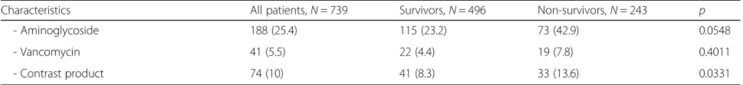
Table 3 Patient characteristics (Continued)
Characteristics All patients, N = 739 Survivors, N = 496 Non-survivors, N = 243 p
- Aminoglycoside 188 (25.4) 115 (23.2) 73 (42.9) 0.0548
- Vancomycin 41 (5.5) 22 (4.4) 19 (7.8) 0.4011
- Contrast product 74 (10) 41 (8.3) 33 (13.6) 0.0331
All data are expressed as median ± 25–75 interquartile or percentage (%)
BMI body mass index, GCS Glasgow coma scale, CKD chronic kidney disease, CAOD chronic arterial occlusive disease, CHF chronic heart failure, TBSA total body surface area,SOFA sequential organ failure assessment, MAP mean arterial pressure, HbCO carboxy hemoglobin, ICU intensive care unit, AKI acute kidney injury, RRT renal replacement therapy, MAKE major associated kidney events, SAPS2 simplified acute physiology score 2
Fig. 2 Unadjusted Kaplan–Meier curve of survival incidence during the 90 days following admission between patients who received hydroxocobalamin and those who did not
Conclusion
In this multicenter observational study of ICU patients with smoke inhalation, administration of hydroxocobala-min was independently associated with AKI and showed no association with survival. The role of hydroxocobala-min following smoke inhalation needs further consider-ation and critical appraisal.
Supplementary information
Supplementary information accompanies this paper athttps://doi.org/10. 1186/s13054-019-2706-0.
Additional file 1 : Table S1. Coding system for smoke inhalation. Additional file 2 : Table S2. Number of patients from each centers. Additional file 3 : Table S3. Multivariate analysis of factors associated with RRT.
Additional file 4 : Table S4. Multivariate analysis of factors associated with major adverse kidney events.
Additional file 5 : Table S5. Comparison between admission lactate quartile.
Additional file 6 : Table S6. Comparison between severely burn and non-severely burn patients.
Abbreviations
AKI:Acute kidney injury; Cis: Confidence intervals; ICU: Intensive care unit; KDIGO: Kidney Disease Improving Global Outcome; MAKE: Major associated kidney events; RRT: Renal replacement therapy; SCreat: Serum creatinine; TBSA: Total body surface area
Acknowledgements None
Authors’ contributions
FD designed the study, collected the data, interpreted the data, and drafted the manuscript. CH collected the data and drafted the manuscript. LD
collected the data and drafted the manuscript. CT collected the data and drafted the manuscript. LMr collected the data and drafted the manuscript. JC collected the data and drafted the manuscript. DA collected the data and drafted the manuscript. EP collected the data and drafted the manuscript. DM collected the data and drafted the manuscript. SW collected the data and drafted the manuscript. DDDL collected the data and drafted the manuscript. AK collected the data and drafted the manuscript. JT collected the data and drafted the manuscript. FR collected the data and drafted the manuscript. KK collected the data and drafted the manuscript. EP collected the data and drafted the manuscript. GS collected the data and drafted the manuscript. FB collected the data and drafted the manuscript. JMC collected the data and drafted the manuscript. TC collected the data and drafted the manuscript. DD collected the data and drafted the manuscript. NT collected the data and drafted the manuscript. BS collected the data and drafted the manuscript. EG collected the data and drafted the manuscript. TL collected the data and drafted the manuscript. AH collected the data and drafted the manuscript. BM collected the data and drafted the manuscript. TL collected the data and drafted the manuscript. VM interpreted the data and drafted the manuscript. RP performed the statistical analysis, interpreted the data, and drafted the manuscript. ML designed the study, collected the data, interpreted the data, supervised the study, and drafted the manuscript. All authors read and approved the final manuscript.
Funding
The study was supported by a grant from the“Fondation des gueules cassées” to Matthieu Legrand.
Availability of data and materials
The datasets used and/or analyzed during the current study are available from the corresponding author on reasonable request.
Ethics approval and consent to participate
The study protocol was approved by our local research ethical committee (Comité de Protection des Personnes 2013/17NICB).
Consent for publication Not applicable. Competing interests
The authors declare that they have no competing interests. Author details
1Department of Anesthesiology and Critical Care and Burn Unit, AP-HP, GH
Saint Louis-Lariboisière, Paris, France.2UMR INSERM 942, Institut National de la Santé et de la Recherche Médicale (INSERM), Paris, France.3F-CRIN,
INICRCT network, Paris, France.4Paris Diderot University, F-75475 Paris,
France.5Burn Center, Percy Military Teaching Hospital, BP 406, 101, avenue
Henri-Barbusse, 92141 Clamart CEDEX, France.6Intensive Care Unit and Hyperbaric Center, Lille University Hospital, F-59037 Lille CEDEX, France.
7General ICU, Service de Réanimation, Hôpital Raymond Poincaré, Laboratory
of Infection and Inflammation, U1173, AP-HP, University of Versailles SQY and INSERM, 104 Boulevard Raymond Poincaré, 92380 Garches, France.8Centre de traitement des grands brûlés Hopital de la Conception APHM, 147 boulevard Baille, 13005 Marseille, France.9Intensive Care Unit, Anaesthesia
and Critical Care Department, Hôtel Dieu—HME, CHU Nantes, Nantes, France.10Centre de traitement des grands brûlés Hopital de Mercy,1 Allée du Château, 57245 Ars-Laquenexy—C.H.R Metz-, Thionville, France.
11Anesthesiology and Critical Care Medicine, Hospices Civils de
Lyon—Université Claude Bernard Lyon 1, Lyon, France.12Anesthesiology and
Critical Care Medicine, CHU Bordeaux, Place Amélie Raba Léon, 33000 Bordeaux, France.13Intensive Care Medicine Department, University of
Montpellier Lapeyronie Hospital, 371, Av Doyen Gaston Giraud, 34295 Montpellier, France.14Intensive Care Medicine Department, CHU de Rennes,
2 rue Henri Le Guilloux, 35033 Rennes CEDEX 9, France.15Service de réanimation médico-chirurgicale, Groupe Hospitalier du Havre—Hôpital Jacques Monod, Montivilliers, France.16Medical Intensive Care Unit, La
Source Hospital, CHR Orléans, Orléans, France.17Department of Perioperative
Medicine, University Hospital of Clermont-Ferrand, Clermont-Ferrand, France.
18Department of Anesthesiology and Critical Care, Rouen University Hospital,
Rouen, France.19Normandie Univ, UNIROUEN, INSERM U1096, Rouen, France. 20Medical Intensive Care Unit, Caen University Hospital, Avenue côte de
Table 4 Multivariate analyses of factors associated with ICU mortality ICU mortality Variable Adjusted odds ratio LCI UCI p Hydroxocobalamin 1.114 0.691 1.797 0.657 Age 1.037 1.023 1.05 < 0.001
Prehospital cardiac arrest 1.091 0.07 17.126 0.95 Severe burn 4.792 2.665 8.617 < 0.001 SOFA score at admission 1.074 0.942 1.225 0.283 Admission plasma lactate 1.256 1.154 1.366 < 0.001 Vasopressors at admission 1.571 0.737 3.346 0.241 Prehospital GCS 1.033 0.961 1.11 0.383 SAPS2 1.039 1.02 1.059 < 0.001 Center 1.084 1.033 1.137 0.001 Interaction between prehospital cardiac arrest and hydroxocobalamin
6.327 0.354 112.993 0.209
ICU intensive care unit, LCI lower confidence interval, UCI upper confidence interval,p p value, SOFA score sequential organ failure assessment score, GCS Glasgow coma scale,SAPS2 simplified acute physiology score 2
Nacre, 14033 Caen CEDEX, France.21Service de Réanimation Médicale,
Centres Hospitaliers Universitaires Grenoble Alpes, Grenoble, France.22Service
de Réanimation Médicale Polyvalente, Centre Hospitalier Public du Cotentin, BP 208, 50102 Cherbourg-Octeville, France.23Service de Réanimation Médicale, Hôpital Européen Georges-Pompidou, Assistance
Publique-Hôpitaux de Paris, Paris, France.24Faculté de Médecine, Université
Paris Descartes, Paris, France.25Département des urgences, service des
urgences, SAMU, CHU de Limoges, 87042 Limoges CEDEX, France.26Inserm CIC 1435, 87042 Limoges, France.27Intensive Care Unit, Saint Louis Hospital,
La Rochelle, France.28Service de réanimation médicale et toxicologie, Hôpital
Lariboisière, Assistance Publique-Hôpitaux de Paris, Paris, France.29Service
d’hépato gastro entérologie Hôpital Cochin, hépato Cochin, Assistance Publique-Hôpitaux de Paris, Paris, France.30Department of Anesthesia and
perioperative care, University of California San Francisco, San Francisco, USA.
31Department of Anesthesiology and Perioperative care Parnassus hospital,
UCSF, San Francisco, USA.
Received: 6 September 2019 Accepted: 16 December 2019
References
1. Lawson-Smith P, Jansen EC, Hyldegaard O. Cyanide intoxication as part of smoke inhalation - a review on diagnosis and treatment from the emergency perspective. Scand J Trauma Resusc Emerg Med. 2011;19:14. 2. Petrikovics I, Budai M, Kovacs K, Thompson DE. Past, present and future of
cyanide antagonism research: from the early remedies to the current therapies. World J Methodol. 2015;5:88–100.
3. Anseeuw K, Delvau N, Burillo-Putze G, De Iaco F, Geldner G, Holmström P, et al. Cyanide poisoning by fire smoke inhalation: a European expert consensus. Eur J Emerg Med. 2013;20:2–9.
4. Legrand M, Michel T, Daudon M, Benyamina M, Ferry A, Soussi S, et al. Risk of oxalate nephropathy with the use of cyanide antidote hydroxocobalamin in critically ill burn patients. Intensive Care Med. 2016;42:1080–1.
5. Legrand M, Mallet V. Intravenous hydroxocobalamin and crystal nephropathy. Nat Rev Nephrol. 2017;13:593.
6. Kellum JA, Lameire N, Aspelin P, Barsoum RS, Burdmann EA, Goldstein SL, et al. Work group membership. Kidney Int. 2012;2:1.
7. Monafo W. Initial management of burns. N Engl J Med. 1996;335(21):1581–6. 8. Chou SH, Lin S-D, Chuang H-Y, Cheng Y-J, Kao EL, Huang M-F. Fiber-optic
bronchoscopic classification of inhalation injury: prediction of acute lung injury. Surg Endosc. 2004;18:1377–9.
9. Bartlett JW, Seaman SR, White IR, Carpenter JR, for the Alzheimer’s Disease Neuroimaging Initiative*. Multiple imputation of covariates by fully conditional specification: accommodating the substantive model. Stat Methods Med Res. 2015;24:462–87.
10. Baud FJ, Borron SW, Mégarbane B, Trout H, Lapostolle F, Vicaut E, et al. Value of lactic acidosis in the assessment of the severity of acute cyanide poisoning. Crit Care Med. 2002;30:2044–50.
11. Baud FJ, Haidar MK, Jouffroy R, Raphalen J-H, Lamhaut L, Carli P. Determinants of lactic acidosis in acute cyanide poisonings. Crit Care Med. 2018;46:e523–9.
12. Kadri SS, Miller AC, Hohmann S, Bonne S, Nielsen C, Wells C, et al. Risk factors for in-hospital mortality in smoke inhalation-associated acute lung injury: data from 68 United States hospitals. Chest. 2016;150:1260–8. 13. Borron SW, Baud FJ, Barriot P, Imbert M, Bismuth C. Prospective study of
hydroxocobalamin for acute cyanide poisoning in smoke inhalation. Ann Emerg Med. 2007;49:794–801.e2.
14. Fortin J-L, Giocanti J-P, Ruttimann M, Kowalski J-J. Prehospital administration of hydroxocobalamin for smoke inhalation-associated cyanide poisoning: 8 years of experience in the Paris fire brigade. Clin Toxicol. 2006;44:37–44. 15. Nguyen L, Afshari A, Kahn SA, McGrane S, Summitt B. Utility and outcomes
of hydroxocobalamin use in smoke inhalation patients. Burns. 2017;43(1): 107–13.
16. EMD Serono, A Division of EMD Inc., Canada. CYANOKIT® Product Monograph. 2014. Available from:http://webprod.hc-sc.gc.ca/dpd-bdpp/ index-eng.jsp. Accessed 30 Sept 2014.
17. Legrand M, Mik EG, Johannes T, Payen D, Ince C. Renal hypoxia and dysoxia after reperfusion of the ischemic kidney. Mol Med Camb Mass. 2008;14:502–16.
18. Legrand M, Almac E, Mik EG, Johannes T, Kandil A, Bezemer R, et al. L-NIL prevents renal microvascular hypoxia and increase of renal oxygen
consumption after ischemia-reperfusion in rats. Am J Physiol Renal Physiol. 2009;296:F1109–17.
19. Soussi S, Dépret F, Benyamina M, Legrand M. Early hemodynamic management of critically ill burn patients. Anesthesiology. 2018;129:583–9.
Publisher’s Note
Springer Nature remains neutral with regard to jurisdictional claims in published maps and institutional affiliations.