HAL Id: hal-02305467
https://hal.sorbonne-universite.fr/hal-02305467
Submitted on 4 Oct 2019HAL is a multi-disciplinary open access archive for the deposit and dissemination of sci-entific research documents, whether they are pub-lished or not. The documents may come from teaching and research institutions in France or abroad, or from public or private research centers.
L’archive ouverte pluridisciplinaire HAL, est destinée au dépôt et à la diffusion de documents scientifiques de niveau recherche, publiés ou non, émanant des établissements d’enseignement et de recherche français ou étrangers, des laboratoires publics ou privés.
In vitro activities of a new fluoroquinolone derivative
highly active against Chlamydia trachomatis
Thi Huyen Vu, Nguyet-Thanh Ha-Duong, Alexandra Aubry, Estelle Capton,
Pierre Fechter, Patrick Plesiat, Philippe Verbeke, Nawal Serradji
To cite this version:
Thi Huyen Vu, Nguyet-Thanh Ha-Duong, Alexandra Aubry, Estelle Capton, Pierre Fechter, et al.. In vitro activities of a new fluoroquinolone derivative highly active against Chlamydia trachomatis. Bioorganic Chemistry, Elsevier, 2019, 83, pp.180-185. �10.1016/j.bioorg.2018.10.033�. �hal-02305467�
1
In vitro Activities of a New Fluoroquinolone Derivative Highly Active against Chlamydia trachomatis.
Thi Huyen Vu,a,b Nguyet-Thanh Ha-Duong,a Alexandra Aubry,c,d Estelle Capton,d Pierre Fechter,e,f Patrick Plésiat,g Philippe Verbeke,h and Nawal Serradji.a*
a
Univ Paris Diderot, Sorbonne Paris Cité, ITODYS, UMR 7086, CNRS, 15 rue Jean Antoine de Baïf, F-75205 Paris, France.
b University of Science and Technology of Hanoi (USTH), Vietnam Academy of Science and Technology,
18 Hoang Quoc Viet, Cau Giay, Hanoi, Vietnam;
c
AP-HP, Hôpital Pitié-Salpêtrière, Centre National de Référence des Mycobactéries et de la Résistance des Mycobactéries aux Antituberculeux, F-75013 Paris, France;
d
Sorbonne Université, INSERM, U1135, Centre d’Immunologie et des Maladies Infectieuses, Cimi-Paris, équipe 13, F-75013, Cimi-Paris, France;
e
CNRS, UMR 7242, Biotechnologie et Signalisation Cellulaire, 67400 Illkirch-Graffenstaden, France;
f
Université de Strasbourg, Institut de Recherche de l'Ecole de Biotechnologie de Strasbourg, 67400 Illkirch-Graffenstaden, France;
g
Centre National de Référence de la résistance aux antibiotiques, Hôpital Jean Minjoz, boulevard Fleming, 25030 Besançon, France;
h
Univ Paris Diderot, Sorbonne Paris Cité, INSERM U1149, Faculté de médecine Xavier Bichat, 16 rue Henri Huchard, F-75018 Paris, France.
*Corresponding author. Email address: serradji@univ-paris-diderot.fr; Tel.: + 33 1 57 27 88 83. Fax: + 33 1 57 27 72 63.
Key words: Inhibitors; 8-hydroxyquinoline; ciprofloxacin; Gram-negative; Gram-positive.
Abstract: Chlamydia trachomatis is a bacterial human pathogen responsible for the development of trachoma, an infection leading to blindness, and is also the cause of the main bacterial sexually transmitted infection worldwide. We designed a new inhibitor of this bacterium with, however, some prerequisites using (i) the iron dependency of the bacterium, (ii) a commercially available broad-spectrum antibiotic and (iii) a short synthetic pathway. The corresponding 8-hydroxyquinoline-ciprofloxacin conjugate was evaluated against a panel of pathogenic bacteria, including C. trachomatis but also the ESKAPE group (Enterococcus faecium, Staphylococcus aureus,
2
species). Its anti-Chlamydia activity is higher than that of ciprofloxacin and seems to be related to
the fluoroquinolone moiety of the molecule, which is also responsible for the complexation of iron(III), as demonstrated by spectrophotometric titration.
Chlamydia trachomatis (C. trachomatis), an obligate intracellular Gram-negative bacterium, is
responsible for the most common sexually transmitted bacterial infection in the world (131 million new cases in 2012, WHO data),1 which causes severe complications leading to serious sequelae in
men and women, including infertility. This infection also facilitates other sexually transmitted infections (STI), including HIV,2,3 and increases the incidence and persistence of human papillomavirus (HPV) infection.4,5 C. trachomatis is also responsible for trachoma, the most common infectious cause of blindness.6 Treatment of C. trachomatis infection requires special considerations related to the bacterial cycle. Indeed, infectious elementary bodies are metabolically inert and insensitive to antibiotics which target bacterial replication (fluoroquinolones) and translation (macrolides, tetracyclines). Effective antibiotics must target the metabolically active reticulate bodies, which replicate in an intracellular inclusion. Lipophilic antibiotics such as azithromycin (octanol-water partition coefficient, log P = 4.02)7 reach very high intracellular concentrations, which explains their good bactericidal activity against Gram-negative bacteria such as C. trachomatis.8,9
Drug-resistant C. trachomatis has rarely been reported.10 However, under the unfavorable conditions produced, for example, by treatment, bacteria can enter into a persistent form, which is viable, non-replicative and less sensitive to antibiotics; it has not yet been isolated in vivo.11 This persistent form can remain for several months/years in infected tissues, causing recurrent infections, chronic inflammation and tissue fibrosis. Consequently, treatment failure, which is observed in 5–23% of cases, could be the result of the re-emergence of the persistent infection.12 Moreover, the persistent form of C. pneumoniae (responsible for respiratory infections) is suspected to spread to other tissues and to be responsible for arthritis, atherosclerosis, endocarditis and asthma.13 Thus, incomplete antibiotic efficacy may be due to a lack of sensitivity of persistent forms of Chlamydiaceae but also to modest intracellular concentrations of the conventionally prescribed drugs. All these elements highlight the importance of seeking new antibiotics against Chlamydiaceae.
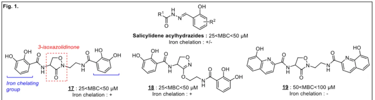
C. trachomatis needs iron to grow. Salicylidene acylhydrazides (Figure 1), inhibitors of the Chlamydial
type III secretion system (T3SS) with iron-chelating properties, inhibit C. trachomatis growth through a mechanism partially involving iron restriction, with minimal bactericidal concentrations (MBC) lower than 50 µM.14 The absence of cytotoxicity to HeLa 229 cells enables their use as vaginal microbiocides.15,16 We previously synthesized 3-isoxazolidinone derivatives, with iron-chelating properties, as equally effective inhibitors of bacterial growth (compounds 17–19; 25 < MBC < 50 µM;
3 Figure 1), without toxicity at 200 µM. However, since the antibacterial activity of 19 is not reversed by excess iron(III), metal chelation cannot be the only mechanism of action of these compounds.17
Fig. 1. Previously described inhibitors of C. trachomatis.17
In the present work, we synthesize a new inhibitor of this bacterium with, however, some prerequisites, using: (i) the iron dependency of the bacterium, (ii) a commercially available broad-spectrum antibiotic and (iii) a short synthetic pathway. We opted for a fluoroquinolone, ciprofloxacin, with the objective of obtaining more active derivatives than the parent antibiotic and, therefore, potentially useful for the treatment of C. trachomatis infection. We have selected conventionally described metal-chelating entities such as catechol, which forms extremely stable complexes with iron(III).18 This very high stability explains why the catechol group is present in many siderophores, molecules which are synthesized by microorganisms to trap iron in the external environment in order to facilitate its intracellular transport.
The 8-hydroxyquinoline entity, present in O-Trensox, a potent synthetic iron chelator,19 also attracted our attention. Both cathechol and 8-hydroxyquinoline were used via the corresponding carboxylic acids (Figure 2).
Fig. 2. Starting materials.
The coupling of ciprofloxacin and 8-hydroxyquinoline-2-carboxylic acid 3 by means of TBTU (2-(1H-benzotriazole-1-yl)-1,1,3,3-tetramethylaminium tetrafluoroborate) and DIEA (N,N-diisopropylethylamine) led to compound 1 (Scheme 1). The catechol-ciprofloxacin conjugate 2 was prepared as previously described.20
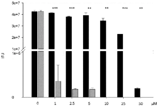
4 The compounds were first screened for cell toxicity. Host cell viability was monitored by Trypan Blue exclusion in the presence of the compounds at different concentrations. No cell toxicity was observed at the concentrations tested (0–200 µM) on either HeLa cells, a tumour cell line, or primary cell cultures of mouse fibroblasts, a non-cancerous mammalian cell line. Compounds were then screened for their capacity to inhibit C. trachomatis growth in HeLa cells. The cells were infected by C.
trachomatis serovar L2 strain as previously described.21 Infection was performed with or without the test molecules (0–50 µM) and with or without iron citrate (200 µM). 72 h post-infection, cell lysates were processed and used to infect new HeLa cells. The reinfection capacity was scored by calculating the Inclusion Forming Unit (IFU) of each cellular lysate. Ciprofloxacin (black bars) was used as external control. The results presented in Figure 3 show that the Minimal Bactericidal Concentration of ciprofloxacin (MBC > 16.5 µg/mL or > 50 µM) is similar to that described in the literature (> 10 µg/mL)22 while compound 1 (grey bars) presents a MBC of 2–5 µg/mL (5–10 µM). Therefore, functionalization of the fluoroquinolone nitrogen by an 8-hydroxyquinoline entity does not inhibit its antibacterial activity. The resulting compound is even more active than the parent molecule, probably due to a gain in lipophilicity. Indeed, its calculated octanol-water partition coefficient (cLog) is higher than that of ciprofloxacin (cLogP(ciprofloxacin) = 1.32 vs cLog P(1) = 3.09).23
5 Fig. 3. Inhibitory effects on C. trachomatis infectious capacity of compounds tested in cellulo (grey bars: compound 1; black bars: ciprofloxacin. Statistically significant differences are noted as follows: ** p<0.01, *** p<0.001).
Under the same conditions, the catechol analogue 2 is inactive (data not shown).
Iron is an essential element for C. trachomatis. However, to date, no siderophores or siderophore receptors have been described in Chlamydiaceae.24 Since catechol is one of the most powerful iron-chelating agents, the inactivity of compound 2 against C. trachomatis suggests that the inhibition induced by compound 1 is not mainly due to iron chelation.
Taking into account the iron-chelating properties of both entities, 8-hydroxyquinoline and the fluoroquinolone, we evaluated the ability of iron(III) to reverse the inhibitory effect of compound 1 by adding exogenous iron citrate (200 µM). The results presented in Figure 4 show that the inhibitory effect of compound 1 is only partially reversed by excess Fe3+ (20%, 35% and 60% decrease at 1, 2.5 and 5 µM, respectively), confirming that iron chelation is not its main antibacterial mechanism.
Fig. 4. Compound 1 inhibitory effect in the presence of excess iron citrate (200 µM). Statistically significant differences are noted as follows: * p<0.05, ** p<0.01, *** p<0.001.
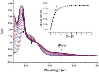
6 To further characterize compound 1, its ability to complex Fe3+ was studied by spectrophotometric titration. Complexation was performed in a H2O/DMSO (1:1; v/v) mixture to avoid precipitation of any
ligand and/or complex. The pH values mentioned are those of aqueous solutions before mixing with DMSO. Compound 1 has two potential sites for metal complexation: the ciprofloxacin carboxylate and keto groups and the 8-hydroxyquinoline part. At pH 2, the addition of FeCl3 to a solution of 1 leads to
a bathochromic shift (red shift) of the - band from 284 to 290 nm and the appearance of a ligand-to-metal charge-transfer (LMCT) band at 450 nm (Figure 5). The latter is identical to the LMCT of a complex between iron(III) and ciprofloxacin described by Fardeau et al.,20 whereas those of the complex between Fe3+ and 8-hydroxyquinoline are observed at 481 and 632 nm (not shown). This result suggests that only the fluoroquinolone part of compound 1 complexes Fe3+ at this pH.
Furthermore, at pH 7.4, iron-exchange experiments between Fe-nitrilotriacetic acid (Fe-NTA), a chelating agent, and 1 or methyl hydroxyquinoline-2-carboxylate, a surrogate of the 8-hydroxyquinoline moiety, show also differences in the LMCT bands (see Figure S1, Supplementary Information).
An isosbestic point at 340 nm indicates the formation of a single iron complex. The inset in Figure 5 presents the plot of the absorbance at 450 nm against the ratio [Fe(III)]/[1]: an increase in the absorbance is followed by a plateau. The two asymptotes intersect at a ratio of 1, implying a 1:1 stoichiometry (metal-ligand) for the complex. We then determined the affinity constant of the complex at pH 2, using Specfit analysis of the spectra; this is low (K11 = 2.5 ± 0.3), which confirms that
iron chelation is not the main antibacterial mechanism of action of this compound.
300 400 500 600 -0,5 0,0 0,5 1,0 1,5 2,0 2,5 3,0 3,5 4,0 0 1 2 3 4 5 6 7 8 0,00 0,05 0,10 0,15 0,20 0,25 A b s a t 4 5 0 n m [Fe]/[1] Abs Wavalength (nm) [Fe]
Fig. 5. Absorption spectra of 1 (10-4 M) in presence of increasing concentrations of FeCl3 (0–10-3 M) at
7 The World Health Organization Sexually Transmitted Infections guidelines suggest treatment of C.
trachomatis infection with one of the following drugs: azithromycin (1 g orally as a single dose) or
doxycycline (100 mg twice a day for 7 days). The in cellulo anti-chlamydia bactericidal activities of both compounds have been published: doxycycline and azithromycin have MBCs of 2.5–5.0 µg/mL and 10–50 µg/mL, respectively.25 Compound 1 is therefore at least as effective in cellulo as these two molecules.
Ciprofloxacin is a broad-spectrum antibiotic. We therefore looked at the ability of compound 1 to inhibit other human pathogens, including Gram-negative and Gram-positive bacteria from the ESKAPE group, an acronym including pathogenic bacteria present in the hospital environment and difficult to treat (Enterococcus faecium, Staphylococcus aureus, Klebsiella pneumoniae, Acinetobacter
baumannii, Pseudomonas aeruginosa and Enterobacter species). Staphylococcus aureus is the most
common staphylococcus strain responsible for human diseases, particularly nosocomial infections, along with Escherichia coli. The minimal inhibitory concentrations (MIC) of the derivatives were then determined (Supplementary Information) (Table 1). Many microbes, including P. aeruginosa strains, are well known to synthesize siderophores to scavenge iron from their environment. In order to facilitate the transport of antibiotics into bacteria, siderophore-conjugates have been described and used in a Trojan-horse strategy.26 Compound 2 was previously described as a new antibiotic following this strategy.20 Indeed, in iron-deficient culture conditions only, compound 2 is active against the P.
aeruginosa DSM 1117 susceptible strain with a MIC of 32 µg/mL, suggesting effective transport of the
corresponding iron(III) complex by bacterial iron-uptake pathways. We found that compound 2 is inactive against P. aeruginosa ATCC-27853, a reference strain (MIC > 128 µg/mL, Table 1), unlike compound 1 (MIC = 4 µg/mL).
While inactive against M. tuberculosis, compound 1 exhibits inhibitory activity against all the other Gram-negative and Gram-positive bacteria tested. For instance, it stops the growth of E. coli (MIC ≤ 0.06 μg/mL), probably by inhibition of its DNA gyrase, one of the bacterial targets of quinolones (IC50
= 8–16 μg/mL; Table 1 and Supplementary Information). In fact, this compound has a panel of interesting antibacterial activities, in that the MICs obtained are in the μg/mL range. However, this molecule is systematically less active than the parent antibiotic, ciprofloxacin, except for S. aureus, especially against laboratory and clinical isolates.
As already observed for C. trachomatis, the catechol analogue 2 is less potent than the 8-hydroxyquinoline derivative 1 on the panel of bacteria tested.
The starting acid 3 was tested in order to evaluate its contribution to the antibacterial activity of compound 1. The results (Table 1) show that whatever the pathogen, this acid is inactive, which suggests that the fluoroquinolone part of 1 is responsible for its efficacy.
8 Table 1. In vitro antibacterial activities (IC50 or MIC) of compounds 1–2, ciprofloxacin and acid 3:
Organism Grama Ciprofloxacin 1 2 3
M. tuberculosis - 12–17b,c >128b >128b >128b E. coli N 1–2.9b 816b 415b >128b E. coli ATCC-25922 N ≤ 0.06d ≤0.06d nd >8d K. pneumoniae ATCC-700603 N 0.25d 4d nd >8d P. aeruginosa ATCC-27853e N 0.25d 4d >128f >8d A. baumannii CIP-7010 N 0.125d 2d nd >8d
S. aureus HG001 (laboratory strain) P 0.125d 0.0625d 5d nd S. aureus ATCC-25923 (clinical
isolate)
P 0.25d 0.125d nd >8d
S. aureus ATCC-700699 (resistant isolate) P >8d >8d nd >8d S. epidermis ATCC-14990 ATCC-35984 P P 0.125d ≤0.06c 0.25d ≤0.06d nd >8d >8d E. faecalis JH2-2 UCN41 P P 2d 1d 8c 8d nd nd >8d >8d E. faecium ATCC-19434T BM-4147 P P 1d 4d 8 d >8d nd nd >8d >8d a P/N : positive/negative. bIC
50 (µg/mL) against wild-type DNA gyrases of M. tuberculosis and E. coli.
c
IC50s slightly higher than those previously determined.27
d
MIC (µg/mL).
e
Similar MIC were obtained against P. aeruginosa PAO1, a laboratory strain (data not shown).
fP. aeruginosa DSM 1117. 20
nd: not determined.
We report here the synthesis of a novel ciprofloxacin derivative by a single-step coupling of the parent antibiotic. This compound has notable antibacterial activity against negative and Gram-positive bacteria, including the obligate intracellular bacterium C. trachomatis. However, only its anti-chlamydial activity is higher than that of the parent antibiotic. This antibacterial effect is only partially reversed by the addition of iron(III), which is complexed by the fluoroquinolone part of the molecule.
Acknowledgments
This work was supported by a Ph.D. grant from the University of Science and Technology of Hanoi (Vu T.H.). The Institut des Humanités de Paris (Paris Diderot), the National Center for Scientific Research
9 (CNRS) and the University Paris Diderot are gratefully thanked for financial support. Sébastien Bellynck is also sincerely thanked for his technical assistance. We thank Dr. John S. Lomas for carefully reading the manuscript.
Supplementary data
Supplementary data associated with this article can be found in the online version.
References and notes
1. Geneva: WHO guidelines for the treatment of Chlamydia trachomatis, 2016.
2. Venkatesh KK, van der Straten A, Cheng H, Montgomery ET, Lurie MN, Chipato T, Ramjee G, Blanchard K, Padian NS, Mayer KH, de Bruyn G. The relative contribution of viral and bacterial sexually transmitted infections on HIV acquisition in southern African women in the Methods for Improving Reproductive Health in Africa study. Int J STD AIDS. 2011; 22(4): 218-224.
3. Peterman TA, Newman DR, Maddox L, Schmitt K, Shiver S. Risk for HIV following a diagnosis of syphilis, gonorrhoea or chlamydia: 328,456 women in Florida, 2000-2011. Int J STD AIDS. 2015; 26(2): 113-119.
4. Seraceni S, De Seta F, Colli C, Del Savio R, Pesel G, Zanin V, D'Agaro P, Contini C, Comar M. High prevalence of hpv multiple genotypes in women with persistent Chlamydia trachomatis infection.
Infect Agent Cancer. 2014; 9: 30.
5. Vriend HJ, Bogaards JA, van Bergen JE, Brink AA, van den Broek IV, Hoebe CJ, King AJ, van der Sande MA, Wolffs PF, de Melker HE, Medical Microbiological Laboratories and the CSI group. Incidence and persistence of carcinogenic genital human papillomavirus infections in young women with or without
Chlamydia trachomatis co-infection. Cancer Med. 2015; 4(10): 1589-1598.
6. Mariotti SP, Pascolini D, Rose-Nussbaumer J. Trachoma: global magnitude of a preventable cause of blindness. Br J Ophthalmol. 2009; 93(5): 563-568.
7. McFarland JW, Berger CM, Froshauer SA, Hayashi SF, Hecker SJ, Jaynes BH, Jefson MR, Kamicker BJ, Lipinski CA, Lundy KM, Reese CP, Vu CB. Quantitative structure-activity relationships among macrolide antibacterial agents: in vitro and in vivo potency against Pasteurella multocida. J Med Chem. 1997; 40(9):1340-1346.
8. McOrist S. Obligate intracellular bacteria and antibiotic resistance. Trends Microbiol. 2000; 8(11): 483-486.
10 9. Samra Z, Rosenberg S, Soffer Y, Dan M. In vitro susceptibility of recent clinical isolates of Chlamydia
trachomatis to macrolides and tetracyclines. Diagn Microbiol Infect Dis. 2001; 39(3):177-179.
10. Sandoz KM, Rockey DD. Antibiotic resistance in Chlamydiae. Future Microbiol. 2010; 5(9): 1427-1442.
11. Hogan RJ, Mathews SA, Mukhopadhyay S, Summersgill JT, Timms P. Chlamydial persistence: beyond the biphasic paradigm. Infect Immun. 2004 ; 72(4) : 1843-1855.
12. Horner PJ. Azithromycin antimicrobial resistance and genital. Chlamydia trachomatis infection: duration of therapy may be the key to improving efficacy. Sex Transm Infect. 2012; 88: 154–6.
13. Hahn DL, Azenabor AA, Beatty WL, Byrne GI. Chlamydia pneumoniae as a respiratory pathogen.
Front Biosci. 2002; 7: e66-e76.
14. Slepenkin A, Enquist PA, Hägglund U, de la Maza LM, Elofsson M, Peterson EM. Reversal of the antichlamydial activity of putative type III secretion inhibitors by iron. Infect Immun. 2007; 75(7): 3478-3489.
15. Chu H, Slepenkin A, Elofsson M, Keyser P, de la Maza LM, Peterson EM. Candidate vaginal microbicides with activity against Chlamydia trachomatis and Neisseria gonorrhoeae. Int J Antimicrob
Agents. 2010; 36(2): 145-150.
16. Ur-Rehman T, Slepenkin A, Chu H, Blomgren A, Dahlgren MK, Zetterström CE, Peterson EM, Elofsson M, Gylfe A. Pre-clinical pharmacokinetics and anti-chlamydial activity of salicylidene acylhydrazide inhibitors of bacterial type III secretion. J Antibiot (Tokyo). 2012; 65(8): 397-404.
17. Abdelsayed S, Ha Duong NT, Hai J, Hémadi M, El Hage Chahine JM, Verbeke P, Serradji N. Design and synthesis of 3-isoxazolidone derivatives as new Chlamydia trachomatis inhibitors. Bioorg Med
Chem Lett. 2014; 24(16): 3854-3860.
18. Karpishin TB, Gebhard MS, Solomon EI, Raymond KN. Spectroscopic studies of the electronic structure of iron(III) tris(catecholates). J Am Chem Soc. 1991; 113: 2977-2984.
19. Baret P, Beguin C, Boukhalfa H, Caris C, Laulhere JP, Pierre JL, Serratrice G. O-TRENSOX: A Promising water-soluble iron chelator (Both FeIII and FeII) potentially suitable for plant nutrition and iron chelation therapy. J Am Chem Soc. 1995; 117: 9760–9761.
11 20. Fardeau S, Dassonville-Klimpt A, Audic N, Sasaki A, Pillon M, Baudrin E, Mullié C, Sonnet P. Synthesis and antibacterial activity of catecholate-ciprofloxacin conjugates. Bioorg Med Chem. 2014; 22: 4049-4060.
21. Dumoux M, Le Gall SM, Habbeddine M, Delarbre C, Hayward RD, Kanellopoulos-Langevin C, Verbeke P. Penicillin kills Chlamydia following the fusion of bacteria with lysosomes and prevents genital inflammatory lesions in C. muridarum-infected mice. PLoS One 2013; 8: e83511.
22. Rumpianesi F, Sambri V, Bertini S, Tamba I, Cevenini R. In vitro activity of ciprofloxacin against
Chlamydia trachomatis and Ureaplasma urealyticum. Chemioterapia. 1984; 3(3): 173-174.
23. Chemdrawbio Ultra version 13.0.03015
24. Stephens RS. Genomic autobiographies of Chlamydiae. In: Stephens RS (ed) Chlamydia. Washington, DC, ASM Press. 1999:9-27.
25. Reveneau N, Crane DD, Fischer E, Caldwell HD. Bactericidal activity of first-choice antibiotics against gamma interferon-induced persistent infection of human epithelial cells by Chlamydia
trachomatis. Antimicrob Agents Chemother. 2005; 49(5):1787-93.
26. Cézard C, Farvacques N, Sonnet P. Chemistry and biology of pyoverdines, Pseudomonas primary siderophores. Curr Med Chem. 2015; 22(2):165-186.
27. Aubry A, Pan XS, Fisher LM, Jarlier V, Cambau E. Mycobacterium tuberculosis DNA gyrase: interaction with quinolones and correlation with antimycobacterial drug activity. Antimicrob Agents
Chemother. 2004; 48(4):1281-1288.
28. Chahine JMEH, Fain D. The mechanism of iron release from transferrin. Slow-proton-transfer-induced loss of nitrilotriacetatoiron(III) complex in acidic media. Eur J Biochem. 1994; 223: 581–587.
29. Pan XS, Fisher LM. Streptococcus pneumoniae DNA gyrase and topoisomerase IV: overexpression, purification, and differential inhibition by fluoroquinolones. Antimicrob Agents Chemother. 1999; 43:1129-1136.
30. Clinical and Laboratory Standards Institute (CLSI, USA). Methods for dilution antimicrobial susceptibility tests for bacteria that grow aerobically; approved standard Mo7-A10.