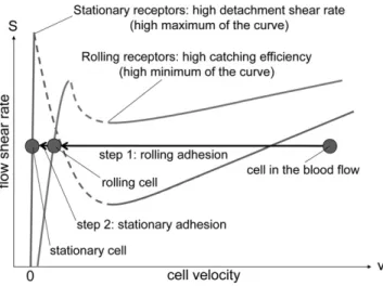
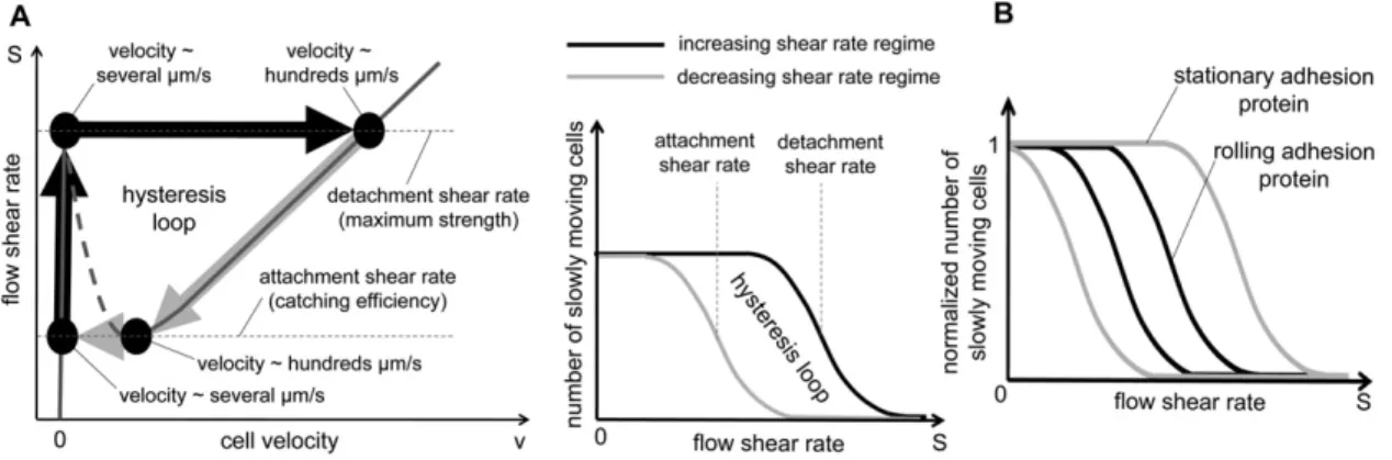
Bistability of Cell Adhesion in Shear Flow
Texte intégral
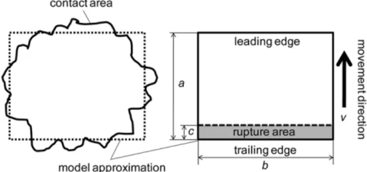
Figure
Documents relatifs
Dodecylphosphate, to which proteins can bind, was selectively adsorbed on these InSnO structures, whereas poly- L L -lysine-g-poly(ethylene glycol) was used to passivate the
The adhesion forces can be splitted in different components : van der Waals, electrostatics and capillary condensation.. This work focuses on capillary condensation as it often can
Notre travail concerne une série de 41 patients présentant une gonarthrose sur genu varum, traités par Ostéotomie tibiale haute de valgisation au service de
lactis bacterial cells immobilized onto AFM tip and cantilever; (B, E) histograms of adhesion forces and (C, F) typical force-distance curves obtained when probing interactions
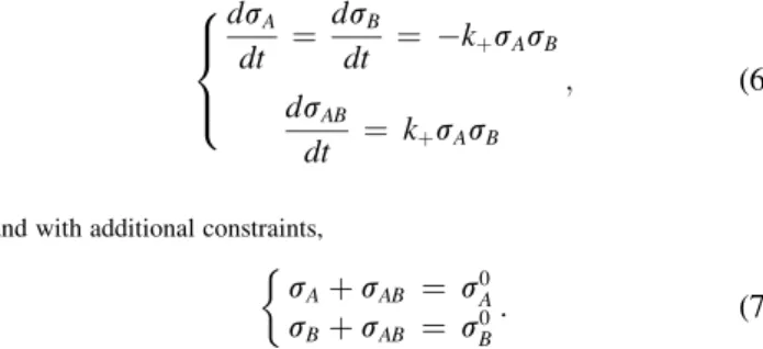
First, when there is no feedback from the cell velocity on the adhesion activity, the model writes as a Cox-Ingersoll-Ross (CIR) process, for which the probability density of the
Using cell biology approaches, we show that recProt01230 is able to adhere to bovine host cells and interacts with proteins from the cell lysate and the "membranes/
Le tracé de chéilorhinoplastie selon Tennisson nous semble particulièrement adapté à la prise en charge des fentes labiales ou labio-palatines unilatérales en mission
In Fig. Computations are always stopped when the stopping criteria is satisfied. When increasing the adhesion rate to a = 0.05, keeping α = 2, see B), the adhesion regions almost
