Assessment of Hip Fracture Risk in Astronauts Exposed to
Long-term Weightlessness
by
Grant Schaffner
B.S., Aeronautics and Astronautics Massachusetts Institute of Technology
1989
M.S., Aeronautics and Astronautics
Massachusetts Institute of Technology
1995
Submitted to the Harvard University - Massachusetts Institute of Technology Division of Health Sciences and Technology
in Partial Fulfillment of the Requirements for the Degree of Doctor of Philosophy
in Medical Engineering at the
Massachusetts Institute of Technology August, 1999
0 1999 Massachusetts Institute of Technology All rights reserved
Author
Harvaf MITbi siof Health Sciences
- A
and Technology
Certified by
bava J. Newman, Ph.D. Associate Professor of Aeronautics and Astronautics Thesis Supervisor
Accepted by
1Iartha L. Gray, Ph.D.
J.W. Kieckhefer Professor of Electrical Engineering
Assessment of Hip Fracture Risk in Astronauts Exposed to
Long-term Weightlessness
by
Grant Schaffner
Submitted to the Harvard University - Massachusetts Institute of Technology Division of Health Sciences and Technology
in Partial Fulfillment of the Requirements for the Degree of Doctor of Philosophy in Medical Engineering
Abstract
Background: A human exploration mission to Mars could take place within 10 years. During
the 6 to 12 month journey astronauts would likely lose bone mineral density (BMD) at a mean rate of 1-2 percent per month in weight-bearing areas, approximately 10 times the rate ated with normal ageing. There exists an important need to quantify the fracture risk associ-ated with this loss. Methods: Using computational modeling, the factor of risk for hip fracture (applied load divided by failure load) was assessed following 0, 6, and 12 months of weight-lessness for: 1) the mid-stance phase of gait, and 2) a fall to the side impacting the greater tro-chanter. Peak applied loading was calculated for Earth and Mars gravity levels using the equations of motion for three-segment models representing locomotion and falls. Mars simu-lations included extravehicular activity (EVA, with spacesuit) and intravehicular activity (IVA). The structural properties of the femur were analyzed using a three-dimensional finite element model derived from quantitative computed tomography scans of a representative cadaveric femur. Space flight associated changes in density, geometry, and muscle strength were incorporated. Results: Peak applied joint contact force ranges for mid-stance were:
1.2-2.5 kN (Earth), 0.9-1.8 kN (Mars IVA), and 1.5-2.4 kN (Mars EVA). Peak applied joint
con-tact forces for fall impact were: 4.2-8.0 kN (Earth), 2.7-5.1 kN (Mars IVA), and 3.1-5.0 kN (Mars EVA). Femoral strength in mid-stance decreased from 5.9-6.1 kN (0 months) to
5.1-5.4 kN (12 months), while femoral strength in fall impact decreased from 4.2-4.4 kN (0
months) to 3.8-4.0 kN (12 months). Typically, the factor of risk for hip fracture was highest for falls in Earth gravity following 12 months of weightlessness (1.12-2.08), and lowest for IVA locomotion in Mars gravity (0.26-0.49). All fall conditions yielded a high likelihood of fracture. Astronauts are advised to take precautions against falling following long duration space flight and could benefit from the temporary use of hip pads.
Thesis Supervisor: Dava J. Newman
Thesis Committee
Dava J. Newman, Ph.D. (Chair / Thesis Supervisor) Associate Professor of Aeronautics and Astronautics Z. Maria Oden, Ph.D.
Assistant Professor, Department of Orthopaedics University of Texas - Houston Medical School
Roger D. Kamm, Ph.D.
Professor of Mechanical Engineering and Bioengineering Mary L. Bouxsein, Ph.D.
Instructor, Department of Orthopaedic Surgery
Acknowledgements
This research was sponsored by a grant from the National Space Biomedical Research Institute (NSBRI).
First of all, I would like to thank my thesis committee: Dava Newman, Maria Oden, Roger Kamm and Mary Bouxsein. I am greatly indebted to them for all the time they devoted to edit-ing this thesis. Their patience and accommodation duredit-ing the hectic finishedit-ing-up period, in particular, went way above and beyond the call of duty. I quite honestly could not have wished for a better committee. I would especially like to thank Dava for all the time and care she put into editing the thesis so thoroughly, and also for all her support and encouragement throughout the six years of graduate school. I am thankful also to Maria for the tremendous amount of help she gave me with the finite element analysis, even spending time on the phone with me while she was on vacation.
Much gratitude goes out also to Dr. Tom Beck and Dr. Chris Ruff of the Johns Hopkins Uni-versity School of Medicine. Their guidance in establishing my research plan was crucial.
I would like to thank everyone at the Orthopaedics Biomechanics Laboratory at Beth Israel
Deaconess Medical Center. Their support, encouragement, and friendship made graduate school a whole lot more enjoyable. In particular, I would like to thank Adolfo and Ara for their devoted and ungrumbling systems administration support. They saw me through many a dark hour. Also Jeanine and Paula, for their help with all things administrative, social, and otherwise.
For my many wonderful friends, I am extremely grateful. They know who they are, and they know how their kindness, thoughtfulness, and humour, made the grad school years go by a whole lot quicker than they might of.
To my darling wife, Candace, I devote my love and enduring thankfulness for her support, her kindness, and especially her patience during this last tough year. I must also not forget Chel-sey (our Brittany Spaniel), for without her taking me for walks and playing ball with me in the lab hallways during the many allnighters of this last month, I might well have lost my sense of humour. Finally, I can not thank my parents enough, for their love, their support, and their unfailing confidence and encouragement throughout the many years of schooling.
In Memory of
Dr. Derek J. Gray
and
Prof. Thomas A. McMahon.
Table of Contents
C hap ter ] ... 13
Introduction 1.1 B ackground ... 13
1.1.1 Measurement of bone mineral density ... 1.1.2 Bone Loss in Spaceflight and Earth-based Analogs ... 1.1.3 Summary of findings from spaceflight and immobilization studies 1.2 Significance Research Aims ... Outline of Thesis . References ... . . . .. . . . .. . . . 2 0 . . . .. . . . 2 1 . . . .. . . . .2 2 . . . .. . . . .2 4 Chapter 2 ... Hip Loading During Locomotion and Falls in Earth Gravity and Mars 2.1 Background ... 2.1.1 Terminology. 2.1.2 Locomotion . 2.1.3 Falls ... 2.2 Methods... 2.2.1 Locomotion . 2.2.2 Falls ... 2.3 Results ... 2.3.1 Locomotion . 2.3.2 Falls ... 2.4 Discussion ... 2.5 References ... ... 27 Gravity ... 28 ... 28 ... 29 ... 34 ... 38 ... 39 ... 5 1 ... 57 ... . 57 ... 69 ... . ... 78 ... 85 C hap ter 3 ... Estimation of Proximal Femur Failure Load during Locomotion and Falls Us-ing Finite Element Analysis 3.1 Background ... 3.2 M ethods ... 3.2.1 Finite Element Model. 3.2.2 Boundary Conditions. 3.2.3 Failure Criteria ... 3.2.4 Analysis ... 3.3 Results ... 3.3.1 Mid-Stance Analysis . 3.3.2 Fall Loading Analysis 3.4 Discussion ... 3.5 References . . .. . . . 1 19 1.3 1.4 1.5 ..13 ..14 ..19 89 ... 89 ... 93 ... 93 ... 100 ... 103 ... 104 ... 105 ... 105 ... 109 ... 113 ... ... ... ... ... . . . . . . . . . . . . . . . . . . . . . . . .... .
Chapter4 ... 123
Modeling Structural Differences in the Proximal Femur Associated with Space Flight Adaptation and Gender 4.1 B ackground ... 123 4.2 M ethods ... 127 4.3 R esults... 132 4.4 D iscussion ... 138 4.5 References ... 144 Chapter 5 ... 147
Factor of Risk for Hip Fracture in Astronauts 5.1 B ackground ... 147
5.2 M ethods... 149
5.3 R esults ... 151
5.4 D iscussion ... 156
5.5 Recommendations for Future Work ... 158
5.6 References ... 159
List of Figures
Figure 1.1: Diagram of overall research plan for thesis. 23 Figure 2.1: A few terms used to describe human gait. 29
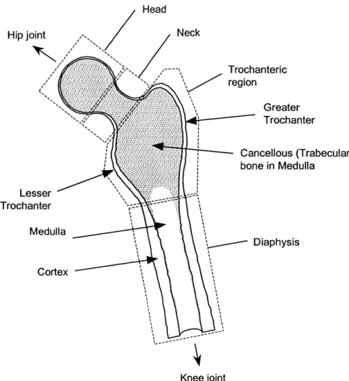
Figure 2.2: Principal regions, and anatomical landmarks of the proximal femur. 30
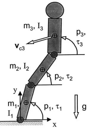
Figure 2.3: Three segment planar model used to simulate human locomotion. 40 Figure 2.4: Hip locus centered over point of support. 44
Figure 2.5: Impedance control of the ankle and knee joints causes the upper body to respond as if there were a virtual spring and damper connecting the hip directly to the ankle. 47
Figure 2.6: Schematic depicting experimental design for locomotion simulations. 50
Figure 2.7: Three segment model used for simulation of human falling to the side. 52 Figure 2.8: Schematic depicting experiment design for fall simulations 56
Figure 2.9: Kinematic ('stick-figure') plot for 50th percentile male running at 4 m/s in a) Earth gravity, and b) Mars gravity. 58
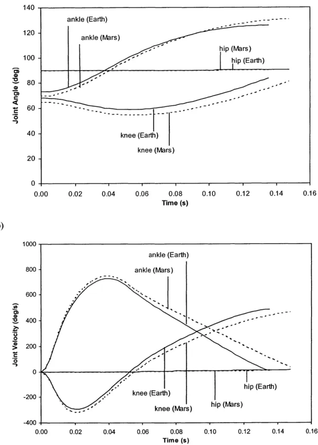
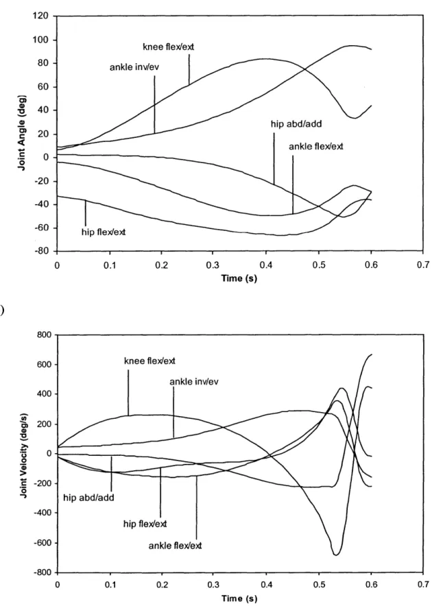
Figure 2.10: Plots of a) joint position, and b) joint velocity for a 50th percentile male running at 4 m/s in Earth gravity and Mars gravity (EVA). 59
Figure 2.11: Time history plots of a) joint acceleration, and b) joint torque for a 50th percentile male running at 4 m/s in Earth gravity and Mars gravity (EVA). 61
Figure 2.12: a) Hip locus plot, and b) time history plot of hip force, for a 50th percentile male running at 4 m/s in Earth gravity and Mars gravity (EVA). 63
Figure 2.13: Time history plot of rate of change of force applied to the hip during locomotion on Earth and on Mars. 64
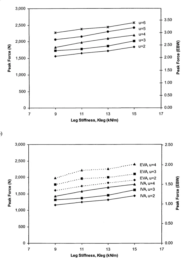
Figure 2.14: Peak hip force as a function of leg stiffness for a 50th percentile male running in a) Earth gravity, and b) Mars gravity. 66
Figure 2.15: Peak hip force as a function of leg stiffness for a 50th percentile female running in a) Earth gravity, and b) Mars gravity. 67
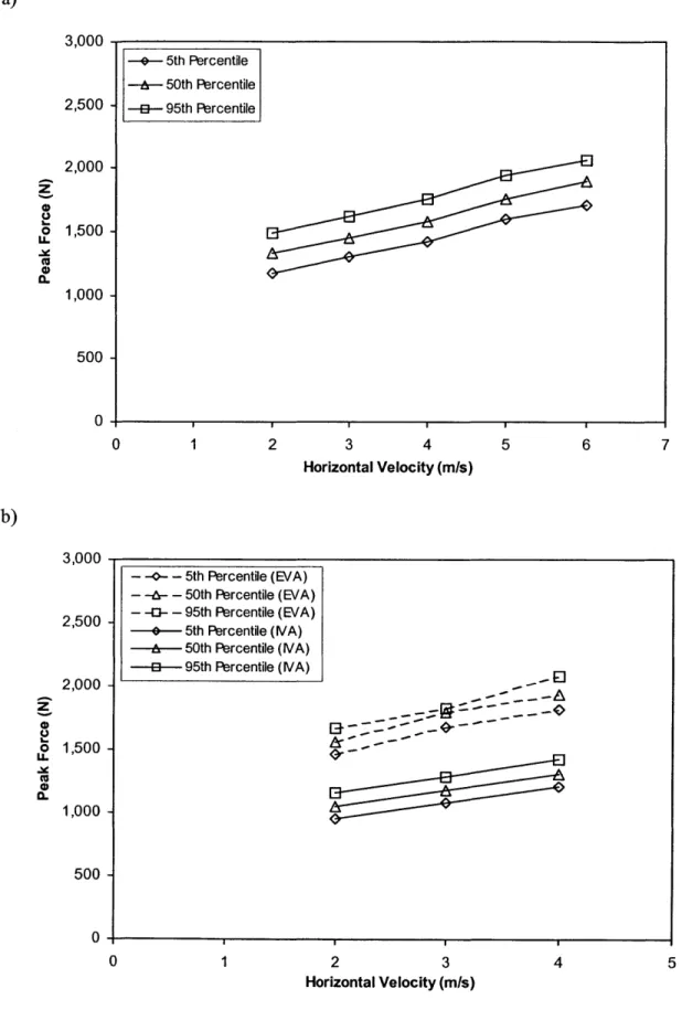
Figure 2.16: Peak hip force as a function of horizontal velocity for males running a) on Earth, and b) on Mars. 68
Figure 2.17: Peak hip force as a function of horizontal velocity for females running a) on Earth, and b) on Mars. 70
Figure 2.18: Kinematic ('stick-figure') plots for a 50th percentile male falling to the side in a) Earth gravity and b) Mars gravity (EVA case). 71
Figure 2.19: Plots of a) joint position and b) joint velocity for 50th percentile male falling in Earth gravity 73
Figure 2.20: Plots of a) joint acceleration and b) joint torque for 50th percentile male falling in Earth gravity 74
Figure 2.21: Plot of hip impact force for 50th percentile male following a fall to the side. 75 Figure 2.22: Body configurations at time of impact (viewed from above) for a 50th percentile
male falling in Earth gravity and Mars gravity (EVA case). The relevant joint angles are indicated with respect to the Earth configuration. 76
Figure 2.23: Peak hip impact force variation over the range of ground contact stiffnesses modeled. 77
Figure 2.24: Peak force exerted on the hip during fall impact (males). 78 Figure 2.25: Peak force exerted on the hip during fall impact (females) 79
Figure 3.1: Process for generating a three-dimensional finite element model of the proximal femur through pQCT scans, based on techniques developed by Oden et al.
(1999). 94
Figure 3.2: a) Sample femur slice after pQCT data has been processed in NIH Image (cuts through lesser trochanter at top left), b) thresholded slice, and c) boundaries
extracted from thresholded slice. 95
Figure 3.3: Creation of geometric femur model in I-DEAS. a) Outer boundary curves imported and stacked. b) Outer curves lofted to create total volume that is partitioned into quarters. c) Inner boundary curves imported and lofted to create
surface that partitions total volume into cortical and cancellous regions. d) Using plane surfaces, the model is partitioned at four points along the length of the bone
giving a final total of 40 volumes. 96
Figure 3.4: Conversion of geometric model into finite element model in I-DEAS. a) Complete model containing 6,400 elements and 25,164 nodes. b) The cortical shell was modeled and meshed as a separate volume. 97
Figure 3.5: Boundary conditions for finite element analysis of locomotion (mid-stance) loading condition. 101
Figure 3.6: Boundary conditions for fall loading case. 102
Figure 3.7: Flowchart depicting application of failure criteria for individual elements and whole bone through the use of a user subroutine included in the ABAQUS run
(based on Selvitelli (1997)). 103
Figure 3.8: Load vs Displacement plot for baseline femur in mid-stance load configuration (muscle forces included). 105
Figure 3.9: Mid-stance loading: Depiction of the deformed femur (white) at point of failure, superimposed on an image of the undeformed femur (grey). The 1, 2, and 3 directions indicated correspond with x, y, and z axes, respectively. 107
Figure 3.10: Mid-stance loading: Contour plot of maximum principal strain. The highest values correspond with areas of greatest tensile strain. 108
Figure 3.11: Mid-stance loading: Pattern of element failure in the proximal femur. Failed elements are indicated in grey. The failure sequence proceeds according to the numbered sequence, with the superior aspect of the femur presented on the left, and the inferior aspect on the right for each set. 109
Figure 3.12: Load vs displacement plot for loading in the mid-stance configuration, but without the application of muscle forces. 110
Figure 3.13: Load vs displacement plot for baseline femur in fall loading configuration. Ill
Figure 3.14: Fall loading: Depiction of the deformed femur (white) at point of failure, superimposed on an image of the undeformed femur (grey). 111
Figure 3.15: Fall loading: Contour plot of minimum principal strain. In this case, the highest negative values correspond with areas of greatest compression. The 1, 2, and 3 directions correspond with the x, y, and z axes, respectively. 112
Figure 3.16: Fall loading: Pattern of element failure in the proximal femur. Failed elements are indicated in grey. The failure sequence proceeds from top to bottom, with the posterior aspect of the femur presented on the left, and the anterior aspect on the right. 114
Figure 3.17: Comparison of failure loads for the three conditions analyzed for the baseline femora. 115
Figure 4.1: Values derived from DXA data on 20 cosmonauts, 7 with data from 2 flights, 27 flights total. BMD and Section Modulus exhibit approximately equal rates of change ranging from -0.75% to -1.50%. Most importantly, the rates of change in endosteal diameter are not offset by corresponding changes in the periosteal diameter, leading to a thinning of the cortical shell.
Source: Beck, T.J., Dept. of Radiology, Johns Hopkins University School of Medicine. 125
Figure 4.2: Method of increasing endosteal diameter to model space flight changes. (Described in text.) 128
9
Figure 4.3: Cortical and medullary (cancellous) element sub-volumes defmed for calculating changes in bone mineral content related to bone resorption at the endosteal
surface. The four sub volumes shown are: a) neck cortex, b) neck medulla, c) trochanteric cortex, and d) trochanteric medulla. 130
Figure 4.4: Space flight modified endosteal boundary in femoral neck. 133
Figure 4.5: Space flight modified endosteal boundary in trochanteric region. 133
Figure 4.6: Space flight modified endosteal boundary in diaphysis. 134 Figure 4.7: Reaction force versus displacement for mid-stance loading. 136
Figure 4.8: Reaction force versus displacement for fall loading. 137
Figure 4.9: Failure load versus duration of weightlessness for all mid-stance analyses. 138
Figure 4.10: Decline in femoral strength in fall loading according to duration of weightlessness. 139
Figure 5.1: Factor of risk for hip fracture in males during mid-stance. 153
Figure 5.2: Factor of risk for hip fracture in females during mid-stance. 153
Figure 5.3: Factor of risk for hip fracture in males during fall loading. 154 Figure 5.4: Factor of risk for hip fracture in females during fall loading. 154
10
Schaffner, G., Ph.D. Thesis 10
List of Tables
Table 1.1 Summary of space flight and bed rest studies of bone loss. 18
Table 2.1 Factors associated with increased risk of hip fracture during a fall. 34 Table 2.2 Summary of peak hip forces in locomotion and fall studies. 37
Table 2.3 Control parameters used in fall simulations. 55
Table 2.4 Body configurations at time of hip impact. (vdK = van den Kroonenberg) 75 Table 3.1 Correlations between in vitro bone strength and bone mineral parameters. 90
Table 3.2 Summary of FEA model generation techniques examined in various studies. 91
Table 3.3 Bone mineral density values obtained using a standard analysis program (Hologic) and custom software (Beck et al., 1990; Mourtada et al., 1996) 94
Table 3.4 Muscle forces applied during locomotion (mid-stance) loading condition. 101
Table 4.1 Minimum, mean and maximum values for rate of change of BMD and endosteal diameter. Note: Maximum is negative for BMD and positive for endosteal diameter. Source: Beck, T.J., Dept. of Radiology, Johns Hopkins University School of Medicine 125
Table 4.2 Comparison of studies assessing long term BMD losses in the femoral neck. 126
Table 4.3 Summary of muscle strength changes associated with unloading during space flight weightlessness and Earth-based analogs. 127
Table 4.4 Changes in BMC and medullary element average density following 6 months and 12 months of weightlessness 135
Table 4.5 Geometric data for calculating medulla element density adjustment 135
Table 5.1 Applied load, failure load, and factor of risk for each category and condition. Applied and failure loads are expressed in Newtons (N). 152
Table 5.2 Equations for calculating factor of risk for hip fracture in males and females according to type of activity and gravitational environment. Note that is the mass of the subject (kg), is the horizontal velocity (mi/s), and is the number of months of weightlessness. Also note that the locomotion equations include the contribution of muscle forces. 155
Schaffner, G., Ph.D. Thesis
11
11 Schaffner, G., Ph.D. Thesis
Abbreviations
AMG automatic mesh generation BMD bone mineral density
BW body weight
c.g. center of gravity
CSMI cross-sectional moment of inertia
CT computed tomography
DXA dual-energy x-ray absorptiometry
EVA extravehicular activity
FEA finite element analysis FEM finite element method
G gravity (usually Earth-normal)
ISS International Space Station IVA intravehicular activity
PLSS portable life support system
PPD proportional plus derivative (control)
QCT quantitative computed tomography
pQCT peripheral quantitative computed tomography
CHAPTER
Introduction
We choose to go to the Moon in this decade and do the other things, not because they are easy, but because they are hard.
- John F. Kennedy
Given the right political motivation and an appropriate investment of resources, astronauts could be exploring the surface of Mars as early as 10 years from now. During the 6 to 12 month journey the astronauts will experience physiological adaptations that are not yet fully understood. Some of the major systems affected include the musculoskeletal, cardiovascular, immune, hematologic, and neurovestibular systems. The changes that occur in these systems, while representing a natural adaptation to weightlessness, could have serious consequences to the individual upon return to a gravitational environment, either on Earth or on Mars. The pur-pose of the body of work described in this thesis is to gain insight into the risks impur-posed by changes in one of these physiological systems, namely, the musculoskeletal system.
1.1 Background
1.1.1 Measurement of bone mineral density
The parameter that is most often used to describe loss of bone mass, stiffness, and strength, is bone mineral density (BMD). This is not a "density" in the traditional engineering
13 Schaffner, G., Ph.D. Thesis
sense, that is, total mass divided by volume (gm/cm3), but is instead an "areal density" of min-eral mass (gm/cm2). Its value is derived from dual-energy X-ray absorptiometry (DXA), in which a two-dimensional projection of a bone is divided up into regions in which the bone-mineral content (BMC, calculated from the amount of X-ray absorptance) is divided by the projected area of the region to yield the BMD value for that region. This technique has several limitations in terms of assessing the strength of bone. Specifically, by collapsing the bone to two dimensions, information about its three dimensional geometry, and three dimensional density distribution are lost. It is thus difficult to reconstruct an engineering model of the bone that takes into account these three-dimensional attributes. To date, all of the measurements of bone mineral loss in the proximal femora of astronauts (and subjects in Earth-based space flight simulations) have used DXA as the sole imaging technique. To assess strength, the BMD values obtained from DXA are correlated with in vitro mechanical tests of cadaveric femora. Applying more accurate techniques of assessing bone strength, such as the three-dimensional finite element analysis described in this thesis, is greatly hindered in the case of space flight studies due to the absense of three dimensional bone information, such as can be obtained by computed tomography (CT), for instance. Many of the limitations of the work described in this thesis stem directly from this lack of three-dimensional data and it is hoped that future space flight bone studies will fill this information gap.
1.1.2 Bone Loss in Spaceflight and Earth-based Analogs
During space flights lasting longer than one month, astronauts undergo significant losses of bone mass and bone mineral density in the weight bearing areas of the skeleton, particularly the spine and lower limbs, as a result of the unloading produced by weightlessness in the microgravity environment (LeBlanc et al., 1998; Holick, 1998; Vico et aL., 1998). Due to the
relatively small number of human subjects who have flown in space, the limited duration of missions to date, and the inaccuracy of early measurement techniques, the problem of bone loss during weightlessness is not yet well quantified or well understood. Enough evidence has been gained, however, to raise concern about the risk of fracture, particularly in the hip, dur-ing skeletal loaddur-ing followdur-ing return to Earth (1 G), durdur-ing activities on the surface of Mars
(3/8 G), or even during strenuous activities performed in weightlessness, such as
extravehicu-lar (EVA) construction of the International Space Station (ISS).
The results of studies conducted during the space flights of the 1960's and early 1970's are highly variable due to poor measurement techniques employed in some cases. Following the Gemini 4, 5, and 7 missions, lasting from 4 to 14 days, investigators noticed a distinct increase in calcium excretion (Lutwak, 1966; Whedon et al., 1967) and initially thought that the astronauts had experienced a dramatic 10-20% loss in calcaneus and metacarpal bone den-sity (Mack and Lachance, 1966; Mack et al., 1967). However, through reevaluation, these losses were reduced to about 2-4% for five astronauts and 9% for one astronaut (Vose, 1974). The 18-day Soyuz 9 mission produced a 8-10% decrease in heel bone density for both cosmo-nauts (Birykov and Krasnykh, 1970). Bone density measurement techniques were improved during the three-man Apollo flights, lasting up to 13 days, but in only one of these flights were investigators able to measure a significant amount of bone loss from the heel (Rambaut et al.,
1975).
Studies conducted during the longer-term Skylab, Salyut and Mir space station missions allowed for better measurement of calcium homeostasis and bone density, but were con-founded by other factors such as variable compliance with prescribed exercise countermea-sures intended to minimize bone loss. Skylab metabolic data indicate that over a three month period, the total negative calcium balance from excess urine and fecal excretion is as much as
25 grams (Rambaut et al., 1979b; Rambaut et al., 1979a), but later estimates reduced this
value to 12.8 grams or 1% of the 1250 grams in the average skeleton (Cann, 1993). Reduced losses during the Skylab 4 mission have been attributed to increased exercise by the astro-nauts. Both US and Soviet investigations estimated that the average bone loss from the calca-neus was 1% per month (Stupakov et al., 1984). A combined US / USSR study of long-term spaceflight, in which quantitative computed tomography (QCT) scans were taken of the spine found no significant loss of density in the vertebral bodies (Oganov et al., 1990), apparently validating the exercise countermeasures. Closer inspection, however, revealed that there was an 8% loss of density in the posterior elements of the vertebrae, which correlated with a 4% loss of volume in the attached muscles, perhaps demonstrating the limited effectiveness of exercise countermeasures in space. Further evidence of this limitation came from QCT scans of one cosmonaut after a 366-day Mir mission, which showed a 10% loss of trabecular bone mass in LI, L2, and L3 vertebrae (Grigoriev et al., 1991). When investigators started looking at other regions in the body, they found even more distressing losses. Most significantly, a quantitative digital radiography (QDR, equivalent to DXA) study of cosmonauts after 4.5-6 month long missions on Mir found bone mineral density (BMD) losses of as much as 14% in the femoral neck and greater trochanter of the hip (Oganov et al., 1992). A study of US astro-nauts found that even in relatively short flights (1 to 2 weeks), the vertebrae L2-4 could lose as much as 3% of baseline BMD (Miyamoto et al., 1998).
The seriousness of the losses in BMD during spaceflight is evident when compared with the losses attributed to aging. On average, the rate of BMD loss for the proximal femur and lumbar vertebrae in men and women over 55 years of age has been estimated to be around
0.5-1% per year (Burger et al., 1998; Greenspan et al., 1996; Ensrud et al., 1995; Jones et al.,
at the rate of about 4% per year and beyond age 75 the risk of hip fracture increases exponen-tially. As mentioned above, the rates of loss from the same skeletal areas during spaceflight are about 1 -2% per month, 10 or more times greater than the rate occurring in normal aging. From another perspective, an estimated loss of 20% in femoral neck BMD during a year of spaceflight would correspond to the average BMD loss in the femoral neck of a woman aging from 50 years to almost 80 years (Looker et al., 1995). While the mechanisms responsible for bone loss in ageing and spaceflight are probably different (LeBlanc and Schneider, 1991), the similarities in the observed changes may be of mutual benefit to the study of either case (Hughes-Fulford, 1991).
Some investigators have used bed rest as an analog for the skeletal unloading experienced in spaceflight. One early study put 90 healthy young men through 5-36 weeks of bed rest and found that not only was there an average 5% loss of calcaneal mineral each month, but that mechanical and biochemical countermeasures were unsuccessful at preventing this loss (Schneider and McDonald, 1984). During another 17 week bed rest study, subjects lost an average of 0.21 0.05% per week of bone mineral density in the femoral neck, and 0.27
+/-0.05% per week in the trochanteric area (LeBlanc et al., 1990). Other studies have shown
sim-ilar mineral losses, slowing of mineralization, and limitations of countermeasures. Some stud-ies have also encountered contradictory results demonstrating the difficultstud-ies of bed rest as an analog for spaceflight (Vico et al., 1987; Zaichick and Morukov, 1998). The combined results of space flight and bed rest studies, in terms of measured BMD loss, are summarized in Table
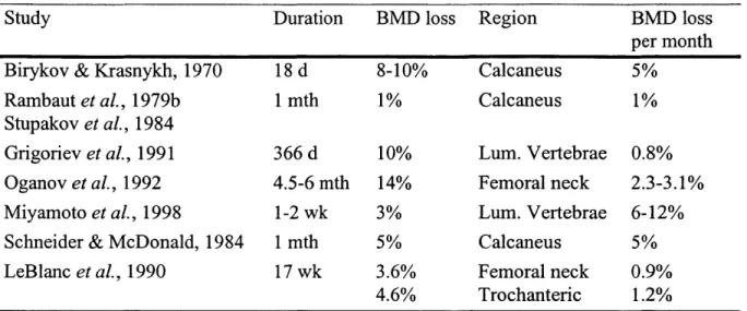
1.1. The last column is a normalized parameter indicating the actual or projected BMD loss
per year.
Results from animal studies of bone mineral loss in spaceflight and immobilization have been highly variable due to differences in study design, duration, and measurement
tech-17
Table 1.1 Summary of space flight and bed rest studies of bone loss.
Study Duration BMD loss Region BMD loss
per month Birykov & Krasnykh, 1970 18 d 8-10% Calcaneus 5%
Rambaut et al., 1979b 1 mth 1% Calcaneus 1%
Stupakov et al., 1984
Grigoriev et al., 1991 366 d 10% Lum. Vertebrae 0.8%
Oganov et al., 1992 4.5-6 mth 14% Femoral neck 2.3-3.1%
Miyamoto et al., 1998 1-2 wk 3% Lum. Vertebrae 6-12%
Schneider & McDonald, 1984 1 mth 5% Calcaneus 5%
LeBlanc et al., 1990 17 wk 3.6% Femoral neck 0.9%
4.6% Trochanteric 1.2%
niques. In addition, much of the variability may be due to the age of the animals, since many of the early studies were carried out on rats that were still growing (less than 5-6 months old). Rats were flown on eight Russian Cosmos biosatellite missions, lasting from 5 days to 22 days, as well as on the US Spacelab 3 mission (7 days). Juvenile male rhesus monkeys were flown on five Cosmos missions and an adult nemestria macaque monkey was flown on an early US mission (Biosatellite 3). Additional studies were carried out on rats and monkeys in various forms of suspension and hypokinesia. Bone formation was found to be reduced in the metaphyses of long bones during the first Cosmos mission (Yagodovsky et al., 1976). The Cosmos 782 and 936 missions resulted in a 40% reduction in the length of the primary spon-giosa (Asling, 1978) and a 30% decrease in the femoral breaking strength (Spector et al.,
1983). In addition, these missions revealed that an arrest line separating normal bone from
defective and hypomineralized bone formed during spaceflight (Turner et al., 1985) and that osteoblast differentiation in non-weight bearing bones was suppressed during weightlessness (Roberts et al., 1981), thus yielding evidence that other bones in the body might be affected in long term flight. Rats flown for 5 days aboard the Cosmos 1514 biologic satellite incurred no measurable change in bone mass (Vico et al., 1987), while calcium excretion studies on
mon-keys on the same flight showed evidence of increased resorption (Cann et al., 1986). After the slightly longer Cosmos 1667 mission, the flight rats showed a greater loss of trabecular bone in the proximal metaphyses of the tibia than rats undergoing tail suspension for the same dura-tion on earth (Kaplansky et al., 1987; Vico et al., 1991). Rats undergoing tail suspension of their hindlimbs for 15 days showed reductions in calcium content to 86.2 ± 2.5% for the tibia,
and 75.5 ± 3.5% for vertebrae (Globus et al., 1984), while those exposed to hypokinesia and
head-down suspension for periods of 35 to 60 days exhibited osteoporosis in the tibial and vertebral spongiosa (Dumova et al., 1986). Other tail suspension studies revealed a reduction of osteoblast number and growth and mineralization rates in the unloaded bones (Morey-Hol-ton and Globus, 1998). Rhesus monkeys flown for 13 days on Cosmos 1887 and 2044, and
11.5 days aboard Cosmos 2229, showed reduced bone mineralization and growth and a
signif-icant decrease in whole body bone mineral content (BMC), with only partial recovery by one month post-flight (Cann et al., 1990; Zerath et al., 1996). In addition, during the Cosmos 2044 mission it was shown that the fracture repair process was impaired in rats (Kaplansky et al.,
1991).
1.1.3 Summary of findings from spaceflight and immobilization studies
The findings of these spaceflight and immobilization studies, conducted over the past 35 years, may be summarized as follows:
* Significant bone loss occurs in humans and animals exposed to weightlessness during spaceflight
* Urine and fecal calcium excretion is increased resulting in a negative calcium balance
- Calcium resorption from bone is increased and absorption from gut is decreased * Bone mineral density decreases
Schaffner, G., Ph.D. Thesis
19
- Critical weight-bearing areas lose density most rapidly and the rate of loss is approxi-mately 1-2% per month
- Non-weight-bearing areas are affected in the long term
* Osteoblast proliferation and activity are reduced while osteoclast activity either remains the same or increases slightly
* Bone growth is slowed * Fracture repair is impaired * Bone strength is reduced
1.2 Significance
Several conclusions may be drawn regarding the significance of these skeletal changes in terms of human spaceflight.
* Astronauts and cosmonauts spending a significant period in weightlessness (> 1 month) will experience a loss of bone strength and a subsequent increase in fracture risk during:
- Activities on Earth (walking / running, falls)
- Intravehicular / extravehicular activity (IVA / EVA) on Mars or in weightlessness * A fracture occurring on Mars (3/8 G) has serious consequences to the individual and
crew due to:
- remoteness (limited medical resources)
- possible inhibition of fracture repair and immune respones associated with weight-lessness
- loss of functionality in terms of the crew member's skills and duties (increased work-load on remaining crew members)
To date, hip fracture has risk has generally been estimated based on correlations between bone mineral density (BMD) and failure load for a given loading condition (usually associated with an specific activity, including traumatic activities such as falls). In the vast majority of
cases, the failure load in this context is obtained through mechanical testing of cadaveric fem-ora. Thus, while BMD correlates reasonably well with bone strength in vitro, actual fracture risk is harder to calculate due to in vivo factors (e.g. body mass). Consequently, there exists a need to better assess the in vivo fracture risk for astronauts performing normal and traumatic activities following a significant period of weightlessness (i.e., more than a month).
1.3 Research Aims
The goals of this thesis may be summarized according to the following aims:
Primary Aim:
To improve the assessment of hip fracture risk in astronauts exposed to long-term weightlessness.
Specific Aim 1:
To estimate the loading applied to the proximal femur during locomotion and falls, both in Earth gravity and Mars gravity.
Specific Aim 2:
To estimate the failure load of the proximal femur during locomotion and falls.
Specific Aim 3:
To model the changes in geometry, bone mineral density and muscle forces related to the proximal femur due to weightlessness, and to account for sex differences.
Specific Aim 4:
To estimate the risk of hip fracture during locomotion and falls in Earth gravity and Mars gravity.
1.4 Outline of Thesis
A diagram of the overall research plan for this thesis is presented in Figure 1.1. There
were two main components to the plan, one aimed at determining the failure load for the prox-imal femur, the other aimed at determining the applied load. The applied loading was calcu-lated by means of dynamic simulations of human locomotion and falls using three-segment models. The equations of motion for these simulations were derived using both a Lagrangian formulation and Kane's method. The failure load was determined through the use of an adjust-able finite element model that served as a representation of the proximal femur for an average male or female adult of the approximate age expected of an astronaut. The model was derived directly from quantitative computerized tomography (QCT) scans of the right femur from a deceased 36 year-old male. The QCT images weer used to derive both geometric information and bone density data. When the applied load (estimated in the dynamics analysis) was divided by the failure load (calculated in the finite element analysis), it yielded the factor of risk for hip fracture ("fracture risk") under the given loading condition (locomotion or fall). The analysis was repeated as the model was adjusted to account for changes in geometry and density due to spaceflight and sex differences. In addition, differences in gravity level (Earth vs Mars) were taken into account during the dynamic simulation.
This work is described in the remaining chapters of this thesis in the following manner:
Chapter 2: This chapter describes the formulation of equations of motion for
three-segment dynamic models used to assess the applied loading in the hip during locomotion and falls. Environments simulated include Earth (1 G) and Mars (3/8 G), as well as both intravehicular activity (IVA) and extra-vehicular activity (EVA). The applied loads calculated were later used in Chapter 5 to determine the factor of risk for fracture.
22
Schaffner, G., Ph.D. Thesis
Chapter 3: Chapter 4: Chapter 5: - 36 y.o. Male - CT - DXA Adjustable Finite Element Model Failure Load
~1i
Space Flight A Incr. endost. diam. Red. trabec. mass Red. musc. strengthGender Equations of Motion * Lagrangian * Kane's method Applied Load
Figure 1.1: Diagram of overall research plan for thesis.
Schaffner, G., Ph.D. Thesis
The construction and analysis of a three-dimensional finite element model of the proximal femur are discussed. The model was analyzed in configurations simulating both locomotion (mid-stance) and a fall to the side impacting the greater trochanter. Failure analysis was carried out through a user subroutine employing maximum and minimum principle strain failure criteria.
The finite element model described in Chapter 3 was modified to account for changes in femoral geometry and bone mineral density that are asso-ciated with space flight and gender differences.
The values calculated for applied loading (Chapter 1) were combined with the corresponding failure load values (Chapters 3 and 4) to arrive at values for factor of risk for hip fracture associated with the various load-ing cases considered. Conclusion of thesis.
3-segment models - locomotion (3 dof) - fall impact (5 dof)
Gravity Level Earth (1g) Mars (3/8g)
Fracture Risk
D- Fapplied FfaiI 231.5 References
Asling, C.W., Histological studies on tibial bone of rats in the 1975 Cosmos 782 flight, in
Final reports of U.S experiments flown on the Soviet satellite Cosmos 782, S.N.
Rosen-zweig and K.A. Souza, Editors. 1978, NASA TM-78525. p. 276-307.
Birykov, N. and Krasnykh, I.G., [Changes in optical density of bone tissue and calcium
metabolism in the cosmonauts]. Kosm Biol Med, 1970. 4: p. 42-45.
Burger, H., de Laet, C.E., van Daele, P.L., Weel, A.E., Witteman, J.C., Hofman, A. and Pols,
H.A., Risk factors for increased bone loss in an elderly population: the Rotterdam study. Am J Epidemiol, 1998. 147(9): p. 871-879.
Cann, C.E., Adaptations of the skeletal system to spaceflight, in Introduction to space life
sci-ence, S. Churchill, Editor. 1993.
Cann, C.E., Calcium metabolism and correlated endocrine measurements in primates during
Cosmos '83, in Final reports of U.S. monkey and rat experiments flown on the Soviet satellite Cosmos 1514, R.C. Mains and E.W. Gomersall, Editors. 1986, NASA
TM-88223. p. 129-144.
Cann, C.E., Rakhmanov, A. and Oganov, V., Radiographic studies of skeletal growth in
pri-mates in Cosmos 1887, in Final reports of the U.S. experiments flown on the Soviet bio-satellite Cosmos 1887, J.P. Connolly, R.E. Grindeland, and R.W. Ballard, Editors.
1990, NASA TM-102254.
Durnova, G.N., Sakharova, Z.F., Kaplanskii, A.S., Ivanov, V.M. and Khaidakov, M.S.,
[Quantitative study of osteoblasts and osteoclasts in the bones of rats during the simula-tion of weightlessness]. Kosm Biol Aviakosm Med, 1986. 20(6): p. 37-41.
Ensrud, K.E., Palermo, L., Black, D.M., Cauley, J., Jergas, M., Orwoll, E.S., Nevitt, M.C., Fox, K.M. and Cummings, S.R., Hip and calcaneal bone loss increase with advancing
age: longitudinal results from the study of osteoporotic fractures. J Bone Miner Res, 1995. 10(11): p. 1778-1787.
Greenspan, S. L., Maitland-Ramsey, L., and Myers, E. (1996). "Classification of osteoporosis in the elderly is dependent on site-specific analysis." Calcif Tissue Int, 58(6), 409-14. Globus, R.K., Bikle, D.D., and Morey-Holton, E., Effects ofsimulated weightlessness on bone
mineral metabolism. Endocrinology, 1984. 114(6): p. 2264-2270.
Grigoriev, A.I., Bugrov, S.A., Bogomolov, V.V., Egorov, A.D., Kozlovskaya, I.B., Pestov I.D., Polyakov, V.V., Tarasov, I.K., Medical results of the Mir year-long mission. Phys-iologist, 1991. 34(Suppl): p. S44-S48.
Holick, M.F., Perspective on the impact of weightlessness on calcium and bone metabolism. Bone, 1998. 22(5 Suppl): p. 105S-1IS.
Hughes-Fulford, M., Altered cellfunction in microgravity. Exp Gerontol, 1991. 26(2-3): p.
247-256.
Jones, G., Nguyen, T., Sambrook, P., Kelly, P.J. and Eisman, J.A., Progressive loss of bone in
the femoral neck in elderly people: longitudinal findings from the Dubbo osteoporosis epidemiology study. BMJ, 1994. 309(6956): p. 691-695.
Kaplansky, A.S., Dumova, G.N., Burkovskaya, T.E. and Vorotnikova, E.V., The effect of
microgravity on bone fracture healing in rats flown on Cosmos 2044. Physiologist,
1991. 34(Suppl): p. S196-S199.
Kaplansky, A.S., Dumova, G.N. and Sakharova, Z.F., [Histomorphometric analysis of the
bones of rats on board the Kosmos 1667 biosatellite]. Kosm Biol Aviakosm Med, 1987. 21(5): p. 25-31.
LeBlanc, A. and Schneider, V., Can the adult skeleton recover lost bone? Exp Gerontol, 1991.
26(2-3): p. 189-201.
LeBlanc, A., L. Shackleford, L. and Schneider, V., Future human bone research in space. Bone, 1998. 22(5 Suppl): p. 113S-116S.
LeBlanc, A.D., Schneider, V.S., Evans, H.J., Engelbretson, D.A. and Krebs, J.M., Bone
min-eral loss and recovery after 17 weeks of bed rest. J Bone Min Res, 1990. 5(8): p. 843-850.
Looker, A.C., Johnston, C.C., Jr, Wahner, H.W., Dunn, W.L., Calvo, M.S., Harris, T.B., Heyse, S.P., Lindsay, R.L., Prevalence oflowfemoral bone density in older U.S. women
from NHANES III. J Bone Min Res, 1995. 10(5): p. 796-802.
Lutwak, L., Chemical analysis of diet, urine, feces and sweat parameters relating to the
cal-cium and nitrogen balance studies during Gemini VIIflight (Exp M7),. 1966.
Mack, P.B. and Lachance, P.A.. Effects of recumbence and space flight on bone density. in
2nd Annual Biomedical Research Conference. 1966. Houston, TX.
Mack, P.B., LaChance, P.A., Vose, G.P. and Vogt, F.B., Bone demineralization offoot and
hand of Gemini-Titan IV, V and VII astronauts during orbital flight. Am J Roentgenol
Rad Therapy Nucl Med, 1967. 100: p. 503-511.
Miyamoto, A., Shigematsu, T., Fukunaga, T., Kawakami, K., Mukai, C. and Sekiguchi, C.,
Medical baseline data collection on bone change with space flight. Bone, 1998. 22(5
Suppl): p. 79S-82S.
Morey-Holton, E.R. and Globus, R.K., Hindlimb unloading ofgrowing rats: a modelfor
pre-dicting skeletal changes during spaceflight. Bone, 1998. 22(5 Suppl): p. 83S-88S.
Oganov, V.S., Cann, C., Rakhmanov, A.S. and Ternovoi S.K., [A computer tomographic
investigation of the muskuloskeletal system of the spine in humans after long-term space flight]. Kosm Biol Aviakosm Med, 1990. 24: p. 20-2 1.
Oganov, V.S., Grigor'ev, A.I., Voronin, L.I., Rakhmanov, A.S., Bakulin, A.V., Schneider,
V.S. and LeBlanc, A.D., [Bone mineral density in cosmonauts after flights lasting 4.5-6
months on the Mir orbital station]. Aviakosm Ekolog Med, 1992. 26(5-6): p. 20-24.
Rambaut, P.C., Skeletal response., in Biomedical results ofApollo, R.S. Johnston, L.F. Diet-lin, and C.A. Berry, Editors. 1975, NASA SP-368: Washington, DC. p. 303-322.
Rambaut, P.C. and Johnston, R.S., Prolonged weightlessness and calcium loss in man. Acta Astronautica, 1979b. 6: p. 1113-1122.
Rambaut, P.C., Leach, C.S. and Whedon, G.D., A study of metabolic balance in crew
mem-bers ofSkylab IV Acta Astronautica, 1979a. 6: p. 1313-1322.
Roberts, W.E., Mozsary, P.G. and Morey, E.R., Suppression of osteoblast differentiation
dur-ing weightlessness. Physiologist, 1981. 24(Suppl): p. S75-S76.
Schneider, V.S. and McDonald, J., Skeletal calcium homeostasis and countermeasures
topre-vent disuse osteoporosis. Calcif Tissue Int, 1984. 36(Suppl 1): p. S144-S15 1.
Spector, M., Arrested bone formation during space flight results in hypomineralized skeletal
defect. Physiologist, 1983. 26(Suppl): p. SIIO-S 11.
Stupakov, G.P., Kazeikin, V.S., Kozlovskii, A.P. and Korolev, V.V., [Evaluation of changes
in human axial skeletal bone structures during long-term space flights]. Kosm Biol
Aviakosm Med, 1984. 18: p. 33-37.
Turner, R.T., Bell, N.H., Duvall, P., Bobyn, J.D., Spector, M., Holton, E.M. and Baylink, D.J.,
Spaceflight results in formation of defective bone. Proc Soc Exp Biol Med, 1985.
180(3): p. 544-549.
Schaffner, G., Ph.D. Thesis
25
Vico, L., Chappard, D., Alexandre, C., Palle, S., Minaire, P., Riffat, G., Morukov, B. and Rakhmanov, S., Effects of a 120 day period of bed-rest on bone mass and bone cell
activities in man: attempts at countermeasure. Bone Miner, 1987. 2(5): p. 383-394.
Vico, L., Chappard, D., Alexandre, C., Palle, S., Minaire, P., Riffat, G., Novikov, V.E. and Bakulin, A.V., Effects of weightlessness on bone mass and osteoclast number in
preg-nant rats after a five-day spaceflight (COSMOS 1514). Bone, 1987. 8(2): p. 95-103.
Vico, L., Lafage-Proust, M.H. and Alexandre, C., Effects ofgravitational changes on the bone
system in vitro and in vivo. Bone, 1998. 22(5 Suppl): p. 95S-100S.
Vico, L., Novikov, V.E., Very, J.M. and Alexandre, C., Bone histomorphometric comparison
of rat tibial metaphysis after 7-day tail suspension vs. 7-day spaceflight. Aviat Space
Environ Med, 1991. 62(1): p. 26-3 1.
Vose, G.P., Review of roentgenographic bone demineralization studies of the Gemini space
flights. Am J Roentgenol Rad Therapy Nucl Med, 1974. 121: p. 1-4.
Whedon, G.D., Lutwak, L. and Neuman, W., Calcium and Nitrogen balance. In: A review of
medical results of Gemini 7 and relatedflights, . 1967, Kennedy Space Center, FL.
Yagodovsky, V.S., Trifaranidi, L.A., and Goroklova, G.P., Space flight effects on skeletal
bones of rats. Aviat Space Environ Med, 1976. 47: p. 734-73 8.
Zaichick, Y. and Morukov, B.V., In vivo bone mineral studies on volunteers during a 370-day
antiorthostatic hypokinesia test. Appi Radiat Isot, 1998. 49(5-6): p. 691-694.
Zerath, E., Novikov, V., Leblanc, A., Bakulin, A., Oganov, V. and Grynpas M., Effects of
spaceflight on bone mineralization in the rhesus monkey. J Appl Physiol, 1996. 81(1): p.
194-200.
26
CHAPTER
Hip Loading During Locomotion
and Falls in Earth Gravity and
Mars Gravity
Man walks the moon but his soul remains riveted to earth. Once upon a time it was the opposite.
- Elie Wiesel
As mentioned in the previous chapter, calculating the factor of risk for hip fracture requires the determination of two parameters: the force applied to the hip joint, and the failure load of the proximal femur. The principal purpose of the work described in this chapter is to determine the applied force for two distinct activities/conditions, namely, locomotion (run-ning) and a fall to the side impacting the greater trochanter. Locomotion was chosen as one of the conditions since it represents a normal activity that could be performed soon after a space flight, either on arrival at Mars or after return to Earth. Fall impact was chosen as the second activity since it represents an abnormal, worst-case, condition; since it is widely believed that approximately 90% of hip fractures on Earth are the result of a fall (Hedlund and Lindgren,
1987; Cummings et al., 1990); and since astronauts are believed to be at greater risk of fall
following space flight due to neurovestibular (balance) compromise and orthostatic intoler-ance (susceptibility to fainting). The peak forces calculated in this chapter will be used in Chapter 5 as the expected applied force in calculating the factor of risk for fracture during the two activities.
Since we are concerned about the fracture risk following return to Earth after a long flight as well as during activity shortly after arrival on Mars, the simulations reported here were per-formed under conditions of both Earth and Mars gravity, and, in the case of a Mars mission, both during intravehicular activity (IVA) and extravehicular activity (EVA). The loadings are calculated by means of two distinct three-link dynamic systems representative of the human body, one used to analyze locomotion and one used to analyze falls. The systems are designed to achieve sufficient accuracy (within 20% of values presented elsewhere in the literature), while still being simple enough that the motion can be controlled without requiring very com-plex motor control schemes.
2.1 Background
A review of the literature on human locomotion and falls reveals that a significant body of
information exists on the musculoskeletal loading experienced during these two activities. A new dynamics simulation study was deemed necessary, however, for two reasons. Firstly, there appears to be little or no information on hip loading during locomotion and falls in reduced gravity. Secondly, the forces reported from calculation and direct measurement in past studies do not have a high degree of consistency.
2.1.1 Terminology
To assist the reader with some of the terminology used in this chapter and other chapters in this thesis, two figures are presented in this section. The first, Figure 2.1, shows the three main stages of the stance phase of human gait. The work in this thesis is focussed on the mid-stance stage. The second, Figure 2.2, shows some of the main regions and anatomical land-marks that will be referred to repeatedly, especially in chapters 3 and 4. It is presented here
because the background text on hip loading makes use of some of the terms. Note in the figure
*---Toe-off Mid-stance Heel-strike
Stance Swing
Leg Leg
Figure 2.1: A few terms used to describe human gait.
that the trochanteric region is a combination of the "trochanteric" and "intertrochanteric" regions conventionally used in analysis of DXA scans.
2.1.2 Locomotion
A number of studies have been performed on the mechanics and energetics of locomotion
in Earth gravity. In general, two types of approaches have been adopted: mathematical models that examine certain aspects of the mechanics or energetics of locomotion, and experimental studies that measure parameters of interest directly.
The majority of studies using mathematical models of locomotion have ranged in com-plexity from simple inverted pendulums, to multi-segment ballistic models, to lumped mass and spring models. Despite their relative simplicity, these models have been successful at cap-turing the most important determinants of gait, such as step lengths, swing-times, and ground reaction forces. Mochon and McMahon (1980) used a three-link mathematical model to
29 Schaffner, G., Ph.D. Thesis
Head
Hip joint Neck
Trochanteric region Greater Trochanter Cancellous (Trabecular) bone in Medulla Lesser Trochanter Medulla Diaphysis Cortex Knee joint
Figure 2.2: Principal regions, and anatomical landmarks of the proximal femur.
examine the swing phase of walking and obtained good correlation with experimental data for swing times, joint angles, and horizontal ground reaction force. Siegler et al. (1982) simulated the stance phase of human locomotion with a model consisting of a concentrated mass sup-ported by two elastic and viscous straight legs. They obtained relatively good correspondence with experimental results, particularly for the vertical ground reaction force. Furthermore, the shape of the force time-history plots agreed well with force plate measurements. McMahon et
al. (1987) further analyzed how changes in the effective vertical stiffness (i.e., the stiffness of
the lower limb that serves to reverse the downward velocity of the body during one contact
30
Schaffner, G., Ph.D. Thesis
period) of the body affect locomotion characteristics. They found that reducing the vertical stiffness, through running with bent knees, reduces transmission of mechanical shock from the foot to the upper body, but requires significantly greater energy utilization. A simple mass-spring model predicted the peak vertical ground reaction force with good accuracy for normal running, but with slightly reduced accuracy for bent-knee running (since the larger joint angles introduce more nonlinearity into vertical stiffness). McMahon and Cheng (1990) further explored the lumped mass and spring model by assuming that the leg acts as a linear spring, but taking into account the angle between the leg and the vertical. The model provided good predictions if the leg stiffness, kieg, was assumed constant, while the effective vertical stiffness increased with increasing forward speed due to a required increase in the initial angle of the leg to the vertical (which results in greater compression of the leg spring). In support of the leg spring model, some studies have sought to characterize the stiffness of the leg spring through experimentation. Farley and Gonzalez (1996) used a treadmill-mounted force plat-form to derive leg spring stiffness in subjects running at 2.5 m/s. The leg spring stiffness was found to vary from 7.0 to 16.3 kN/m between the lowest and highest stride frequencies. Viale
et al. (1998) also used force platform measurements in conjunction with a spring-mass model
and obtained a leg spring stiffness of 13.0 kN/m for running.
Studies aimed at assessing the actual loading in the hip joint during walking, running, and other activities have generally used indirect approaches to determine the forces and moments present, either through inverse dynamics applied to experimental measurements, or through mathematical models. Crowninshield et al. (1978) generated kinematic data through photog-raphy of subjects performing movements and combined this with kinetic data from a force platform in an inverse dynamics analysis to determine the intersegmental forces and moments at the hip, knee, and ankle. They found that a 69 kg subject walking at 1.0 m/s experiences a
vertical joint contact force in the hip of 4.0 to 5.0 times body weight (BW). Recently, van den Bogert et al. (1999) found similar results using a slightly different approach, which measured acceleration of the upper body through a body-mounted accelerometer system in nine male subjects. Intersegmental force and moment at the hip joint during walking, running, and skiing were obtained through inverse dynamics. The peak joint contact force during walking at 1.5 m/s was 2.5 times BW, while running at 3.5 m/s produced a peak joint contact force of 5.2 times BW.
It was not until the introduction of instrumented hip prostheses, however, that actual mea-surements of in vitro hip loading could be obtained. Bergmann et al. (1993) measured hip forces and moments in two patients. In the first patient, they found that median peak forces increased with walking speed from 2.8 times body weight (BW) at 0.3 m/s (1 km/h) to about 4.8 times BW at 1.4 m/s (5 km/h). The forces increased to about 5.5 times BW during jogging and very fast walking. In the second patient, the median forces at 0.8 m/s (3 km/h) were about 4.1 times BW. Bassey et al. (1997) measured compressive axial force in an instrumented tita-nium implant during 'osteogenic' (encouraging bone formation) exercises (slow jumping, fast continuous jumping, or jogging). The implant forces (equivalent to the hip forces described by Bergmann et al.) were 2.5 to 4.0 times BW, and were also significantly related to muscle activity.
Studies of locomotion in reduced gravity are not nearly as numerous as those in Earth gravity conditions. Some of the early studies were conducted during the late 1960's in support of the Apollo moon landings (Margaria, 1966; Margaria and Cavagna, 1967; Hewes, 1968; Hewes, 1969). These studies were mainly concerned with gait strategies, energy require-ments, and the feasibility of locomotion in a reduced gravity environment (the moon, 1/6
(0.17) Earth gravity) while wearing a pressurized space suit.
More recent studies have sought to understand the mechanics and energetics of reduced gravity locomotion in greater scientific detail. He et al. (1991) performed experiments in which subjects ran on a treadmill instrumented with a force platform while reduced gravity was simulated using a suspension device that applied a nearly constant vertical force to the subjects. Four subjects ran at speeds of 2.0-6.0 m/s in conditions of Earth gravity and at 3.0 m/s in six simulated fractions of Earth gravity, ranging from 0.2 to 0.7 G. A leg stiffness of
11.3 kN/m applied to their linear spring-mass model produced good agreement with
experi-mental results. They found that leg stiffness and the vertical excursion of the center of mass during the flight phase of running did not change with increased forward speed or gravity. Furthermore, their model successfully predicted the decreasing peak force observed under conditions simulating reduced gravity. Newman et al. (1994) used an underwater treadmill with a ballasting harness to simulate partial gravity environments. They found that a loping gait was used over a wide range of speeds (1.5 m/s to 2.3 m/s). Furthermore, peak vertical force and stride frequency were significantly reduced as the gravity level was reduced, while ground contact time was independent of gravity.
Several studies have focussed more exclusively on the energetics of reduced gravity loco-motion. Farley and McMahon (1992) found that reducing gravity decreases the energetic cost for running much more than for walking, with the result that running is actually more efficient than walking for low levels of gravity. The same partial gravity simulation system used by Newman et al. was used by Wickman and Luna (1996) to assess energy requirements imposed on astronauts when supporting the mass of a protective suit and portable life support system
(PLSS). They found that energy expenditure, calculated from measured oxygen consumption,
was positively correlated with gravity level, speed, and load size. They predicted that individ-uals in average physical condition could walk for 8 h on the Moon with up to 170% of their
33
body mass, and on Mars with up to 50% of their body mass for the same duration. They also predicted that added mass (i.e., for a space suit) could be beneficial for bone mass mainte-nance in reduced gravity environments, but that some supplemental bone mass maintemainte-nance measures would still be required for most individuals. Cavagna et al. (1998) employed an
air-craft undergoing gravity-reducing flight profiles to investigate the mechanics of walking on Mars. The optimal walking speed on Mars was found to be 0.9 m/s (3.4 km/h), compared to
1.5 m/s (5.5 km/h) on Earth. Furthermore, the walk-run transition on Mars is predicted to
occur near the optimal walking speed on Earth and the work done per unit distance to move the center of mass would be about half that on Earth, which agrees with the findings of New-man et al. (1994).
2.1.3 Falls
It has been well established that 90 percent of all hip fractures are the result of a fall (Anonymous, 1985). While less than 10 percent of falls result in hip fracture, susceptible patients can sustain a hip fracture even from mild trauma (Goh et al., 1996). The factors
asso-ciated with an increased risk of hip fracture during a fall are listed in Table 2.1 (Goh et al.,
1996; Greenspan et al., 1998; Schwartz et al., 1998).
Table 2.1 Factors associated with increased risk of hip fracture during a fall.
1) Reduced BMD in the proximal femur
2) Fall direction (to the side, versus any other direction)
3) Direct impact (without hitting knee first, or catching oneself with an outstretched arm)
4) Impairment in mobility
5) Reduced soft tissue covering the greater trochanter (thin vs obese patients)
6) Poor physical conditioning
Impact direction has also been shown to correlate with failure load. Ford et al. (1996) used