Publisher’s version / Version de l'éditeur:
Biomacromolecules, 16, 1, pp. 319-325, 2014-12-01
READ THESE TERMS AND CONDITIONS CAREFULLY BEFORE USING THIS WEBSITE.
https://nrc-publications.canada.ca/eng/copyright
Vous avez des questions? Nous pouvons vous aider. Pour communiquer directement avec un auteur, consultez la première page de la revue dans laquelle son article a été publié afin de trouver ses coordonnées. Si vous n’arrivez pas à les repérer, communiquez avec nous à PublicationsArchive-ArchivesPublications@nrc-cnrc.gc.ca.
Questions? Contact the NRC Publications Archive team at
PublicationsArchive-ArchivesPublications@nrc-cnrc.gc.ca. If you wish to email the authors directly, please see the first page of the publication for their contact information.
NRC Publications Archive
Archives des publications du CNRC
This publication could be one of several versions: author’s original, accepted manuscript or the publisher’s version. / La version de cette publication peut être l’une des suivantes : la version prépublication de l’auteur, la version acceptée du manuscrit ou la version de l’éditeur.
For the publisher’s version, please access the DOI link below./ Pour consulter la version de l’éditeur, utilisez le lien DOI ci-dessous.
https://doi.org/10.1021/bm501516r
Access and use of this website and the material on it are subject to the Terms and Conditions set forth at
Cationic poly(2-aminoethylmethacrylate) and
poly(N-(2-aminoethylmethacrylamide) modified cellulose nanocrystals: synthesis,
characterization, and cytotoxicity
Hemraz, Usha D.; Campbell, Kendra A.; Burdick, James S.; Ckless, Karina;
Boluk, Yaman; Sunasee, Rajesh
https://publications-cnrc.canada.ca/fra/droits
L’accès à ce site Web et l’utilisation de son contenu sont assujettis aux conditions présentées dans le site LISEZ CES CONDITIONS ATTENTIVEMENT AVANT D’UTILISER CE SITE WEB.
NRC Publications Record / Notice d'Archives des publications de CNRC:
https://nrc-publications.canada.ca/eng/view/object/?id=307fd0bf-1b8e-4f0a-8efa-ecb9e43abb64 https://publications-cnrc.canada.ca/fra/voir/objet/?id=307fd0bf-1b8e-4f0a-8efa-ecb9e43abb64Cationic Poly(2-aminoethylmethacrylate) and
Poly(N‑(2-aminoethylmethacrylamide) Modified Cellulose Nanocrystals:
Synthesis, Characterization, and Cytotoxicity
Usha D. Hemraz,
†,‡Kendra A. Campbell,
§James S. Burdick,
#Karina Ckless,
#Yaman Boluk,
†and Rajesh Sunasee*
,#,§†
Department of Civil & Environmental Engineering, University of Alberta and National Institute for Nanotechnology, National Research Council, 11421 Saskatchewan Drive, Edmonton, Alberta T6G 2M9, Canada
‡
National Research Council, 6100 Royalmount Avenue, Montreal, Quebec H4P 2R2, Canada
§
Department of Chemistry, Cape Breton University, 1250 Grand Lake Road, Sydney, Nova Scotia B1P 6L2, Canada
#
Department of Chemistry, State University of New York at Plattsburgh, Plattsburgh, New York 12901, United States
*
S Supporting InformationABSTRACT: Cellulose nanocrystals (CNCs) continue to gain increasing attention in the materials community as sustainable nanoparticles with unique chemical and mechanical properties. Their nanoscale dimensions, biocompatibility, biodegradability, large surface area, and low toxicity make them promising materials for biomedical applications. Here, we disclose a facile synthesis of poly(2-aminoethylmethacry-late) (poly(AEM)) and poly(N-(2-aminoethylmethacryla-mide) (poly(AEMA)) CNC brushes via the surface-initiated single-electron-transfer living radical polymerization technique. The resulting modified CNCs were characterized for their
chemical and morphological features using a combination of analytical, spectroscopic, and microscopic techniques. Zeta potential measurements indicated a positive surface charge, and further proof of the cationic nature was confirmed by gold deposition as evidenced by electron microscopy. The cytotoxicity of these cationic modified CNCs was evaluated utilizing a 3-(4,5-dimethylthiazol-2-yl)-2,5-diphenyltetrazolium bromide (MTT) assay in two different cell lines, J774A1 (mouse monocyte cells) and MCF-7 (human breast adenocarcinoma cells). The results indicated that none of the cationic modified CNCs decreased cell viability at low concentrations, which could be suitable for biomedical applications.
■
INTRODUCTIONCellulose nanocrystals (CNCs) are biosourced nontoxic nanomaterials of great research interest in both academic and industrial sectors. These rigid rod-like cellulose crystals, obtained from the acidic hydrolysis of native cellulose, display remarkable strength and physicochemical properties with several potential applications.1−5 Their unique properties and future commercialization prospects have recently led to the industrial production of CNCs in both USA and Canada.6The ability to chemically modify CNCs through surface reactive hydroxyl groups opens up various potential applications, which are otherwise unachievable with unmodified CNCs. As a result, covalent or noncovalent chemical surface functionalization of CNCs continues to be an exciting area of research.7−9Previous
works have mainly focused on modifying CNCs for use as reinforcing agents in nanocomposites.2,10,11 However, CNCs have recently emerged as promising materials for biomedical applications owing to some of their interesting attributes such as nanoscale dimensions, biocompatibility, biodegradability, large surface area, and low toxicity.12−15For instance, CNCs’
surfaces were chemically modified to append fluorescent molecules13,16−19 for bioimaging12 and pH sensing.20 These modified CNCs were used for protein/enzyme immobilization studies due to their large surface area (150−250 m2/g) and
nonporous structure.21−23CNCs were explored as a potential drug delivery carrier, where they were capable of binding to ionized and hydrophobic drugs and undergoing controlled release.24CNCs have also been used to prepare colloidal silver
suspensions for antimicrobial properties.25 Although these initial studies look promising, a great deal of work is still required to fully understand the successful integration of CNCs in the biological systems.
Our interest in surface modification of CNCs for potential bioapplications led us to the design of cationic CNCs.26There
are only a few reports on cationization of CNCs, with the first reported pathway by Gray and co-workers that described a
one-Received: October 13, 2014 Revised: November 26, 2014 Published: December 1, 2014
step method to introduce positive charges on the surface of CNCs via a nucleophilic addition of alkali-activated cellulose hydroxyl groups to the epoxy moiety of epoxypropyltrimethy-lammonium chloride.27A similar approach was disclosed by Ni and co-workers, who, instead of performing cationization in an aqueous suspension, used a semidry method, resulting in an improved degree of substitution and charge density.28 Thiele-mans and co-workers used heterogeneous click chemistry to attach cationic imidazolium groups on the surface CNCs for ion-exchange applications.29The same group recently reported the preparation of cationized pyridinium CNCs with a high degree of substitution via a one-pot esterification/nucleophilic displacement reaction.30 In this work, our intent was to graft
cationic polymers from CNC surfaces by the surface-initiated single-electron-transfer living radical polymerization (SI-SET-LRP) approach using cationic monomers, namely, 2-amino-ethyl methacrylate (AEM) hydrochloride and 2-amino2-amino-ethyl- 2-aminoethyl-methacrylamide (AEMA) hydrochloride. The choice of these monomers was guided in part by our future interest in developing cationic CNC-based materials for biomedical applications. Furthermore, cationic poly(AEM) is a well-known polymer that has been used in conjunction with other polymers for in vitro delivery of genes to COS cells,31,32 while poly(AEM) homopolymers with well-defined chain lengths were recently designed for DNA vaccine delivery to dendritic cells.33Poly(AEMA) has been employed as a cationic core in the synthesis of nanogels for the design of plasmid DNA,34 protein,35and siRNA36delivery vehicles. The resulting cationic CNCs were characterized for their chemical and morphological features using a combination of analytical, spectroscopic, and microscopic techniques. The negative surface of unmodified CNCs, previously obtained via sulfuric acid mediated hydrolysis, was rendered cationic after the polymerization process. We further showed that these cationic CNC materials could be exploited as a nanoplatform for gold deposition for potential biomedical applications. The cytotoxicity of these cationic modified CNCs was evaluated with two different cell lines.
■
EXPERIMENTAL SECTIONMaterials. Freeze-dried CNCs were obtained from Alberta Innovates Technology Futures (AITF), which were produced by a sulfuric-acid-mediated hydrolysis of cellulose. A detailed preparation method is described elsewhere.37 Ethylenediamine, 1,3-diaminopro-pane, hydroquinone, methacrylic anhydride, tetrahydrofuran (THF, 99%), triethylamine (TEA), 4-dimethylaminopyridine (DMAP), 2-bromoisobutyryl bromide, N,N,N′,N″,N″-pentamethyldiethylenetri-amine (PMDETA), 2-propanol, copper(I) bromide, hydrochloric acid, methanol, ethanol, acetone, and deuterium oxide were obtained from Sigma-Aldrich. 2-Aminoethyl methacrylate hydrochloride was obtained from Polysciences Inc. Uranyl acetate (dihydrate, 99.6%) and 400 mesh copper carbon TEM grids were purchased from Electron Microscopy Sciences. Gold nanoparticles (AuNPs, 2 nm) were obtained from Nanocs. Cell lines (mouse monocyte (macrophage) cells (J774A1) and human breast adenocarcinoma cells (MCF-7)) were obtained from ATCC.
Synthesis of Cationic Monomer (AEMA Hydrochloride). AEMA hydrochloride was synthesized according to a reported mild and protecting-group-free method with slight modifications.38 Ethyl-enediamine (16 mL, 239 mmol) in MeOH (100 mL) was added to a stirred solution of ethylenediamine hydrochloride (30 g, 226 mmol) in distilled water (100 mL) at 0 °C. The resulting mixture was stirred for 1 h. Methacrylic anhydride (68 mL, 459 mmol) and a few crystals of hydroquinone in MeOH (50 mL) were subsequently added dropwise to the reaction mixture. The ice-bath was then removed, and stirring
was maintained for 12 h in the dark at room temperature. Concentrated hydrochloric acid (40 mL) was added dropwise to the reaction mixture, and stirring was continued for 1 h. The solvent was removed under a vacuum, and the crude creamish residue was thoroughly washed with acetone. Recrystallization of the crude product using 2-propanol afforded the desired product as a white solid in 55% yield.1H NMR (D
2O, 400 MHz) δ 1.84 (s, 3H), 3.05 (t, J
= 5.9 Hz, 2H), 3.48 (t, J = 5.9 Hz, 2H), 5.38 (s, 1H), 5.65 (s, 1H).13C
NMR (D2O, 100 MHz) δ 17.9, 37.0, 39.3, 122.1, 138.5, 172.5.
Preparation of Initiator Modified CNC (CNC-BriB). The initiator modified CNCs were prepared according to a previously reported procedure with slight modifications.39 Freeze-dried CNCs were reacted with 2-bromoisobutyryl bromide at two different ratios with respect to anhydroglucose units (AGU) in CNCs ([Br]/[AGU]), 5:3 for CNC-BriB-1 and 5:12 for CNC-BriB-2. Freeze-dried CNCs (1.00 g, 6.17 mmol of AGU, equivalent to 18.5 mmol of OH groups) was dispersed in dry THF and stirred continuously in a 250 mL flask under N2gas at room temperature. TEA (2.3 mL, 16.5 mmol) was
then added followed by DMAP (2.26 g, 18.5 mmol). 2-Bromoisobutyryl bromide (1.25 mL, 10.0 mmol for CNC-BriB-1; 0.313 mL, 2.50 mmol for CNC-BriB-2) was added dropwise, and the resulting suspension was stirred at room temperature for 24 h. The mixture was centrifuged (12 000 rpm at 10 °C for 20 min) twice with dry THF, twice with 1:1 THF:EtOH, and once with acetone. The white precipitate was dried.
Synthesis of Cationized CNCs. CNC-BriB was grafted with poly(AEM) and poly(AEMA) via SI-SET-LRP at different ratios of monomer to AGU: [AEM/AGU] = 60:3 and [AEMA/AGU] = 50:3 and 60:3. A typical general procedure for the preparation of CNC-g-poly(AEMA) is as follows:40CNC-BriB (350 mg) was dispersed in a H2O:MeOH solvent mixture (100 mL, 1:1) in a 250 mL Schlenk flask.
The reaction mixture was degassed under N2gas before addition of
AEMA hydrochloride (8.23 g, 50 mmol for CNC-g-poly(AEMA)-1; 9.88 g, 60 mmol for CNC-g-poly(AEMA)-2); and 0.143 g, 1 mmol for copper(I) bromide). The suspension was degassed again before addition of PMDETA (0.209 mL, 1 mmol), and the resulting mixture was stirred for 24 h. The resulting poly(AEMA) grafted CNC was then centrifuged (3 × 12000 rpm at 10 °C for 20 min) with 1:3 H2O:MeOH. The residual solid was resuspended in water and
extensively dialyzed against deionized water for 1 week. The suspension was then freeze-dried to yield a white flaky solid. The syntheses of other cationic CNCs are described in detail in the Supporting Information.
Fourier Transform Infrared Spectroscopy (FT-IR). FTIR spectra were acquired on a Varian FTIR spectrophotometer (FTS-7000) using freeze-dried samples at room temperature under continuous nitrogen purging. Spectra in the range 4000−400 cm−1
were obtained with a resolution of 4 cm−1by cumulating 32 scans. KBr
pellets were prepared by grinding and compressing about 2% of the samples in KBr.
Dynamic Light Scattering (DLS) and Zeta Potential Measurements.After equilibrating CNC suspensions (0.1 wt %) at room temperature for 10 min, suspensions were placed in a temperature-regulated cell at 25.0 ± 0.1 °C. DLS measurements were conducted using a Malvern Zetasizer Nano-S instrument working at a 173° scattering angle. This instrument is equipped with a 4.0 mW He−Ne laser (λ = 633 nm) and an Avalanche photodiode detector. Comparative effective hydrodynamic diameters and size distributions were obtained using DLS. The dhyd(hydrodynamic size) from DLS
reported are effective diameters based upon the intensity of scattered light. Zeta (ζ) potential measurements were performed using the same instrument. Duplicates were measured for each sample, and averages are reported.
X-ray Photoelectron Spectroscopy (XPS) Analysis. The XPS measurements were performed on an AXIS 165 spectrometer. The base pressure in the analytical chamber was lower than 5 × 10⁻⁸Pa. A
monochromatic Al Kα source (hν = 1486.6 eV) was used at a power of 210 W. The analysis spot was 400 × 700 μm2, and the takeoff angle
was 90°. The resolution of the instrument is 0.55 eV for Ag 3d peaks and 0.70 eV for Au 4f peaks. The survey scans were collected for
Biomacromolecules Article
dx.doi.org/10.1021/bm501516r | Biomacromolecules 2015, 16, 319−325
binding energies spanning from 1100 to 0 eV with an analyzer pass energy of 160 eV and a step of 0.4 eV. The high-resolution spectra were run with a pass energy of 20 eV. Electron flooding was used to compensate sample charging, and the data were calibrated for C 1s at 284.8 eV. Vision-2 instrument software was applied to process the data. Compositions were calculated from the major elemental peaks in survey spectra using Shirley or linear background and sensitivity factors provided by the database.
Gold Deposition.To a stirred solution of unmodified or cationic CNCs (1 mL, 1% suspension in Nanopure water), 2 mL of AuNPs was added (2 nm, 0.01%). The colloidal suspension was stirred for 48 h, after which the resulting mixture was then centrifuged (14 000 rpm). The wet cake obtained was washed with Nanopure water to remove any free Au NPs.
Cell Culture and Cytotoxicity Assay. Mouse monocyte (macrophage) cells (J774A1) were cultured in RPMI 1640 medium supplemented with 10% FBS, penicillin, streptomycin, and L
-glutamine. Human breast adenocarcinoma cells (MCF-7) were cultured in DMEM medium, supplemented with 10% FBS, essential amino acids (0.1 mM), insulin (10 μg/mL), sodium pyruvate (1 mM), penicillin, streptomycin, and L-glutamine. Both cell lines were
incubated at 37 °C in a 5% CO2-supplemented atmosphere for at
least 24 h before the appropriate treatments. The cytotoxicity of the unmodified CNC and its derivatives was assessed by a 3-(4,5-dimethylthiazol-2-yl)-2,5-diphenyltetrazolium bromide (MTT) assay. The cytotoxicity assay using MTT is based on the conversion of MTT into formazan crystals by living cells, which determines mitochondrial activity. Since for most cell populations the total mitochondrial activity is related to the number of viable cells, this assay is broadly used to measure the in vitro toxicity effects of drugs on cell lines.41J774A1 and MCF-7 cells were seeded in 96-well plates at a density of 5 × 10⁵cells/
mL. After 24 h, they were treated with unmodified CNC and its derivatives at 25, 50, and 100 μg/mL, for 24 h. All compounds were dispersed in sterile phosphate buffered saline (PBS). After the treatment, the medium containing the CNCs was removed and the wells were filled with fresh culture medium with 0.5 mg/mL MTT. After 30 min for J774A1 and 45 min for MCF-7, the medium was removed and the insoluble crystals were solubilized with 200 μL/well of DMSO. The absorbance was measured at 570 nm using a Synergy H1 Hybrid Multimode Microplate Reader (BioTek). Data are reported as % of control (nontreated cells, 100% cell viability) and are the mean values ± standard deviation of at least two independent experiments performed in triplicate. Data were analyzed by two-way ANOVA and post hoc Dunnett’s multiple comparison test (GraphPad Prism), and significant differences were considered at p < 0.05.
■
RESULTS AND DISCUSSIONCovalent Immobilization of Initiator Sites onto CNCs. Freeze-dried CNCs were first chemically modified to introduce initiator sites for subsequent SI-SET-LRP. Hydroxyl groups on the surface of CNC were esterified with 2-bromoisobutyryl bromide in dry THF at room temperature in the presence of a
base (Et3N) and DMAP as catalyst (Scheme 1). The number of
initiator sites on the cellulosic surface was varied by reacting CNC with 2-bromoisobutyryl bromide at different ratios with respect to the AGU (Table 1). The FT-IR spectra of the
initiator modified CNCs (CNC-BriB) revealed carbonyl stretching bands at 1742 cm−1, typical for an ester functionality
(Figure 1B). The peak at 3340 cm−1was almost unchanged due
to the presence of a high percentage of internal hydroxyl groups, which were unaffected by the esterification reaction.
Next, the surface properties of unmodified CNC and CNC-BriB suspensions were analyzed using ζ-potential measure-ments at neutral pH. The magnitude of the ζ values is a good indicator of the stability of the colloidal suspensions before and after surface modifications, with more positive than +30 mV or more negative than −30 mV generally accepted as stable suspensions. For unmodified CNCs, the negative surface charge (ζ = −58.2 ± 0.5 mV) is mainly due to the sulfate groups resulting from the acid hydrolysis procedure. After covalent immobilization of initiators on CNCs, a slight decrease in magnitude of ζ-potentials was observed as compared to unmodified CNCs (Table 1). The measurements were in good agreement with the ratio of initiator to AGU used in the experimental procedure, implying that a higher initiator content would result in a more elevated degree of substitution on the surface of the CNCs. For instance, CNC-BriB-2 ([Br]/[AGU] = 5:12) had a ζ value of −56.7 ± 1.1 mV, compared to −45.8 ± 1.1 mV found for CNC-BriB-1 ([Br]/[AGU] = 5:3). DLS was used to measure the size of the colloidal particles, which were found to be in the nanometer range, and in agreement with the STEM data (Figure 4 and Figure S4 (Supporting Informa-tion)). Although DLS is intended for spherical particles, and CNCs are rod-shaped, the measurements can be used to Scheme 1. Synthetic Route for Grafting of Cationic Polymers from the Surface of CNC via Esterification Followed by the Surface-Initiated Single-Electron-Transfer Living Radical Polymerization Technique
Table 1. DLS and ζ-Potentials for Unmodified and Modified CNCs
sample diameter (nm)hydrodynamic ζ-potentials(mV) unmodified CNC 87.2 ± 1.2 −58.2 ± 0.5 CNC-BriB-1, [Br]/[AGU] = 5:3 83.7 ± 0.7 −45.8 ± 1.1 CNC-BriB-2, [Br]/[AGU] = 5:12 85.9 ± 0.3 −56.7 ± 1.1 CNC-g-poly(AEM)-1, [AEM]/[AGU] = 60:3 161.6 ± 5.0 +56.4 ± 0.9 CNC-g-poly(AEM)-2, [AEM]/[AGU] = 60:3 154.6 ± 3.6 +45.2 ± 1.3 CNC-g-poly(AEMA)-1, [AEM]/[AGU] = 50:3 193.1 ± 4.6 +47.4 ± 4.6 CNC-g-poly(AEMA)-2, [AEM]/[AGU] = 60:3 183.4 ± 4.0 +43.9 ± 0.1 321
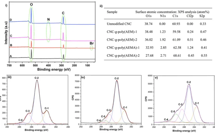
compare relative sizes of CNCs. XPS analysis of freeze-dried CNC-BriB samples further supported successful esterification reactions. When compared to unfunctionalized CNCs, a peak at 69.5 eV corresponding to the binding energy of bromine (Br 3d) was observed in addition to the typical peaks for carbon (C 1s) and oxygen (O 1s) (Figure 2: (i) A, B). Peaks
corresponding to trace amounts of sulfur were also detected due to the presence of sulfate groups. Overall, both FT-IR and XPS confirm the successful covalent attachment of the initiators on the surface of CNC.
Synthesis and Characterization of Cationic CNCs. A graf ting-f rom approach (Scheme 1) was employed to polymer-ize cationic AEM and AEMA using BriB-1 and CNC-BriB-2 via SI-SET-LRP. This polymerization technique has recently been used for the grafting of thermoresponsive poly(NiPAAm) brushes from the surface of cellulose nano-crystals.39,40 The resulting poly(AEM) and CNC-g-poly(AEMA) samples were extensively purified by centrifuga-tion and dialysis to ensure efficient removal of unreacted reagents, monomers, and homopolymers prior to all analyses. FT-IR analysis of the purified freeze-dried cationic CNC-g-poly(AEM) samples displayed a typical ester carbonyl peak at 1726 cm−1(Figure 1C). When compared to the carbonyl peak
of CNC-BriB, CNC-g-poly(AEM) showed a higher intensity ester carbonyl peak at a slightly lower frequency value. CNC-g-poly(AEMA) samples displayed new FT-IR peaks typical for the amide stretch (1638 cm−1) and amine bend (1538 cm−1)
(Figure 1D). FT-IR results indicate successful graf ting from polymerization. The cationic nature of the polymer modified CNCs was investigated using potential measurements. The ζ-potentials of the four samples ranged from about +40 to +55 mV, confirming successful cationization of CNCs. This positive surface charge will not only ensure the formation of a well-dispersed colloidal suspension but will also allow the bioconjugation of CNCs with negatively charged biomolecules.
Figure 1.FT-IR of unmodified CNC (A), CNC-BriB-1 (B), CNC-g-poly(AEM)-1 (C), and CNC-g-poly(AEMA)-1 (D).
Figure 2.XPS data on unmodified and modified CNCs: (i) survey scan spectra of unmodified CNC (A), CNC-BriB-1 (B), CNC-g-poly(AEM)-1 (C), and CNC-g-poly(AEMA)-1 (D); (ii) atomic composition (%) obtained by low resolution XPS and deconvolution of (iii) unmodified CNC, (iv) CNC-g-poly(AEM)-1, and (v) CNC-g-poly(AEMA)-1.
Biomacromolecules Article
dx.doi.org/10.1021/bm501516r | Biomacromolecules 2015, 16, 319−325
To further confirm the grafting of cationic poly(AEM) and poly(AEMA) from the surface of CNC and gain more insight on the nature of the surface grafts, XPS of freeze-dried CNC-g-poly(AEM) and CNC-g-poly(AEMA) samples was carried out to obtain the relative intensities of carbon, oxygen, and nitrogen on the surface of CNCs. While carbon and oxygen were predominant in all samples at around 285 and 532 eV, nitrogen peaks were detected for the cationic polymer modified CNCs at a characteristic emission peak of about 399 eV. This change in atomic composition was due to the presence of polymer brushes on the surface of CNCs, with higher nitrogen content for samples, which initially had higher [Br]/[AGU] ratios. While XPS is used to measure the surface atomic composition, elemental analysis provides the total atomic composition. With grafting only occurring on the exposed hydroxyl groups, the atomic composition of nitrogen using both techniques should ideally be in agreement. The nitrogen atomic composition obtained for the samples by elemental analysis was in the expected range (Table S1, Supporting Information). Using elemental analysis, the calculated weight percent of polymer chains on g-poly(AEM)-1, g-poly(AEM)-2, CNC-g-poly(AEMA)-1, and CNC-g-poly(AEMA)-2 was 32, 20, 19, and 16%, respectively (see the Supporting Information). Although the monomer concentrations in CNC-g-poly-(AEM)-1 and CNC-g-poly(AEM)-2 were the same, the polymer content on the grafted samples was different due to the different initial initiator loading.40 The difference in the calculated weight percent of poly(AEM) and poly(AEMA) grafted CNCs could be due to the challenge in executing controlled polymerization of these monomers, whereby unprotected amine containing monomers are known to undergo unwanted side reactions.42 In both techniques, trace amounts of sulfur were observed due to the sulfate ester groups, with peaks at 167 eV in XPS. We observed a general increase in the percentage of carbon both by XPS and elemental analysis, due to the presence of polymer grafts on the CNCs’ surface. High-resolution XPS was used to deconvolute the C 1s signal for C in CNC, CNC-g-poly(AEM)-1, and CNC-g-poly-(AEMA)-1. Symmetric Gaussian components with Shirley or linear background were used for the curve fitting procedure for C 1s.43 For the unmodified CNC, deconvolution resulted in four peaks, which corresponded to C-1 (CC and/or CH), C-2 (CO, which has been shown to derive mainly from cellulose), C-3 (CO or OCO), and C-4 (OCO). For the polymer modified cationic CNCs, an additional peak (C-5) for the CN bond was observed. Although we have assigned C-4 and C-5 through deconvolution, the CN and OCO peaks are known to appear in the same binding energy region and may overlap.44
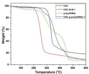
In our attempts to further elucidate the physicochemical properties of these cationic CNC materials, we investigated the thermal stabilities of the unmodified, initiator modified, and polymer modified CNCs by thermogravimetric analysis (Figure 3 and Figure S3, Supporting Information). The weight loss profiles for CNC, CNC-BriB-1, poly(AEMA), and CNC-g-poly(AEMA)-1 are illustrated in Figure 3. The initial weight loss for the samples below 120 °C is attributed to evaporation of adsorbed water, which is usually still present despite freeze-drying the samples. Unmodified CNC displayed a degradation profile with an onset temperature of about 300 °C, typical of CNCs derived from sulfuric acid hydrolysis. A decrease in thermal stability of the initiator modified CNCs was observed, compared to the unmodified CNCs. The thermal stability was
improved after grafting of cationic AEMA from CNC-BriB-1, thereby confirming the presence of polymer brushes on the surface of CNCs. The thermal degradation profile of CNC-g-poly(AEMA)-1 possessed characteristics from both CNCs and poly(AEMA).
Gold Deposition. The successful grafting and cationic nature of these polymer modified CNCs were evaluated by visualizing the morphologies of the unmodified and surface modified CNCs using STEM. The size of the rod-like particles was found to be around 100−200 nm in length and 10−20 nm in diameter. For the cationic polymer modified CNCs (Figure 4B and Figure S4A, Supporting Information), well-dispersed and rod-shaped nanoparticles were observed, implying that surface chemical functionalization did not affect the structural and morphological integrity of the CNCs. Since the width of
Figure 3. Thermograms of unmodified CNC and modified CNC derivatives.
Figure 4.STEM images of samples dispersed in water: unmodified CNC and CNC-g-poly(AEMA)-1 before (A, B) and after (C, D) gold deposition, respectively. The samples (A, B) were stained with uranyl acetate for better contrast but were unstained for the gold deposition (C, D). Scale bar = 200 nm.
the CNC nanoparticles and the derivatives were fairly uniform, it was possible to measure the diameters before and after functionalization. The STEM images (Figure 4A,B and Figure S4A, Supporting Information) revealed a diameter of 11.8 ± 2.4 nm for the unmodified CNC,40 16.4 ± 2.7 nm for CNC-g-poly(AEMA)-1, and 14.8 ± 1.7 nm for CNC-g-poly(AEM)-1, thus confirming the successful grafting. Next, the surface charge of the nanofibers was evaluated upon incubation of the colloidal suspensions of CNCs and the modified derivatives with AuNPs. Upon visualization, it was observed that AuNPs were randomly distributed over the grid and had minimal interaction with the CNC rods for the unmodified CNCs (Figure 4C). This was due to the electrostatic repulsive interactions between the negatively charged CNC rods (ζ = −58.2 mV) and the negatively charged AuNPs (ζ = −30 to −40 mV, as provided by the supplier). On the other hand, the AuNPs were found to deposit on the cationic rod-like particles (Figure 4D). The heterocoagulation of AuNPs onto cationic CNC particles was controlled by Brownian motions and oppositely charged surface potentials. As a result of the reduced net charge of the colloidal suspensions, the cationic CNCs were found to aggregate in the presence of the AuNPs (Figure 4D and Figure S4B, Supporting Information).
Cytotoxicity Assay.A recent ecotoxicology study involving rainbow trout hepatocytes and nine aquatic species showed that unmodified CNCs had low toxicity potential with low environmental risk.14 MTT and LDH studies against nine mammalian cell lines also demonstrated the lack of cytotoxicity of CNCs, rendering them attractive materials for nanomedical uses.45 Given the potential bioapplications of our cationic CNCs, the cytotoxicity of unmodified and cationic CNCs was assessed using MTT assay on two different cell lines, J774A1 (mouse monocyte cells) and MCF-7 (human breast adenocarcinoma cells) (Figure 5). The results indicated that, in J774A1 (Figure 5A), cationic CNC derivatives appeared to decrease cell viability only at the highest concentration (100 μg/mL). Surprisingly, in MCF-7 cells, these cationic CNC derivatives increased the conversion of MTT into formazan, even at the lowest concentration (Figure 5B). This unexpected increase in MTT reduction might be due to a potential effect in the mitochondrial activity, which is not necessarily associated with increases in cell proliferation or cell viability. A further investigation of this intriguing effect is currently underway to confirm whether these modified CNCs induce cell proliferation in breast cancer cell lines.
■
CONCLUSIONSIn this work, we have presented a comprehensive account of the synthesis of poly(2-aminoethyl methacrylate) (poly(AEM)) and poly(N-(2-aminoethylmethacrylamide) (poly(AEMA)) CNCs via a living radical polymerization approach. The resulting rod-like CNC-g-poly(AEM) and CNC-g-poly(AEMA) particles were characterized using a combination of analytical, spectroscopic, and microscopic techniques. The anionic surfaces of unmodified CNCs were rendered cationic after the polymerization process. Gold nanoparticles were deposited on the cationic CNC surfaces, as illustrated by STEM analysis. The results of MTT assay indicated that none of the cationic modified CNCs decreased cell viability at low concentrations, which are more suitable for biomedical applications. Furthermore, the apparent increase in cell viability of MCF-7 cells caused by these compounds, in particular at higher concentration, demands further investigation. In addition,
thorough in vivo testing will be required before realizing the full potential of these novel cationic modified CNCs.
■
ASSOCIATED CONTENT*
S Supporting InformationExperimental procedure, NMR spectra, elemental analysis, thermographs, STEM images, and calculations for polymer weight percentage in cationic CNC samples. This material is available free of charge via the Internet at http://pubs.acs.org.
■
AUTHOR INFORMATION Corresponding Author*E-mail: raje sh.sunasee@plattsburgh.edu. Phone: +15185642703. Fax: +15185643169.
Notes
The authors declare no competing financial interest.
■
ACKNOWLEDGMENTSThe authors thank SUNY Plattsburgh for a Presidential research award, the Alberta Centre for Surface Engineering and Science, University of Alberta for XPS measurements, National Institute for Nanotechnology, and National Research Council of Canada for supporting this project.
■
REFERENCES(1) Habibi, Y.; Lucia, L. A.; Rojas, O. J. Chem. Rev. 2010, 110, 3479− 3500.
(2) Eichhorn, S. J. Soft Matter 2011, 7, 303−315.
(3) Klemm, D.; Kramer, F.; Moritz, S.; Lindström, T.; Ankerfors, M.; Gray, D.; Dorris, A. Angew. Chem., Int. Ed. 2011, 50, 5438−5466.
(4) Peng, B. L.; Dhar, N.; Liu, H. L.; Tam, K. C. Can. J. Chem. Eng. 2011, 89, 1191−1206.
Figure 5.Cytotoxicity of unmodified and cationic CNCs on J774A1 (A) and MCF-7 (B) cells. After 24 h of treatment, cell viability was determined by MTT assay. Data are expressed as percentage of control (nontreated cells 100%, viability) using the mean values and standard deviations from triplicate experiments. *p < 0.05 compared to control.
Biomacromolecules Article
dx.doi.org/10.1021/bm501516r | Biomacromolecules 2015, 16, 319−325
(5) Moon, R. J.; Martini, A.; Nairn, J.; Simonsen, J.; Youngblood, J.
Chem. Soc. Rev. 2011, 40, 3941−3994.
(6) USDA, Forest Service, Forest Products Laboratory, Wisconsin, USA (www.fpl.fs.fed.us); CelluForce, Quebec, Canada (www. celluforce.com); FPInnovations, Quebec, Canada (www. fpinnovations.ca); Alberta Innovates Technology Futures, Alberta, Canada, www.albertatechfutures.ca.
(7) Eyley, S.; Thielemans, W. Nanoscale 2014, 6, 7764−7779. (8) Habibi, Y. Chem. Soc. Rev. 2014, 43, 1519−1542.
(9) Hemraz, U. D.; Sunasee, R. Functionalization of Nanocrystalline Cellulose Surfaces. In Dekker Encyclopedia of Nanoscience and
Nanotechnology, 3rd ed.; Lyshevski, S. E., Ed.; CRC Press: Boca
Raton, FL, 2014; Vol. II.
(10) Favier, V.; Chanzy, H.; Cavaillé, J. Y. Macromolecules 1995, 28, 6365−6367.
(11) Favier, V.; Canova, G. R.; Cavaillé, J. Y.; Chanzy, H.; Dufresne, A.; Gauthier, C. Polym. Adv. Technol. 1995, 6, 351−355.
(12) Dong, S.; Roman, M. J. Am. Chem. Soc. 2007, 129, 13810− 13811.
(13) Mahmoud, K. A.; Mena, J. A.; Male, K. B.; Hrapovic, S.; Kamen, A.; Luong, J. H. T. ACS Appl. Mater. Interfaces 2010, 2, 2924−2932.
(14) Kovacs, T.; Naish, V.; O’Connor, B.; Blaise, C.; Gagné, F.; Hall, L.; Trudeau, V.; Martel, P. Nanotoxicology 2010, 4, 255−270.
(15) Lam, E.; Male, K. B.; Chong, J. H.; Leung, A. C. W.; Luong, J. H. T. Trends Biotechnol. 2012, 30, 283−290.
(16) Yang, Q.; Pan, X. J. Appl. Polym. Sci. 2010, 117, 3639−3644. (17) Filpponen, L.; Sadeghifar, H.; Argyropoulos, D. S. Nanomater.
Nanotechnol. 2011, 1, 34−43.
(18) Huang, J.-L.; Li, C.-J.; Gray, D. G. ACS Sustainable Chem. Eng. 2013, 1, 1160−1164.
(19) Abitbol, T.; Palermo, A.; Moran-Mirabal, J. M.; Cranston, E. D.
Biomacromolecules 2013, 14, 3278−3284.
(20) Nielsen, L. J.; Eyley, S.; Thielemans, W.; Aylott, J. W. Chem.
Commun. 2010, 4, 8929−8931.
(21) Yang, R.; Tan, H.; Wei, F.; Wang, S. Biotechnology 2008, 7, 233− 241.
(22) Mahmoud, K. A.; Male, K. B.; Hrapovic, S.; Luong, J. H. T. ACS
Appl. Mater. Interfaces 2009, 1, 1383−1386.
(23) Incani, V.; Danumah, C.; Boluk, Y. Cellulose 2013, 20, 191−200. (24) Jackson, J. K.; Letchford, K.; Wasserman, B. Z.; Ye, L.; Hamad, W.; Burt, H. M. Int. J. Nanomed. 2011, 6, 321−330.
(25) Drogat, N.; Sol, V.; Memmi, A.; Saad, N.; Koerkamp, C. K.; Bressollier, P.; Krausz, P. J. Nanopart. Res. 2011, 13, 1557−1562.
(26) Hemraz, U. D.; Boluk, Y.; Sunasee, R. Can. J. Chem. 2013, 91, 974−981.
(27) Hasani, M.; Cranston, E. D.; Westman, G.; Gray, D. G. Soft
Matter 2008, 4, 2238−2244.
(28) Zaman, M.; Xiao, H.; Chibante, F.; Ni, Y. Carbohydr. Polym. 2012, 89, 163−170.
(29) Eyley, S.; Thielemans, W. Chem. Commun. 2011, 47, 4177− 4179.
(30) Jasmani, L.; Eyley, S.; Wallbridge, R.; Thielemans, W. Nanoscale 2013, 5, 10207−10211.
(31) Dubruel, P.; Christiaens, B.; Rosseneu, M.; Vandekerckhove, J.; Grooten, J.; Goossens, V.; Schacht, E. Biomacromolecules 2004, 5, 379−388.
(32) Zhu, C. H.; Jung, S.; Si, G. Y.; Cheng, R.; Meng, F. H.; Zhu, X. L.; Park, T. G.; Zhong, Z. Y. J. Polym. Sci., Part A: Polym. Chem. 2010,
48, 2869−2877.
(33) Ji, W.; Panus, D.; Palumbo, R. N.; Tang, R.; Wang, C.
Biomacromolecules 2011, 12, 4373−4385.
(34) Sunasee, R.; Watanaarsakit, P.; Ahmed, M.; Lollmahomed, F.; Narain, R. Bioconjugate Chem. 2012, 23, 1925−1933.
(35) Bhuchar, N.; Sunasee, R.; Ishihara, K.; Thundat, T.; Narain, R.
Bioconjugate Chem. 2012, 23, 75−83.
(36) Ahmed, M.; Wattanaarsakit, P.; Narain, R. Polym. Chem. 2013, 4, 3829−3836.
(37) Boluk, Y.; Lahiji, R.; Zhao, L.; McDermott, M. T. Colloid Surf., A 2011, 377, 297−303.
(38) Deng, Z.; Bouchekif, H.; Babooram, K.; Housni, A.; Choytun, N.; Narain, R. J. Polym. Sci., Part A: Polym. Chem. 2008, 46, 4984− 4996.
(39) Zoppe, J. O.; Habibi, Y.; Rojas, O. J.; Venditti, R. A.; Johansson, L. S.; Efimenko, K.; Osterberg, M.; Laine, J. Biomacromolecules 2010,
11, 2683−2691.
(40) Hemraz, U. D.; Lu, A.; Sunasee, R.; Boluk, Y. J. Colloid Interface
Sci. 2014, 430, 157−165.
(41) van Meerloo, J.; Kaspers, G. J.; Cloos, J. Methods Mol. Biol. 2011,
731, 237−245.
(42) Alidedeoglu, A. H.; York, A. W.; McCormick, C. L.; Morgan, S. E. J. Polym. Sci., Part A: Polym. Chem. 2009, 47, 5405−5415.
(43) Johansson, L. S.; Campbell, J. M. Surf. Interface Anal. 2004, 36, 1018−1022.
(44) Barazzouk, S.; Daneault, C. Nanomaterials 2012, 2, 187−205. (45) Dong, S.; Hirani, A. A.; Colacino, K. R.; Lee, Y. W.; Roman, M.
NanoLIFE 2012, 2, 1−11.
![Table 1. DLS and ζ -Potentials for Unmodi fi ed and Modi fi ed CNCs sample hydrodynamic diameter (nm) ζ-potentials(mV) unmodi fi ed CNC 87.2 ± 1.2 − 58.2 ± 0.5 CNC-BriB-1, [Br]/[AGU] = 5:3 83.7 ± 0.7 − 45.8 ± 1.1 CNC-BriB-2, [Br]/[AGU] = 5:12 85.9 ± 0.3 −56.7](https://thumb-eu.123doks.com/thumbv2/123doknet/14095831.465103/4.938.96.848.145.337/table-potentials-unmodi-sample-hydrodynamic-diameter-potentials-unmodi.webp)