Publisher’s version / Version de l'éditeur:
International Journal of Cardiology, 149, 3, pp. 315-322, 2010-03-03
READ THESE TERMS AND CONDITIONS CAREFULLY BEFORE USING THIS WEBSITE.
https://nrc-publications.canada.ca/eng/copyright
Vous avez des questions? Nous pouvons vous aider. Pour communiquer directement avec un auteur, consultez la
première page de la revue dans laquelle son article a été publié afin de trouver ses coordonnées. Si vous n’arrivez pas à les repérer, communiquez avec nous à PublicationsArchive-ArchivesPublications@nrc-cnrc.gc.ca.
Questions? Contact the NRC Publications Archive team at
PublicationsArchive-ArchivesPublications@nrc-cnrc.gc.ca. If you wish to email the authors directly, please see the first page of the publication for their contact information.
NRC Publications Archive
Archives des publications du CNRC
This publication could be one of several versions: author’s original, accepted manuscript or the publisher’s version. / La version de cette publication peut être l’une des suivantes : la version prépublication de l’auteur, la version acceptée du manuscrit ou la version de l’éditeur.
For the publisher’s version, please access the DOI link below./ Pour consulter la version de l’éditeur, utilisez le lien DOI ci-dessous.
https://doi.org/10.1016/j.ijcard.2010.02.009
Access and use of this website and the material on it are subject to the Terms and Conditions set forth at
Defects in myoglobin oxygenation in KATP-deficient mouse hearts under normal and stress conditions characterized by near infrared spectroscopy and imaging
Jilkina, Olga; Glogowski, Miriam; Kuzio, Bozena; Zhilkin, Peter A.; Gussakovsky, Eugene; Kupriyanov, Valery V.
https://publications-cnrc.canada.ca/fra/droits
L’accès à ce site Web et l’utilisation de son contenu sont assujettis aux conditions présentées dans le site LISEZ CES CONDITIONS ATTENTIVEMENT AVANT D’UTILISER CE SITE WEB.
NRC Publications Record / Notice d'Archives des publications de CNRC: https://nrc-publications.canada.ca/eng/view/object/?id=31e42bc7-7fa3-4b52-b70f-ca0b8addeb1e https://publications-cnrc.canada.ca/fra/voir/objet/?id=31e42bc7-7fa3-4b52-b70f-ca0b8addeb1e
Olga Jilkina, Miriam Glogowski, Bozena Kuzio, Peter A. Zhilkin, Eugene Gussakovsky, Valery V. Kupriyanov
Defects in myoglobin oxygenation in KATP-deficient mouse hearts under normal and stress conditions characterized by near infrared spectroscopy and imaging
Abstract
BackgroundDisruption of ATP-sensitive potassium (KATP) channel activity results in the development of dilated cardiomyopathy in response to different forms of stress, likely due to the underlying metabolic defects. To further understand the role of Kir6.2-containing channels in the
development of cardiac disease, we analysed the left ventricular (LV) wall oxygenation and the physiologic responses induced by acute stress in non-dilated Kir6.2−/− hearts.
Methods
Control (C57BL6) and Kir6.2−/− mouse hearts were perfused in constant flow Langendorff mode with Krebs–Henseleit buffer. Myocardial oxygenation was evaluated using a newly developed technique, near infrared spectroscopic imaging (NIRSI) of the myoglobin (Mb) oxygen saturation parameter (OSP, ratio of oxy- to total Mb).
Results
2,4-dinitrophenol (DNP, 50-µM) and isoproterenol (0.1-µM) failed to produce a transient vasodilatory response and caused a significant diastolic pressure increase in Kir6.2−/− hearts. DNP strongly suppressed contractile function in both groups and induced severe mean OSP decreases in Kir6.2−/− hearts. Isoproterenol-induced decreases in OSP were similar despite the lack of contractile function stimulation in the Kir6.2−/− group. The index of OSP spatial
heterogeneity (relative dispersion, RD) was lower by 15% in the Kir6.2−/− group at the baseline conditions. Recovery after stress caused reduction of RD values by 20% (DNP) and 8%
(isoproterenol) in controls; however, these values did not change in the Kir6.2−/− group.
Conclusions
1) NIRSI can be used to analyse 2-D dynamics of LV oxygenation in rodent models of cardiomyopathy; 2) Kir6.2-containing KATP channels play an important role in maintaining myocardial oxygenation balance under acute stress conditions and in post-stress recovery.
Keywords: Near infrared spectroscopic imaging; Kir6.2 knockout; Langendorff-perfused mouse heart; Heterogeneity of myoglobin oxygen saturation; 2,4-dinitrophenol; Isoproterenol
1. Introduction
Progressive forms of cardiomyopathy and heart failure suffer from a mismatch between
myocardial oxygen demand and supply leading to the oxygenation deficits that can be detected using direct measurements of ventilation and gas exchange [1] and [2]. This problem has been identified in different forms of cardiomyopathy and can be caused by microvessel abnormalities, and also by metabolic defects (diabetes mellitus, mutations in components of mitochondrial metabolism, cardiac structural proteins, and potassium channels, etc.) [3], [4], [5] and [6]. It is not clear, however, whether metabolic defects per se can result in a shifted oxygenation balance in the myocardium in early forms of cardiomyopathy, before hypertrophy and dilated
cardiomyopathy develop.
Presently, several cardiac imaging modalities based on magnetic resonance imaging (MRI), X-ray-computer tomography (CT), positron emission tomography (PET), and
Doppler-ultrasonography have become available for evaluation of the intramural macro- and micro-perfusion flow [7]. Metabolic activities in the myocardium have been visualized by fluorescence imaging of endogenous (nicotinamide adenine dinucleotide, NADH) and exogenous (e.g., tetramethyl-rhodamine ethyl ester, TMRM) chromophores, and, non-invasively, by PET [8], [9],
[10] and [11]. The spatial resolution varied from several mm (PET) to tens of μm (MRI, CT, and fluorescence imaging) thus allowing demonstration of spatial heterogeneities in the flow and metabolism. None of these methods, however, measure myocardial tissue oxygenation. At the same time, optical methods are capable of evaluating cardiomyocyte oxygenation by monitoring changes in the optical density related to the absorption by oxy- and deoxy-forms of haem-containing molecules: extracellular haemoglobin (Hb) and intracellular myoglobin (Mb)
[12]. These changes can be detected in the visible and near infrared (NIR) spectral range. Previously, oxygen-dependent changes of Mb in intact myocardium have been detected by visible–NIR point spectroscopy in rodent and swine hearts [12], [13], [14], [15], [16] and [17]
and NIR spectroscopic imaging (NIRSI) in swine hearts [18] and [19]. The depth of NIR light penetration into the tissue can reach several mm [19]. Thus, in a rodent heart, not only
epicardial/subepicardial, but also transmural layers can be probed. The NIR range is also preferable because there is less interference with the peaks arising from other tissues chromophores (cytochromes and flavoproteins).
Currently, only rodent (mostly mouse) models of metabolic heart diseases have a well-established genetic background thus urging development of oxygenation imaging techniques with sufficient spatial resolution for hearts that are less than 0.2 g. In this work, we evaluated intracellular tissue oxygenation in Kir6.2−/− mouse hearts. These animals have a disabling mutation in the potassium-conducting subunit (Kir6.2) of the sarcolemmal ATP-sensitive potassium (KATP) channel [20]. The KATP channel is closed and therefore ―silent‖ in
cardiomyocytes under normal conditions [14] and [21] and Kir6.2−/− mice have a normal life span and seem healthy under non-stress conditions. However when subjected to chronic stress or increased hemodynamic load, they develop dilated cardiomyopathy leading to heart failure and can die suddenly, probably due to aberrations in the components of energy metabolism
manifested under stress conditions [14], [22], [23] and [24]. In response to acute metabolic insult, significant regional hypoxia was also revealed in Kir6.2−/− hearts [14]. We hypothesized
therefore that metabolic defects in KATP-deficient hearts may coexist with widespread oxygenation deficits under acute stress.
In human population studies, mutations in Kir6.2 (E23K variant) have been overrepresented in heart failure patients [25]. E23K mutation represents a glutamic acid to lysine substitution at codon 23 of the KCNJ11 gene encoding Kir6.2 protein, which alters the high-fidelity KATP gating. Homozygotes in E23K polymorphism have impaired exercise stress response.
Importantly, E23K Kir6.2 polymorphism is commonly occurring in Caucasian population and has been linked to increased susceptibility to diabetes type 2 in humans [26].
Thus, our goals were two-fold: 1) to test how efficient is non-contact 2-dimensional near infrared spectroscopic imaging (NIRSI) for evaluation of tissue oxygenation in a rodent heart with a metabolic heart disease (i.e., a diffuse rather than a localized type of tissue damage); and 2) to investigate whether defects in KATP channel are associated with widespread oxygenation deficits in mammalian myocardium under acute stress. In addition, we analysed spatial distribution patterns of the myocardial tissue oxygenation.
2. Materials and methods
All procedures in this study conform with the ―Guide to the Care and Use of Experimental Animals‖ published by the Canadian Council on Animal Care (2nd edition, Ottawa, On, 1993). All experiments were approved by the institutional Animal Care Committee.
2.1. Animals
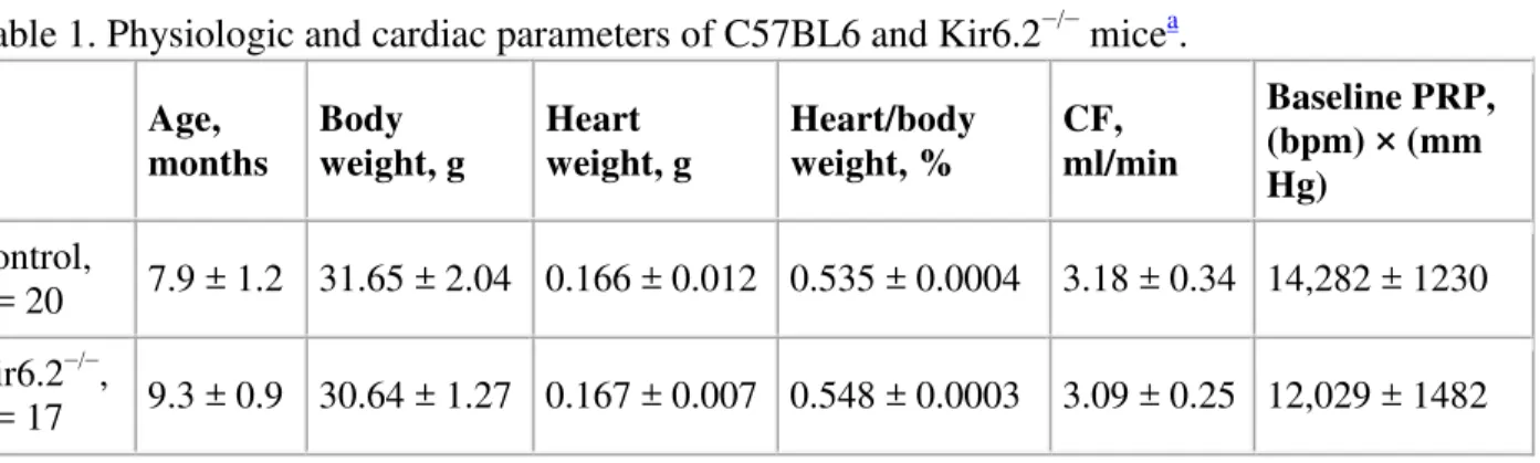
Kir6.2−/− mice were generated in the laboratory of Dr. S. Seino by disruption of the Kir6.2 gene encoding a pore-forming subunit of the KATP channel and backcrossed for several generations into the C57BL6 strain [20]. Dr. S. Seino kindly gave us permission to use Kir6.2−/−mice (via Dr. J.-M. Renaud at the University of Ottawa). Kir6.2−/− and control C57BL6 animals were bred at the Institute for Biodiagnostics animal facility. To confirm the presence of the mutation in the breeders, the polymerase chain reaction (PCR) was performed according to the protocol described elsewhere [20]. All animals were kept under standard conditions and no statistically significant differences in their heart or body weight were detected (Table 1). The mature Kir6.2−/− animals were generally healthy under normal no-stress conditions and neither early mortality nor gross cardiac abnormalities associated with dilated cardiomyopathy were detected. Their hearts were not dilated and the heart-to-body ratio was not increased.
Table 1. Physiologic and cardiac parameters of C57BL6 and Kir6.2−/− micea.
Age, months Body weight, g Heart weight, g Heart/body weight, % CF, ml/min Baseline PRP, (bpm) × (mm Hg) Control, n = 20 7.9 ± 1.2 31.65 ± 2.04 0.166 ± 0.012 0.535 ± 0.0004 3.18 ± 0.34 14,282 ± 1230 Kir6.2−/−, n = 17 9.3 ± 0.9 30.64 ± 1.27 0.167 ± 0.007 0.548 ± 0.0003 3.09 ± 0.25 12,029 ± 1482
Age, months Body weight, g Heart weight, g Heart/body weight, % CF, ml/min Baseline PRP, (bpm) × (mm Hg) pb value N.S. N.S. N.S. N.S. N.S. N.S. a
Means ± S.E.M. are presented. CF, coronary flow; PRP, pressure-rate-product. b
Kir6.2−/− group versus control group.
2.2. Heart perfusion and experimental protocols
Langendorff-perfusion of isolated hearts from male and female C57BL6 and Kir6.2−/− mice (Table 1) was carried out under constant flow conditions as described in our previous publication
[14]. The spontaneously beating hearts were perfused in a dry mode, i.e., non-submerged in the buffer. Modified Krebs–Henseleit buffer (KHB) contained (in mM): 25 NaHCO3, 118 NaCl, 4.7 KCl, 2.5 CaCl2, 1.2 MgSO4, 0.5 EDTA, 11 glucose, and 1.5 Na-pyruvate, aerated with a mixture of 95% O2 and 5% CO2 at 36 °C, pH 7.4. Typical parameters of spontaneously beating control hearts were: heart rate (HR), 250 bpm; left ventricular systolic (LVSP), 70 mm Hg; left ventricular end-diastolic (LVEDP), 10 mm Hg; and perfusion pressure (PP), 75 mm Hg. The pressure-rate-product (PRP) was calculated as a product of developed pressure
(LVDP = LVSP − LVEDP) and HR. After a 10–15 min equilibration period, stress agents, 2,4-dinitrophenol (DNP, 50 µM, Sigma, St. Louis MO, U.S.A.) or isoproterenol (0.1 µM, Sigma, St. Louis MO, U.S.A.) were infused for 24 min, then the infusion was stopped and hearts recovered for 24 min. After that, 10-min no-flow global ischemia was introduced at the end of the protocol for calibration purposes.
2.3. Visible–NIR point spectroscopy
Spectra at selected points on the heart surface were acquired in the range of 400–1100 nm with an increment of 1 nm using an integration time of 0.1 s and an averaging factor of 120.
Broadband visible/NIR light from a fiber optic illuminator Oriel model 77501 (Stratford, CT) was transmitted to the heart through one arm of a bifurcated fiber optic bundle. The common illumination/collection probe tip was placed in a contact with the left ventricle perpendicular to the tissue plane with zero distance between them, which allowed the collection of predominantly diffuse reflected light through the other arm of the fiber bundle to the model PDA-512
spectrometer (Control Developments Inc. (South Bend, IN)). The light from the light source was delivered by a single fiber optic filament of 650-μm in diameter. The reflected light was
collected by 36 fiber optic filaments of 15-μm diameter randomly surrounding the illuminating filament [17].
Pseudo-optical density (POD) for any given wavelength was calculated according to the formula: (1) where Is is the intensity of the light diffusely reflected by the sample in the back direction and I0 is the intensity of the light reflected by Spectralon® as a standard.
In the visible spectral range, a decrease in Mb oxygenation results in an increase in the deoxy-Mb peak at 550 nm (POD550) and a decrease in the oxy-Mb peak at 580 nm (POD580) [13] and
[14] (Fig. 1A). In the NIR range, deoxy-Mb and oxy-Mb give rise to peaks with maxima at 760 and about 920 nm, respectively (Figs. 1B and 2). The 760-nm peak is very distinct, while the 920-nm peak is rather broad and is difficult to resolve in a complex spectrum [27]. The water peak is centered at 970 nm.
Full-size image (43K) Fig. 1.
Diffuse reflectance point spectroscopy measurements. Pseudo-optical density (POD) spectra of a representative Langendorff-perfused control heart under normal (1) and ischemic (2) conditions. Curve 3 is the difference between a normal and an ischemic spectrum. POD was determined according to Eq. (1) (see Materials and methods for details). Panels A and B show the same spectra in two wavelength ranges: visible in panel A and NIR in panel B. The peaks
corresponding to oxy- (580-nm), deoxy-Mb (550-nm), and a reduced form of cytochrome c oxidase (605-nm) in the visible part of the spectra are depicted in panel A. The peaks corresponding to deoxy-Mb (760-nm) and water (970-nm) in the NIR region are depicted in panel B. The NIR peak of oxy-Mb, which is very broad and centered at 920 nm, is difficult to resolve visually from the other bands. An offset was applied for clarity of spectra presentation.
Full-size image (19K) Fig. 2.
POD spectra in a selected pixel in a NIR spectroscopic image. Extracted (solid) and
reconstructed fit (dashed) spectra of a representative control mouse heart are shown. The fit spectra were calculated by summing up the products of multiplication of the absorption
coefficients by the respective absorptivities of oxy- and deoxy-Mb, and water, at all wavelengths, with a 10-nm increment, offset by a constant. The fit was determined by the least squares
method. The position of distinct 760-nm deoxy-Mb and 970-nm water peaks are clearly seen, while a 920-nm oxy-Mb peak is very broad and is difficult to resolve visually in complex spectra. Amplitude of the deoxy-Mb peak (Adeoxy-Mb), relative to the nearest minima at 720 and 800 nm, is depicted.
2.4. NIRSI of mouse hearts
Images were acquired using a CCD-array camera equipped with AF-60 Nikon microlens and a liquid crystal tuneable filter (LCTF, Cambridge Research and Instrumentation, Woburn, MA, USA) operating in the range from 650 to 1050 nm with a 10-nm increment. The images were non-gated because the amplitude of a mouse heart motion was negligible in the isovolumic preparations as a sealed water-filled latex balloon inserted into the left ventricular cavity for the pressure measurements prevented any significant volume changes. The hearts were positioned approximately 10 cm away from the lens providing the field of view of 20 × 20 mm. The hearts were illuminated with Fiber Optic Illuminator, Model 190 (Fisher Scientific). Scanning time for the full spectral range for each image was 4 min. The reflectance standard (Kodak Gray Card, Eastman Kodak, Rochester, NY, USA) was placed in front of each heart at the end of the
experiments to keep the illumination patterns and the distance to the standard similar to those for each heart. As a result, a POD spectrum calculated according to formula (1) was available for each of 256 × 256 pixel providing 80 µm nominal in-plane resolution.
Dynamic image acquisition was performed as follows: in DNP protocols, images were taken at the end of an equilibration period (baseline conditions), at 10–14 and 20–24 min of DNP infusion, at 20–24 min of a recovery period, and at 6–10 min of no-flow ischemia (Fig. 3C). In isoproterenol protocols, images were taken at the end of an equilibration period, at 5–9, 12–16, and 20–24 min of isoproterenol infusion, at 20–24 min of a recovery period, and at 6–10 min of no-flow ischemia (Fig. 3F).
Full-size image (145K) Fig. 3.
The effects of DNP and isoproterenol on left ventricular end-diastolic pressure (LVEDP, panels A and D), pressure-rate-product (PRP, panels B and E), and perfusion pressure (PP, panels C and F) in the Langendorff-perfused control and Kir6.2−/− hearts. NIR imaging sessions were as marked (panels C and F). * p for control versus Kir6.2−/− hearts at the same stage of the protocol is shown. # p versus baseline value for control hearts is shown for selected time points.
2.5. Calculation of oxygen saturation parameter (OSP)
Absorption coefficients of oxy-Mb and deoxy-Mb (coxy and cdeoxy, respectively), as well as the water contribution to POD in each pixel were determined by fitting the acquired POD spectra to the weighed sum of the known absorptivities of these chromophores by the least squares method (Fig. 2) [18] and [27]. OSP (ratio of oxy- to total myoglobin) in each pixel was calculated by using the absorption coefficients:
(2)
2.6. Calculation of an increase in the amplitude of deoxy-Mb peak
Hypoxia resulted in an increase in the amplitude of the 760-nm deoxy-Mb peak (Adeoxy-Mb) relative to the two nearest apparent POD minima at 720 and 800 nm (Fig. 2), which was calculated according to the formula:
(3) where POD760, POD720, and POD800
correspond to POD at 760, 720, and 800 nm, respectively. A similar approach was used previously to evaluate the change in the Mb oxygenation in a rat heart by calculating the absorbance difference in the visible range between the oxy-Mb peak at 580 nm and the nearest absorbance minimum at 620 nm [12].
2.7. Analysis of OSP spatial heterogeneity
A region of interest, ROI, was drawn manually in the first image of a well-oxygenated heart to include the visible surface (mostly of the LV lateral wall) and exclude the artifacts and the borders. The same ROI was automatically applied to all images in the sequence, except for the last, ischemic one, which was smaller due the efflux of the perfusate under no-flow conditions. Thus, to correct for the shrinkage of the ischemic heart, the ROI was selected manually and included the same area as in the previous images. A mean OSP ± STDEV was calculated for each selected ROI. To evaluate OSP spatial heterogeneity, a relative dispersion (RD) of OSP, (a ratio of STDEV over the mean OSP) was calculated [28].
2.8. Statistical analysis
In each group separately (control and Kir6.2−/− hearts), a paired t-test (which is used when one group of units has been tested twice, a ―repeated measures‖ t-test) was applied for each condition
versus baseline data — to evaluate the effect of the stress agent (either DNP or isoproterenol, either treatment or recovery). The p value is marked with # and & for the control and Kir6.2−/− groups, respectively.
The two groups were compared to each other at specific time points by using a t-test assuming equal variances (unpaired, or ―independent samples‖ t-test, which is used when two separate samples are compared) — to evaluate the difference between the groups; in this case, p value was marked with *. In addition, an F-test was used to demonstrate that the two groups have the same variances.
Differences were considered statistically significant when p < 0.05. The data are presented as means ± S.E.M.
3. Results
3.1. Physiologic response of perfused control and Kir6.2−/− hearts to stress
DNP and isoproterenol caused a significant increase in LVEDP in Kir6.2−/− hearts, which persisted after the washout, while changes in the control hearts were minimal (Fig. 3A and D). DNP strongly suppressed contractile function (PRP) in both groups (to 20%), which
recovered only partially upon washout (Fig. 3B). In control hearts, isoproterenol increased PRP to 200% during the first 3 min; the stimulation was sustained at 150% over the treatment period, returning to the baseline once the infusion of isoproterenol stopped (Fig. 3E). However, in Kir6.2−/− hearts, isoproterenol only transitorily increased PRP to 175%; the function declined to 50% during recovery.
DNP and isoproterenol caused an initial transient vasodilation (a decrease in PP) followed by vasoconstriction (an increase in PP) in the control group (Fig. 3C and F). In the Kir6.2−/− hearts, PP increased during the first 5–10 min of DNP and isoproterenol infusion, and then stabilized at the elevated level; initial vasodilation was not observed.
3.2. Effects of DNP and isoproterenol on LV oxygenation in perfused control and Kir6.2−/− hearts
Before any intervention, tissue oxygenation of control and Kir6.2−/− hearts was similar: the mean OSP values were 0.829 ± 0.011 (control, n = 20) and 0.830 ± 0.009 (Kir6.2−/−, n = 17).
DNP infusion uncoupled oxidative phosphorylation in the mitochondria [29] and increased oxygen consumption in the cardiomyocytes thus resulting in hypoxic changes in the perfused hearts due to constant CF. Hypoxia was confirmed by visible range point spectroscopy (Fig. 4) and NIRSI (Fig. 5). During DNP-treatment, the 580-nm peak corresponding to the oxygenated form of Mb decreased, while the 550-nm peak (deoxy-Mb) increased; the 605-nm peak
corresponding to the reduced form of cytochrome c oxidase was also observed (Fig. 4). The hypoxic changes were reversed during the recovery period and re-appeared during ischemia.
Full-size image (24K) Fig. 4.
Visible range POD spectra of a representative control heart subjected to metabolic stress by DNP (50 µM). In selected DNP-treated hearts, before the NIRS imaging session, POD point
spectroscopy measurements were taken and regional hypoxia was confirmed. The heart was perfused at normal conditions for approximately 15 min, treated with DNP for 24 min, allowed to recover for 24 min, and subjected to global no-flow ischemia for 10 min. Vertical dashed lines show positions of the principle light-absorption bands of deoxy-Mb (550-nm), oxy-Mb (580-nm), and a reduced form of cyt c oxidase (605-nm).
Full-size image (240K) Fig. 5.
OSP (panel A) and Adeoxy-Mb (panel B) images of a representative control heart. The heart was perfused at normal conditions for approximately 15 min, treated with DNP (50 µM) for 24 min, allowed to recover for 24 min, and subjected to global no-flow ischemia for 10 min. OSP or
Adeoxy-Mb images of mouse hearts were created in false colors to reflect the degree of oxygenation of the left ventricular wall. The bright blue spot in the middle of the images was an artifact caused by glare; the bright red line on the top of the heart was an artifact produced by a suture used to tie the heart to the cannulae. A drop of the effluent is visible at the heart apex. Please note that the color changes are in reversed order on panels A and B.
We compared two NIRS image-constructing algorithms based on calculations of: (i) OSP and (ii)
Adeoxy-Mb in each pixel. Both methods demonstrated comparable results. Oxygenation of the LV wall decreased in response to DNP, returned to the baseline level upon termination of DNP
infusion, and decreased even further during no-flow ischemia (Fig. 5A). Changes in Adeoxy-Mb (indicative of hypoxia) were exactly the opposite (Fig. 5B). On average, the OSP method proved to be more robust. Thus, only a trend was demonstrated for changes of mean Adeoxy-Mb during DNP-treatment (not shown), while in the same group of hearts, changes of the mean OSP were highly significant (Fig. 6A). DNP-induced OSP decreases were greater in Kir6.2−/− hearts.
Full-size image (43K) Fig. 6.
Effect of DNP (50 µM, panel A) and isoproterenol (ISO, 0.1 µM, panel B) on the mean OSP in control and Kir6.2−/− hearts. # p versus baseline value for control hearts is shown. & p versus baseline value for Kir6.2−/− hearts is shown. * p for control versus Kir6.2−/− hearts at the same stage of the protocol is shown. N.S. — not significant. ko, knockout of Kir6.2.
Isoproterenol stimulation increased oxygen demand; however, oxygen delivery was clamped by the constant CF thus resulting in the partial tissue hypoxia. This was confirmed by visible range spectroscopy (not shown) and OSP imaging (Fig. 6B). On average, there was no statistically significant difference in isoproterenol-induced mean OSP changes between Kir6.2−/− and control hearts.
3.3. Effect of stress and subsequent recovery on OSP spatial distribution and heterogeneity
OSP values in most pixels in the OSP images were between 0.5 and 1. Thus, OSP distribution histograms (number of pixels versus pixel intensity distributed between 20 bins) were plotted in this range (Fig. 7). During the interventions, not only the mean OSP and the position of the major OSP peak shifted, but also the shape of the curve and the width of the peak changed thus
Full-size image (25K) Fig. 7.
OSP distribution histograms for the same heart as in Fig. 4. The heart was perfused at normal conditions, treated with DNP (50 µM) for 10 and 20 min, allowed to recover for 20 min, and subjected to global no-flow ischemia.
To evaluate these changes, we calculated an index of the OSP spatial heterogeneity, its relative dispersion (RD). At the baseline conditions, RD was lower by 15% in the KATP-deficient hearts: 0.076 ± 0.003, (Kir6.2−/−, n = 17) versus 0.088 ± 0.005 (control, n = 20), p < 0.03. In response to the hypoxic insult (DNP or isoproterenol), changes in RD were observed in the individual hearts. Nevertheless, on average, RD did not change significantly in both groups despite a shift in the oxygen supply/demand balance (not shown). However, removal of stress agents (DNP and isoproterenol) reduced RD values in the control group by 20 and 8%, respectively, while the values did not change in the Kir6.2−/− group (Table 2).
Table 2. The effects of DNP- and isoproterenol-induced post-stress recovery on OSP heterogeneity (RD of OSP) in control and Kir6.2−/− hearts perfused in a constant flow Langendorff modea. Group RD at baseline conditions RD at post-stress recoveryb p value DNP-treatment, control, n = 9 0.087 ± 0.006 0.069 ± 0.003 p < 0.02c DNP-treatment, Kir6.2−/−, n = 8 0.073 ± 0.009 0.067 ± 0.012 N.S.d Isoproterenol-treatment, control, n = 11 0.089 ± 0.004 0.082 ± 0.005 p < 0.05 c Isoproterenol-treatment, Kir6.2−/−, n = 9 0.079 ± 0.006 0.077 ± 0.005 N.S. d a
OSP, oxygen saturation parameter; RD, relative dispersion; 2,4-dintrophenol (DNP). b
Changes in RD during recovery were measured at 20 min after the infusion of stress agents (DNP (50-μM) or isoproterenol (0.1-μM)) was stopped. Data are presented as means ± S.E.M. c
p recovery versus baseline for control hearts. d
4. Discussion
4.1. 2-D imaging of tissue oxygenation in a rodent heart with a metabolic disease
Imaging of tissue oxygenation (rather than evaluation of perfusion or metabolism deficits) was performed in a mouse heart with the Kir6.2 deficiency. Currently, genetically-constructed models of cardiac metabolic defects are available only in rodents (typically, mice). Therefore, methodological aspects of NIRSI in a small (less than 0.2 g) heart were addressed. We compared two NIR image-constructing algorithms based on (i) OSP and (ii) Adeoxy-Mb calculations. To the advantage of Adeoxy-Mb imaging, it can be used in a fast acquisition mode, potentially with an increased number of co-added scans, as it requires measurements at only three wavelengths, while the OSP imaging requires full spectral range data. However, the accuracy of the Adeoxy-Mb method was inferior to that of the OSP method probably because the changes in the oxy-Mb and water peaks were ignored. In addition, the measured parameter, Adeoxy-Mb, depends on the light pathlength, while OSP does not, because it represents the ratio of the absorption coefficients. Therefore, Adeoxy-Mb measurements are sensitive to the experimental setup and changes in the tissue optical properties (thickness of the sample, presence of oedema, haematoma, scarring, etc.).
It has to be noted that the OSP method provides a relative OSP value, which remains well above zero even during the no-flow ischemia, as discussed previously [30]. There could be several reasons for this discrepancy. First, Hb is absent from the sample and Mb may remain partially oxygenated even at very low PO2[16] and [31]. Second, the epicardial contamination by the oxygen from the surrounding air is significant for very small hearts [32]. Nevertheless, a strong positive correlation between the OSP and PO2 in-situ and in-vivo in swine hearts has been demonstrated [27] and [33]. In comparison to spectroscopy, NIR OSP imaging has longer acquisition times and a lower number of co-added scans (3 versus 120) thus resulting in a lower signal-to noise ratio. However, NIR imaging provides 2-D picture of any side of the heart and increases the depth of tissue penetration relative to the visible range spectroscopy. The dynamics of mean OSP changes were qualitatively in agreement with the visible range spectroscopy
measurements acquired under the same physiological conditions in the same animal models [14], thus validating the NIR OSP measurements in mouse hearts. However, the previous
measurements were restricted to (i) a limited region of the left ventricle and (ii) superficial layers only.
4.2. Role of cardiac KATP channel in myocardial wall oxygenation under stress
We demonstrated for the first time that the balance between oxygen delivery and consumption was shifted in Kir6.2−/− hearts under stress. We cannot exclude that the lack of the initial vasodilatory response in Kir6.2−/− hearts (Fig. 3C and F) may have resulted in insufficient increase in oxygen delivery when the demand was increased. However, this is unlikely as both types of hearts were under constant (and equal, see Table 1) CF conditions. A decrease in LV oxygenation observed in this particular type of metabolic disease in the hearts that have not yet develop cardiac hypertrophy was, most likely, caused by the underlying metabolic defects in the
cardiomyocytes [14], [23], [34] and [35] thus underscoring yet another role of the cardiac KATP channels under stress.
The two types of stress, namely metabolic by DNP and β-adrenergic by isoproterenol, differed in their effects on heart oxygenation and contractile function: DNP increased oxygen demand due to uncoupling and suppressing contractile function, while isoproterenol stimulated oxygen demand by stimulating contractile function. Kir6.2−/− hearts were irreversibly damaged by DNP and could not be stimulated by isoproterenol to the same degree as the control hearts. Opening of the sarcolemmal KATP channels directly by a decrease in [ATP]/[ADP] (caused by DNP) as well as by a concurrent release of adenosine would lead to a decrease in intracellular Ca2+ due to sarcolemma hyperpolarization in all Kir6.2-containing cells (e.g., cardiomyocytes and smooth muscle cells) and hence, transitory vasodilation (Fig. 3C). The effect of isoproterenol is different, as it increases Ca2+ cycling and therefore stimulates contractile function in control hearts [36]. However, in Kir6.2−/− hearts, primary defects in ionic homeostasis under stress, along with moderately compromised baseline mitochondrial function [23] resulted in 1) increased sensitivity to even moderate uncoupling by DNP and 2) toxic reaction to stimulation by isoproterenol. This was particularly evident from the greater increases in LVEDP (Fig. 3A and D) thus implying lower ATP/ADP ratio [14], and higher cytoplasmic Ca2+, which negatively affected the mitochondrial function in the KATP-deficient hearts. This may help explain the irreversible damage to the contractile function and the oxygenation deficits.
4.3. Heterogeneity of Mb oxygenation in Kir6.2−/− and control hearts
There are several sources of the observed ―lateral‖ spatial heterogeneity of OSP (in contrast to the transmural one): (a) coronary flow distribution can be heterogeneous due to non-uniform longitudinal distribution of capillaries and vascular tone [28]. (b) Oxygen diffusion from the vascular lumen to the cytoplasm may be heterogeneous due to variability in vascular
permeability and diffusion distance between capillaries and myocytes [37]. The latter depends on the interstitial volume that is known to increase in crystalloid-perfused hearts due to edema [38], which again can be non-uniform. (c) Oxygen consumption is usually linearly dependent on cardiac workload, which may vary not only transmurally, but also longitudinally due to different mechanical stress for muscle fibers in different locations. All these factors could contribute to 15% lower basal heterogeneity of Mb oxygenation in Kir6.2−/− hearts.
We analysed the composite signal from the epi/subepicardial and transmural layers. In this mode of acquisition, the contribution from the transmural layers that display greater microflow
heterogeneity [38] was attenuated due to high absorbance, and scattering in the cardiac tissue. Additional factors, such as averaging of OSP over a 4-min interval, neglecting the small motion of the isovolumic hearts during the cardiac cycle, and perfusion of mouse hearts with buffer rather than blood (thus, at higher perfusion rates) likely resulted in an RD decrease [39]. Some measurement artifacts may have contributed to the observed heterogeneity. However, we analysed the sequential changes in individual hearts under constant flow and illumination conditions, and without changes in the setup. Therefore, the heterogeneity in the images due to errors of the measurements did not vary during the experiments and the observed changes in the RD of OSP in each heart were caused by physiologic factors. Lack of changes during application
of DNP and isoproterenol in both groups implies that the above factors (a, b, and c) either did not change or canceled each other out during stress.
The post-stress (recovery) decrease in RD by 8% (isoproterenol) and 20% (DNP) in control group and the lack of changes in the Kir6.2−/− group are puzzling. KATP deficiency may affect the heterogeneity of oxygenation through the mechanisms that include either changes in the oxygen consumption (via Kir6.2−/− myocytes) or microvascular oxygen delivery (via Kir6.2−/−
endothelial cells) [40]. For instance, oxygen uptake by more severely damaged Kir6.2−/− myocytes could be increased (uncoupling), decreased (respiratory chain defect) or unchanged. Correspondingly, cardiomyocytes' oxygenation might decrease, increase, or remain unchanged thus contributing to the oxygenation heterogeneity. However, a decrease in post-stress RD could also be due to changes in the oxygen delivery to the cells. The differences in PP patterns in Kir6.2−/− hearts imply a shifted balance between vasodilatory and vasoconstrictory responses governed by the complex interplay of cardiomyocytes' adenosine release, endothelial NO and reactive oxygen species (ROS) release. Indeed, deficiency in Kir6.2 abolishes response of the endothelial cells to shear stress [41] and therefore may affect the permeability of the
microvessels to oxygen and homogeneity of the tissue oxygenation.
4.4. Spatial resolution
Cardiomyocytes are approximately 20 μm in length [37] thus, the optical resolution of 100 μm achieved in this work corresponds to the area that includes several cells that could be perfused by 3–7 smallest capillaries ( 10-μm diameter) [42]. Judging by visual inspection, the
heterogeneity was random. Previously, macroscopic (several hundred μm in width) sharp anoxic zones during high-flow hypoxia and acidosis were demonstrated by NADH fluorescence
photography in working, buffer-perfused rat hearts [8]. We did not observe such phenomena. The differences could be due to: 1) differences in the species and heart preparation, 2) less severe metabolic challenges, and 3) parameters assayed (Mb oxygen saturation versus pyridine
nucleotide fluorescence).
4.5. Conclusions and implications
This work shows that the NIRSI technique can be used to evaluate 2-D patterns of oxygenation (rather than perfusion or metabolism) in small (less than 0.2 g) hearts in genetically-created animal models of cardiomyopathy. Our NIRSI approach helped illustrate the critical role of the sarcolemmal KATP channel in maintaining the oxygenation balance in myocardium in acute stress and post-stress recovery. Similar defects in the human cardiac KATP channel may also result in compromised myocardial oxygenation in stress and post-stress recovery.
Acknowledgments
This research was supported, in part, by an operating grant from the Manitoba Health Research Council to O.J. We would like to thank Dr. S. Seino for the permission to use Kir6.2−/− mice; Dr. J.-M. Renaud for the gift of Kir6.2−/− and C57BL6 breeding pairs; Ms. C. Kellar and Ms. C. Makowsky for excellent animal care; Drs. A. R. Shaw and L. Leonardi for helpful discussions and suggestions regarding NIRSI measurements; Dr. R. Summers for consultations on statistical
methods; and Dr. J. Rendell for his help with the manuscript preparation. The authors of this manuscript have certified that they comply with the Principles of Ethical Publishing in the International Journal of Cardiology [43].
References
[1] D.M. Mancini, H. Eisen, W. Kussmaul, R. Mull, L.H. Edmunds Jr and J.R. Wilson, Value of peak exercise oxygen consumption for optimal timing of cardiac transplantation in ambulatory patients with heart failure, Circulation 83 (1991), pp. 778–786. View Record in Scopus | Cited By in Scopus (718)
[2] R. Arena, J. Myers and M. Guazzi, The clinical and research applications of aerobic capacity and ventilatory efficiency in heart failure: an evidence-based review, Heart Fail Rev 13 (2008), pp. 245–269. Full Text via CrossRef | View Record in Scopus | Cited By in Scopus (48)
[3] J.D. Rossen, Abnormal microvascular function in diabetes: relationship to diabetic cardiomyopathy, Coron Artery Dis 7 (1996), pp. 133–138. Full Text via CrossRef | View Record in Scopus | Cited By in Scopus (6)
[4] J.B. Gavin, L. Maxwell and S.G. Edgar, Microvascular involvement in cardiac pathology, J
Mol Cell Cardiol 30 (1998), pp. 2531–2540. Abstract | PDF (183 K) | View Record in Scopus
| Cited By in Scopus (45)
[5] K. Carvajal and R. Moreno-Sánchez, Heart metabolic disturbances in cardiovascular diseases, Arch Med Res 34 (2003), pp. 89–99. Article | PDF (236 K) | View Record in Scopus | Cited By in Scopus (58)
[6] R. Karch, F. Neumann and R. Ullrich et al., The spatial pattern of coronary capillaries in patients with dilated, ischemic, or inflammatory cardiomyopathy, Cardiovasc Pathol 14 (2005), pp. 135–144. Article | PDF (497 K) | View Record in Scopus | Cited By in Scopus (12) [7] W.R. Bauer, K.H. Hiller and P. Galuppo et al., Fast high-resolution magnetic resonance imaging demonstrates fractality of myocardial perfusion in microscopic dimensions, Circ Res 88 (2001), pp. 340–346. View Record in Scopus | Cited By in Scopus (36)
[8] C. Steenbergen, G. Deleeuw, C. Barlow, B. Chance and J.R. Williamson, Heterogeneity of the hypoxic state in perfused rat heart, Circ Res 41 (1977), pp. 606–615. View Record in Scopus
| Cited By in Scopus (41)
[9] F.C. Visser, Imaging of cardiac metabolism using radiolabelled glucose, fatty acids and acetate, Coron Artery Dis 12 (Suppl 1) (2001), pp. S12–S18. View Record in Scopus | Cited By in Scopus (12)
[10] M. Matsumoto-Ida, M. Akao, T. Takeda, M. Kato and T. Kita, Real-time 2-photon imaging of mitochondrial function in perfused rat hearts subjected to ischemia/reperfusion, Circulation
114 (2006), pp. 1497–1503. Full Text via CrossRef | View Record in Scopus | Cited By in Scopus (22)
[11] J.D. Schipke, The heterogeneities of the heart, Basic Res Cardiol 96 (2001), pp. 515–516. Full Text via CrossRef | View Record in Scopus | Cited By in Scopus (0)
[12] K. Ito, S. Nioka and B. Chance, Oxygen dependence of energy state and cardiac work in the perfused rat heart, Adv Exp Med Biol 277 (1990), pp. 449–457. View Record in Scopus | Cited By in Scopus (6)
[13] C. Du, G.A. MacGowan, D.L. Farkas and A.P. Koretsky, Calcium measurements in perfused mouse heart: quantitating fluorescence and absorbance of Rhod-2 by application of photon migration theory, Biophys J 80 (2001), pp. 549–561. Article | PDF (266 K) | View Record in Scopus | Cited By in Scopus (16)
[14] O. Jilkina, B. Kuzio, J. Rendell, B. Xiang and V.V. Kupriyanov, K+ transport and energetics in Kir6.2−/− mouse hearts assessed by 87Rb and 31P magnetic resonance and optical spectroscopy,
J Mol Cell Cardiol 41 (2006), pp. 893–901. Article | PDF (588 K) | View Record in Scopus |
Cited By in Scopus (3)
[15] O. Jilkina, B. Kuzio and V.V. Kupriyanov, Potassium fluxes, energy metabolism, and oxygenation in intact diabetic rat hearts under normal and stress conditions, Can J Physiol
Pharmacol 86 (2008), pp. 710–725. Full Text via CrossRef | View Record in Scopus | Cited By in Scopus (0)
[16] K.A. Schenkman, D.R. Marble, D.H. Burns and E.O. Feigl, Optical spectroscopic method for in vivo measurements of cardiac myoglobin oxygen saturation, Appl Spectrosc 53 (1999), pp. 332–338. Full Text via CrossRef | View Record in Scopus | Cited By in Scopus (23)
[17] E. Gussakovsky, O. Jilkina, Y. Yang and V. Kupriyanov, Hemoglobin plus myoglobin concentrations and near infrared light pathlength in phantom and pig hearts determined by diffuse reflectance spectroscopy, Anal Biochem 382 (2008), pp. 107–115. Article | PDF (344 K) | View Record in Scopus | Cited By in Scopus (5)
[18] S.P. Nighswander-Rempel, M. Hewko, V.V. Kupriyanov and H.H. Mantsch, Regional variations in myocardial tissue oxygenation mapped by near-infrared spectroscopic imaging, J
Mol Cell Cardiol 34 (2002), pp. 1195–1203. Abstract | PDF (929 K) | View Record in Scopus
| Cited By in Scopus (14)
[19] S.P. Nighswander-Rempel, V.V. Kupriyanov and R.A. Shaw, Assessment of optical path length in tissue using neodymium and water absorptions for application to near-infrared spectroscopy, J Biomed Opt 10 (2005), p. 024023. Full Text via CrossRef | View Record in Scopus | Cited By in Scopus (1)
[20] T. Miki, K. Nagashima and F. Tashiro et al., Defective insulin secretion and enhanced insulin action in KATP channel-deficient mice, Proc Natl Acad Sci USA 95 (1998), pp. 10402– 10406. Full Text via CrossRef | View Record in Scopus | Cited By in Scopus (267)
[21] S. Seino, T. Iwanaga, K. Nagashima and T. Miki, Diverse roles of KATP channels learned from Kir6.2 genetically engineered mice, Diabetes 49 (2000), pp. 311–318. Full Text via CrossRef | View Record in Scopus | Cited By in Scopus (92)
[22] L.V. Zingman, D.M. Hodgson, P.H. Bast, G.C. Kane, C. Perez-Terzic, D. Pucar and R.J. Gumina et al., Terzic A Kir6.2 is required for adaptation to stress, Proc Natl Acad Sci USA 99 (2002), pp. 13278–13283. Full Text via CrossRef | View Record in Scopus | Cited By in Scopus (139)
[23] X. Hu, X. Xu and Y. Huang et al., Disruption of sarcolemmal ATP-sensitive potassium channel activity impairs the cardiac response to systolic overload, Circ Res 103 (2008), pp. 1009–1017. Full Text via CrossRef | View Record in Scopus | Cited By in Scopus (4) [24] G.C. Kane, A. Behfar and R.B. Dyer et al., KCNJ11 gene knockout of the Kir6.2 KATP channel causes maladaptive remodeling and heart failure in hypertension, Hum Mol Genet 15 (2006), pp. 2285–2297. Full Text via CrossRef | View Record in Scopus | Cited By in Scopus (39)
[25] S. Reyes, S. Park, B.D. Johnson, A. Terzic and T.M. Olson, KATP channel Kir6.2 E23K variant overrepresented in human heart failure is associated with impaired exercise stress response, Hum Genet 14 (Aug 2009) Epub ahead of print.
[26] M.J. Riedel, D.C. Steckley and P.E. Light, Current status of the E23K Kir6.2
polymorphism: implications for type-2 diabetes, Hum Genet 116 (2005), pp. 133–145. Full Text
via CrossRef | View Record in Scopus | Cited By in Scopus (34)
[27]Shaw RA, Kupriyanov VV, Jilkina O, Sowa MG. ―Near-infrared in vivo spectroscopic
imaging: Biomedical research and clinical applications‖. In Raman, IR, and NIR Chemical
Imaging. S. Sasic and Y. Ozaki, Eds. John Wiley & Sons, 2010.
[28] J.B. Bassingthwaighte, M.A. Malone and T.C. Moffett et al., Validity of microsphere depositions for regional myocardial flows, Am J Physiol 253 (1987), pp. H184–H193.
[29] S.G. McLaughlin and J.P. Dilger, Transport of protons across membranes by weak acids,
Physiol Rev 60 (1980), pp. 825–963.
[30] V.V. Kupriyanov, S. Nighswander-Rempel and B. Xiang, Mapping regional oxygenation and flow in pig hearts in vivo using near-infrared spectroscopic imaging, J Mol Cell Cardiol 37 (2004), pp. 947–957. Article | PDF (823 K) | View Record in Scopus | Cited By in Scopus (20)
[31] D.M. Mancini, L. Bolinger, H. Li, K. Kendrick, B. Chance and J.R. Wilson, Validation of near-infrared spectroscopy in humans, J Appl Physiol 77 (1994), pp. 2740–2747. View Record in Scopus | Cited By in Scopus (295)
[32] D.S. Loiselle, Exchange of oxygen across the epicardial surface distorts estimates of
myocardial oxygen consumption, J Gen Physiol 94 (1989), pp. 567–590. Full Text via CrossRef
| View Record in Scopus | Cited By in Scopus (3)
[33] S.P. Nighswander-Rempel, V.V. Kupriyanov and R.A. Shaw, Regional cardiac tissue oxygenation as a function of blood flow and pO2: a near-infrared spectroscopic imaging study, J
Biomed Opt 11 (2006), p. 054004. Full Text via CrossRef | View Record in Scopus | Cited By in Scopus (2)
[34] R.J. Gumina, D.F. O'Cochlain and C.E. Kurtz et al., KATP channel knockout worsens myocardial calcium stress load in vivo and impairs recovery in stunned heart, Am J Physiol 292 (2000), pp. H1706–H1713.
[35] J. Zlatkovic, D.K. Arrell, G.C. Kane, T. Miki, S. Seino and A. Terzic, Proteomic profiling of KATP channel-deficient hypertensive heart maps risk for maladaptive cardiomyopathic
outcome, Proteomics 9 (2009), pp. 1314–1325. Full Text via CrossRef | View Record in Scopus
| Cited By in Scopus (11)
[36] R.W. Tsien, B.P. Bean, P. Hess, J.B. Lansman, B. Nilius and M.C. Nowycky, Mechanisms of calcium channel modulation by beta-adrenergic agents and dihydropyridine calcium agonists,
J Mol Cell Cardiol 18 (1986), pp. 691–710. Abstract | PDF (1345 K) | View Record in Scopus | Cited By in Scopus (146)
[37] E. Takahashi and K. Doi, Impact of diffusional oxygen transport on oxidative metabolism in the heart, Jpn J Physiol 48 (1998), pp. 243–252. Full Text via CrossRef | View Record in
Scopus | Cited By in Scopus (9)
[38] T. Matsumoto, H. Tachibana and T. Asano et al., Pattern differences between distributions of microregional myocardial flows in crystalloid- and blood-perfused rat hearts, Am J Physiol
286 (2004), pp. H1331–H1338. View Record in Scopus | Cited By in Scopus (6)
[39] R.S. Conway and H.R. Weiss, Dependence of spatial heterogeneity of myocardial blood flow on mean blood flow rate in the rabbit heart, Cardiovasc Res 19 (1985), pp. 160–168. Full Text via CrossRef | View Record in Scopus | Cited By in Scopus (7)
[40] A. Morrissey, E. Rosner and J. Lanning et al., Immunolocalization of KATP channel subunits in mouse and rat cardiac myocytes and the coronary vasculature, BMC Physiol 5 (2005), p. 1. Full Text via CrossRef
[41] S. Chatterjee, I. Levitan, Z. Wei and A.B. Fisher, KATP channels are an important component of the shear-sensing mechanism in the pulmonary microvasculature,
Microcirculation 13 (2006), pp. 633–644. Full Text via CrossRef | View Record in Scopus |
Cited By in Scopus (16)
[42] R.S. Balaban and A. Arai, Function, metabolic, and flow heterogeneity of the heart: the view is getting better, Circ Res 88 (2001), pp. 265–267. View Record in Scopus | Cited By in Scopus (14)
[43] A.J. Coats, Ethical authorship and publishing, Int J Cardiol 131 (2009), pp. 149–150. Article | PDF (81 K) | View Record in Scopus | Cited By in Scopus (1281)