Splenic duplication: a rare cause of acute upper
gastrointestinal bleeding
Pankaj Sharma,
1,2Hatem Alkadhi,
2Christoph Gubler,
3Peter Bauerfeind,
3Thomas Pfammatter
21
Department of Radiodiagnosis, Delhi State Cancer Institute, Delhi, India
2
Institute of Diagnostic and Interventional Radiology, University Hospital Zurich, Raemistr. 100, 8091 Zurich, Switzerland
3
Clinic of Gastroenterology and Hepatology, University Hospital Zurich, Zurich, Switzerland
Abstract
Acute gastrointestinal bleeding represents a common medical emergency. We report the rare case of acute upper gastrointestinal bleeding caused by varices in the gastric fundus secondary to splenic duplication. Splenic duplication has been only rarely reported in the litera-ture, and no case so far has described the associated complication of gastrointestinal bleeding, caused by venous drainage of the upper spleen via varices in the gastric fundus. We describe the imaging findings from endoscopy, endosonography, computed tomography (CT), flat-panel CT, and angiography in this rare con-dition and illustrate the effective role of intra-arterial embolization.
Key words: Splenic duplication—Varices— Arteriovenous—Polysplenia—Embolization—GI bleeding
Acute gastrointestinal (GI) bleeding represents a com-mon medical emergency with an annual incidence of 40– 150 episodes per 100,000 persons for upper GI hemor-rhage and 20–27 episodes per 100,000 persons for lower GI hemorrhage. GI bleeding is usually classified as upper or lower based on whether the bleeding source is proxi-mal or distal to the ligament of Treitz. Typical reasons for acute GI bleeding include varices, ulcers, aorto-enteric fistula, angiodysplasia, and tumors [1].
Despite advances in the radiologic work-up of GI bleeding, including the use of recent multi-detector row computed tomography (CT) machines, often extensive
and repetitive diagnostic imaging studies may fail to reveal the source of hemorrhage even in patients with the common underlying diseases mentioned above.
We report an unusual case of splenic duplication with acute upper GI bleeding due to drainage of the veins from one spleen through varices in the gastric fundus. Bleeding could be controlled by embolizing the arterial branch feeding this spleen using polyvinyl alcohol par-ticles.
Case report
A 46-year-old male patient was referred for diagnostic work-up and therapy of acute severe upper GI bleeding. He was admitted to another hospital 1 week earlier after the acute episode where the suspicion of acute hemor-rhagic gastroenteritis was made. Initial hemoglobin (Hb) level was 71 g/L, which increased after blood transfusion to 99 g/L. Upon admission to our hospital, the physical examination showed an overweight patient (28.9 kg/m2) with normal temperature (36.8°C), normal blood pres-sure (109/71 mmHg), and normal pulse (72/min). Bowel sounds were normal over all quadrants, and there was no palpable abdominal mass. Laboratory tests were all normal.
Endoscopy in combination with endosonography was performed demonstrating isolated submucosal varices in the gastric fundus with blood flow (Fig.1), however, without active bleeding. Contrast-enhanced CT in the portal-venous phase showed two, wedged-shaped spleens located next to each other (Fig.2). No additional abnormality was noted. There were no signs of active bleeding in CT. Subsequently, conventional angiography was performed confirming two spleens which were fed separately by a superomedial and an inferolateral branch of the splenic artery, with each artery supplying one spleen (Fig.3). Flat-panel CT images reconstructed after selective arterial injection into the superomedial branch
Correspondence to:Hatem Alkadhi; email: [email protected]
ª Springer Science+Business Media, LLC 2012 Published online: 4 April 2012
A
bdominal
I
maging
Abdom Imaging (2013) 38:163–166 DOI: 10.1007/s00261-012-9884-4
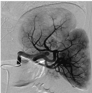
showed enhancement of the upper spleen and venous drainage into varices in the gastric fundus (Fig.4), eventually reaching the splenic and portal vein. There-after, embolization of the upper spleen was performed with injection of polyvinyl alcohol particles (300–500 lm particles, Cook Inc.) after intra-arterial application of mepivacaine HCl 1% for pain reduction. Angiography after embolization showed occlusion of the superomedial splenic artery branch along with devascularization of the upper spleen (Fig.5).
Endoscopy and endosonography performed 2 days after embolization showed less prominent submucosal gastric varices with considerably reduced flow. The patient was discharged home in a good clinical condi-tion 3 days after admission.
Discussion
The spleen is a mesodermal derivative which first appears as a condensation of mesenchymal cells inside the dorsal mesogastrium at the end of the fourth embryonic week. The mesenchymal cells of the splenic primodium differ-entiate to form the capsule, connective tissue framework, and splenic parenchyma. Failure of the individual clumps of mesenchymal cells to fuse properly results in congenital anomalies of the spleen like polysplenia, accessory spleen and splenic duplication [2]. Additional congenital anomalies [3] of the spleen include wandering spleen, splenic cleft and lobulation.
One differential diagnosis in our patient is polysplenia [2], which usually is characterized by equally sized splenic tissue with round shape. Patients with polysplenia Fig. 2. A, B Axial and coronal computed tomography image
obtained in the portal-venous phase showing two spleens being located adjacent to each other (asterisks). Note com-munication of the veins from the superomedially located spleen with fundal gastric varices (arrow).
Fig. 1. Endoscopic and endosonographic (inlay) image demonstrating submucosal varices in the gastric fundus being the source of acute upper gastrointestinal bleeding.
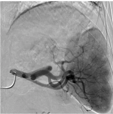
Fig. 3. Angiography image showing division of the splenic artery into two branches (arrow): superomedial and infero-lateral splenic arteries, with each artery supplying the two separate spleens.
additionally show abnormalities such as congenital heart disease, short pancreas, abnormal arrangement of the solid abdominal organs or anomalies of the inferior vena cava. In our patient, the two spleens were not round but rather wedge-shaped. Also, no additional abnormality was noted in our patient, and two different splenic artery branches were seen entering the hilum of each spleen separately, which altogether best fits to splenic duplica-tion.
The imaging findings of splenic duplication were first described in 2009 by Kim et al. [4], reporting a patient with small-cell lung carcinoma. During CT work-up for metastatic disease, two similarly shaped spleens lying next to each other were noted, with the medial portions of the spleens in contact. Unfortunately, no information about the arterial supply and venous drainage was pro-vided by the authors. Similar to the case from Kim et al. [4], we found two similarly shaped spleens which were lying immediately adjacent to each other. In our patient, each spleen was supplied by a separate splenic artery branch while only one (i.e., the inferolateral) spleen drained normally via the splenic vein into the portal system. The other (i.e., the superolateral) spleen had a separate drainage via varices in the gastric fundus, eventually reaching the portal vein.
The incidence of hemorrhage from gastric varices ranges from 10%–70% [5]. Isolated gastric varices sec-ondary to splenic pathology have been implicated as cause of upper gastrointestinal blood loss and include
wandering spleen [6], splenic thrombosis [7] and splenic arteriovenous fistula [8]. To the best of our knowledge, the patient reported herein represents the first case of upper GI bleeding resulting from the presence of splenic duplication with venous drainage via gastric varices, which most probably caused the acute GI bleeding.
In splenic duplication, enlarged splenic volume may result in increased blood flow and venous drainage. Collateral circulation develops via the short gastric, gastroepiploic, and short gastric vein, and left-sided portal hypertension with formation of gastric varices along the greater curvature and fundus of stomach may develop [9].
The optimal treatment of gastric varices remains controversial [10]. Interventional radiologic techniques [11], being important alternatives to traditional surgical approaches, have recently assumed an important role in the treatment algorithm for various splenic disease causing upper GI hemorrhage. The embolic agents most commonly used for splenic arterial embolization are gelatin sponge pledgets, polyvinyl alcohol particles, and coils. Palsson et al. [12] stated that partial splenic arterial embolization preserves functional splenic tissue and produces durable effects on hematologic parameters, prevents variceal hemorrhage, and improves clinical status. Injection of thrombin [13] has been also suggested as effective treatment for gastric variceal hemorrhage.
In our patient, injection of polyvinyl alcohol particles into the superomedial splenic artery branch resulted in Fig. 4. Flat panel computed tomography image after
intra-arterial injection of contrast media into the superomedial splenic artery showing the patchy enhancement of the upper spleen and venous drainage through submucosal gastric varices (arrows).
Fig. 5. Post-embolization angiography showing occlusion of the superomedial splenic artery and devascularization of the upper spleen. Note persistent normal enhancement of the inferiorly located spleen.
complete occlusion of the branch. Restriction of blood inflow to the superomedial spleen led to decompression of the gastric varices. The patient responded well to the intervention and had no major complication on follow-up. Also, the inferolaterally placed spleen was preserved with most likely decreased patient morbidity.
In conclusion, we report the rare cause of splenic duplication causing acute upper GI bleeding via drainage of one spleen via varices in the gastric fundus. Splenic arterial intervention proved to be a quick, safe and effective method of controlling this unusual cause of bleeding.
References
1. Scheffel H, Pfammatter T, Wildi S, et al. (2007) Acute gastroin-testinal bleeding: detection of source and etiology with multi-detector-row CT. Eur Radiol 17:1555–1565
2. Moore KL, Persuad TVN (1998) The digestive system. In: Moore KL, Persuad TVN (eds) The developing human: clinically orientated embryology, 6th edn. Philadelphia: Saunders, pp 217–245 3. Gayer G, Zissin R, Apter S, Atar S, et al. (2001) CT findings in
congenital anomalies of the spleen. Br J Radiol 74:767–772
4. Kim SW, Shin HC, Kim IY, Bae S-B, et al. (2009) Duplication of the spleen with a short pancreas. Br J Radiol 82:42–43
5. Little AG, Mossa AR (1981) Gastrointestinal haemorrhage from left sided portal hypertension: an unappreciated complication of pancreatitis. Am J Surg 141:153–158
6. Sorgen RA, Robbins DI (1980) Bleeding gastric varices secondary to wandering spleen. Gastrointest Radiol 5:25–27
7. Smyth R, Parks RW, Diamond T (2001) Gastric variceal haem-orrhage successfully managed by splenectomy—a case report and literature review. Ulster Med J 70:54–55
8. Keller FS, Rosch J, Dotter CT (1980) Bleeding from esophageal varices exacerbated by splenic arteriovenous fistula: complete transcatheter obliterative therapy. Cardiovasc Interv Radiol 3:97– 102
9. Mossa AR, Gadd MA (1985) Isolated splenic vein thrombosis. World J Surg 9:384–390
10. Ryan BM, Stockbrugger RW, Ryan JM (2004) A pathophysiologic, gastroenterologic, and radiologic approach to the management of gastric varices. Gastroenterology 126:1175–1189
11. Madoff DC, Denys A, Wallace MJ, Murthy R, et al. (2005) Splenic arterial interventions: anatomy, indications, technical consider-ations, and potential complications. Radiographics 25:191–211 12. Palsson B, Hellen M, Forsberg AM, Alwmark A (2003) Partial
splenic embolization: long term outcome. Langenbecks Arch Surg 387:421–426
13. Williams SGJ, Peters RA, Westaby (1994) Thrombin—an effective treatment for gastric variceal haemorrhage. Gut 35:1287–1289